Systematic Review

In Vivo Application of Silica-Derived Inks for Bone Tissue Engineering: A 10-Year Systematic Review

1
Univ. Bordeaux, INSERM, BIOTIS, U1026, F-33000 Bordeaux, France
2
Division of Endodontics and Restorative Dentistry, Department of Oral Function, Kyushu Dental University, 2-6-1 Manazuru, Kokurakita-ku, Kitakyushu 803-8580, Japan
3
Faculty of Dentistry, University of Bordeaux, 146 rue Léo Saignat, F-33076 Bordeaux, France
4
CHU de Bordeaux, Pôle de Médecine et Chirurgie Bucco-Dentaire, Place Amélie Raba Léon, F-33076 Bordeaux, France
5
Laboratory of Biomaterials, Department of Regeneration Science and Engineering, Institute for Frontier Life and Medical Sciences, Kyoto University, Kyoto 606-8397, Japan
*
Author to whom correspondence should be addressed.
Bioengineering 2022, 9(8), 388; https://doi.org/10.3390/bioengineering9080388
Submission received: 22 July 2022 / Revised: 10 August 2022 / Accepted: 12 August 2022 / Published: 15 August 2022

Abstract

:
As the need for efficient, sustainable, customizable, handy and affordable substitute materials for bone repair is critical, this systematic review aimed to assess the use and outcomes of silica-derived inks to promote in vivo bone regeneration. An algorithmic selection of articles was performed following the PRISMA guidelines and PICO method. After the initial selection, 51 articles were included. Silicon in ink formulations was mostly found to be in either the native material, but associated with a secondary role, or to be a crucial additive element used to dope an existing material. The inks and materials presented here were essentially extrusion-based 3D-printed (80%), and, overall, the most investigated animal model was the rabbit (65%) with a femoral defect (51%). Quality (ARRIVE 2.0) and risk of bias (SYRCLE) assessments outlined that although a large majority of ARRIVE items were “reported”, most risks of bias were left “unclear” due to a lack of precise information. Almost all studies, despite a broad range of strategies and formulations, reported their silica-derived material to improve bone regeneration. The rising number of publications over the past few years highlights Si as a leverage element for bone tissue engineering to closely consider in the future.

1. Introduction

Bone defects, whether due to infection, tumor, trauma, surgery, aging or genetic disorders, are a major health issue, affecting hundreds of millions of people every year [1]. Although a human bone graft remains the gold standard therapy, the demand outmatches the supply. A wide range of synthetic biomaterials has been developed to overcome limitations associated with the use of autografts and allografts such as morbidity on the donor site for autografts, and the lack of availability and risks of disease transmission, infection and immunogenicity for allografts [2]. Xenografts and xenogenic products are an uprising therapeutic field but also tend to suffer from graft drawbacks. Synthetic materials offer a more affordable alternative; however, they can be criticized because of their limited osteoconductivity, inadequate mechanical properties, unpredictable dissolution kinetics and poor reproduction of bone architecture [3]. Hence, the optimum formulation to overcome these hurdles is still to be found. Moreover, the development of tissue engineering has led to the design of new materials and strategies aiming at bone tissue regeneration instead of bone tissue replacement [4].
During the last decades, silica-based materials have gained a rising potential in hard tissue engineering since their first application in 1971 [5,6] and have been widely used for medical applications [7].
Silicon (Si) is the eighth-most common element in the universe by mass. More than 90% of the Earth’s crust is composed of silicate minerals, making silicon the second most abundant element in the crust (about 28%) after oxygen [8]. Many studies have shown the important biological role of Si in humans [9] and revealed its presence at various rates in many tissues within the body, such as the liver, kidney, lungs, muscle, bone, cartilage and other connective tissues [10].
At the bone level, silicon is an essential nutrient for tissue formation. In the early 1970s, a deficiency of Si was demonstrated to be involved in bone formation disorders [11,12]. Many authors reported enhanced bioactivity of biomaterials due to the presence of Si, which promotes collagen type I synthesis, osteoblastic differentiation and bone repair [13]. Indeed, Si plays a key role in connective tissue metabolism, particularly in bone and cartilage [14]. In osteoporosis and osteopenia, the reduced proliferation and activity of osteoblasts were found to be correlated to the loss of the biological availability of Si [15]. Other experimental studies have shown the role of Si in the remodeling process of bone and suggested its effect on the inhibition of the physiological resorption mechanisms [16,17].
These findings highlight the obvious interest and high potential of silicon inclusion in biomaterials used for bone regeneration.
Easily available, and suitable for malleable preparations, silicon-derived materials crossed the path of additive manufacturing [18]. Developed in the second part of the 20th century [19], the application of this field to biology covers various technologies, used for fabricating 3D objects in mineral and/or organic elements [20,21]. In bone tissue engineering, sustaining the required mechanical functions of bone is one of the historical and critical challenges [22]. As such, material composition must be compatible with strategies of construct modeling, such as extrusion-based bioprinting, the most common approach [23]. Another crucial point is to induce a favorable biological response to restore or supply the lost bone function. Often, the desired outcome is to locally obtain the mineralization of the tissue replacing the bone defect, which will eventually be remodeled as a bone tissue as mature and physiologically competent as native bone. Therefore, research is heavily focused on developing suitable materials that bring a satisfying answer on these two levels, exhibiting robust mechanical properties with a high biocompatibility and strong regenerative potential. Thus, in the scope of identifying the most efficient materials for bone tissue engineering, investigations of silicon-derived materials were extensively conducted, as it has been widely demonstrated to be an essential component of different types of biomaterials such as bioactive ceramics in bone repair/regeneration [24].
In bone regeneration and repair applications, silicon has often been used as a substitute or a dopant in calcium phosphate-based ceramics since it has a positive impact on the performance of conventional biomaterials, related to the surface chemical structure, mechanical strength, bioactivity and biocompatibility [25]. Moreover, the current trend has shifted from traditional biocompatible materials to bioactive materials, to promote a specific biological response between the material and the tissue, leading to the development of a bond between them [26].
Indeed, several research studies have been conducted in the past [27,28], notably, regarding physiologically relevant compounds such as hydroxyapatite [29] or collagen [30], but none specifically concerned with the use of silicon.
Of the numerous materials and strategies assayed, some printable silicon-containing formulations went through the in vitro characterization to evaluate their in vivo bone repair potential. Thus, this systematic review proposes to report on the last decade of in vivo applications based on (bio)printed silicon strategies for bone tissue engineering.

2. Materials and Methods

2.1. Protocol

The systematic review was conducted according to the Preferred Reporting Items for Systematic Reviews and Meta-Analysis (PRISMA) guidelines [31], using the Population, Intervention, Comparison and Outcome (PICO) methods [32] to define the search strategy. The protocol was registered in PROSPERO database (CRD42022311293).

2.2. Focused Question

The following focused question was defined: “Which silica-based ink formulations have been shown effective for bone tissue regeneration?”

2.3. Selection Criteria

In vivo preclinical studies involving the use of silica-based ink for bone regeneration were included. Only studies published in English with their abstracts available on the database were considered. In vitro and ex vivo studies, case reports and reviews were excluded.

2.4. Search Strategy

The search strategy was developed based on the PICO reporting system (Table 1). A structured electronic search for articles published in English was conducted up to and including 10 April 2022 using the electronic databases MEDLINE (PubMed), Scopus and Web of Science.
The following search combination was used:
PubMed: ((silic*[Title/Abstract]) AND (print*[Title/Abstract]) AND (bone[Title/Abstract])) NOT (silicone[Title/Abstract]) AND ((in vivo[Title/Abstract])
Scopus: (TITLE-ABS-KEY (silic* AND print* AND bone AND NOT silicone) AND TITLE-ABS-KEY (in AND vivo))
Web of Science: silic* (Topic) AND print* (Topic) AND bone (Topic) NOT silicone (Topic) AND in vivo (Topic)
The keyword “silicone” was intentionally removed from the research outcome, being already described as a poor, detrimental material for bone repair [33]. Thus, all silicone-based approaches were excluded from the research scope.
Additional articles complemented the electronic search after manually screening the list of references of all publications selected. The searches were re-run just before the final analyses to retrieve the most recent studies eligible for inclusion.

2.5. Screening Methods and Data Extraction

All references were imported into a Zotero database. After eliminating the duplicated ones, references were exported to an Excel sheet to perform the screening phases. The articles were selected by two reviewers (NT and OK) working independently. The selection was performed in two phases, based on inclusion and exclusion criteria: (1) title/abstract screening, (2) full-text screening. The two lists of selected references were then compared and all disagreements were resolved by discussion, or, if persistent, by a third reviewer (RD).
Prioritized exclusion criteria were as follows: (1) during title/abstract screening, no application in bone regeneration, reviews, in vitro or ex vivo studies, clinical trials, cases reports, no silica-based material ink, (2) during full-text screening, no relevant outcome measure reported, no control group.
For eligible studies that matched with inclusion criteria, full text was screened to extract data by two reviewers using the data extraction form and was summarized in the form of Excel-based extraction tables. Data were extracted from text, tables and graphs. Extraction information included but was not limited to: data for evidence synthesis (study design, animal model, intervention of interest, primary and secondary outcomes), as well as information required for assessment of study quality and assessment of risk of bias. In case of missing data, the corresponding authors were contacted via email. Any disagreement between reviewers was resolved by discussion with the third reviewer (RD).

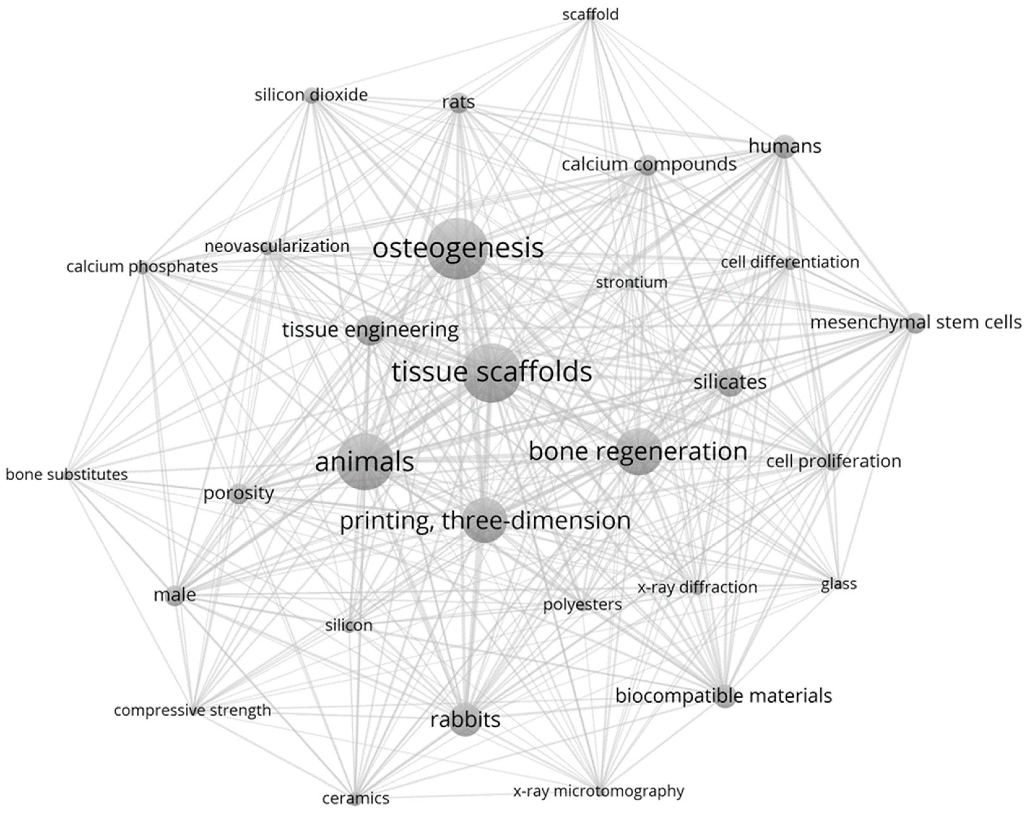
2.6. Science Mapping Analysis

Science mapping analysis of scientific domains was performed from the list of included articles by using keyword co-occurrence networking on VOSviewer (free software, version 1.6.18, Centre for Science and Technology Studies, Leiden University, Leiden, The Netherlands, 2022). Network analysis of the keywords was generated from the matrix of retrieved papers [34] (threshold value at 4). The terms–document matrix allowed to measure document similarities between clusters of topics.

2.7. Quality Assessment and Risk of Bias

The methodological quality and risk of bias of the studies included were assessed by two independent reviewers.
The ARRIVE 2.0 guidelines for reporting animal research were used to evaluate the quality of the studies. The items evaluated according to ARRIVE 2.0 [35] (“reported”, “not reported”, “unclear”) were: (1) study design, (2) sample size, (3) inclusion and exclusion criteria, (4) randomization, (5) blinding, (6) outcome measures, (7) statistical methods, (8) experimental animal, (9) experimental procedures, (10) results, (11) abstract, (12) background, (13) objectives, (14) ethical statement, (15) housing and husbandry, (16) animal care and monitoring, (17) interpretation/scientific implications, (18) generalizability/translation, (19) protocol registration, (20) data access, (21) declaration of interests.
The risk of bias was evaluated using the SYRCLE (Systematic review Center for Laboratory Animal Experimentation) tool for animal studies [36], using the following criteria: (1) random sequence generation, (2) baseline characteristics, (3) allocation concealment, (4) random housing, (5) blinding of participants and personnel, (6) random outcome assessment, (7) blinding of outcome assessment, (8) incomplete outcome data, (9) selective outcome reporting, (10) other risk of bias. Three categories of potential bias judgments were used: “low risk of bias”, “high risk of bias”, “unclear risk of bias”.
Two reviewers (NT and OK) were involved in assessment of risk of bias and study quality. When no consensus was reached, discrepancies were resolved by a third reviewer (RD).

2.8. Data Analysis

Data analysis was performed in a descriptive way due to heterogeneity of the included studies in terms of animal model, bone defect model, interventions and outcomes. No meta-analysis was considered for this systematic review. Summary tables were intended to provide the most relevant information on study characteristics and findings. In particular, the descriptive analysis explored the relationships between studies and described the impact of various silica-based ink formulations on bone regeneration process.

3. Results

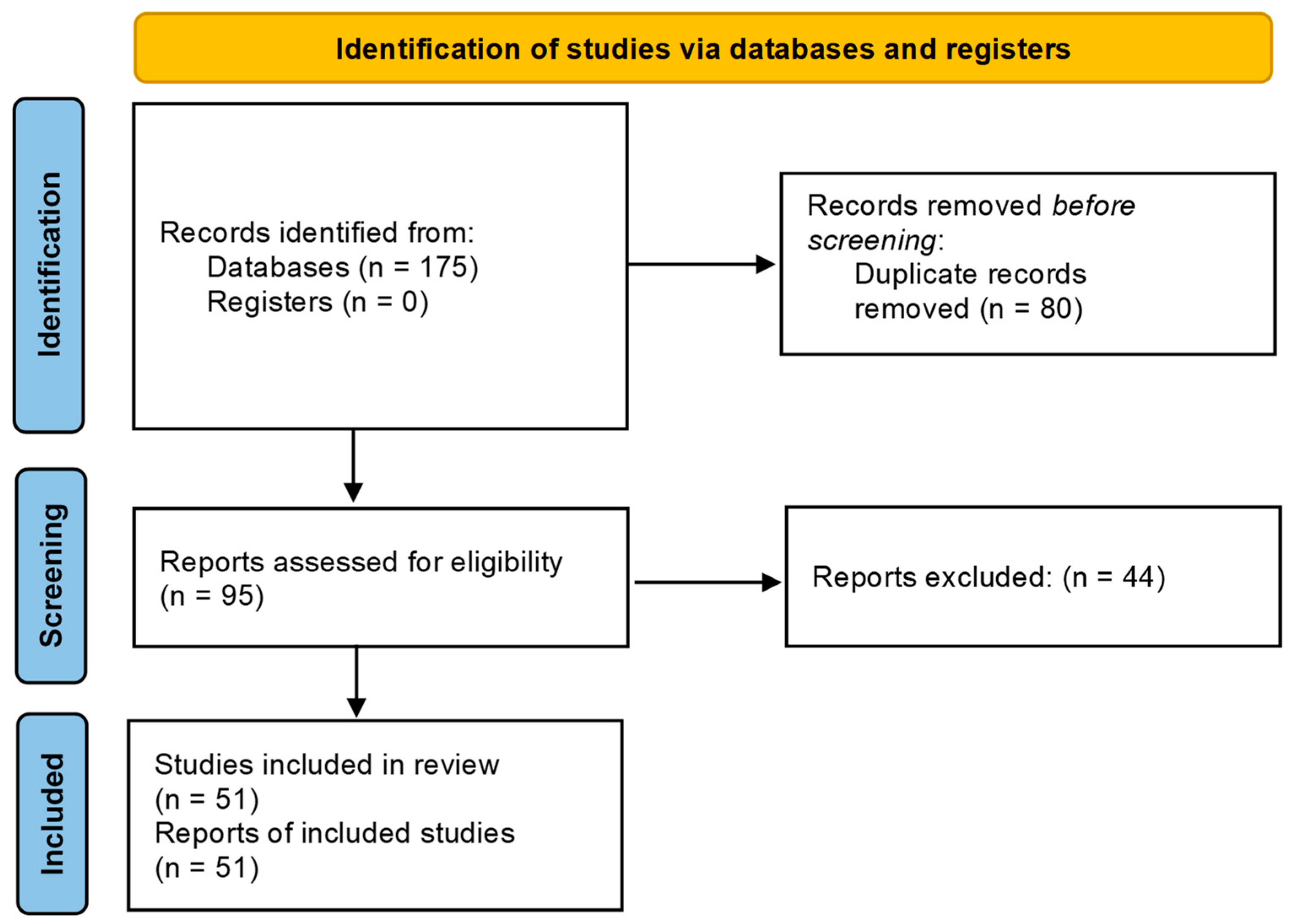
3.1. Systematic Review following PRISMA Guidelines

After application of the inclusion and exclusion criteria, 175 articles were selected initially.
The search using the PICO model, yielded 43 articles from MEDLINE/PubMed, 62 articles from Scopus and 70 articles from Web Of Science within the past 10 years. After the removal of duplicates/triplicates and preliminary analysis of the abstracts, 95 articles were selected for full-text analysis and evaluation, after which 44 articles were excluded and 51 articles were included in the review (Figure 1).
The spatial representation of the relationships between the keywords was displayed through a science map (Figure 2). The core, most recurrent keywords were (by decreasing rank): “tissue scaffold”, “animals”, “osteogenesis”, “printing, three-dimensional” and “bone regeneration”.
This occurrence representation map highlighted three silicon forms used within the scoped articles: silicon, silicon dioxide and silicates. With one form excluding the others, those keywords had no link strength between each other, and were located in opposite areas. Of the material formulation-related keywords drawn by the occurrence map, silicon was observed to be closest to ceramics, while silicon dioxide was associated to calcium phosphate and silicates was near polyesters and calcium compounds.
The most occurring physical characteristics were porosity, compressive strength and X-ray diffraction. “Mesenchymal stem cells” was among the top 30 most recurring words/phrases and the only cellular population mentioned, and the most represented biological analyses were neovascularization, cell proliferation and differentiation.

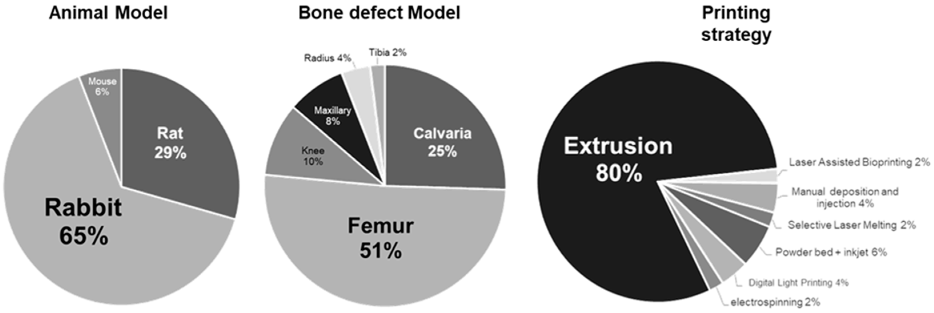
3.2. Study Characteristics

In the scoped articles, three animal models were assessed (Figure 3): rabbit (65%), rat (29%) and mouse (6%). Interestingly, no large animals (sheep, goat) were involved. It is worthy to note that the number of articles was here correlated to the size of the model. Two preferential bone models were used: femur (51%) and calvaria (25%). In this review, only bone regeneration was considered as a crucial criterion, and no specific consideration was given to mechanical assessment. However, except calvaria and maxillary, all bone models in this review are associated with motion and superior load-bearing capacity.
Seven assembling strategies were performed in the articles gathered for this review. An overwhelming number of studies employed extrusion-related techniques (80%), and the six other strategies shared the other 20%. Within this minority, inkjet-associated strategies were the most representative [37,38,39] (6% of total), while digital light processing [40,41] and manual deposit or injection [42,43] were both 4%. Electrospinning [44], laser-assisted bioprinting [45] and selective laser melting [46] shared equally the 6% remaining (2% each). The trend of the studies assessing silicon-based inks was here to obtain an extrudable paste, mostly mixed with polyvinyl alcohol (PVA), that went through a sintering process after the printing step.
To summarize, the most recurrent combination of models and approaches assessed in the present review were: rabbit–femur–extrusion. The probability of retrieving a study (among the scoped articles) investigating the extrusion of a scaffold implanted in a femoral defect of rabbit was calculated and found to be P(e×f×r) = ((0.8 × 0.51 × 0.65) = 0.2652 (26.25%)). In this study, P(e×f×r) × N(articles) = 13.52 articles should therefore theoretically associate those three designs. Seventeen articles (33.3% of total) were actually found to present those characteristics, giving a 6.8% difference between the predicted value and the actual number of articles assessed.
The main data from the articles selected are shown in Table 2 and Figure 4, and additive information may be found in the Supplementary Data Table.
The list of articles was divided into two main categories, for silicon was either part of the native material (1), or was added as an external component of the initial material assessed (2). In each category, two additional subcategories were created to sort the studies depending on the fact that the silicon has either a primary role (a), or a secondary role (b) over the bone regeneration assessed (Table 3).
By creating these four categories, all the articles retrieved via the algorithm employed were able to be sorted without the possibility of an article belonging to more than one category. Closest articles were then gathered by focus of interest, creating final subcategories with the largest size possible.
Three main sources of Si were identified: tetraethyl orthosilicate (TEOS) (37%), pure SiO2 (29%) and wollastonite (CaSiO3) (20%). The remaining 14% of sources were shared by Ca2Si, Ca3Si, Na2SiO3 and RSiO3/2, or were of undefined origin. According to the sorted articles, TEOS and CaSiO₃ were principally involved as scaffold basal constituents, while SiO2 free forms were mostly presented as doping agents.

3.3. Outcomes

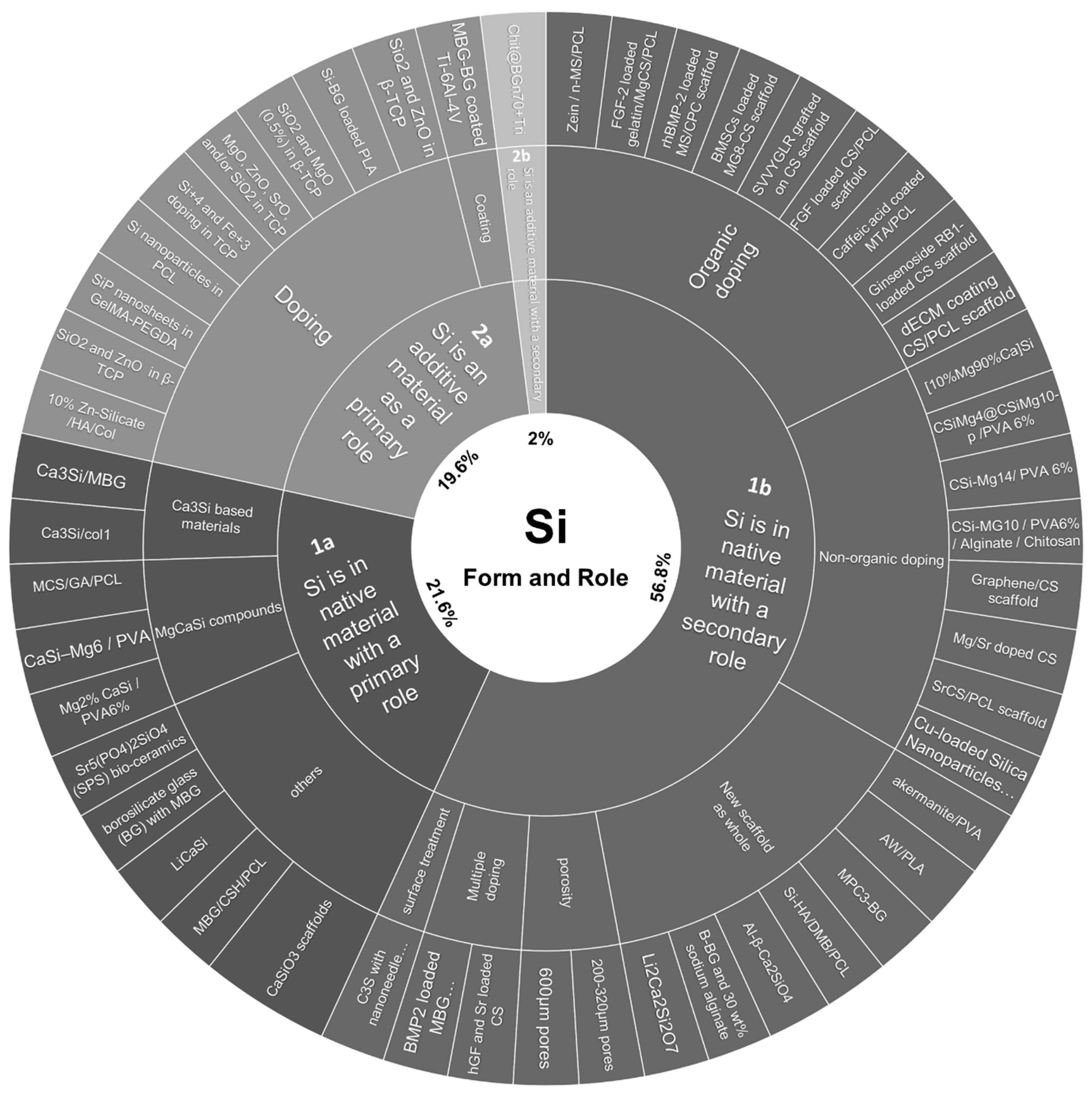
3.3.1. Articles’ Sorting and Main Outcomes

  • Category 1—Si is in the native material
With 78.4% of the total studied articles sorted in this category, the use of Si as a native part of the investigated material was the leading category.
-
1a—Si is in the native material with a primary role
Within articles including Si as a native part of the material, 21.6% of the total (11 out of 51) articles placed silicon as a primary element of focus. The compositions of the investigated materials were variable with monocalcium-based inks [47,48], tricalcium-based inks [45,55]; glasses also involved [53,54]; elements such as magnesium [28,29,30], strontium [52] or lithium [56] were also core components of calcium–silicate materials. Controls were usually either an empty defect, β-TCP or titanium scaffolds. Only Touya et al. [45], using laser-assisted bioprinting in a mouse calvaria defect model, reported an absence of enhanced tissue mineralization despite encouraging in vitro results, whether associated with cells or not. Overall, the outcome was the improvement in bone repair compared to the control.
-
1b—Si is in the native material with a secondary role
The largest subcategory, including 56.8% of the total (29 out of 51) articles reviewed, could be divided into three main focuses: doping an existing scaffold, assessing a new material formula (in which Si is of minor interest for the authors) or investigating physical differences between scaffolds with the same composition. Scaffolds were doped by either organic, non-organic or mixed elements with coating or loading strategies.
Two articles implanted materials loaded with cells. Shen et al. [62] reported no significant difference in bone formation between loading or not their magnesium and calcium–silicate scaffold with bone marrow stromal cells. Wang et al. [74] used gingival fibroblast and observed an improved bone regeneration compared to their sole implants.
Plant-derived organic compounds [58,59,65], animal growth factors [60,63,64,73], osteopontin-derived peptide [61] and decellularized matrix [57] were investigated. Overall, they all triggered an enhanced bone regeneration compared to their respective non-loaded controls.
Most non-organic doping involved magnesium [67,69,70,71,72], while others included copper [44], strontium [66] or graphene [68]. All of the studies reported an improvement in bone regeneration associated with loaded materials compared to their controls.
In several studies [43,75,76,77,78,79,80], Si was part of the native material, but not considered as a determining factor in the bone repair evaluated. Although Meseguer et al. [78] only performed qualitative observations, all studies reported a bone repair enhancement.
For three studies, physical properties of the material assessed constituted the focus of the investigations: porosity [40,41] and surface texture [81] were highlighted to be determining parameters for bone repair response.
This category illustrates the use of Si as a structural/vehicle agent.
  • Category 2—Si is an additive material
Among the scoped articles, 21.6% of the total were sorted in this category.
-
2a—Si is an additive material with a primary role
Si was used as a determining additive material in 19.6% of total studied articles.
All but one of the articles in this category reported the use of Si as a doping agent [37,38,39,42,82,83,84,85,86] meant to be released through time in the implant micro-environment. Zhang et al. [46] presented the coating of a titanium scaffold with a bioglass. All articles reported an improved bone regeneration compared to their siliconless counterparts (for Zhang et al., at the periphery of the implant).
-
2b—Si is an additive material with a secondary role
Only one (2% of total) article was sorted in this category [87], as the element of focus was a tri-molecule dexamethasone-FGF2-phenamil loading in a chitosan/bioglass-coated PCL scaffold. Silicon was associated to the coated bioglass on the PCL scaffold, and was one ingredient out of a vast association of doping agents. This study also reported an improved bone regeneration.

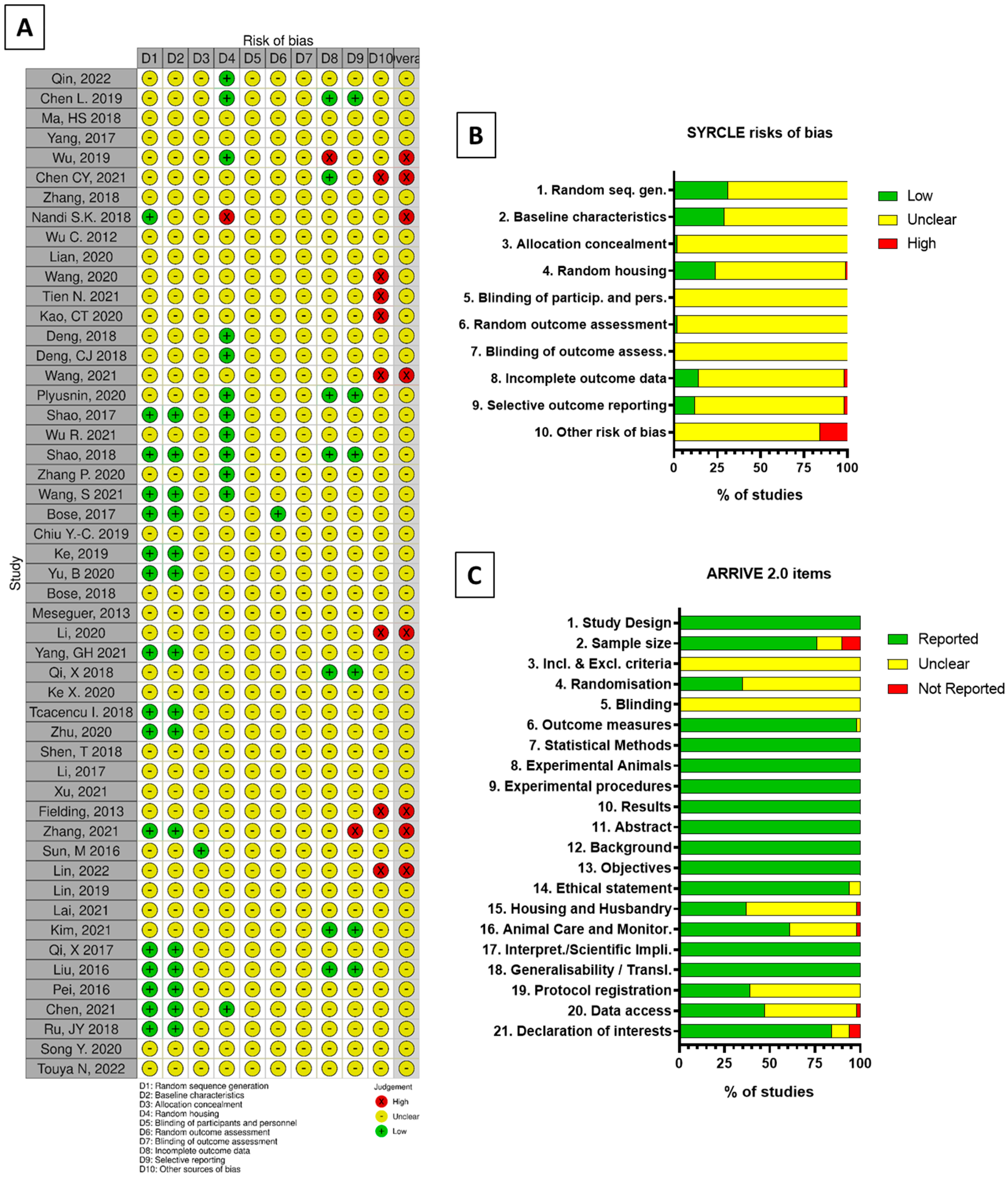
3.3.2. Quality Assessment and Risk of Bias

Results from the quality assessment of the studies selected for detailed analysis are shown in Figure 5.
All scoped articles were analyzed according to SYRCLE and ARRIVE 2.0 methods. As ARRIVE is, to date, not associated with any graphical representation, the performed quality assessment was depicted inspired by the SYRCLE display tool robvis [88].
According to SYRCLE evaluation, 86.5% of answers to each risk of bias were “Unclear” as the information needed was either missing or incomplete for most studies, while 11.5% were attributed as “Low” when information was unequivocal and 2% as “High”. If one or more item of any article was filled by a “High” potential risk of bias, the whole article was systematically considered to be also of “High” potential risk of bias, regardless of the element that led to the decision to attribute the initial “High”. Two categories were identified as most involved in sorting High risks of bias: the animal random housing (cat. 4) and the treatment of outcome data (cat. 8–9). Randomness of housing could not be ensured as it was not necessarily specified in many studies. Some studies presented differences between the number of animals used displayed in the methods section, and the number of animals displayed in the results section without justifying it.
With the ARRIVE 2.0 method, 75% of items were “Reported”, 25% were “Unclear” and only 1% were found to be “Not reported”; the categories with most missing or partially reported data were the inclusion and exclusion criteria, and the blinding.
Overall, the articles were found to be of satisfying quality but associated with risks of bias. However, those risks could be mostly attributed to a lack of information provided by authors rather than clearly misconducted experiments.

4. Discussion

Silica-based materials exist in various forms, depending on the purity of sole Si, or complexation, resulting in a hybrid material. Even though Si was found in the literature to be part of natural metabolism and to benefit from a high renal clearance [89], some forms of silica may exert toxicity or deleterious effects at the scale of the whole organism [90,91]. None of the articles in this review seemed to employ such Si derivatives. Investigating the possibility to use Si as a drug delivery carrier, the size of 70 nm and below [92] of the Si particles applied to skin was found to be responsible for systemic exposure in mice and exerted mutagenic activity [93]. Even though such findings were consistent with the inhalation or digestion of such a size of nanoparticles [94,95], the literature insists on the good compatibility of these sizes with the dermal route even at a 2000 mg/kg dose [96]. In this review, the Si nanoparticles’ smallest reported form was of 100 nm diameter [84], and no systemic effect was reported. However, Si content within the ink formulations investigated was unassessable due to the ink preparation process. Although the initial source and proportion of Si or Si-based derivatives within the material was indicated in many studies, the washing, drying and sintering steps contributed to affect the material mass or volume, and therefore the element’s contents. Moreover, in different articles, the purpose of Si was to be released through time. Hence, measures of the native material’s Si content regarding this strategy were irrelevant, and the focus on the outcome, i.e., the bone repair compared to appropriate controls, constituted the principal focus of this review.
Indeed, Si, beyond being cytocompatible, was reported to exert a bioactive effect, by enhancing angiogenesis in endothelial cells [97], and enhancing mineralization [98] in osteogenic lineages. These two critical events being tightly associated to an efficient bone repair, the direct impact of Si upon them was investigated.
However, other articles excluded from our scope used alternative strategies. For instance, in a study by Li et al. [99] (rejected for assessing only the subcutaneous implantation of material), the strategy relied on the support of activated and polarized macrophages to stimulate angiogenesis consecutive to bone defect creation. Mediated by Si ions, M2 macrophages were polarized. Associated with HUVEC, they significantly improved angiogenesis and enhanced bone repair subcutaneously. The immune system could therefore be a first order actor to modulate the tissue repair response after a trauma, of which silicon may offer a therapeutic interest.
With the emergence of biofabrication strategies for tissue engineering, the development of compatible materials on both technological and therapeutic aspects has increased, especially recently [23]. Indeed, more than half of the recovered articles through the algorithm were published in 2019 or later, highlighting the uprising place of material inks developed in bone tissue engineering. Among the initially gathered articles, 51 studies remained in the scope. The article sorting was designed in the perspective of best answering the original question “Which silica-based ink formulations have been shown effective for bone tissue regeneration?”. Categories were designed to avoid duplicates, and no article was found to belong to multiple subcategories. This sorting could help decipher relevant trends of the use of Si in recent Si-based materials.
Indeed, it was outlined that in ~60% of articles, the role of silicon was not the determining aspect of the study. Overall, Si was considered as holding a structural role within printed materials. For the doped materials, when Si was often brought alongside with Zn, Mg or other elements, it was not possible to formally consider Si as the unique discriminated agent of bone repair enhancement, except for two studies. A trend of 96% of studies nevertheless reported a bone repair improvement in the formulations involving Si.
This review focused on 3D printing-associated materials. Although the printing of silicium is not new, its application essentially used to belong to the electronic [100], security [101] or glass [102] industries.
Overall, 80% of articles used the extrusion-based printing technique. This trend was found to be highly consistent with the outcome of a recent systematic review dedicated to hydrogel bioprinting [23] that found extrusion to be used in 76% of 393 articles. It also agreed to a previous review over the privileged place of extrusion [103], in spite of a huge difference between the percentage of each printing technique involved (also first rank with 36.18% of 351 articles), in which the presence of cells within the biomaterial was mandatory in the search algorithm. Extrusion seems then to be by far the gold standard technique in non-involving cells’ strategies, which is consistent with the cells’ sensitivity to shear stress and associated loss of viability [104]. Moreover, due to the low viscosity nature of most materials investigated here and the inherent constraints of ink deposition this low viscosity triggered, extrusion was the most relevant tool to perform an efficient, reproducible printing process. Overall, a very limited number of studies investigated the association of cells [45,62,74] to the Si-derived material as part of the engineered final product to implant, and most of the publications relied on the host response rather than an enhanced biological activity due to additive cells. Only Wang et al. [74] found the addition of cells to their material to significantly improve bone formation.
In this review, the in vivo models investigated only concerned small animals. The rabbit appeared to be the most recurring target to assess bone repair, among other animal species or bone defects. This result goes against previous findings, as rat was reported by O’Loughlin et al. [105] (2008) to be the preferred model of fracture healing investigation. Some bone models possess analogous examples in humans or other species. Assessing various bones can therefore target a larger number of finer, physiologically equal models [106]. Hence, investigating the broadest range of animals and bones is crucial to provide an extensive and more truthful physiological bone response to implanted engineered biomaterials. However, in the effort to identify a baseline model to assess bone repair, thus providing a more standardized approach to enable comparison between studies and new materials, the diversity of animal and bone models might appear as a hurdle. The choice of a small animal model, beyond physiological fidelity, surgical accessibility or translation potential towards larger models, is associated with greater material advantages [107]. Indeed, smaller animals are more available and affordable in terms of basal caretaking costs per capita (purchase, housing, feeding, monitoring and overall costs of experiment-associated materials). Questioning here the bone regeneration potential of silica-based (bio)inks, the rabbit model appeared, hence, to be the most pragmatic and suitable model and logically federated the largest number of studies. Preclinical studies following the identification of the most outstanding materials regarding bone repair should consider the implantation in larger mammal models, as the studies presented here can only provide an in vivo first glimpse of a small model response.
Tightly bound to the animal species model choice, the baseline characteristics are also essential. Even though the outcome (a fully-filled, mature mineralized tissue with equal —if not superior—mechanical capacities regarding native tissue consecutively to creation of a bone defect) could be expected to be the major interest, the healing dynamics involved, the physiological performance and well-being of biological material over time should also be considered. In the scope of reaching the most reliable bone model to standardize the research of bone tissue engineering, extensive attention should then be brought with priority to the baseline characteristics of studied animals. First, the age of animals should be systematically reported, for this variable has a tight relationship with bone repair following a trauma [108,109]. Regarding gender, if fracture incidence was reported to be different through gender and age [110], there is no consensus in the literature regarding an influence of the sex over bone repair. Even though several studies in mice reported that males had a faster bone healing, they were unable to formally discriminate this result from the fact that studied males had more weight than females, or a superior activation of signaling pathways [111]. Overweight and obesity were indeed reported to delay and reduce bone healing post trauma [112,113].
To summarize, the age, weight and sex of animals should at least be reported systematically in each study. For example, as rabbit was the most abundant model in the articles scoped here (65% of total), it is proposed here to select specimens (either male of female) of 28–30 weeks of age for skeletal maturity [114,115] (specifically based on the growth plate closure age over life expectancy age), preferentially with homogeneous weights.
The choice of the defect location in animals is of major interest regarding healing dynamics through time. Each bone has its own indications for the creation of a defect [116], and studies may exhibit different approaches. Each model offers different relevant defects in terms of the physiological response and mechanical consequences, aiming or not for the creation of a critical-size defect [117]. For instance, flat bones were reported to heal same size defects faster than long bones [118]. Moreover, there seems to be an absence of consensus concerning the size of critical defects in humans [119], which adds challenge to the establishment of a universal defect for in vivo investigations. In this review, the question was to provide an extensive sight of the silica-derived inks regarding the enhancement of bone repair consecutive to the creation of a defect, beyond natural healing. Therefore, although of great importance, the size of the defect here was not determining, and this review focused on the ink formulation employed and the final outcome compared to appropriate controls. In this review, the majority of defects were associated with long bones, especially the femur (51% of articles). Regarding the most representative animal used, femoral defects created in rabbit varied from 5 × 5 mm to 6 × 10 mm cylindrical defects (33.3% of total articles). The most abundant defects (6 out of 17 (11.7% of total articles)) were associated with a 6 × 6 mm size [59,60,64,65,66,80]. Thus, in the challenge of proposing a consensual model regarding the development of silica-based inks for bone repair, the prevalence of 6 × 6 mm cylindrical femoral defects in rabbits in this review should be highlighted.
It was challenging for bone repair assessment to be quantitatively compared through the screened articles due to the heterogeneity in animal/bone defect/healing time models. First, the most recurrent techniques displayed to qualitatively appreciate and quantitatively measure bone regeneration were, respectively, histological staining and micro-computed tomography (micro-CT). Indeed, 78% (40 out of 51) of articles investigated bone repair with both histology and micro-CT. Most of the time, histological analysis remained qualitative, and the tissue organization and interaction with the implanted/remodeled material were described and compared to controls. In this regard, outcomes presented in this review were limited to the quality of bone repair in the presence of silica-derived materials.
Regarding articles’ quality and risk of bias, SYRCLE methodology highlighted a dramatic lack of animal procedure transparency and availability; ethical statements or animal procedures, as incorporated in studied articles, were unable to provide enough information to fill the required items. Most studies cited an ethical authority that provided an approval to conduct an in vivo experiment. However, until the full experimental procedure documents become available as a Supplementary File attached to any animal experiment-related publication, the tools for risk of bias detection will widely remain irrelevant. We hereby emphasize the absence of animal experiment-related protocols attached to publications. A better transparency will improve publications at many levels, especially in terms of the reliability and reproducibility of conducted experiments. Unfortunately, likely due to potential patents and/or the intellectual property involved with publications, most of the animal procedure complete protocols remain unavailable, and therefore the displayed elements within most manuscripts cannot be considered as whole.
Concerning the regeneration process, the immune status of the animal model was questioned. It was considered that, in animals with an altered immune system, the conclusions of observed results could be inconsistent with a normal physiological response involving immune system key mechanisms [120,121]. Therefore, studies with immune-compromised animals were categorized as having an “Unclear” risk of bias.
Overall, although of great interest, the SYRCLE risk of bias assessment seemed not to represent the best tool to judge the quality of studies assessing the development of material formulations, for a majority of blinding-related aspects were conflictual with the implantation of a material versus an empty defect. ARRIVE 2.0 methodology was perceived as the opposite of SYRCLE regarding the proportion of items found to meet the requirement of the guidelines with the reported/unclear/not reported system. However, this scoring was not able to underline the weaknesses of some studies. For example, an inappropriate methodology employed regarding statistical hypotheses and tests could be performed in an article, or conflicts of interests could be displayed. According to the scoring system, the items 7-Statistical Methods and 21-declaration of interests would be considered reported and, consequently, provide a pledge to quality.
In spite of aiming for a single purpose, bone repair, this review highlighted the plurality in strategies: from the scaffold/ink preparation process to the delivery of the material. Most studies involved processes with several intermediary steps such as soaking, drying, sintering, etc., to generate a scaffold, while few were able to deliver a final product by one-step assembling and in situ printing [43,45]. Although the tissue engineering definition [122] depicted a trinity involving cells, adjuvants and support materials, almost none of the articles here investigated focused on a strategy relying on the use of cells, and most used an inorganic material-associated approach. This is consistent with a pragmatic will of obtaining a standardized, patient adjustable, quickly available elaborated material.
This manufacturing aspect raises the question of the materials’ availability for patients regarding the acceptable delay from the moment of demand versus the moment of delivery. Moreover, the questions about the strategy underlying the formulation are crucial, for the materials can be supposed to immediately replace a bone on a mechanical perspective with poor physiological respect, or to be associated with a longer (closer to natural) healing time but aim to restore the bone tissue as if it was native. Still regarding formulation, targeted physiological aspects can be crucial, choosing to stimulate vascularization, mineralization and innervation, or to first prevent a microbiological contamination hazard while keeping the composition as simple as possible, with a defined architecture and physicochemical properties expected to address physiologically-specific needs.
From a translation-to-clinic perspective, the manufacturing of new materials must therefore overcome considerable challenges. The scale up from the in vitro evaluation of materials with in vivo assessment in small animal models to in vivo therapeutic implantation to large mammals with respect to the laws and regulations for health seems yet to remain for a minority of products [123,124]. The increasing number and diversity of 3D printing approaches this last decade might help to bring more materials and applications to the clinic, and research should be continued in this effort.

5. Conclusions

Involved in bone tissue repair strategies since the 1970s, silicon and its derivatives recently grew in interest in the additive manufacturing and bioprinting fields. In this review, silicon appeared to be a versatile element to improve bone repair, whether as a constitutive or as a functionalizing, additive element. This review highlighted the dominant place of extrusion printing and the privileged place of rabbit and the femur, respectively, as animal and bone defect models for bone tissue engineering regarding the development of biocompatible inks suited for 3D printing. Considering the trend of an increasing number of publications related to the in vivo application of silica-derived inks for bone tissue engineering, it is expected that future printed patient-designed prostheses will integrate Si within the manufacturing process.

Supplementary Materials

Author Contributions

Conceptualization, O.K.; methodology, O.K.; software, A.N.; validation, A.W., C.K. and Y.T.; formal analysis, R.D.; investigation, N.T.; resources, N.T. and O.K.; data curation, N.T. and O.K.; writing—original draft preparation, N.T.; writing—review and editing, O.K.; visualization, N.T.; supervision, O.K.; project administration, O.K.; funding acquisition, O.K. and R.D. All authors have read and agreed to the published version of the manuscript.

Funding

This international collaboration between French and Japanese research teams is part of a more global research project funded by the JSPS (Japan Society for the Promotion of Science), the Foundation ARC (French Foundation on Cancer Research), the French Embassy in Japan (“Exploration Japon” Program) and IdEX—University of Bordeaux (Initiative of Excellence). The Japan Society for the Promotion of Science—ID No. P22101—17 December 2021. The foundation ARC-n° ARCMOB2021080004095—14 December 2021. The French Embassy in Japan—“Exploration Japon” Program-18 February 2020. IdEX—University of Bordeaux-n°191203_001—3 February 2020.

Institutional Review Board Statement

Not applicable.

Informed Consent Statement

Not applicable.

Data Availability Statement

All data gathered for this review were obtained from the scoped and cited articles. No new data were created or analyzed in this study.

Conflicts of Interest

The authors declare no conflict of interest. The funders had no role in the design of the study; in the collection, analyses, or interpretation of data; in the writing of the manuscript; or in the decision to publish the results.

References

  1. Hernigou, P.; Scarlat, M.M. Growth in Musculoskeletal Pathology Worldwide: The Role of Société Internationale de Chirurgie Orthopédique et de Traumatologie and Publications. Int. Orthop. (SICOT) 2022, 46, 1913–1920. [Google Scholar] [CrossRef] [PubMed]
  2. Porter, J.R.; Ruckh, T.T.; Popat, K.C. Bone Tissue Engineering: A Review in Bone Biomimetics and Drug Delivery Strategies. Biotechnol. Prog. 2009, 25, 1539–1560. [Google Scholar] [CrossRef] [PubMed]
  3. García-Gareta, E.; Coathup, M.J.; Blunn, G.W. Osteoinduction of Bone Grafting Materials for Bone Repair and Regeneration. Bone 2015, 81, 112–121. [Google Scholar] [CrossRef] [PubMed]
  4. Hench, L.L.; Polak, J.M. Third-Generation Biomedical Materials. Science 2002, 295, 1014–1017. [Google Scholar] [CrossRef]
  5. Hench, L.L.; Splinter, R.J.; Allen, W.C.; Greenlee, T.K. Bonding Mechanisms at the Interface of Ceramic Prosthetic Materials. J. Biomed. Mater. Res. 1971, 5, 117–141. [Google Scholar] [CrossRef]
  6. Götz, W.; Tobiasch, E.; Witzleben, S.; Schulze, M. Effects of Silicon Compounds on Biomineralization, Osteogenesis, and Hard Tissue Formation. Pharmaceutics 2019, 11, 117. [Google Scholar] [CrossRef]
  7. Vallet-Regí, M.; Balas, F. Silica Materials for Medical Applications. Open Biomed. Eng. J. 2008, 2, 1–9. [Google Scholar] [CrossRef]
  8. Clarke, F.W.; Washington, H.S. The Composition of the Earth’s Crust. Prof. Pap. 1924. [Google Scholar] [CrossRef]
  9. Jugdaohsingh, R. Silicon and Bone Health. J. Nutr. Health Aging 2007, 11, 99–110. [Google Scholar]
  10. Schwarz, K. A Bound Form of Silicon in Glycosaminoglycans and Polyuronides. Proc. Natl. Acad. Sci. USA 1973, 70, 1608–1612. [Google Scholar] [CrossRef]
  11. Carlisle, E.M. Silicon: A Possible Factor in Bone Calcification. Science 1970, 167, 279–280. [Google Scholar] [CrossRef] [PubMed]
  12. Schwarz, K.; Milne, D.B. Growth-Promoting Effects of Silicon in Rats. Nature 1972, 239, 333–334. [Google Scholar] [CrossRef] [PubMed]
  13. Gao, T.; Aro, H.T.; Ylänen, H.; Vuorio, E. Silica-Based Bioactive Glasses Modulate Expression of Bone Morphogenetic Protein-2 MRNA in Saos-2 Osteoblasts in Vitro. Biomaterials 2001, 22, 1475–1483. [Google Scholar] [CrossRef]
  14. Reffitt, D.M.; Ogston, N.; Jugdaohsingh, R.; Cheung, H.F.J.; Evans, B.A.J.; Thompson, R.P.H.; Powell, J.J.; Hampson, G.N. Orthosilicic Acid Stimulates Collagen Type 1 Synthesis and Osteoblastic Differentiation in Human Osteoblast-like Cells in Vitro. Bone 2003, 32, 127–135. [Google Scholar] [CrossRef]
  15. Hench, L.L. 10—Biological Implications. In Sol-Gel Silica; Hench, L.L., Ed.; William Andrew Publishing: Westwood, NJ, USA, 1998; pp. 117–132. ISBN 978-0-8155-1419-0. [Google Scholar]
  16. Hott, M.; de Pollak, C.; Modrowski, D.; Marie, P.J. Short-Term Effects of Organic Silicon on Trabecular Bone in Mature Ovariectomized Rats. Calcif. Tissue Int. 1993, 53, 174–179. [Google Scholar] [CrossRef]
  17. Hielsen, F.H.; Poellot, R. Dietary Silicon Affects Bone Turnover Differently in Ovariectomized and Sham-Operated Growing Rats. J. Trace Elem. Exp. Med. 2004, 17, 137–149. [Google Scholar] [CrossRef]
  18. Zhou, X.; Zhang, N.; Mankoci, S.; Sahai, N. Silicates in Orthopedics and Bone Tissue Engineering Materials. J. Biomed. Mater. Res. Part A 2017, 105, 2090–2102. [Google Scholar] [CrossRef]
  19. Korpela, M.; Riikonen, N.; Piili, H.; Salminen, A.; Nyrhilä, O. Additive manufacturing—Past, present, and the future. In Technical, Economic and Societal Effects of Manufacturing 4.0: Automation, Adaption and Manufacturing in Finland and Beyond; Collan, M., Michelsen, K.-E., Eds.; Springer International Publishing: Cham, Switzerland, 2020; pp. 17–41. ISBN 978-3-030-46103-4. [Google Scholar]
  20. Qu, H. Additive Manufacturing for Bone Tissue Engineering Scaffolds. Mater. Today Commun. 2020, 24, 101024. [Google Scholar] [CrossRef]
  21. Naveau, A.; Smirani, R.; Catros, S.; De Oliveira, H.; Fricain, J.-C.; Devillard, R. A Bibliometric Study to Assess Bioprinting Evolution. Appl. Sci. 2017, 7, 1331. [Google Scholar] [CrossRef]
  22. Yu, X.; Tang, X.; Gohil, S.V.; Laurencin, C.T. Biomaterials for Bone Regenerative Engineering. Adv. Healthc. Mater. 2015, 4, 1268–1285. [Google Scholar] [CrossRef]
  23. Pedroza-González, S.C.; Rodriguez-Salvador, M.; Pérez-Benítez, B.E.; Alvarez, M.M.; Santiago, G.T. Bioinks for 3D Bioprinting: A Scientometric Analysis of Two Decades of Progress. Int. J. Bioprinting 2021, 7, 333. [Google Scholar] [CrossRef] [PubMed]
  24. Arcos, D.; Vallet-Regí, M. Sol-Gel Silica-Based Biomaterials and Bone Tissue Regeneration. Acta Biomater. 2010, 6, 2874–2888. [Google Scholar] [CrossRef] [PubMed]
  25. Khan, A.F.; Saleem, M.; Afzal, A.; Ali, A.; Khan, A.; Khan, A.R. Bioactive Behavior of Silicon Substituted Calcium Phosphate Based Bioceramics for Bone Regeneration. Mater. Sci. Eng. C Mater. Biol. Appl. 2014, 35, 245–252. [Google Scholar] [CrossRef] [PubMed]
  26. Al-Harbi, N.; Mohammed, H.; Al-Hadeethi, Y.; Bakry, A.S.; Umar, A.; Hussein, M.A.; Abbassy, M.A.; Vaidya, K.G.; Berakdar, G.A.; Mkawi, E.M.; et al. Silica-Based Bioactive Glasses and Their Applications in Hard Tissue Regeneration: A Review. Pharmaceuticals 2021, 14, 75. [Google Scholar] [CrossRef] [PubMed]
  27. Wu, X.; Walsh, K.; Hoff, B.L.; Camci-Unal, G. Mineralization of Biomaterials for Bone Tissue Engineering. Bioengineering 2020, 7, 132. [Google Scholar] [CrossRef] [PubMed]
  28. Wang, C.; Huang, W.; Zhou, Y.; He, L.; He, Z.; Chen, Z.; He, X.; Tian, S.; Liao, J.; Lu, B.; et al. 3D Printing of Bone Tissue Engineering Scaffolds. Bioact. Mater. 2020, 5, 82–91. [Google Scholar] [CrossRef] [PubMed]
  29. Han, Y.; Wei, Q.; Chang, P.; Hu, K.; Okoro, O.V.; Shavandi, A.; Nie, L. Three-Dimensional Printing of Hydroxyapatite Composites for Biomedical Application. Crystals 2021, 11, 353. [Google Scholar] [CrossRef]
  30. Marques, C.F.; Diogo, G.S.; Pina, S.; Oliveira, J.M.; Silva, T.H.; Reis, R.L. Collagen-Based Bioinks for Hard Tissue Engineering Applications: A Comprehensive Review. J. Mater. Sci. Mater. Med. 2019, 30, 32. [Google Scholar] [CrossRef]
  31. Page, M.J.; McKenzie, J.E.; Bossuyt, P.M.; Boutron, I.; Hoffmann, T.C.; Mulrow, C.D.; Shamseer, L.; Tetzlaff, J.M.; Akl, E.A.; Brennan, S.E.; et al. The PRISMA 2020 Statement: An Updated Guideline for Reporting Systematic Reviews. Syst. Rev. 2021, 10, 89. [Google Scholar] [CrossRef]
  32. Huang, X.; Lin, J.; Demner-Fushman, D. Evaluation of PICO as a Knowledge Representation for Clinical Questions. AMIA Annu. Symp. Proc. 2006, 2006, 359–363. [Google Scholar]
  33. Minamikawa, Y.; Peimer, C.A.; Ogawa, R.; Howard, C.; Sherwin, F.S. In Vivo Experimental Analysis of Silicone Implants on Bone and Soft Tissue. J. Hand Surg. Am. 1994, 19, 575–583. [Google Scholar] [CrossRef]
  34. Waltman, L.; van Eck, N.J.; Noyons, E.C.M. A Unified Approach to Mapping and Clustering of Bibliometric Networks. J. Informetr. 2010, 4, 629–635. [Google Scholar] [CrossRef]
  35. Percie du Sert, N.; Hurst, V.; Ahluwalia, A.; Alam, S.; Avey, M.T.; Baker, M.; Browne, W.J.; Clark, A.; Cuthill, I.C.; Dirnagl, U.; et al. The ARRIVE Guidelines 2.0: Updated Guidelines for Reporting Animal Research. PLoS Biol. 2020, 18, e3000410. [Google Scholar] [CrossRef]
  36. Cochrane Handbook for Systematic Reviews of Interventions. Available online: https://training.cochrane.org/handbook (accessed on 8 June 2022).
  37. Bose, S.; Tarafder, S.; Bandyopadhyay, A. Effect of Chemistry on Osteogenesis and Angiogenesis Towards Bone Tissue Engineering Using 3D Printed Scaffolds. Ann. Biomed. Eng. 2017, 45, 261–272. [Google Scholar] [CrossRef] [PubMed]
  38. Bose, S.; Banerjee, D.; Robertson, S.; Vahabzadeh, S. Enhanced In Vivo Bone and Blood Vessel Formation by Iron Oxide and Silica Doped 3D Printed Tricalcium Phosphate Scaffolds. Ann. Biomed. Eng. 2018, 46, 1241–1253. [Google Scholar] [CrossRef]
  39. Nandi, S.K.; Fielding, G.; Banerjee, D.; Bandyopadhyay, A.; Bose, S. 3D Printed β-TCP Bone Tissue Engineering Scaffolds: Effects of Chemistry on in Vivo Biological Properties in a Rabbit Tibia Model. J. Mater. Res. 2018, 33, 1939–1947. [Google Scholar] [CrossRef]
  40. Wu, R.; Li, Y.; Shen, M.; Yang, X.; Zhang, L.; Ke, X.; Yang, G.; Gao, C.; Gou, Z.; Xu, S. Bone Tissue Regeneration: The Role of Finely Tuned Pore Architecture of Bioactive Scaffolds before Clinical Translation. Bioact. Mater. 2021, 6, 1242–1254. [Google Scholar] [CrossRef]
  41. Qin, H.; Wei, Y.; Han, J.; Jiang, X.; Yang, X.; Wu, Y.; Gou, Z.; Chen, L. 3D Printed Bioceramic Scaffolds: Adjusting Pore Dimension Is Beneficial for Mandibular Bone Defects Repair. J. Tissue Eng. Regen. Med. 2022, 16, 409–421. [Google Scholar] [CrossRef]
  42. Yang, G.H.; Yeo, M.; Choi, E.; Kang, D.; Kim, M.; Nam, Y.; Gwak, S.-J.; Yoo, H.H.; Park, M.-J.; Jung, B.; et al. Investigating the Physical Characteristics and Cellular Interplay on 3D-Printed Scaffolds Depending on the Incorporated Silica Size for Hard Tissue Regeneration. Mater. Des. 2021, 207, 109866. [Google Scholar] [CrossRef]
  43. Li, C.; Hao, W.; Wu, C.; Li, W.; Tao, J.; Ai, F.; Xin, H.; Wang, X. Injectable and Bioactive Bone Cement with Moderate Setting Time and Temperature Using Borosilicate Bio-Glass-Incorporated Magnesium Phosphate. Biomed. Mater. 2020, 15, 045015. [Google Scholar] [CrossRef]
  44. Lian, M.; Han, Y.; Sun, B.; Xu, L.; Wang, X.; Ni, B.; Jiang, W.; Qiao, Z.; Dai, K.; Zhang, X. A Multifunctional Electrowritten Bi-Layered Scaffold for Guided Bone Regeneration. Acta Biomater. 2020, 118, 83–99. [Google Scholar] [CrossRef] [PubMed]
  45. Touya, N.; Devun, M.; Handschin, C.; Casenave, S.; Ahmed Omar, N.; Gaubert, A.; Dusserre, N.; De Oliveira, H.; Kérourédan, O.; Devillard, R. In Vitroandin Vivocharacterization of a Novel Tricalcium Silicate-Based Ink for Bone Regeneration Using Laser-Assisted Bioprinting. Biofabrication 2022, 14, 024104. [Google Scholar] [CrossRef] [PubMed]
  46. Zhang, G.; Zhao, P.; Lin, L.; Qin, L.; Huan, Z.; Leeflang, S.; Zadpoor, A.A.; Zhou, J.; Wu, L. Surface-Treated 3D Printed Ti-6Al-4V Scaffolds with Enhanced Bone Regeneration Performance: An in Vivo Study. Ann. Transl. Med. 2021, 9, 39. [Google Scholar] [CrossRef] [PubMed]
  47. Ma, H.; Li, T.; Huan, Z.; Zhang, M.; Yang, Z.; Wang, J.; Chang, J.; Wu, C. 3D Printing of High-Strength Bioscaffolds for the Synergistic Treatment of Bone Cancer. NPG Asia Mater. 2018, 10, 31–44. [Google Scholar] [CrossRef]
  48. Wu, C.; Fan, W.; Zhou, Y.; Luo, Y.; Gelinsky, M.; Chang, J.; Xiao, Y. 3D-Printing of Highly Uniform CaSiO3 Ceramic Scaffolds: Preparation, Characterization and in Vivo Osteogenesis. J. Mater. Chem. 2012, 22, 12288–12295. [Google Scholar] [CrossRef]
  49. Zhang, Y.; Yu, W.; Ba, Z.; Cui, S.; Wei, J.; Li, H. 3D-Printed Scaffolds of Mesoporous Bioglass/Gliadin/Polycaprolactone Ternary Composite for Enhancement of Compressive Strength, Degradability, Cell Responses and New Bone Tissue Ingrowth. Int. J. Nanomed. 2018, 13, 5433–5447. [Google Scholar] [CrossRef] [PubMed]
  50. Shao, H.; Ke, X.; Liu, A.; Sun, M.; He, Y.; Yang, X.; Fu, J.; Liu, Y.; Zhang, L.; Yang, G.; et al. Bone Regeneration in 3D Printing Bioactive Ceramic Scaffolds with Improved Tissue/Material Interface Pore Architecture in Thin-Wall Bone Defect. Biofabrication 2017, 9, 025003. [Google Scholar] [CrossRef]
  51. Wang, S.; Huang, Z.; Liu, L.; Shi, Z.; Liu, J.J.J.; Li, Z.; Hao, Y. Design and Study of in Vivo Bone Formation Characteristics of Biodegradable Bioceramic. Mater. Des. 2021, 212, 110242. [Google Scholar] [CrossRef]
  52. Deng, C.; Zhu, H.; Li, J.; Feng, C.; Yao, Q.; Wang, L.; Chang, J.; Wu, C. Bioactive Scaffolds for Regeneration of Cartilage and Subchondral Bone Interface. Theranostics 2018, 8, 1940–1955. [Google Scholar] [CrossRef]
  53. Qi, X.; Wang, H.; Zhang, Y.; Pang, L.; Xiao, W.; Jia, W.; Zhao, S.; Wang, D.; Huang, W.; Wang, Q. Mesoporous Bioactive Glass-Coated 3D Printed Borosilicate Bioactive Glass Scaffolds for Improving Repair of Bone Defects. Int. J. Biol. Sci. 2018, 14, 471–484. [Google Scholar] [CrossRef]
  54. Qi, X.; Pei, P.; Zhu, M.; Du, X.; Xin, C.; Zhao, S.; Li, X.; Zhu, Y. Three Dimensional Printing of Calcium Sulfate and Mesoporous Bioactive Glass Scaffolds for Improving Bone Regeneration in Vitro and in Vivo. Sci. Rep. 2017, 7, 42556. [Google Scholar] [CrossRef] [PubMed]
  55. Pei, P.; Qi, X.; Du, X.; Zhu, M.; Zhao, S.; Zhu, Y. Three-Dimensional Printing of Tricalcium Silicate/Mesoporous Bioactive Glass Cement Scaffolds for Bone Regeneration. J. Mater. Chem. B 2016, 4, 7452–7463. [Google Scholar] [CrossRef] [PubMed]
  56. Chen, L.; Deng, C.; Li, J.; Yao, Q.; Chang, J.; Wang, L.; Wu, C. 3D Printing of a Lithium-Calcium-Silicate Crystal Bioscaffold with Dual Bioactivities for Osteochondral Interface Reconstruction. Biomaterials 2019, 196, 138–150. [Google Scholar] [CrossRef]
  57. Wu, Y.-H.A.; Chiu, Y.-C.; Lin, Y.-H.; Ho, C.-C.; Shie, M.-Y.; Chen, Y.-W. 3D-Printed Bioactive Calcium Silicate/Poly-ε-Caprolactone Bioscaffolds Modified with Biomimetic Extracellular Matrices for Bone Regeneration. Int. J. Mol. Sci. 2019, 20, 942. [Google Scholar] [CrossRef]
  58. Chen, C.-Y.; Shie, M.-Y.; Lee, A.K.-X.; Chou, Y.-T.; Chiang, C.; Lin, C.-P. 3D-Printed Ginsenoside Rb1-Loaded Mesoporous Calcium Silicate/Calcium Sulfate Scaffolds for Inflammation Inhibition and Bone Regeneration. Biomedicines 2021, 9, 907. [Google Scholar] [CrossRef] [PubMed]
  59. Tien, N.; Lee, J.-J.; Lee, A.K.-X.; Lin, Y.-H.; Chen, J.-X.; Kuo, T.-Y.; Shie, M.-Y. Additive Manufacturing of Caffeic Acid-Inspired Mineral Trioxide Aggregate/Poly-ε-Caprolactone Scaffold for Regulating Vascular Induction and Osteogenic Regeneration of Dental Pulp Stem Cells. Cells 2021, 10, 2911. [Google Scholar] [CrossRef]
  60. Kao, C.-T.; Chen, Y.-J.; Huang, T.-H.; Lin, Y.-H.; Hsu, T.-T.; Ho, C.-C. Assessment of the Release Profile of Fibroblast Growth Factor-2-Load Mesoporous Calcium Silicate/Poly-ε-Caprolactone 3D Scaffold for Regulate Bone Regeneration. Processes 2020, 8, 1249. [Google Scholar] [CrossRef]
  61. Zhu, M.; He, H.; Meng, Q.; Zhu, Y.; Ye, X.; Xu, N.; Yu, J. Osteopontin Sequence Modified Mesoporous Calcium Silicate Scaffolds to Promote Angiogenesis in Bone Tissue Re.egeneration. J. Mater. Chem. B 2020, 8, 5849–5861. [Google Scholar] [CrossRef]
  62. Shen, T.; Dai, Y.; Li, X.; Xu, S.; Gou, Z.; Gao, C. Regeneration of the Osteochondral Defect by a Wollastonite and Macroporous Fibrin Biphasic Scaffold. ACS Biomater. Sci. Eng. 2018, 4, 1942–1953. [Google Scholar] [CrossRef]
  63. Li, C.; Jiang, C.; Deng, Y.; Li, T.; Li, N.; Peng, M.; Wang, J. RhBMP-2 Loaded 3D-Printed Mesoporous Silica/Calcium Phosphate Cement Porous Scaffolds with Enhanced Vascularization and Osteogenesis Properties. Sci. Rep. 2017, 7, 41331. [Google Scholar] [CrossRef]
  64. Lai, W.-Y.; Chen, Y.-J.; Lee, A.K.-X.; Lin, Y.-H.; Liu, Y.-W.; Shie, M.-Y. Therapeutic Effects of the Addition of Fibroblast Growth Factor-2 to Biodegradable Gelatin/Magnesium-Doped Calcium Silicate Hybrid 3D-Printed Scaffold with Enhanced Osteogenic Capabilities for Critical Bone Defect Restoration. Biomedicines 2021, 9, 712. [Google Scholar] [CrossRef] [PubMed]
  65. Ru, J.; Wei, Q.; Yang, L.; Qin, J.; Tang, L.; Wei, J.; Guo, L.; Niu, Y. Zein Regulating Apatite Mineralization, Degradability, in Vitro Cells Responses and in Vivo Osteogenesis of 3D-Printed Scaffold of n-MS/ZN/PCL Ternary Composite. RSC Adv. 2018, 8, 18745–18756. [Google Scholar] [CrossRef] [PubMed]
  66. Chiu, Y.-C.; Shie, M.-Y.; Lin, Y.-H.; Lee, A.K.-X.; Chen, Y.-W. Effect of Strontium Substitution on the Physicochemical Properties and Bone Regeneration Potential of 3D Printed Calcium Silicate Scaffolds. Int. J. Mol. Sci. 2019, 20, 2729. [Google Scholar] [CrossRef] [PubMed]
  67. Lin, Y.-H.; Lee, A.K.-X.; Ho, C.-C.; Fang, M.-J.; Kuo, T.-Y.; Shie, M.-Y. The Effects of a 3D-Printed Magnesium-/Strontium-Doped Calcium Silicate Scaffold on Regulation of Bone Regeneration via Dual-Stimulation of the AKT and WNT Signaling Pathways. Mater. Sci. Eng. C Mater. Biol. Appl. 2022, 133, 112660. [Google Scholar] [CrossRef]
  68. Lin, Y.-H.; Chuang, T.-Y.; Chiang, W.-H.; Chen, I.-W.P.; Wang, K.; Shie, M.-Y.; Chen, Y.-W. The Synergistic Effects of Graphene-Contained 3D-Printed Calcium Silicate/Poly-ε-Caprolactone Scaffolds Promote FGFR-Induced Osteogenic/Angiogenic Differentiation of Mesenchymal Stem Cells. Mater. Sci. Eng. C Mater. Biol. Appl. 2019, 104, 109887. [Google Scholar] [CrossRef]
  69. Ke, X.; Qiu, J.; Wang, X.; Yang, X.; Shen, J.; Ye, S.; Yang, G.; Xu, S.; Bi, Q.; Gou, Z.; et al. Modification of Pore-Wall in Direct Ink Writing Wollastonite Scaffolds Favorable for Tuning Biodegradation and Mechanical Stability and Enhancing Osteogenic Capability. FASEB J. 2020, 34, 5673–5687. [Google Scholar] [CrossRef]
  70. Sun, M.; Liu, A.; Shao, H.; Yang, X.; Ma, C.; Yan, S.; Liu, Y.; He, Y.; Gou, Z. Systematical Evaluation of Mechanically Strong 3D Printed Diluted Magnesium Doping Wollastonite Scaffolds on Osteogenic Capacity in Rabbit Calvarial Defects. Sci. Rep. 2016, 6, 34029. [Google Scholar] [CrossRef]
  71. Chen, Y.; Huang, J.; Liu, J.; Wei, Y.; Yang, X.; Lei, L.; Chen, L.; Wu, Y.; Gou, Z. Tuning Filament Composition and Microstructure of 3D-Printed Bioceramic Scaffolds Facilitate Bone Defect Regeneration and Repair. Regen. Biomater. 2021, 8, rbab007. [Google Scholar] [CrossRef]
  72. Shao, H.; Sun, M.; Zhang, F.; Liu, A.; He, Y.; Fu, J.; Yang, X.; Wang, H.; Gou, Z. Custom Repair of Mandibular Bone Defects with 3D Printed Bioceramic Scaffolds. J. Dent. Res. 2018, 97, 68–76. [Google Scholar] [CrossRef]
  73. Wang, H.; Deng, Z.; Chen, J.; Qi, X.; Pang, L.; Lin, B.; Adib, Y.T.Y.; Miao, N.; Wang, D.; Zhang, Y.; et al. A Novel Vehicle-like Drug Delivery 3D Printing Scaffold and Its Applications for a Rat Femoral Bone Repairing in Vitro and in Vivo. Int. J. Biol. Sci. 2020, 16, 1821–1832. [Google Scholar] [CrossRef]
  74. Wang, C.-Y.; Chiu, Y.-C.; Lee, A.K.-X.; Lin, Y.-A.; Lin, P.-Y.; Shie, M.-Y. Biofabrication of Gingival Fibroblast Cell-Laden Collagen/Strontium-Doped Calcium Silicate 3D-Printed Bi-Layered Scaffold for Osteoporotic Periodontal Regeneration. Biomedicines 2021, 9, 431. [Google Scholar] [CrossRef] [PubMed]
  75. Deng, C.; Yang, Q.; Sun, X.; Chen, L.; Feng, C.; Chang, J.; Wu, C. Bioactive Scaffolds with Li and Si Ions-Synergistic Effects for Osteochondral Defects Regeneration. Appl. Mater. Today 2018, 10, 203–216. [Google Scholar] [CrossRef]
  76. Zhang, P.; Yang, K.; Zhou, Z.; Zhu, X.; Li, W.; Cao, C.; Zhou, K.; Liao, L.; Ai, F. Customized Borosilicate Bioglass Scaffolds With Excellent Biodegradation and Osteogenesis for Mandible Reconstruction. Front. Bioeng. Biotechnol. 2020, 8, 610284. [Google Scholar] [CrossRef] [PubMed]
  77. Yu, B.; Fu, S.; Kang, Z.; Zhu, M.; Ding, H.; Luo, T.; Zhu, Y.; Zhang, Y. Enhanced Bone Regeneration of 3D Printed β-Ca2SiO4 Scaffolds by Aluminum Ions Solid Solution. Ceram. Int. 2020, 46, 7783–7791. [Google Scholar] [CrossRef]
  78. Meseguer-Olmo, L.; Vicente-Ortega, V.; Alcaraz-Baños, M.; Calvo-Guirado, J.L.; Vallet-Regí, M.; Arcos, D.; Baeza, A. In-Vivo Behavior of Si-Hydroxyapatite/Polycaprolactone/DMB Scaffolds Fabricated by 3D Printing. J. Biomed. Mater. Res. A 2013, 101, 2038–2048. [Google Scholar] [CrossRef]
  79. Tcacencu, I.; Rodrigues, N.; Alharbi, N.; Benning, M.; Toumpaniari, S.; Mancuso, E.; Marshall, M.; Bretcanu, O.; Birch, M.; McCaskie, A.; et al. Osseointegration of Porous Apatite-Wollastonite and Poly(Lactic Acid) Composite Structures Created Using 3D Printing Techniques. Mater. Sci. Eng. C Mater. Biol. Appl. 2018, 90, 1–7. [Google Scholar] [CrossRef]
  80. Liu, A.; Sun, M.; Yang, X.; Ma, C.; Liu, Y.; Yang, X.; Yan, S.; Gou, Z. Three-Dimensional Printing Akermanite Porous Scaffolds for Load-Bearing Bone Defect Repair: An Investigation of Osteogenic Capability and Mechanical Evolution. J. Biomater. Appl. 2016, 31, 650–660. [Google Scholar] [CrossRef]
  81. Yang, C.; Wang, X.; Ma, B.; Zhu, H.; Huan, Z.; Ma, N.; Wu, C.; Chang, J. 3D-Printed Bioactive Ca3SiO5 Bone Cement Scaffolds with Nano Surface Structure for Bone Regeneration. ACS Appl. Mater. Interfaces 2017, 9, 5757–5767. [Google Scholar] [CrossRef]
  82. Plyusnin, A.; Kulkova, J.; Arthurs, G.; Jalava, N.; Uppstu, P.; Moritz, N. Biological Response to an Experimental Implant for Tibial Tuberosity Advancement in Dogs: A Pre-Clinical Study. Res. Vet. Sci. 2020, 128, 183–196. [Google Scholar] [CrossRef]
  83. Ke, D.; Tarafder, S.; Vahabzadeh, S.; Bose, S. Effects of MgO, ZnO, SrO, and SiO2 in Tricalcium Phosphate Scaffolds on in Vitro Gene Expression and in Vivo Osteogenesis. Mater. Sci. Eng. C Mater. Biol. Appl. 2019, 96, 10–19. [Google Scholar] [CrossRef]
  84. Xu, C.; Chang, Y.; Xu, Y.; Wu, P.; Mu, C.; Nie, A.; Qu, Y.; Duan, D.; Guo, X.; Liu, Z.; et al. Silicon-Phosphorus-Nanosheets-Integrated 3D-Printable Hydrogel as a Bioactive and Biodegradable Scaffold for Vascularized Bone Regeneration. Adv. Healthc. Mater. 2022, 11, e2101911. [Google Scholar] [CrossRef] [PubMed]
  85. Fielding, G.; Bose, S. SiO2 and ZnO Dopants in Three-Dimensionally Printed Tricalcium Phosphate Bone Tissue Engineering Scaffolds Enhance Osteogenesis and Angiogenesis in Vivo. Acta Biomater. 2013, 9, 9137–9148. [Google Scholar] [CrossRef] [PubMed]
  86. Song, Y.; Wu, H.; Gao, Y.; Li, J.; Lin, K.; Liu, B.; Lei, X.; Cheng, P.; Zhang, S.; Wang, Y.; et al. Zinc Silicate/Nano-Hydroxyapatite/Collagen Scaffolds Promote Angiogenesis and Bone Regeneration via the P38 MAPK Pathway in Activated Monocytes. ACS Appl. Mater. Interfaces 2020, 12, 16058–16075. [Google Scholar] [CrossRef]
  87. Kim, H.-S.; Lee, J.-H.; Mandakhbayar, N.; Jin, G.-Z.; Kim, S.-J.; Yoon, J.-Y.; Jo, S.B.; Park, J.-H.; Singh, R.K.; Jang, J.-H.; et al. Therapeutic Tissue Regenerative Nanohybrids Self-Assembled from Bioactive Inorganic Core/Chitosan Shell Nanounits. Biomaterials 2021, 274, 120857. [Google Scholar] [CrossRef] [PubMed]
  88. McGuinness, L.A.; Higgins, J.P.T. Risk-of-Bias VISualization (Robvis): An R Package and Shiny Web App for Visualizing Risk-of-Bias Assessments. Res. Synth. Methods 2021, 12, 55–61. [Google Scholar] [CrossRef] [PubMed]
  89. Reffitt, D.M.; Jugdaohsingh, R.; Thompson, R.P.; Powell, J.J. Silicic Acid: Its Gastrointestinal Uptake and Urinary Excretion in Man and Effects on Aluminium Excretion. J. Inorg. Biochem. 1999, 76, 141–147. [Google Scholar] [CrossRef]
  90. Croissant, J.G.; Butler, K.S.; Zink, J.I.; Brinker, C.J. Synthetic Amorphous Silica Nanoparticles: Toxicity, Biomedical and Environmental Implications. Nat. Rev. Mater. 2020, 5, 886–909. [Google Scholar] [CrossRef]
  91. Chen, L.; Liu, J.; Zhang, Y.; Zhang, G.; Kang, Y.; Chen, A.; Feng, X.; Shao, L. The Toxicity of Silica Nanoparticles to the Immune System. Nanomedicine 2018, 13, 1939–1962. [Google Scholar] [CrossRef]
  92. Hudson, S.; Padera, R.F.; Langer, R.; Kohane, D.S. The biocompatibility of mesoporous silicates. Biomaterials 2008, 29, 4045–4055. [Google Scholar] [CrossRef]
  93. Nabeshi, H.; Yoshikawa, T.; Matsuyama, K.; Nakazato, Y.; Matsuo, K.; Arimori, A.; Isobe, M.; Tochigi, S.; Kondoh, S.; Hirai, T.; et al. Systemic Distribution, Nuclear Entry and Cytotoxicity of Amorphous Nanosilica Following Topical Application. Biomaterials 2011, 32, 2713–2724. [Google Scholar] [CrossRef]
  94. Berg, J.M.; Romoser, A.A.; Figueroa, D.E.; Spencer West, C.; Sayes, C.M. Comparative Cytological Responses of Lung Epithelial and Pleural Mesothelial Cells Following in Vitro Exposure to Nanoscale SiO2. Toxicol. In Vitro 2013, 27, 24–33. [Google Scholar] [CrossRef] [PubMed]
  95. Sergent, J.-A.; Paget, V.; Chevillard, S. Toxicity and Genotoxicity of Nano-SiO2 on Human Epithelial Intestinal HT-29 Cell Line. Ann. Occup. Hyg. 2012, 56, 622–630. [Google Scholar] [CrossRef] [PubMed]
  96. Ryu, H.J.; Seong, N.; So, B.J.; Seo, H.; Kim, J.; Hong, J.-S.; Park, M.; Kim, M.-S.; Kim, Y.-R.; Cho, K.-B.; et al. Evaluation of Silica Nanoparticle Toxicity after Topical Exposure for 90 Days. Int. J. Nanomed. 2014, 9, 127–136. [Google Scholar] [CrossRef]
  97. Monte, F.; Cebe, T.; Ripperger, D.; Ighani, F.; Kojouharov, H.V.; Chen, B.M.; Kim, H.K.W.; Aswath, P.B.; Varanasi, V.G. Ionic Silicon Improves Endothelial Cells’ Survival under Toxic Oxidative Stress by Overexpressing Angiogenic Markers and Antioxidant Enzymes. J. Tissue Eng. Regen Med. 2018, 12, 2203–2220. [Google Scholar] [CrossRef] [PubMed]
  98. Kim, E.-J.; Bu, S.-Y.; Sung, M.-K.; Choi, M.-K. Effects of Silicon on Osteoblast Activity and Bone Mineralization of MC3T3-E1 Cells. Biol. Trace Elem. Res. 2013, 152, 105–112. [Google Scholar] [CrossRef] [PubMed]
  99. Li, T.; Peng, M.; Yang, Z.; Zhou, X.; Deng, Y.; Jiang, C.; Xiao, M.; Wang, J. 3D-Printed IFN-γ-Loading Calcium Silicate-β-Tricalcium Phosphate Scaffold Sequentially Activates M1 and M2 Polarization of Macrophages to Promote Vascularization of Tissue Engineering Bone. Acta Biomater. 2018, 71, 96–107. [Google Scholar] [CrossRef]
  100. Samimi Gharaie, S.; Seyfoori, A.; Khun Jush, B.; Zhou, X.; Pagan, E.; Godau, B.; Akbari, M. Silicate-Based Electro-Conductive Inks for Printing Soft Electronics and Tissue Engineering. Gels 2021, 7, 240. [Google Scholar] [CrossRef]
  101. Nair, K.S.; Abhilash, P.; Surendran, K.P. Silica-Based Organic-Inorganic Hybrid Fluorescent Ink for Security Applications. ACS Omega 2019, 4, 2577–2583. [Google Scholar] [CrossRef]
  102. Cooperstein, I.; Shukrun, E.; Press, O.; Kamyshny, A.; Magdassi, S. Additive Manufacturing of Transparent Silica Glass from Solutions. ACS Appl. Mater. Interfaces 2018, 10, 18879–18885. [Google Scholar] [CrossRef]
  103. Ng, W.L.; Chua, C.K.; Shen, Y.-F. Print Me An Organ! Why We Are Not There Yet. Prog. Polym. Sci. 2019, 97, 101145. [Google Scholar] [CrossRef]
  104. Boularaoui, S.; Al Hussein, G.; Khan, K.A.; Christoforou, N.; Stefanini, C. An Overview of Extrusion-Based Bioprinting with a Focus on Induced Shear Stress and Its Effect on Cell Viability. Bioprinting 2020, 20, e00093. [Google Scholar] [CrossRef]
  105. O’Loughlin, P.F.; Morr, S.; Bogunovic, L.; Kim, A.D.; Park, B.; Lane, J.M. Selection and Development of Preclinical Models in Fracture-Healing Research. J. Bone Jt. Surg. 2008, 90 (Suppl. S1), 79–84. [Google Scholar] [CrossRef] [PubMed]
  106. Taguchi, T.; Lopez, M.J. An Overview of de Novo Bone Generation in Animal Models. J. Orthop. Res. 2021, 39, 7–21. [Google Scholar] [CrossRef] [PubMed]
  107. McGovern, J.A.; Griffin, M.; Hutmacher, D.W. Animal Models for Bone Tissue Engineering and Modelling Disease. Dis. Models Mech. 2018, 11, dmm033084. [Google Scholar] [CrossRef] [PubMed]
  108. Clark, D.; Nakamura, M.; Miclau, T.; Marcucio, R. Effects of Aging on Fracture Healing. Curr. Osteoporos. Rep. 2017, 15, 601–608. [Google Scholar] [CrossRef] [PubMed]
  109. Gibon, E.; Lu, L.; Goodman, S.B. Aging, Inflammation, Stem Cells, and Bone Healing. Stem Cell Res. Ther. 2016, 7, 44. [Google Scholar] [CrossRef]
  110. Singer, B.R.; McLauchlan, G.J.; Robinson, C.M.; Christie, J. Epidemiology of Fractures in 15,000 Adults: The Influence of Age and Gender. J. Bone Joint Surg. Br. 1998, 80, 243–248. [Google Scholar] [CrossRef]
  111. Haffner-Luntzer, M.; Fischer, V.; Ignatius, A. Differences in Fracture Healing Between Female and Male C57BL/6J Mice. Front. Physiol. 2021, 12, 712494. [Google Scholar] [CrossRef]
  112. Hou, J.; He, C.; He, W.; Yang, M.; Luo, X.; Li, C. Obesity and Bone Health: A Complex Link. Front. Cell Dev. Biol. 2020, 8, 600181. [Google Scholar] [CrossRef]
  113. Gao, F.; Lv, T.-R.; Zhou, J.-C.; Qin, X.-D. Effects of Obesity on the Healing of Bone Fracture in Mice. J. Orthop. Surg. Res. 2018, 13, 145. [Google Scholar] [CrossRef]
  114. Masoud, I.; Shapiro, F.; Kent, R.; Moses, A. A Longitudinal Study of the Growth of the New Zealand White Rabbit: Cumulative and Biweekly Incremental Growth Rates for Body Length, Body Weight, Femoral Length, and Tibial Length. J. Orthop. Res. 1986, 4, 221–231. [Google Scholar] [CrossRef] [PubMed]
  115. Kilborn, S.H.; Trudel, G.; Uhthoff, H. Review of Growth Plate Closure Compared with Age at Sexual Maturity and Lifespan in Laboratory Animals. Contemp. Top. Lab. Anim. Sci. 2002, 41, 21–26. [Google Scholar] [PubMed]
  116. Li, Y.; Chen, S.-K.; Li, L.; Qin, L.; Wang, X.-L.; Lai, Y.-X. Bone Defect Animal Models for Testing Efficacy of Bone Substitute Biomaterials. J. Orthop. Translat. 2015, 3, 95–104. [Google Scholar] [CrossRef] [PubMed]
  117. Schemitsch, E.H. Size Matters: Defining Critical in Bone Defect Size! J. Orthop. Trauma 2017, 31 (Suppl. S5), S20–S22. [Google Scholar] [CrossRef]
  118. Lim, J.; Lee, J.; Yun, H.-S.; Shin, H.-I.; Park, E.K. Comparison of Bone Regeneration Rate in Flat and Long Bone Defects: Calvarial and Tibial Bone. Tissue Eng. Regen Med. 2013, 10, 336–340. [Google Scholar] [CrossRef]
  119. Nauth, A.; Schemitsch, E.; Norris, B.; Nollin, Z.; Watson, J.T. Critical-Size Bone Defects: Is There a Consensus for Diagnosis and Treatment? J. Orthop. Trauma 2018, 32, S7. [Google Scholar] [CrossRef]
  120. Rapp, A.E.; Bindl, R.; Recknagel, S.; Erbacher, A.; Müller, I.; Schrezenmeier, H.; Ehrnthaller, C.; Gebhard, F.; Ignatius, A. Fracture Healing Is Delayed in Immunodeficient NOD/Scid-IL2Rγcnull Mice. PLoS ONE 2016, 11, e0147465. [Google Scholar] [CrossRef]
  121. Reinke, S.; Geissler, S.; Taylor, W.R.; Schmidt-Bleek, K.; Juelke, K.; Schwachmeyer, V.; Dahne, M.; Hartwig, T.; Akyüz, L.; Meisel, C.; et al. Terminally Differentiated CD8+ T Cells Negatively Affect Bone Regeneration in Humans. Sci. Transl. Med. 2013, 5, 177ra36. [Google Scholar] [CrossRef]
  122. Langer, R.; Vacanti, J.P. Tissue Engineering. Science 1993, 260, 920–926. [Google Scholar] [CrossRef]
  123. Sallent, I.; Capella-Monsonís, H.; Procter, P.; Bozo, I.Y.; Deev, R.V.; Zubov, D.; Vasyliev, R.; Perale, G.; Pertici, G.; Baker, J.; et al. The Few Who Made It: Commercially and Clinically Successful Innovative Bone Grafts. Front. Bioeng. Biotechnol. 2020, 8, 952. [Google Scholar] [CrossRef]
  124. Govoni, M.; Vivarelli, L.; Mazzotta, A.; Stagni, C.; Maso, A.; Dallari, D. Commercial Bone Grafts Claimed as an Alternative to Autografts: Current Trends for Clinical Applications in Orthopaedics. Materials 2021, 14, 3290. [Google Scholar] [CrossRef] [PubMed]
Figure 1. Flow diagram of study identification following PRISMA guidelines.
Bioengineering 09 00388 g001
Figure 2. Science mapping analysis of scientific domains: keyword co-occurrence networks among the included articles. Each node size is proportionate to its degree and link’s thickness represents the tie strength.
Bioengineering 09 00388 g002
Figure 3. Overall animal/bone defect models and printing strategies repartition among the reviewed articles. Rabbit, femur and extrusion constituted the majority of the articles’ focuses.
Bioengineering 09 00388 g003
Figure 4. Overall silicon form and role repartition among the scoped articles.
Bioengineering 09 00388 g004
Figure 5. Visualizing risk of bias assessments of studied articles following (A,B) SYRCLE and (C) ARRIVE 2.0 guidelines with robvis tool.
Bioengineering 09 00388 g005
Table 1. Systematic search strategy (PICO model).
Category
PopulationAnimals with created bone defect
InterventionPrinted silica-based ink
ComparisonUntreated or controls
OutcomeResults of bone regeneration
Search combinationPubMed: ((silic*[Title/Abstract]) AND (print*[Title/Abstract]) AND (bone[Title/Abstract])) NOT (silicone[Title/Abstract]) AND ((in vivo[Title/Abstract])
Scopus: (TITLE-ABS-KEY (silic* AND print* AND bone AND NOT silicone) AND TITLE-ABS-KEY (in AND vivo))
Web of Science: silic* (Topic) AND print* (Topic) AND bone (Topic) NOT silicone (Topic) AND in vivo (Topic)
LanguageEnglish
Electronic databasesMEDLINE/PubMed, Scopus, Web Of Science
Table 2. Bone repair outcomes of the scoped articles sorted by silicon form, role and investigated formulation.
Si FormSi Role% of ArticlesMain FocusNumber of ArticlesOutcomePublications
Si is in native materialwith a primary role21.6%CaSiO3 scaffolds2
-
improved bone regeneration (vs. CaSiO3/Fe composite or pure Fe scaffold)
-
improved bone regeneration (vs. B-TCP)
Ma 2018 [47]
Wu 2012 [48]
MgCaSi compounds3
-
MCS/GA/PCL: 30% MCS formulation improved bone regeneration (vs. 0 and 15%)
-
CaSi–Mg6/PVA: Double layer CSi/PVA improved bone regeneration at 12 weeks (vs earlier times and vs. CaSi-Mg6/PVA single composites)
-
Mg2% CaSi/PVA6%: improved bone regeneration (vs. Ti scaffold)
Zhang 2018 [49]
Shao 2017 [50]
Wang 2021 [51]
Sr5(PO4)2SiO4 (SPS) bio-ceramics1improved ICRS score (vs. CTR and TCP). Improved neobone structural organizationDeng 2018 [52]
borosilicate glass (BG) with MBG1improved bone regeneration (vs. BG alone)Qi 2018 [53]
MBG/CSH/PCL160% MBG: improved bone regeneration (vs. lower % BG or CSH/PCL alone)Qi 2017 [54]
C3S materials2
-
C3S/MBG: improved bone regeneration (vs. C3S alone and vs. ctrl)
-
C3S/Col1: no significant bone regeneration (vs. ctrl), or by adding cells (SCAPs)
Pei 2016 [55]
Touya 2022 [45]
LiCaSi1L2C4S: improved bone regeneration (vs. β-TCP)Chen 2019 [56]
with a secondary role (1/2)56.8%Organic doping9
-
dECM coating CS/PCL scaffold: improved bone regeneration (vs. CS/PCL alone)
-
Ginsenoside Rb1-loaded CS scaffold: improved bone regeneration (vs. CS alone)
-
Caffeic acid coated MTA/PCL: improved bone regeneration (vs. no coating)
-
FGF-loaded CS/PCL scaffold: improved bone regeneration (vs. CS/PCL alone)
-
SVVYGLR grafted on CS scaffold: improved bone regeneration (vs. CS alone)
-
BMSCs-loaded Mg8-CS scaffold: no significant difference in bone formation (vs. no BMSCs loading)
-
rhBMP-2-loaded MS/CPC scaffold: improved bone regeneration (vs. CS alone)
-
FGF-2-loaded gelatin/MgCS/PCL: improved bone regeneration (vs. gelatin/MgCS/PCL vs. MgCS/PCL)
-
Zein/n-MS/PCL: improved bone regeneration (20%ZN/30%n-MS/50%PCL vs. 10%ZN/30%n-MS/60%PCL vs. 0%ZN/30%n-MS/70%PCL)
Wu 2019 [57]
Chen 2021 [58]
Tien 2021 [59]
Kao 2020 [60]
Zhu 2020 [61]
Shen 2018 [62]
Li 2017 [63]
Lai 2021 [64]
Ru 2018 [65]
Si is in native materialwith a secondary role (2/2) Non-organic doping8
-
Cu-loaded silica nanoparticles in PLGA: improved bone regeneration (vs. SN alone or PLGA alone)
-
SrCS/PCL scaffold: improved bone regeneration (vs. CS/PCL alone)
-
Mg/Sr-doped CS: improved bone regeneration (vs. MgCS or SrCS)
-
Graphene/CS scaffold: improved bone regeneration (vs. 0 graphene)
-
Csi-Mg10/PVA6%/alginate/chitosan: improved bone regeneration (vs. Csi alone)
-
Csi-Mg14/PVA 6%: improved bone regeneration (vs. every inferior Csi-Mg compound)
-
CsiMg4@CsiMg10-p/PVA 6%: improved bone regeneration (vs. CsiMg4@CsiMg10 vs. CsiMg10@CsiMg4 vs. CsiMg10@CsiMg4-p)
-
(10%Mg90%Ca)Si: 8 weeks lower but 16 weeks improved bone regeneration (vs. bredigite and vs. pure CaSi)
Lian 2020 [44]
Chiu 2019 [66]
Lin 2022 [67]
Lin 2019 [68]
Ke 2020 [69]
Sun 2016 [70]
Chen 2021 [71]
Shao 2018 [72]
Multiple doping2
-
BMP2-loaded MBG-coated 1393BG: improved bone regeneration (vs. 1393 alone) (BMP2-loaded > MBG + 1393)
-
hGF and Sr-loaded CS: improved bone regeneration (vs. Sr loading alone and vs. CS alone)
Wang 2020 [73]
Wang 2021 [74]
New scaffold as whole7
-
LCS (Li2Ca2Si2O7): improved bone regeneration and ICRS (vs. ctrl and β-TCP)
-
B-BG and 30 wt% sodium alginate: improved bone regeneration (vs. HA and ctrl)
-
Al-β-Ca2SiO4: improved bone regeneration (15%Al vs. C2S alone)
-
Si-HA/DMB/PCL: qualitatively appreciated bone colonization
-
MPC3-BG: improved bone regeneration (vs. MPC alone and ctrl)
-
AW/PLA: improved bone regeneration (vs. AW alone or PLA alone)
-
akermanite (Ca(2)MgSi(2)O(7))/PVA: improved bone regeneration (vs. β-TCP)
Deng 2018 [75]
Zhang 2020 [76]
Yu 2020 [77]
Meseguer 2013 [78]
Li 2020 [43]
Tcacencu 2018 [79]
Liu 2016 [80]
porosity2
-
600 µm pores: most bone ingrowth, good mechanical strength and excellent Mg2+ ion release potential (vs. 480 and 720 µm pores)
-
200–320 µm pores: slower degradability but improved bone regeneration (at 16 weeks vs. 450–600 µm pores)
Qin 2022 [41]
Wu 2021 [40]
surface treatment1C3S with nanoneedle surface structure: improved bone regeneration (vs. no treatment)Yang 2017 [81]
Si is an additive materialwith a primary role19.6%Doping9
-
SiO2 and ZnO in β-TCP scaffolds: improved bone regeneration (vs. no doping)
-
Si-BG-loaded PLA scaffolds: equal integration and bone regeneration (vs. Ti6Al4V and vs. empty defects)
-
SiO2 and MgO (0.5%) in β-TCP scaffolds: improved bone regeneration (vs. no doping)
-
MgO, ZnO, SrO and/or SiO2 in TCP: Mg/Si-doped scaffold improved bone regeneration (vs. all others and pure TCP)
-
Si + 4 and Fe + 3 doping in TCP: enhanced blood vessels and osteoid tissue formation (vs. TCP alone)
-
100 nm Si particles in PCL: enhanced bone regeneration (vs. 500 and 800 nm particles)
-
SiP nanosheets in GelMA-PEGDA: enhanced angiogenic activity and osteogenic induction for bone regeneration (vs. BP/GelMA-PEGDA and GelMA-PEGDA alone)
-
SiO2 (0.5%) and ZnO (0.25%) in β-TCP scaffolds: improved bone regeneration (vs. no doping)
-
10% Zn-Silicate/HA/Col: improved bone regeneration (vs. 10% Zn-Silicate/HA/Col + P38 inhibitor and 10% Zn-Silicate/HA…+ Col ERK1/2 inhibitor)
Nandi 2018 [39]
Plyusnin 2020 [82]
Bose 2017 [37]
Ke 2019 [83]
Bose 2018 [38]
Yang 2021 [42]
Xu 2021 [84]
Fielding 2013 [85]
Song 2020 [86]
Coating1
-
MBG-BG-coated Ti-6Al-4V: improved bone regeneration (vs. no coating)
Zhang 2021 [46]
with a secondary role2%Chitosan + BGn70 + Tri1Chit@BGn70 + Tri (tri-molecule dexamethasone-FGF2-phenamil-loaded chitosan/bioglass-coated PCL scaffold): improved bone regeneration (vs Chit@BGn70 vs. ctrl)Kim 2021 [87]
Table 3. Decision table of articles sorting.
CategorySi Is in the Native MaterialSi Is Primary in the Study
1aYesYes
1bYesNo
1cNoYes
1dNoNo
Publisher’s Note: MDPI stays neutral with regard to jurisdictional claims in published maps and institutional affiliations.

