
The Biological Effects of 3D Resins Used in Orthodontics: A Systematic Review

 , ,
, ,  , , , and
Abstract
:1. Introduction
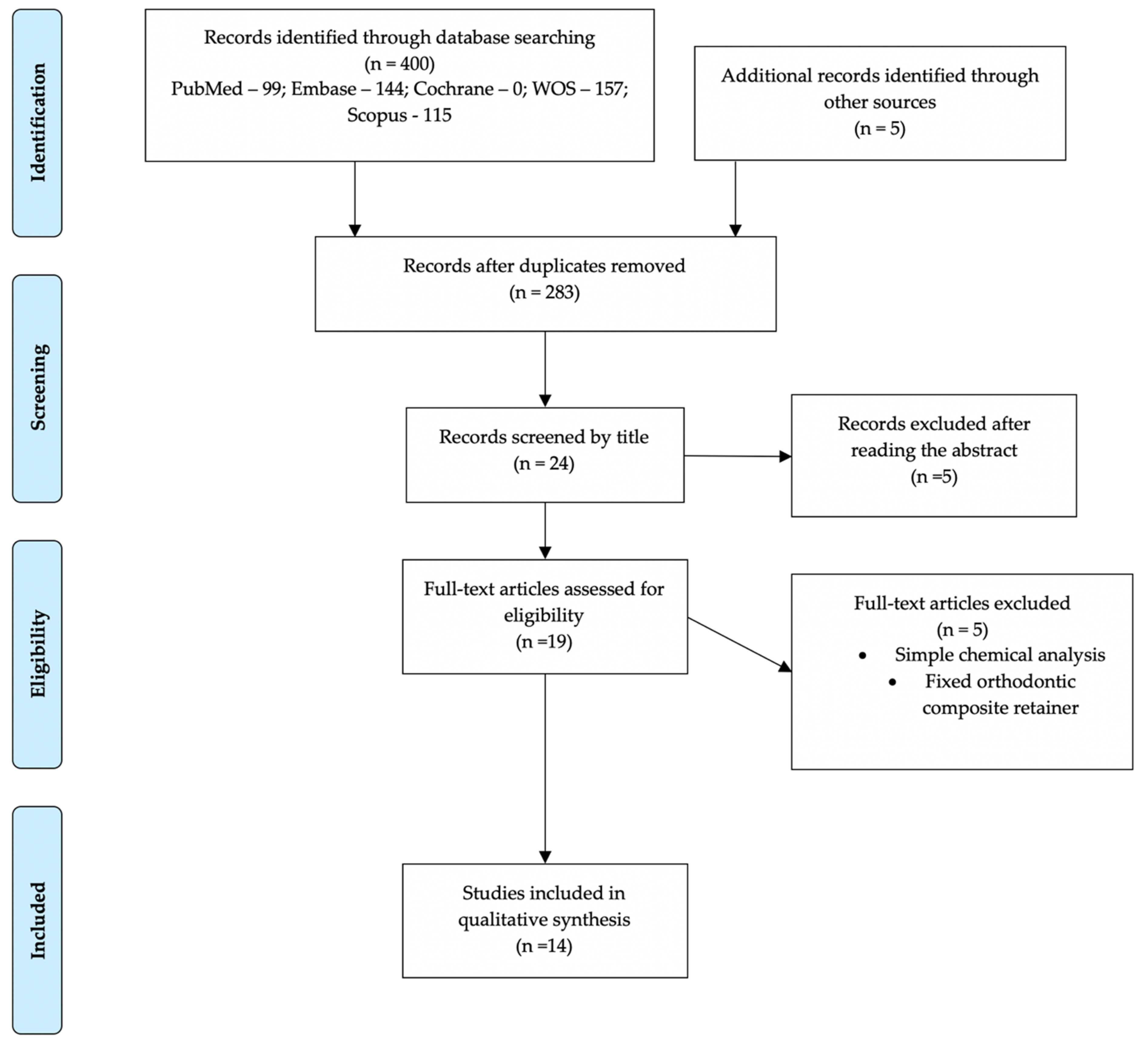
2. Materials and Methods
3. Results
3.1. Cytotoxicity Evaluation
3.1.1. In Vitro Studies
3.1.2. In Vivo Studies
3.1.3. Clinical Studies
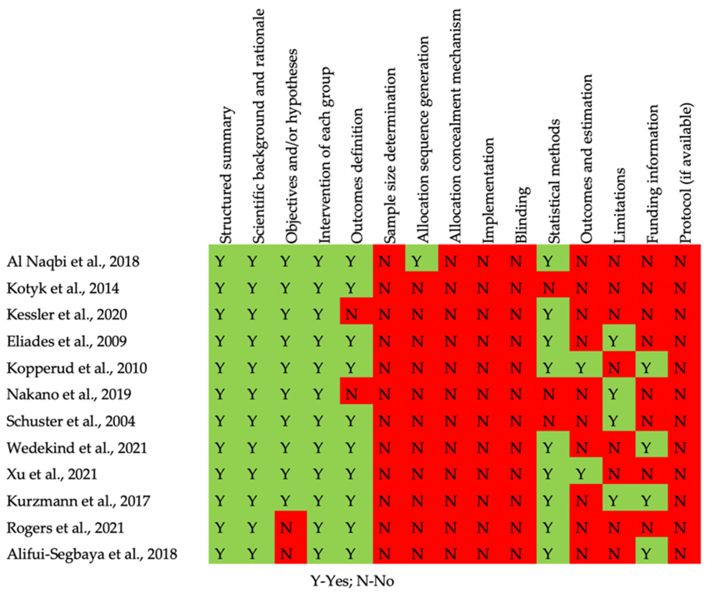
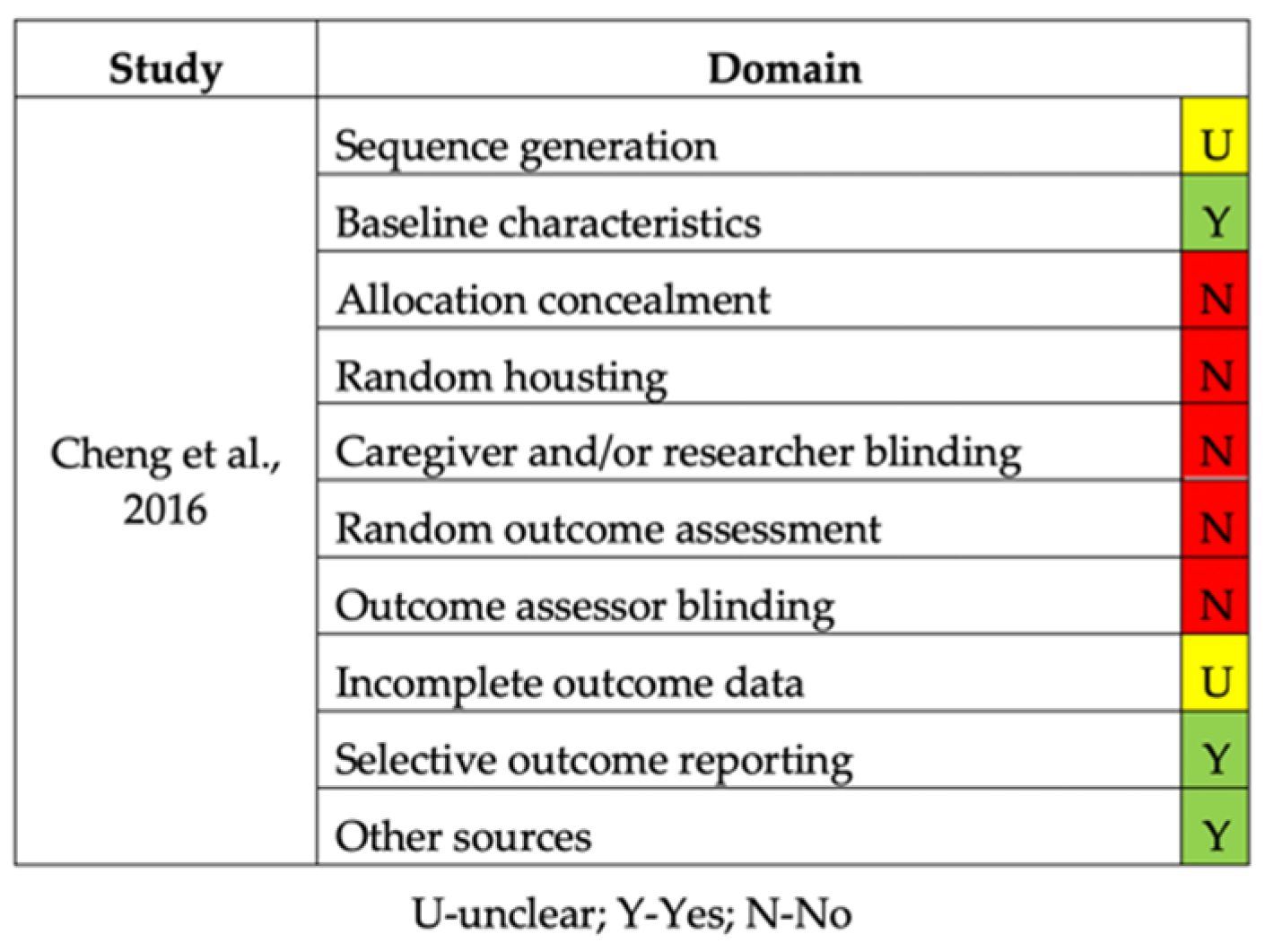
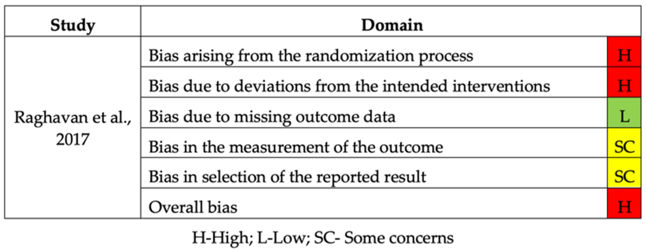
3.2. Risk of Bias
4. Discussion
5. Conclusions
Author Contributions
Funding
Institutional Review Board Statement
Informed Consent Statement
Data Availability Statement
Conflicts of Interest
References
- Kassam, S.K.; Stoops, F.R. Are clear aligners as effective as conventional fixed appliances? Evid. Based Dent. 2020, 21, 30–31. [Google Scholar] [CrossRef]
- Weir, T. Clear aligners in orthodontic treatment. Aust. Dent. J. 2017, 62, 58–62. [Google Scholar] [CrossRef] [PubMed] [Green Version]
- Maspero, C.; Tartaglia, G.M. 3D Printing of Clear Orthodontic Aligners: Where We Are and Where We Are Going. Materials 2020, 13, 5204. [Google Scholar] [CrossRef]
- Borda, A.F.; Garfinkle, J.S.; Covell, D.A.; Wang, M.; Doyle, L.; Sedgley, C.M. Outcome assessment of orthodontic clear aligner vs fixed appliance treatment in a teenage population with mild malocclusions. Angle Orthod. 2020, 90, 485–490. [Google Scholar] [CrossRef]
- Zheng, M.; Liu, R.; Ni, Z.; Yu, Z. Efficiency, effectiveness and treatment stability of clear aligners: A systematic review and meta-analysis. Orthod. Orthod. Res. 2017, 20, 127–133. [Google Scholar] [CrossRef] [PubMed]
- Tamer, I.; Oztas, E.; Marsan, G. Orthodontic Treatment with Clear Aligners and The Scientific Reality Behind Their Marketing: A Literature Review. Turkish J. Orthod. 2019, 32, 241–246. [Google Scholar] [CrossRef] [PubMed]
- Cardoso, P.C.; Espinosa, D.G.; Mecenas, P.; Flores-Mir, C.; Normando, D. Pain level between clear aligners and fixed appliances: A systematic review. Prog. Orthod. 2020, 21, 3. [Google Scholar] [CrossRef] [PubMed] [Green Version]
- Ke, Y.; Zhu, Y.; Zhu, M. A comparison of treatment effectiveness between clear aligner and fixed appliance therapies. BMC Oral Health 2019, 19, 24. [Google Scholar] [CrossRef]
- Rossini, G.; Parrini, S.; Castroflorio, T.; Deregibus, A.; Debernardi, C.L. Efficacy of clear aligners in controlling orthodontic tooth movement: A systematic review. Angle Orthod. 2015, 85, 881–889. [Google Scholar] [CrossRef] [Green Version]
- Eliades, T.; Pratsinis, H.; Athanasiou, A.E.; Eliades, G.; Kletsas, D. Cytotoxicity and estrogenicity of Invisalign appliances. Am. J. Orthod. Dentofac. Orthop. 2009, 136, 100–103. [Google Scholar] [CrossRef] [PubMed]
- Rogers, H.B.; Zhou, L.T.; Kusuhara, A.; Zaniker, E.; Shafaie, S.; Owen, B.C.; Duncan, F.E.; Woodruff, T.K. Dental resins used in 3D printing technologies release ovo-toxic leachates. Chemosphere 2021, 270, 129003. [Google Scholar] [CrossRef] [PubMed]
- Tartaglia, G.M.; Mapelli, A.; Maspero, C.; Santaniello, T.; Serafin, M.; Farronato, M.; Caprioglio, A. Direct 3D Printing of Clear Orthodontic Aligners: Current State and Future Possibilities. Materials 2021, 14, 1799. [Google Scholar] [CrossRef]
- Park, J.-H.; Lee, H.; Kim, J.-W.; Kim, J.-H. Cytocompatibility of 3D printed dental materials for temporary restorations on fibroblasts. BMC Oral Health 2020, 20, 157. [Google Scholar] [CrossRef] [PubMed]
- Saeed, F.; Muhammad, N.; Khan, A.S.; Sharif, F.; Rahim, A.; Ahmad, P.; Irfan, M. Prosthodontics dental materials: From conventional to unconventional. Mater. Sci. Eng. C 2020, 106, 110167. [Google Scholar] [CrossRef]
- Ali, U.; Karim, K.J.B.A.; Buang, N.A. A Review of the Properties and Applications of Poly (Methyl Methacrylate) (PMMA). Polym. Rev. 2015, 55, 678–705. [Google Scholar] [CrossRef]
- Aretxabaleta, M.; Xepapadeas, A.B.; Poets, C.F.; Koos, B.; Spintzyk, S. Comparison of additive and subtractive CAD/CAM materials for their potential use as Tübingen Palatal Plate: An in-vitro study on flexural strength. Addit. Manuf. 2021, 37, 101693. [Google Scholar] [CrossRef]
- Shin, J.-W.; Kim, J.-E.; Choi, Y.-J.; Shin, S.-H.; Nam, N.-E.; Shim, J.-S.; Lee, K.-W. Evaluation of the Color Stability of 3D-Printed Crown and Bridge Materials against Various Sources of Discoloration: An In Vitro Study. Materials 2020, 13, 5359. [Google Scholar] [CrossRef]
- Li, P.; Schille, C.; Schweizer, E.; Kimmerle-Müller, E.; Rupp, F.; Heiss, A.; Legner, C.; Klotz, U.E.; Geis-Gerstorfer, J.; Scheideler, L. Selection of extraction medium influences cytotoxicity of zinc and its alloys. Acta Biomater. 2019, 98, 235–245. [Google Scholar] [CrossRef]
- Atalayin, C.; Armagan, G.; Konyalioglu, S.; Kemaloglu, H.; Tezel, H.; Ergücü, Z.; Keser, A.; Dagci, T.; Önal, B. The protective effect of resveratrol against dentin bonding agents-induced cytotoxicity. Dent. Mater. J. 2015, 34, 766–773. [Google Scholar] [CrossRef] [Green Version]
- Engelmann, J.; Leyhausen, G.; Leibfritz, D.; Geurtsen, W. Effect of TEGDMA on the intracellular glutathione concentration of human gingival fibroblasts. J. Biomed. Mater. Res. 2002, 63, 746–751. [Google Scholar] [CrossRef]
- Schweikl, H.; Spagnuolo, G.; Schmalz, G. Genetic and Cellular Toxicology of Dental Resin Monomers. J. Dent. Res. 2006, 85, 870–877. [Google Scholar] [CrossRef]
- Sancar, A.; Lindsey-Boltz, L.A.; Ünsal-Kaçmaz, K.; Linn, S. Molecular Mechanisms of Mammalian DNA Repair and the DNA Damage Checkpoints. Annu. Rev. Biochem. 2004, 73, 39–85. [Google Scholar] [CrossRef] [PubMed] [Green Version]
- Samuelsen, J.T.; Dahl, J.E.; Karlsson, S.; Morisbak, E.; Becher, R. Apoptosis induced by the monomers HEMA and TEGDMA involves formation of ROS and differential activation of the MAP-kinases p38, JNK and ERK. Dent. Mater. 2007, 23, 34–39. [Google Scholar] [CrossRef]
- Faggion, C.M. Guidelines for Reporting Pre-clinical In Vitro Studies on Dental Materials. J. Evid. Based Dent. Pract. 2012, 12, 182–189. [Google Scholar] [CrossRef] [PubMed]
- Kopperud, H.M.; Kleven, I.S.; Wellendorf, H. Identification and quantification of leachable substances from polymer-based orthodontic base-plate materials. Eur. J. Orthod. 2011, 33, 26–31. [Google Scholar] [CrossRef] [Green Version]
- Nakano, H.; Kato, R.; Kakami, C.; Okamoto, H.; Mamada, K.; Maki, K. Development of Biocompatible Resins for 3D Printing of Direct Aligners. J. Photopolym. Sci. Technol. 2019, 32, 209–216. [Google Scholar] [CrossRef] [Green Version]
- Kurzmann, C.; Janjić, K.; Shokoohi-Tabrizi, H.; Edelmayer, M.; Pensch, M.; Moritz, A.; Agis, H. Evaluation of Resins for Stereolithographic 3D-Printed Surgical Guides: The Response of L929 Cells and Human Gingival Fibroblasts. Biomed. Res. Int. 2017, 2017, 4057612. [Google Scholar] [CrossRef] [PubMed] [Green Version]
- Kessler, A.; Reichl, F.-X.; Folwaczny, M.; Högg, C. Monomer release from surgical guide resins manufactured with different 3D printing devices. Dent. Mater. 2020, 36, 1486–1492. [Google Scholar] [CrossRef] [PubMed]
- Kotyk, M.W.; Wiltshire, W.A. An investigation into bisphenol-A leaching from orthodontic materials. Angle Orthod. 2014, 84, 516–520. [Google Scholar] [CrossRef]
- Al Naqbi, S.R.; Pratsinis, H.; Kletsas, D.; Eliades, T.; Athanasiou, A.E. In Vitro Assessment of Cytotoxicity and Estrogenicity of Vivera® Retainers. J. Contemp. Dent. Pract. 2018, 19, 1163–1168. [Google Scholar] [CrossRef]
- Schuster, S.; Eliades, G.; Zinelis, S.; Eliades, T.; Bradley, T.G. Structural conformation and leaching from in vitro aged and retrieved Invisalign appliances. Am. J. Orthod. Dentofac. Orthop. 2004, 126, 725–728. [Google Scholar] [CrossRef] [PubMed]
- Xu, Y.; Xepapadeas, A.B.; Koos, B.; Geis-Gerstorfer, J.; Li, P.; Spintzyk, S. Effect of post-rinsing time on the mechanical strength and cytotoxicity of a 3D printed orthodontic splint material. Dent. Mater. 2021, 37, e314–e327. [Google Scholar] [CrossRef] [PubMed]
- Wedekind, L.; Güth, J.-F.; Schweiger, J.; Kollmuss, M.; Reichl, F.-X.; Edelhoff, D.; Högg, C. Elution behavior of a 3D-printed, milled and conventional resin-based occlusal splint material. Dent. Mater. 2021, 37, 701–710. [Google Scholar] [CrossRef]
- Alifui-Segbaya, F.; Bowman, J.; White, A.R.; Varma, S.; Lieschke, G.J.; George, R. Toxicological assessment of additively manufactured methacrylates for medical devices in dentistry. Acta Biomater. 2018, 78, 64–77. [Google Scholar] [CrossRef]
- Chen, S.; Li, S.; Fang, D.; Bai, Y. Quantification of metal trace elements in orthodontic polymeric aligners and retainers by inductively coupled plasma mass spectrometry (ICP-MS). Int. J. Clin. Exp. Med. 2016, 9, 16273–16282. [Google Scholar]
- Raghavan, A.S.; Pottipalli Sathyanarayana, H.; Kailasam, V.; Padmanabhan, S. Comparative evaluation of salivary bisphenol A levels in patients wearing vacuum-formed and Hawley retainers: An in-vivo study. Am. J. Orthod. Dentofac. Orthop. 2017, 151, 471–476. [Google Scholar] [CrossRef]
- Iliadi, A.; Koletsi, D.; Papageorgiou, S.N.; Eliades, T. Safety Considerations for Thermoplastic-Type Appliances Used as Orthodontic Aligners or Retainers. A Systematic Review and Meta-Analysis of Clinical and In-Vitro Research. Materials 2020, 13, 1843. [Google Scholar] [CrossRef] [Green Version]
- Huang, D.; Xu, Y.; Lei, F.; Yu, X.; Ouyang, Z.; Chen, Y.; Jia, H.; Guo, X. Degradation of polyethylene plastic in soil and effects on microbial community composition. J. Hazard. Mater. 2021, 416, 126173. [Google Scholar] [CrossRef] [PubMed]
- Viera, J.S.C.; Marques, M.R.C.; Nazareth, M.C.; Jimenez, P.C.; Sanz-Lázaro, C.; Castro, Í.B. Are biodegradable plastics an environmental rip off? J. Hazard. Mater. 2021, 416, 125957. [Google Scholar] [CrossRef]
- Jiang, B.; Kauffman, A.E.; Li, L.; McFee, W.; Cai, B.; Weinstein, J.; Lead, J.R.; Chatterjee, S.; Scott, G.I.; Xiao, S. Health impacts of environmental contamination of micro- and nanoplastics: A review. Environ. Health Prev. Med. 2020, 25, 29. [Google Scholar] [CrossRef] [PubMed]


| Authors, Year | Study Design | Fabrication Technique | Resin Composition | Cell Line | Sample Size (n) | Test Group | Time | Assay Type | Results | Conclusions |
|---|---|---|---|---|---|---|---|---|---|---|
| Eliades T. et al, 2009 [10] | Extract Technique | Thermoformed | Invisalign appliances | Cytotoxicity Human gingival fibroblasts; Estrogenicity MCF-7: Estrogen-sensitivive MDA-MB-231 human breats adenocarcinoma—estrogen-insensitive. | 3 sets of aligners; n = 6 (96 aligner eluents per group). | Test group: invisalign at 5%, 10%, 20%; Control group: Vehicle at 5%, 10%, 20%. | 2 months | Cytotoxicity (by modification of the MTT assay); Estrogenicity (assays involved 2 cell lines: MCF-7 and MDA-MB-231). | Cytotoxicity (optical density of human gingival fibroblasts); Estrogenicity was assessed by the proliferation of MCF-7 and MDA-MB-231. | No cytotoxicity or estrogenic activity of Invisalign appliances was documented in this in vitro assay. |
| Kurzmann C. et al., 2017 [27] | Direct and indirect contact | Resins for Stereolithographic 3D-Printed | Clear resin (FLGPCL02), Dental SG resin (FLDGOR01) | L929 cell line, Human gingiva fibroblasts | n = 96-well culture plates | Test group: Clear (exposed to printed Clear resin) and Dental SG (exposed to Dental SG resin); Control group: W/O (untreated control). | 24 h | Macroscopic and scanning electron microscopy. | When exposed to the materials, the cellular activity of L929 cells and gingival fibroblasts was observed. | The impact of Clear and Dental SG resins depends on the processing stage of the material. |
| Rogers H. et al., 2021 [11] | Ex vivo + in vitro (direct and indirect) | 3D-printed using Form 2 SLA printers | Dental SG (DSG-FLDGOR01, Lot Nos. XN232N05, XK244N01, XK242N01, XK25N01, XH084N05) and Dental LT Clear (DLT-FLDLCL01, Lot Nos. XK484N02, XH043N02, XK29N02). | Mouse oocytes | n = 540 | Test group: DSG and DLT wells; Control group: polystyrene control. | 168 h | Mass spectroscopy | Exposure to DSG and DLT was proved to induce rapid mammalian oocyte degeneration in vitro. | The use of two 3DP resins revealed severe reproductive toxicity. |
| Kessler A. et al., 2020 [28] | Extract technique | 3D-printed using Rapidshape D20 II (RS), Solflex 350 (SF), Form2 (Form). | 3Delta Guide (UDMA, TMPTA, TPO); Freeprint Splint (Acrylated resin, Aliphatic urethane acrylate, TPGDA, THFMA, TPO); Fotodent Guide (BIS-EMA, Acrylresin, HEMA, HPMA, Monoester with 1,2-Propandiol, TPO); Nextdent SG (Methacrylic oligomers, Phosphine oxide); V-printed SG (BIS-EMA, UDMA, TPO). | Chemical analysis: Eluted in methanol and water for 3 days. | n = 4 | Not reported | 3 days | Finnigan Trace GC ultra gas chromatograph connected to a DSQ mass spectrometer. | The elution in methanol (total of twelve) and water (total of four) detected the release of substances. | The material and the printing device have a significant influence on the release of monomers from 3D-printed surgical guides. |
| Kotyk M. et al., 2014 [29] | Extract tecnhique | Thermoformed | Biocryl Essix (prethermoformed and thermoformed); Biocryl Retainer (prethermoformed and thermoformed); Dentsply Raintree Essix (prethermoformed), Dentsply Essix (thermoformed), Invisalign aligner (unused and used). | Chemical analysis: Eluted in artificial saliva; Bisphenol-A (BPA) leached from orthodontic materials. | n = 8 retainer materials, cut into pieces of an unspecified number. | Not reported | 2 weeks | Gas chromatography/mass spectroscopy (GC-MS). | In the first 3 days of artificial saliva immersion, BPA leaching was observed. | - BPA was found to leach from thermoformed Biocryl acrylic resin retainer material; - BPA was below the reference dose for daily intake; - evidence suggests the patient BPA exposure should be minimized or even eliminated. |
| Naqbi A. et al., 2018 [30] | Indirect contact (extract tecnhique) | Thermoformed | Vivera retainers (from the manufacturer and after retrieved from patients). | Estrogen-sensitive MCF-7 Estrogen-insensitive MDA-MB-231. | n = 12 (6 for each of the two groups; 48 aligner eluents per group). | Test group: retainers sterilized with gamma-irradiation, retainers sterilized with autoclaving; Control group: retainers not subjected to any sterilization mode. | 14 days | Cytotoxicity and Estrogenicity. | No significant proliferation of MCF-7, and MDA-MB-231 cells were induced by the three samples. | - Vivera retainers did not seem to exhibit cytotoxicity or estrogenic activity; - Vivera retainers can be used as part-time removable oral appliances following the manufacturer’s instructions. |
| Schuster S. et al., 2004 [31] | Mechanical test | Thermoformed | Invisalign appliances | Mechanical analysis | n = 10 samples of aligners before intraoral placement and after retrieval; n = 12 samples of same aligners after placement intraorally for 22hours for 2 weeks. | Not reported | 2 weeks | Reflection microscopy, FTIR, scanning electron microscopy, Vickers hardness, Gas chromatography-mass spectroscopy (GC-MS). | Retrieved Invisalign appliance shows a morphological variation (Reflection microscopy, FTIR, scanning electron microscopy, Vickers hardness). Substance leaching (GC-MS): no residual monomers or oxidative byproducts were detected. | No definitive consensus on the reactivity and biological properties can be established. |
| Xu Y. et al., 2021 [32] | Mechanical test Direct contact test + extract test | Stereolithographically (SLA) printed | Dental LT Clear resin (UDMA, HEMA, EGDMA, HPA) | L929 mouse fibroblasts | n = 12 | Not reported | Mechanical test—12 h Direct and indirect—12 h, 24 h, 72 h | Flexural strength test Scanning electron microscopy Metabolic activity. | No alterations were detected on the samples for less than 1 h. When post-rising prolonged to 12 h could be observed surface fissures. | The removal of cytotoxic methacrylate monomers by post rinsing could be achieved in 5 min. Further extending the post-rinsing time did not improve the cytocompatibility but rather reduced the flexural strength of the SLA-printed acrylic. - If the 3D printed material is mistakenly post-rinsed overnight(12 h), the resulting surface defects and strength reduction may not be acceptable. |
| Wedekind L. et al., 2021 [33] | Indirect contact | Additive manufacturing (3D-printing: SHERAprint-ortho plus); Subtractive manufacturing (SHERAeco-disc PM20); Conventional manufacturing (SHERAORTHOMER). | Polymethyl methacrylate (PMMA) (THFMA, BDDMA, TPGDA). | Human gingival fibroblasts | Not reported | Each sample eluted with water and methanol | 24 h and 72 h | GC/MS analysis XTT based cell viability assay. | With the solvent methanol, the released components exceeded the cytotoxic concentrations; In water eluates, only THFMA was determined from SHERAprint-ortho plus in concentrations of non-cytotoxic levels. | With the solvent methanol, released components from the investigated splint materials exceeded cytotoxic concentrations in HGFs calculated for a worst-case scenario in splint size. In the water eluates, only the methacrylate THFMA could be determined from SHERAprint-ortho plus in concentrations below cytotoxic levels in HGFs. Therefore, in the physiological (water/saliva) situation, a health risk is of minor relevance. |
| Alifui-Segbaya F. et al., 2018 [34] | Indirect contact (extract tecnhique) | EnvisionTec’s digital processing (DLP) and Formlab’s reverse stereolithography (SL) systems | E-Denture (ED), E-Guard (EG), Dental SG (DSG) methacrylates. | Zebrafish embryo model | n = 10 | - E-Denture (ED); - E-Guard (EG); - Dental SG (DSG) methacrylates; - control. | 96 h and 120 h | FTIR spectroscopy | Biocompatibility was influenced by physicochemical characteristics of materials. | - Despite the twofold increase in DC (%) for nTx EG, it was unsafe in zebrafish bioassays; hence there is a limited correlation between conversion rate and biological performance. - The study concludes that it is preferable to use approved materials, apposite manufacturing parameters, and post-processing techniques that together ensure optimal results for medical devices. |
| Kopperud H. et al., 2011 [25] | Extract tecnhique | Heat-cure (Orthocryl), Light-cured (Triad VLC), Thermoplastic (Biocryl C, Essix A+, Essix Embrace) resins. | Methyl methacrylate (MMA), acetonitrile, ammonium acetate, 2,4-dinitro-phenylhydrazine (DNPH), distilled water, 2-hydroxyethyl methacrylate (2-HEMA), methanol and UDMA. | Chemical analysis: Eluted in formaldehyde | n = 5 | Not reported | 10 days | Gas chromatography/mass spectroscopy (GC-MS) and liquid chromatography/mass spectrometry. | Leaching methacrylate monomers from prefabricated thermoplastic plates are lower than those from powder-and-liquid-based material and from paste material. | Orthodontic prefabricated thermoplastic plates should be preferred. |
| Nakano H. et al., 2019 [26] | Indirect contact | 3D-printed | Acrylic-epoxy hybrid light-curing resins | Not reported | n = 8 | Okamoto Chemicals (3D-1M: 1); NextDent (Ortho Clear); ISO20795-2 | 24 h and 72 h | Cellular toxicity LDH-test Cell Viability WST1 test Mechanical experiments Stereolithography. | Have successfully developed a 3D biocompatible resin, without cellular toxicity but with not yet ideal mechanical properties. | Achieved a biocompatible 3D-printed resin that releases no toxic materials to humans or the environment. |
| Authors, Year | Study Design | Sample Size (n) | Test Groups | Fabrication Technique | Resin Composition | Outcome Time | Study Measure Outcome | Results | Conclusions |
|---|---|---|---|---|---|---|---|---|---|
| Chen S. et al., 2016 [35] | In vivo | Mini-screw implant + thermoplastic sample Wistar (n = 80). | Test group of aligner (n = 30); Test group of retainer (n = 30); Control group (n = 10); Blank group (n = 10). | Thermoformed | - Invisalign Smart Track aligners; - Erkodur retainers. | T1:28 days; T2: 56 days; T3: 112 days. | 0.5 mL blood samples (rat orital vein); Inductively coupled plasma mass spectrometry (ICP-MS). | - Al, Ti, V, Cr, Fe, Co, Ni, Cu, Zn were detected in polymeric retainers; - Al, Ni, Zn, Sn were detected in polymeric aligners. | - The metal elements in polymeric materials evaluated in blood did not exceed toxic values; - The identified element levels decrease after 2 weeks. |
| Authors, Year | Study Design | Sample Size (n, Sex) | Control Group | Intervention Group (Time of Use) | Fabrication Technique | Resin Composition | Outcome Time (Hours) | Study Measure Outcome | Results | Conclusions |
|---|---|---|---|---|---|---|---|---|---|---|
| Raghavan A. et al., 2017 [36] | RCT | n = 45: G1(n = 15); G2(n = 15); G3(n = 15. Sex: not reported | Not reported | T0: before placement; T1: 1h after placement; T2: 7 days after placement; T3: 30 days after placement. | G1: Biostar vacuum thermoforming system (VFR); G2: Hawley retainerHeat cure method; G3: Hawley retainerChemical cure method (in both arches). | G1: Essix ACE Plastic Vaccum-formed retainers (VFRs); G2: DPI Heat CureHawley retainers (compression molding technique); G3: DPI Cold Curechemical cure (“sprinkle” on technique). | 180 saliva samples T0: Group 1—0.00001 ± 0.0001, Group 2—0.00006 ± 0.00004, Group 3—0.00009 ± 0.00006; T1: Group 1—1.20236 ± 0.35643, Group 2—0.00091 ± 0.00081, Group 3—0.06031 ± 0.02550; T2: Group 1—2.38420 ± 1.79714, Group 2—0.00045 ± 0.00008, Group 3—0.00363 ± 0.00050; T3: Group 1—0.020396 ± 0.08709, Group 2—0.00067 ± 0.001410, Group 3—0.00934 ± 0.00237. | BPA levels in the saliva | BPA levels: G1: T1 > T0 (+1.20 ppm); T2 > T1 (+1.18 ppm); T3 < T2 (−2.18 ppm). G2 and G3: T1 > T0; T2 < T3; T3 > T2. | - Increases BPA levels in saliva in all groups after placement of the retainers; - BPA levels were found to be larger in VFRs, followed by Hawley retainers by chemical cure, and finally Hawley retainers by heat cure; - VFRs increase BPA levels after placement to 1 week but decrease after 1 month; - Hawleys retainers (heat and chemical) decrease after placement to 1 week but increase after 1 month. |
Publisher’s Note: MDPI stays neutral with regard to jurisdictional claims in published maps and institutional affiliations. |
© 2022 by the authors. Licensee MDPI, Basel, Switzerland. This article is an open access article distributed under the terms and conditions of the Creative Commons Attribution (CC BY) license (https://creativecommons.org/licenses/by/4.0/).
Share and Cite
Francisco, I.; Paula, A.B.; Ribeiro, M.; Marques, F.; Travassos, R.; Nunes, C.; Pereira, F.; Marto, C.M.; Carrilho, E.; Vale, F. The Biological Effects of 3D Resins Used in Orthodontics: A Systematic Review. Bioengineering 2022, 9, 15. https://doi.org/10.3390/bioengineering9010015
Francisco I, Paula AB, Ribeiro M, Marques F, Travassos R, Nunes C, Pereira F, Marto CM, Carrilho E, Vale F. The Biological Effects of 3D Resins Used in Orthodontics: A Systematic Review. Bioengineering. 2022; 9(1):15. https://doi.org/10.3390/bioengineering9010015
Chicago/Turabian StyleFrancisco, Inês, Anabela Baptista Paula, Madalena Ribeiro, Filipa Marques, Raquel Travassos, Catarina Nunes, Flávia Pereira, Carlos Miguel Marto, Eunice Carrilho, and Francisco Vale. 2022. "The Biological Effects of 3D Resins Used in Orthodontics: A Systematic Review" Bioengineering 9, no. 1: 15. https://doi.org/10.3390/bioengineering9010015