Next Issue
Volume 9, February
Previous Issue
Volume 8, December


Bioengineering, Volume 9, Issue 1 (January 2022) – 41 articles

Cover Story (view full-size image): 
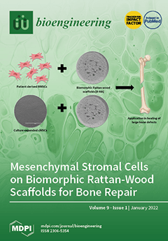
Bone healing post-trauma presents significant health challenges. Impaired bone healing may result in severe complications requiring multiple surgical interventions. The use of biomaterial scaffolds to aid bone repair and healing has been found to be helpful in these situations. Biomorphic rattan-wood scaffolds (B-HA) present characteristics resembling the structure of the bone. Our study evaluated the ability of donor-derived bone marrow mesenchymal stromal cells (BMSCs) and culture-expanded mesenchymal stromal cells (cMSCs) to attach, survive and express their genes on these B-HA scaffolds to potentially aid bone repair. View this paper
  • Issues are regarded as officially published after their release is announced to the table of contents alert mailing list.
  • You may sign up for e-mail alerts to receive table of contents of newly released issues.
  • PDF is the official format for papers published in both, html and pdf forms. To view the papers in pdf format, click on the "PDF Full-text" link, and use the free Adobe Reader to open them.
Order results
Result details
Section
Select all
Export citation of selected articles as:
30 pages, 814 KiB  
Review
Targeting Ocular Drug Delivery: An Examination of Local Anatomy and Current Approaches
by Emily Dosmar, Julia Walsh, Michael Doyel, Katlynn Bussett, Adekite Oladipupo, Sabri Amer and Katherine Goebel
Bioengineering 2022, 9(1), 41; https://doi.org/10.3390/bioengineering9010041 - 17 Jan 2022
Cited by 24 | Viewed by 6165
Abstract
Ocular drug delivery remains the focus of much modern research. Primary routes of administration include the surface, the intravitreal space, the subretinal space, and the subconjunctival space, each with its own series of unique challenges, limitations, and advantages. Each of these approaches requires [...] Read more.
Ocular drug delivery remains the focus of much modern research. Primary routes of administration include the surface, the intravitreal space, the subretinal space, and the subconjunctival space, each with its own series of unique challenges, limitations, and advantages. Each of these approaches requires careful consideration of the local anatomy, physical barriers, and key cells as well as the interface between the anatomy and the drug or drug system being delivered. While least invasive, the topical route poses a challenge with the many physical barriers that prevent drug penetration into the eye; while injection into the intravitreal, subretinal, and subconjunctival spaces are direct and targeted but limited due to the many internal clearance mechanisms and potential for damage to the eye. Polymeric-based, sustained-release drug delivery systems have been identified as a potential solution to many of these challenges; however, the design and successful implementation of a sustained-release system that is well-tolerated, bioactive, biocompatible, and degradable remains, in many cases, only in the early stages. The drugs and biomaterials in question also require special attention as small chemical changes could result in vastly different outcomes. This paper explores the anatomy and key cells of these four primary drug delivery routes as well as the interface between drug and drug delivery systems and the anatomy, reviewing the recent developments and current state of research in each area. Finally, this paper also examines the frequently used drugs and biomaterials found in ocular drug delivery and summarizes the primary interactions observed. Full article
(This article belongs to the Topic Human Anatomy and Pathophysiology)
Show Figures

Figure 1

22 pages, 4882 KiB  
Article
In Silico Study to Enhance Delivery Efficiency of Charged Nanoscale Nasal Spray Aerosols to the Olfactory Region Using External Magnetic Fields
by Benjamin Li and Yu Feng
Bioengineering 2022, 9(1), 40; https://doi.org/10.3390/bioengineering9010040 - 16 Jan 2022
Cited by 2 | Viewed by 2772
Abstract
Various factors and challenges are involved in efficiently delivering drugs using nasal sprays to the olfactory region to treat central nervous system diseases. In this study, computational fluid dynamics was used to simulate nasal drug delivery to (1) examine effects on drug deposition [...] Read more.
Various factors and challenges are involved in efficiently delivering drugs using nasal sprays to the olfactory region to treat central nervous system diseases. In this study, computational fluid dynamics was used to simulate nasal drug delivery to (1) examine effects on drug deposition when various external magnetic fields are applied to charged particles, (2) comprehensively study effects of multiple parameters (i.e., particle aerodynamic diameter; injection velocity magnitude, angle, and position; magnetic force strength and direction), and (3) determine how to achieve the optimal delivery efficiency to the olfactory epithelium. The Reynolds-averaged Navier–Stokes equations governed airflow, with a realistic inhalation waveform implemented at the nostrils. Particle trajectories were modeled using the one-way coupled Euler–Lagrange model. A current-carrying wire generated a magnetic field to apply force on charged particles and direct them to the olfactory region. Once drug particles reached the olfactory region, their diffusion through mucus to the epithelium was calculated analytically. Particle aerodynamic diameter, injection position, and magnetic field strength were found to be interconnected in their effects on delivery efficiency. Specific combinations of these parameters achieved over 65-fold higher drug delivery efficiency compared with uniform injections with no magnetic fields. The insight gained suggests how to integrate these factors to achieve the optimal efficiency. Full article
(This article belongs to the Special Issue Computational Fluid Dynamics in Medicine and Biology)
Show Figures

Figure 1

21 pages, 4695 KiB  
Article
Online Measurement System for Dynamic Flow Bioreactors to Study Barrier Integrity of hiPSC-Based Blood–Brain Barrier In Vitro Models
by Jihyoung Choi, Sanjana Mathew, Sabrina Oerter, Antje Appelt-Menzel, Jan Hansmann and Tobias Schmitz
Bioengineering 2022, 9(1), 39; https://doi.org/10.3390/bioengineering9010039 - 16 Jan 2022
Cited by 8 | Viewed by 3251
Abstract
Electrochemical impedance spectroscopy (EIS) is a noninvasive, reliable, and efficient method to analyze the barrier integrity of in vitro tissue models. This well-established tool is used most widely to quantify the transendothelial/epithelial resistance (TEER) of Transwell-based models cultured under static conditions. However, dynamic [...] Read more.
Electrochemical impedance spectroscopy (EIS) is a noninvasive, reliable, and efficient method to analyze the barrier integrity of in vitro tissue models. This well-established tool is used most widely to quantify the transendothelial/epithelial resistance (TEER) of Transwell-based models cultured under static conditions. However, dynamic culture in bioreactors can achieve advanced cell culture conditions that mimic a more tissue-specific environment and stimulation. This requires the development of culture systems that also allow for the assessment of barrier integrity under dynamic conditions. Here, we present a bioreactor system that is capable of the automated, continuous, and non-invasive online monitoring of cellular barrier integrity during dynamic culture. Polydimethylsiloxane (PDMS) casting and 3D printing were used for the fabrication of the bioreactors. Additionally, attachable electrodes based on titanium nitride (TiN)-coated steel tubes were developed to perform EIS measurements. In order to test the monitored bioreactor system, blood–brain barrier (BBB) in vitro models derived from human-induced pluripotent stem cells (hiPSC) were cultured for up to 7 days. We applied equivalent electrical circuit fitting to quantify the electrical parameters of the cell layer and observed that TEER gradually decreased over time from 2513 Ω·cm2 to 285 Ω·cm2, as also specified in the static control culture. Our versatile system offers the possibility to be used for various dynamic tissue cultures that require a non-invasive monitoring system for barrier integrity. Full article
Show Figures

Figure 1

21 pages, 4607 KiB  
Article
Alginate Hydrogel Microtubes for Salivary Gland Cell Organization and Cavitation
by Matthew Jorgensen, Pujhitha Ramesh, Miriam Toro, Emily Evans, Nicholas Moskwa, Xulang Zhang, Susan T. Sharfstein, Melinda Larsen and Yubing Xie
Bioengineering 2022, 9(1), 38; https://doi.org/10.3390/bioengineering9010038 - 15 Jan 2022
Cited by 10 | Viewed by 3383
Abstract
Understanding the different regulatory functions of epithelial and mesenchymal cell types in salivary gland development and cellular organization is essential for proper organoid formation and salivary gland tissue regeneration. Here, we demonstrate a biocompatible platform using pre-formed alginate hydrogel microtubes to facilitate direct [...] Read more.
Understanding the different regulatory functions of epithelial and mesenchymal cell types in salivary gland development and cellular organization is essential for proper organoid formation and salivary gland tissue regeneration. Here, we demonstrate a biocompatible platform using pre-formed alginate hydrogel microtubes to facilitate direct epithelial–mesenchymal cell interaction for 3D salivary gland cell organization, which allows for monitoring cellular organization while providing a protective barrier from cell-cluster loss during medium changes. Using mouse salivary gland ductal epithelial SIMS cells as the epithelial model cell type and NIH 3T3 fibroblasts or primary E16 salivary mesenchyme cells as the stromal model cell types, self-organization from epithelial–mesenchymal interaction was examined. We observed that epithelial and mesenchymal cells undergo aggregation on day 1, cavitation by day 4, and generation of an EpCAM-expressing epithelial cell layer as early as day 7 of the co-culture in hydrogel microtubes, demonstrating the utility of hydrogel microtubes to facilitate heterotypic cell–cell interactions to form cavitated organoids. Thus, pre-formed alginate microtubes are a promising co-culture method for further understanding epithelial and mesenchymal interaction during tissue morphogenesis and for future practical applications in regenerative medicine. Full article
(This article belongs to the Special Issue Material and Engineering-Based Approaches for Organoids)
Show Figures

Graphical abstract

15 pages, 2784 KiB  
Article
Comparative Effects of Basic Fibroblast Growth Factor Delivery or Voluntary Exercise on Muscle Regeneration after Volumetric Muscle Loss
by Caroline Hu, Bugra Ayan, Gladys Chiang, Alex H. P. Chan, Thomas A. Rando and Ngan F. Huang
Bioengineering 2022, 9(1), 37; https://doi.org/10.3390/bioengineering9010037 - 14 Jan 2022
Cited by 7 | Viewed by 3123
Abstract
Volumetric muscle loss (VML) is associated with irreversibly impaired muscle function due to traumatic injury. Experimental approaches to treat VML include the delivery of basic fibroblast growth factor (bFGF) or rehabilitative exercise. The objective of this study was to compare the effects of [...] Read more.
Volumetric muscle loss (VML) is associated with irreversibly impaired muscle function due to traumatic injury. Experimental approaches to treat VML include the delivery of basic fibroblast growth factor (bFGF) or rehabilitative exercise. The objective of this study was to compare the effects of spatially nanopatterned collagen scaffold implants with either bFGF delivery or in conjunction with voluntary exercise. Aligned nanofibrillar collagen scaffold bundles were adsorbed with bFGF, and the bioactivity of bFGF-laden scaffolds was examined by skeletal myoblast or endothelial cell proliferation. The therapeutic efficacy of scaffold implants with either bFGF release or exercise was examined in a murine VML model. Our results show an initial burst release of bFGF from the scaffolds, followed by a slower release over 21 days. The released bFGF induced myoblast and endothelial cell proliferation in vitro. After 3 weeks of implantation in a mouse VML model, twitch force generation was significantly higher in mice treated with bFGF-laden scaffolds compared to bFGF-laden scaffolds with exercise. However, myofiber density was not significantly improved with bFGF scaffolds or voluntary exercise. In contrast, the scaffold implant with exercise induced more re-innervation than all other groups. These results highlight the differential effects of bFGF and exercise on muscle regeneration. Full article
(This article belongs to the Special Issue Extracellular Matrix in Musculoskeletal Regeneration)
Show Figures

Figure 1

17 pages, 4136 KiB  
Article
Flexible Strain-Sensitive Silicone-CNT Sensor for Human Motion Detection
by Natalia A. Demidenko, Artem V. Kuksin, Victoria V. Molodykh, Evgeny S. Pyankov, Levan P. Ichkitidze, Victoria A. Zaborova, Alexandr A. Tsymbal, Svetlana A. Tkachenko, Hassan Shafaei, Ekaterina Diachkova and Alexander Yu. Gerasimenko
Bioengineering 2022, 9(1), 36; https://doi.org/10.3390/bioengineering9010036 - 13 Jan 2022
Cited by 20 | Viewed by 3925
Abstract
This article describes the manufacturing technology of biocompatible flexible strain-sensitive sensor based on Ecoflex silicone and multi-walled carbon nanotubes (MWCNT). The sensor demonstrates resistive behavior. Structural, electrical, and mechanical characteristics are compared. It is shown that laser radiation significantly reduces the resistance of [...] Read more.
This article describes the manufacturing technology of biocompatible flexible strain-sensitive sensor based on Ecoflex silicone and multi-walled carbon nanotubes (MWCNT). The sensor demonstrates resistive behavior. Structural, electrical, and mechanical characteristics are compared. It is shown that laser radiation significantly reduces the resistance of the material. Through laser radiation, electrically conductive networks of MWCNT are formed in a silicone matrix. The developed sensor demonstrates highly sensitive characteristics: gauge factor at 100% elongation −4.9, gauge factor at 90° bending −0.9%/deg, stretchability up to 725%, tensile strength 0.7 MPa, modulus of elasticity at 100% 46 kPa, and the temperature coefficient of resistance in the range of 30–40 °C is −2 × 10⁻³. There is a linear sensor response (with 1 ms response time) with a low hysteresis of ≤3%. An electronic unit for reading and processing sensor signals based on the ATXMEGA8E5-AU microcontroller has been developed. The unit was set to operate the sensor in the range of electrical resistance 5–150 kOhm. The Bluetooth module made it possible to transfer the received data to a personal computer. Currently, in the field of wearable technologies and health monitoring, a vital need is the development of flexible sensors attached to the human body to track various indicators. By integrating the sensor with the joints of the human hand, effective movement sensing has been demonstrated. Full article
Show Figures

Graphical abstract

19 pages, 2333 KiB  
Article
The Impact of the Extracellular Matrix Environment on Sost Expression by the MLO-Y4 Osteocyte Cell Line
by Robert T. Brady, Fergal J. O’Brien and David A. Hoey
Bioengineering 2022, 9(1), 35; https://doi.org/10.3390/bioengineering9010035 - 13 Jan 2022
Cited by 3 | Viewed by 3188
Abstract
Bone is a dynamic organ that can adapt its structure to meet the demands of its biochemical and biophysical environment. Osteocytes form a sensory network throughout the tissue and orchestrate tissue adaptation via the release of soluble factors such as a sclerostin. Osteocyte [...] Read more.
Bone is a dynamic organ that can adapt its structure to meet the demands of its biochemical and biophysical environment. Osteocytes form a sensory network throughout the tissue and orchestrate tissue adaptation via the release of soluble factors such as a sclerostin. Osteocyte physiology has traditionally been challenging to investigate due to the uniquely mineralized extracellular matrix (ECM) of bone leading to the development of osteocyte cell lines. Importantly, the most widely researched and utilized osteocyte cell line: the MLO-Y4, is limited by its inability to express sclerostin (Sost gene) in typical in-vitro culture. We theorised that culture in an environment closer to the in vivo osteocyte environment could impact on Sost expression. Therefore, this study investigated the role of composition and dimensionality in directing Sost expression in MLO-Y4 cells using collagen-based ECM analogues. A significant outcome of this study is that MLO-Y4 cells, when cultured on a hydroxyapatite (HA)-containing two-dimensional (2D) film analogue, expressed Sost. Moreover, three-dimensional (3D) culture within HA-containing collagen scaffolds significantly enhanced Sost expression, demonstrating the impact of ECM composition and dimensionality on MLO-Y4 behaviour. Importantly, in this bone mimetic ECM environment, Sost expression was found to be comparable to physiological levels. Lastly, MLO-Y4 cells cultured in these novel conditions responded accordingly to fluid flow stimulation with a decrease in expression. This study therefore presents a novel culture system for the MLO-Y4 osteocyte cell line, ensuring the expression of an important osteocyte specific gene, Sost, overcoming a major limitation of this model. Full article
Show Figures

Figure 1

18 pages, 2152 KiB  
Article
Riboflavin and Its Effect on Dentin Bond Strength: Considerations for Clinical Applicability—An In Vitro Study
by Franziska Beck and Nicoleta Ilie
Bioengineering 2022, 9(1), 34; https://doi.org/10.3390/bioengineering9010034 - 13 Jan 2022
Cited by 5 | Viewed by 2217
Abstract
Bioactive collagen crosslinkers propose to render the dentin hybrid layer less perceptive to hydrolytic challenge. This study aims to evaluate whether bond strength of dental resin composite to dentin benefits from riboflavin (RB)-sensitized crosslinking when used in a clinically applicable protocol. A total [...] Read more.
Bioactive collagen crosslinkers propose to render the dentin hybrid layer less perceptive to hydrolytic challenge. This study aims to evaluate whether bond strength of dental resin composite to dentin benefits from riboflavin (RB)-sensitized crosslinking when used in a clinically applicable protocol. A total of 300 human dentin specimens were prepared consistent with the requirements for a macro-shear bond test. RB was applied on dentin, either incorporated in the primer (RBp) of a two-step self-etch adhesive or as an aqueous solution (RBs) before applying the adhesive, and blue light from a commercial polymerization device was used for RB photoactivation. Bonding protocol executed according to the manufacturer’s information served as control. Groups (n = 20) were tested after 1 week, 1 month, 3 months, 6 months or 1 year immersion times (37 °C, distilled water). The different application methods of RB significantly influenced bond strength (p < 0.001) with a medium impact (η2p = 0.119). After 1 year immersion, post hoc analysis identified a significant advantage for RB groups compared to RBp (p = 0.018), which is attributed to a pH-/solvent-dependent efficiency of RB-sensitized crosslinking, stressing the importance of formulation adjustments. We developed an application protocol for RB-sensitized crosslinking with emphasis on clinical applicability to test its performance against a gold-standard adhesive, and are confident that, with a few adjustments to the application solution, RB-sensitized crosslinking can improve the longevity of adhesive restorations in clinics. Full article
Show Figures

Figure 1

13 pages, 652 KiB  
Review
Wearables for Biomechanical Performance Optimization and Risk Assessment in Industrial and Sports Applications
by Sam McDevitt, Haley Hernandez, Jamison Hicks, Russell Lowell, Hamza Bentahaikt, Reuben Burch, John Ball, Harish Chander, Charles Freeman, Courtney Taylor and Brock Anderson
Bioengineering 2022, 9(1), 33; https://doi.org/10.3390/bioengineering9010033 - 13 Jan 2022
Cited by 18 | Viewed by 4964
Abstract
Wearable technologies are emerging as a useful tool with many different applications. While these devices are worn on the human body and can capture numerous data types, this literature review focuses specifically on wearable use for performance enhancement and risk assessment in industrial- [...] Read more.
Wearable technologies are emerging as a useful tool with many different applications. While these devices are worn on the human body and can capture numerous data types, this literature review focuses specifically on wearable use for performance enhancement and risk assessment in industrial- and sports-related biomechanical applications. Wearable devices such as exoskeletons, inertial measurement units (IMUs), force sensors, and surface electromyography (EMG) were identified as key technologies that can be used to aid health and safety professionals, ergonomists, and human factors practitioners improve user performance and monitor risk. IMU-based solutions were the most used wearable types in both sectors. Industry largely used biomechanical wearables to assess tasks and risks wholistically, which sports often considered the individual components of movement and performance. Availability, cost, and adoption remain common limitation issues across both sports and industrial applications. Full article
Show Figures

Figure 1

14 pages, 3472 KiB  
Article
A Bioprinted Heart-on-a-Chip with Human Pluripotent Stem Cell-Derived Cardiomyocytes for Drug Evaluation
by Alan Faulkner-Jones, Victor Zamora, Maria P. Hortigon-Vinagre, Wenxing Wang, Marcus Ardron, Godfrey L. Smith and Wenmiao Shu
Bioengineering 2022, 9(1), 32; https://doi.org/10.3390/bioengineering9010032 - 13 Jan 2022
Cited by 17 | Viewed by 3851
Abstract
In this work, we show that valve-based bioprinting induces no measurable detrimental effects on human-induced pluripotent stem cell-derived cardiomyocytes (hiPSC-CMs). The aim of the current study was three-fold: first, to assess the response of hiPSC-CMs to several hydrogel formulations by measuring electrophysiological function; [...] Read more.
In this work, we show that valve-based bioprinting induces no measurable detrimental effects on human-induced pluripotent stem cell-derived cardiomyocytes (hiPSC-CMs). The aim of the current study was three-fold: first, to assess the response of hiPSC-CMs to several hydrogel formulations by measuring electrophysiological function; second, to customise a new microvalve-based cell printing mechanism in order to deliver hiPSC-CMs suspensions, and third, to compare the traditional manual pipetting cell-culture method and cardiomyocytes dispensed with the bioprinter. To achieve the first and third objectives, iCell2 (Cellular Dynamics International) hiPSC-CMs were used. The effects of well-known drugs were tested on iCell2 cultured by manual pipetting and bioprinting. Despite the results showing that hydrogels and their cross-linkers significantly reduced the electrophysiological performance of the cells compared with those cultured on fibronectin, the bio-ink droplets containing a liquid suspension of live cardiomyocytes proved to be an alternative to standard manual handling and could reduce the number of cells required for drug testing, with no significant differences in drug-sensitivity between both approaches. These results provide a basis for the development of a novel bioprinter with nanolitre resolution to decrease the required number of cells and to automate the cell plating process. Full article
(This article belongs to the Special Issue Advances in 3D Printing Tissue Engineering)
Show Figures

Graphical abstract

12 pages, 1283 KiB  
Article
Maxillary Transverse Deficit: A Retrospective Study of Two Biologically Oriented Devices through a Digital Workflow
by Graziano Montaruli, Simona Virgilio, Michele Laurenziello, Michele Tepedino and Domenico Ciavarella
Bioengineering 2022, 9(1), 31; https://doi.org/10.3390/bioengineering9010031 - 12 Jan 2022
Cited by 3 | Viewed by 2629
Abstract
The aim of this retrospective study was to compare the efficiency of two biologically oriented devices in achieving maxillary expansion: Rapid Palatal Expander (RPE) and Nitanium Palatal Expander-2 (NPE-2). Thirty-six subjects, divided in two equal groups, were included in this study. Maxillary dental [...] Read more.
The aim of this retrospective study was to compare the efficiency of two biologically oriented devices in achieving maxillary expansion: Rapid Palatal Expander (RPE) and Nitanium Palatal Expander-2 (NPE-2). Thirty-six subjects, divided in two equal groups, were included in this study. Maxillary dental arches were scanned using Trios 3 shape®, in order to perform a digital analysis of 3D models. The models were analyzed using Autodesk Fusion 360® and Meshmixer®. All data obtained from analysis of pre-treatment and post-treatment models were processed using Prism® software. The anterior arch width, the posterior arch width, the palate height, and palatal surface were measured to evaluate differences between the devices. A D’Agostino–Pearson normality test was done to check the data. A non-parametric t-test was used to compare the anterior and posterior arch width between the two groups, while a parametric t-test was used to compare the palatal height measurements between the two groups. The p-value was calculated. The limit value fixed was 0.05. Palatal width and surface showed a significant increase in both groups, but no significant changes in palatal height were found. The data processed showed that there were no significant differences between the devices (ΔREP−ΔNPE) in variation of anterior arch width, there were no significant differences in variation of posterior arch width and there were no significant differences in variation of palatal height. The comparison between the two groups showed that both methods were equally effective in correcting transverse defect. Full article
Show Figures

Figure 1

16 pages, 854 KiB  
Review
Metallurgical Tests in Endodontics: A Narrative Review
by Alessio Zanza, Marco Seracchiani, Rodolfo Reda, Gabriele Miccoli, Luca Testarelli and Dario Di Nardo
Bioengineering 2022, 9(1), 30; https://doi.org/10.3390/bioengineering9010030 - 12 Jan 2022
Cited by 23 | Viewed by 3230
Abstract
Since there are no reviews of the literature on this theme, the aim of this narrative review is to summarize the metallurgical tests used in endodontics, pointing out their functional use and their pros and cons and giving readers a user-friendly guide to [...] Read more.
Since there are no reviews of the literature on this theme, the aim of this narrative review is to summarize the metallurgical tests used in endodontics, pointing out their functional use and their pros and cons and giving readers a user-friendly guide to serve as an orientation aid in the plethora of metallurgical tests. With this purpose, a literature search for articles published between January 2001 and December 2021 was conducted, using the electronic database PubMed to collect all published articles regarding the metallurgical tests used in endodontics for the evaluation of NiTi rotary instruments. The search was conducted using the following keywords: “metallurgy”, “differential scanning calorimetry” (DSC), “X-ray diffraction” (XRD), “atomic force microscopy” (AFM), “energy-dispersive X-ray spectroscopy” (EDS), “focused ion beam analysis” (FIB) and “Auger electron spectroscopy” (AES) combined with the term “endodontics” or “NiTi rotary instruments”. Considering the inclusion and exclusion criteria, of the 248 articles found, only 81 were included in the narrative review. According to the results, more than 50% of the selected articles were published in one of the two most relevant journals in endodontics: International Endodontic Journal (22.2%) and Journal of Endodontics (29.6%). The most popular metallurgical test was DSC, with 43 related articles, followed by EDS (33 articles), AFM (22 articles) and XRD (21 articles). Few studies were conducted using other tests such as FIB (2 articles), micro-Raman spectroscopy (4 articles), metallographic analysis (7 articles) and Auger electron spectroscopy (2 articles). Full article
Show Figures

Figure 1

16 pages, 2664 KiB  
Article
Radiation-Induced Alterations in Proliferation, Migration, and Adhesion in Lens Epithelial Cells and Implications for Cataract Development
by Graysen Vigneux, Jake Pirkkanen, Taylor Laframboise, Hallie Prescott, Sujeenthar Tharmalingam and Christopher Thome
Bioengineering 2022, 9(1), 29; https://doi.org/10.3390/bioengineering9010029 - 12 Jan 2022
Cited by 11 | Viewed by 2523
Abstract
The lens of the eye is one of the most radiosensitive tissues. Although the exact mechanism of radiation-induced cataract development remains unknown, altered proliferation, migration, and adhesion have been proposed as factors. Lens epithelial cells were exposed to X-rays (0.1–2 Gy) and radiation [...] Read more.
The lens of the eye is one of the most radiosensitive tissues. Although the exact mechanism of radiation-induced cataract development remains unknown, altered proliferation, migration, and adhesion have been proposed as factors. Lens epithelial cells were exposed to X-rays (0.1–2 Gy) and radiation effects were examined after 12 h and 7 day. Proliferation was quantified using an MTT assay, migration was measured using a Boyden chamber and wound-healing assay, and adhesion was assessed on three extracellular matrices. Transcriptional changes were also examined using RT-qPCR for a panel of genes related to these processes. In general, a nonlinear radiation response was observed, with the greatest effects occurring at a dose of 0.25 Gy. At this dose, a reduction in proliferation occurred 12 h post irradiation (82.06 ± 2.66%), followed by an increase at 7 day (116.16 ± 3.64%). Cell migration was increased at 0.25 Gy, with rates 121.66 ± 6.49% and 232.78 ± 22.22% greater than controls at 12 h and 7 day respectively. Cell adhesion was consistently reduced above doses of 0.25 Gy. Transcriptional alterations were identified at these same doses in multiple genes related to proliferation, migration, and adhesion. Overall, this research began to elucidate the functional changes that occur in lens cells following radiation exposure, thereby providing a better mechanistic understanding of radiation-induced cataract development. Full article
Show Figures

Figure 1

24 pages, 2436 KiB  
Review
Organ on Chip Technology to Model Cancer Growth and Metastasis
by Giorgia Imparato, Francesco Urciuolo and Paolo Antonio Netti
Bioengineering 2022, 9(1), 28; https://doi.org/10.3390/bioengineering9010028 - 11 Jan 2022
Cited by 23 | Viewed by 5256
Abstract
Organ on chip (OOC) has emerged as a major technological breakthrough and distinct model system revolutionizing biomedical research and drug discovery by recapitulating the crucial structural and functional complexity of human organs in vitro. OOC are rapidly emerging as powerful tools for oncology [...] Read more.
Organ on chip (OOC) has emerged as a major technological breakthrough and distinct model system revolutionizing biomedical research and drug discovery by recapitulating the crucial structural and functional complexity of human organs in vitro. OOC are rapidly emerging as powerful tools for oncology research. Indeed, Cancer on chip (COC) can ideally reproduce certain key aspects of the tumor microenvironment (TME), such as biochemical gradients and niche factors, dynamic cell–cell and cell–matrix interactions, and complex tissue structures composed of tumor and stromal cells. Here, we review the state of the art in COC models with a focus on the microphysiological systems that host multicellular 3D tissue engineering models and can help elucidate the complex biology of TME and cancer growth and progression. Finally, some examples of microengineered tumor models integrated with multi-organ microdevices to study disease progression in different tissues will be presented. Full article
(This article belongs to the Special Issue Biomaterials Approaches for Disease Modeling)
Show Figures

Figure 1

16 pages, 1551 KiB  
Systematic Review
Speech- and Language-Based Classification of Alzheimer’s Disease: A Systematic Review
by Inês Vigo, Luis Coelho and Sara Reis
Bioengineering 2022, 9(1), 27; https://doi.org/10.3390/bioengineering9010027 - 11 Jan 2022
Cited by 17 | Viewed by 3994
Abstract
Background: Alzheimer’s disease (AD) has paramount importance due to its rising prevalence, the impact on the patient and society, and the related healthcare costs. However, current diagnostic techniques are not designed for frequent mass screening, delaying therapeutic intervention and worsening prognoses. To be [...] Read more.
Background: Alzheimer’s disease (AD) has paramount importance due to its rising prevalence, the impact on the patient and society, and the related healthcare costs. However, current diagnostic techniques are not designed for frequent mass screening, delaying therapeutic intervention and worsening prognoses. To be able to detect AD at an early stage, ideally at a pre-clinical stage, speech analysis emerges as a simple low-cost non-invasive procedure. Objectives: In this work it is our objective to do a systematic review about speech-based detection and classification of Alzheimer’s Disease with the purpose of identifying the most effective algorithms and best practices. Methods: A systematic literature search was performed from Jan 2015 up to May 2020 using ScienceDirect, PubMed and DBLP. Articles were screened by title, abstract and full text as needed. A manual complementary search among the references of the included papers was also performed. Inclusion criteria and search strategies were defined a priori. Results: We were able: to identify the main resources that can support the development of decision support systems for AD, to list speech features that are correlated with the linguistic and acoustic footprint of the disease, to recognize the data models that can provide robust results and to observe the performance indicators that were reported. Discussion: A computational system with the adequate elements combination, based on the identified best-practices, can point to a whole new diagnostic approach, leading to better insights about AD symptoms and its disease patterns, creating conditions to promote a longer life span as well as an improvement in patient quality of life. The clinically relevant results that were identified can be used to establish a reference system and help to define research guidelines for future developments. Full article
Show Figures

Figure 1

35 pages, 9651 KiB  
Review
A Review on Damage and Rupture Modelling for Soft Tissues
by Sai Naga Sri Harsha Chittajallu, Ashutosh Richhariya, Kwong Ming Tse and Viswanath Chinthapenta
Bioengineering 2022, 9(1), 26; https://doi.org/10.3390/bioengineering9010026 - 10 Jan 2022
Cited by 12 | Viewed by 3667
Abstract
Computational modelling of damage and rupture of non-connective and connective soft tissues due to pathological and supra-physiological mechanisms is vital in the fundamental understanding of failures. Recent advancements in soft tissue damage models play an essential role in developing artificial tissues, medical devices/implants, [...] Read more.
Computational modelling of damage and rupture of non-connective and connective soft tissues due to pathological and supra-physiological mechanisms is vital in the fundamental understanding of failures. Recent advancements in soft tissue damage models play an essential role in developing artificial tissues, medical devices/implants, and surgical intervention practices. The current article reviews the recently developed damage models and rupture models that considered the microstructure of the tissues. Earlier review works presented damage and rupture separately, wherein this work reviews both damage and rupture in soft tissues. Wherein the present article provides a detailed review of various models on the damage evolution and tear in soft tissues focusing on key conceptual ideas, advantages, limitations, and challenges. Some key challenges of damage and rupture models are outlined in the article, which helps extend the present damage and rupture models to various soft tissues. Full article
Show Figures

Figure 1

15 pages, 1853 KiB  
Article
Functionalizing Fibrin Hydrogels with Thermally Responsive Oligonucleotide Tethers for On-Demand Delivery
by Chase S. Linsley, Kevin Sung, Cameron White, Cara A. Abecunas, Bill J. Tawil and Benjamin M. Wu
Bioengineering 2022, 9(1), 25; https://doi.org/10.3390/bioengineering9010025 - 10 Jan 2022
Cited by 3 | Viewed by 1775
Abstract
There are a limited number of stimuli-responsive biomaterials that are capable of delivering customizable dosages of a therapeutic at a specific location and time. This is especially true in tissue engineering and regenerative medicine applications, where it may be desirable for the stimuli-responsive [...] Read more.
There are a limited number of stimuli-responsive biomaterials that are capable of delivering customizable dosages of a therapeutic at a specific location and time. This is especially true in tissue engineering and regenerative medicine applications, where it may be desirable for the stimuli-responsive biomaterial to also serve as a scaffolding material. Therefore, the purpose of this study was to engineer a traditionally non-stimuli responsive scaffold biomaterial to be thermally responsive so it could be used for on-demand drug delivery applications. Fibrin hydrogels are frequently used for tissue engineering and regenerative medicine applications, and they were functionalized with thermally labile oligonucleotide tethers using peptides from substrates for factor XIII (FXIII). The alpha 2-plasmin inhibitor peptide had the greatest incorporation efficiency out of the FXIII substrate peptides studied, and conjugates of the peptide and oligonucleotide tethers were successfully incorporated into fibrin hydrogels via enzymatic activity. Single-strand complement oligo with either a fluorophore model drug or platelet-derived growth factor-BB (PDGF-BB) could be released on demand via temperature increases. These results demonstrate a strategy that can be used to functionalize traditionally non-stimuli responsive biomaterials suitable for on-demand drug delivery systems (DDS). Full article
(This article belongs to the Special Issue Biomaterials Approaches for Disease Modeling)
Show Figures

Figure 1

14 pages, 3605 KiB  
Article
Unraveling the Mechanism of Platelet Aggregation Suppression by Monoterpenoids
by Liliya E. Nikitina, Roman S. Pavelyev, Ilmir R. Gilfanov, Sergei V. Kiselev, Zulfiya R. Azizova, Alexander A. Ksenofontov, Pavel S. Bocharov, Elena V. Antina, Vladimir V. Klochkov, Ayzira F. Timerova, Ilfat Z. Rakhmatullin, Olga V. Ostolopovskaya, Mohammed A. Khelkhal, Sergei V. Boichuk, Aigul R. Galembikova, Natalia S. Andriutsa, Larisa L. Frolova, Alexander V. Kutchin and Airat R. Kayumov
Bioengineering 2022, 9(1), 24; https://doi.org/10.3390/bioengineering9010024 - 10 Jan 2022
Cited by 5 | Viewed by 2910
Abstract
Platelet aggregation causes various diseases and therefore challenges the development of novel antiaggregatory drugs. In this study, we report the possible mechanism of platelet aggregation suppression by newly synthesized myrtenol-derived monoterpenoids carrying different heteroatoms (sulphur, oxygen, or nitrogen). Despite all tested compounds suppressed [...] Read more.
Platelet aggregation causes various diseases and therefore challenges the development of novel antiaggregatory drugs. In this study, we report the possible mechanism of platelet aggregation suppression by newly synthesized myrtenol-derived monoterpenoids carrying different heteroatoms (sulphur, oxygen, or nitrogen). Despite all tested compounds suppressed the platelet aggregation in vitro, the most significant effect was observed for the S-containing compounds. The molecular docking confirmed the putative interaction of all tested compounds with the platelet’s P2Y12 receptor suggesting that the anti-aggregation properties of monoterpenoids are implemented by blocking the P2Y12 function. The calculated binding force depended on heteroatom in monoterpenoids and significantly decreased with the exchanging of the sulphur atom with oxygen or nitrogen. On the other hand, in NMR studies on dodecyl phosphocholine (DPC) as a membrane model, only S-containing compound was found to be bound with DPC micelles surface. Meanwhile, no stable complexes between DPC micelles with either O- or N-containing compounds were observed. The binding of S-containing compound with cellular membrane reinforces the mechanical properties of the latter, thereby preventing its destabilization and subsequent clot formation on the phospholipid surface. Taken together, our data demonstrate that S-containing myrtenol-derived monoterpenoid suppresses the platelet aggregation in vitro via both membrane stabilization and blocking the P2Y12 receptor and, thus, appears as a promising agent for hemostasis control. Full article
Show Figures

Graphical abstract

14 pages, 645 KiB  
Article
Motor Nerve Conduction Block Estimation in Demyelinating Neuropathies by Deconvolution
by Luca Mesin, Edoardo Lingua and Dario Cocito
Bioengineering 2022, 9(1), 23; https://doi.org/10.3390/bioengineering9010023 - 10 Jan 2022
Cited by 2 | Viewed by 2905
Abstract
A deconvolution method is proposed for conduction block (CB) estimation based on two compound muscle action potentials (CMAPs) elicited by stimulating a nerve proximal and distal to the region in which the block is suspected. It estimates the time delay distributions by CMAPs [...] Read more.
A deconvolution method is proposed for conduction block (CB) estimation based on two compound muscle action potentials (CMAPs) elicited by stimulating a nerve proximal and distal to the region in which the block is suspected. It estimates the time delay distributions by CMAPs deconvolution, from which CB is computed. The slow afterwave (SAW) is included to describe the motor unit potential, as it gives an important contribution in case of the large temporal dispersion (TD) often found in patients. The method is tested on experimental signals obtained from both healthy subjects and pathological patients, with either Chronic Inflammatory Demyelinating Polyneuropathy (CIDP) or Multifocal Motor Neuropathy (MMN). The new technique outperforms the clinical methods (based on amplitude and area of CMAPs) and a previous state-of-the-art deconvolution approach. It compensates phase cancellations, allowing to discriminate among CB and TD: estimated by the methods of amplitude, area and deconvolution, CB showed a correlation with TD equal to 39.3%, 29.5% and 8.2%, respectively. Moreover, a significant decrease of percentage reconstruction errors of the CMAPs with respect to the previous deconvolution approach is obtained (from a mean/median of 19.1%/16.7% to 11.7%/11.2%). Therefore, the new method is able to discriminate between CB and TD (overcoming the important limitation of clinical approaches) and can approximate patients’ CMAPs better than the previous deconvolution algorithm. Then, it appears to be promising for the diagnosis of demyelinating polyneuropathies, to be further tested in the future in a prospective clinical trial. Full article
Show Figures

Figure 1

19 pages, 8363 KiB  
Article
CFD-Based and Experimental Hydrodynamic Characterization of the Single-Use Bioreactor XcellerexTM XDR-10
by Diana Kreitmayer, Srikanth R. Gopireddy, Tomomi Matsuura, Yuichi Aki, Yuta Katayama, Takuya Nakano, Takuma Eguchi, Hirofumi Kakihara, Koichi Nonaka, Thomas Profitlich, Nora A. Urbanetz and Eva Gutheil
Bioengineering 2022, 9(1), 22; https://doi.org/10.3390/bioengineering9010022 - 08 Jan 2022
Cited by 5 | Viewed by 2488
Abstract
Understanding the hydrodynamic conditions in bioreactors is of utmost importance for the selection of operating conditions during cell culture process development. In the present study, the two-phase flow in the lab-scale single-use bioreactor XcellerexTM XDR-10 is characterized for working volumes from 4.5 [...] Read more.
Understanding the hydrodynamic conditions in bioreactors is of utmost importance for the selection of operating conditions during cell culture process development. In the present study, the two-phase flow in the lab-scale single-use bioreactor XcellerexTM XDR-10 is characterized for working volumes from 4.5 L to 10 L, impeller speeds from 40 rpm to 360 rpm, and sparging with two different microporous spargers at rates from 0.02 L min1 to 0.5 L min1. The numerical simulations are performed with the one-way coupled Euler–Lagrange and the Euler–Euler models. The results of the agitated liquid height, the mixing time, and the volumetric oxygen mass transfer coefficient are compared to experiments. For the unbaffled XDR-10, strong surface vortex formation is found for the maximum impeller speed. To support the selection of suitable impeller speeds for cell cultivation, the surface vortex formation, the average turbulence energy dissipation rate, the hydrodynamic stress, and the mixing time are analyzed and discussed. Surface vortex formation is observed for the maximum impeller speed. Mixing times are below 30 s across all conditions, and volumetric oxygen mass transfer coefficients of up to 22.1 h1 are found. The XDR-10 provides hydrodynamic conditions which are well suited for the cultivation of animal cells, despite the unusual design of a single bottom-mounted impeller and an unbaffled cultivation bioreactor. Full article
(This article belongs to the Section Biochemical Engineering)
Show Figures

Figure 1

18 pages, 3121 KiB  
Article
Impact of Electrospun Piezoelectric Core–Shell PVDFhfp/PDMS Mesh on Tenogenic and Inflammatory Gene Expression in Human Adipose-Derived Stem Cells: Comparison of Static Cultivation with Uniaxial Cyclic Tensile Stretching
by Walter Baumgartner, Petra Wolint, Silvan Hofmann, Cléa Nüesch, Maurizio Calcagni, Marzia Brunelli and Johanna Buschmann
Bioengineering 2022, 9(1), 21; https://doi.org/10.3390/bioengineering9010021 - 08 Jan 2022
Cited by 8 | Viewed by 2316
Abstract
Specific microenvironments can trigger stem cell tenogenic differentiation, such as specific substrates or dynamic cell cultivation. Electrospun meshes composed by core–shell fibers (random or aligned; PDMS core; piezoelectric PVDFhfp shell) were fabricated by coaxial electrospinning. Elastic modulus and residual strain were assessed. Human [...] Read more.
Specific microenvironments can trigger stem cell tenogenic differentiation, such as specific substrates or dynamic cell cultivation. Electrospun meshes composed by core–shell fibers (random or aligned; PDMS core; piezoelectric PVDFhfp shell) were fabricated by coaxial electrospinning. Elastic modulus and residual strain were assessed. Human ASCs were seeded on such scaffolds either under static conditions for 1 week or with subsequent 10% dynamic stretching for 10,800 cycles (1 Hz, 3 h), assessing load elongation curves in a Bose® bioreactor system. Gene expression for tenogenic expression, extracellular matrix, remodeling, pro-fibrotic and inflammatory marker genes were assessed (PCR). For cell-seeded meshes, the E modulus increased from 14 ± 3.8 MPa to 31 ± 17 MPa within 3 h, which was not observed for cell-free meshes. Random fibers resulted in higher tenogenic commitment than aligned fibers. Dynamic cultivation significantly enhanced pro-inflammatory markers. Compared to ASCs in culture flasks, ASCs on random meshes under static cultivation showed a significant upregulation of Mohawk, Tenascin-C and Tenomodulin. The tenogenic commitment expressed by human ASCs in contact with random PVDFhfp/PDMS paves the way for using this novel highly elastic material as an implant to be wrapped around a lacerated tendon, envisioned as a functional anti-adhesion membrane. Full article
(This article belongs to the Special Issue Multifunctional Scaffolds for Musculoskeletal Regeneration)
Show Figures

Figure 1

9 pages, 1046 KiB  
Article
Influence of Enamel Exposure to Acidic Drink on Shear Bond Strength of Different Fissure Sealants
by Riccardo Beltrami, Marco Colombo, Andrea Cavada, Sofia Panizzi, Claudio Poggio and Andrea Scribante
Bioengineering 2022, 9(1), 20; https://doi.org/10.3390/bioengineering9010020 - 08 Jan 2022
Cited by 3 | Viewed by 1404
Abstract
In the present study, we evaluated the influence of bovine enamel exposure to acidic drinks (Coca-Cola, Coca-Cola Company, Milano, Italy, pH = 2.37) on shear bond strength of three sealants (Fissurit; Grandio Seal and Admira Fusion—Voco Gmbh, Cuxhaven, Germany). For each sealant, two [...] Read more.
In the present study, we evaluated the influence of bovine enamel exposure to acidic drinks (Coca-Cola, Coca-Cola Company, Milano, Italy, pH = 2.37) on shear bond strength of three sealants (Fissurit; Grandio Seal and Admira Fusion—Voco Gmbh, Cuxhaven, Germany). For each sealant, two adhesive techniques were tested to investigate the impact of the adhesive application on shear bond strength of sealants after immersion in acidic drink and in the control: Group 1—Control: enamel surface was not in contact with acid drinks, acid etching application and self-adhesive technique for fissure sealant; Group 2—enamel surface was not in contact with acid drinks, acid etching, and adhesive applications, an etch-and-rinse technique for fissure sealant; Group 3—enamel surface was immersed in acid drink, acid etching application and self-adhesive technique for fissure sealant; Group 4—enamel surface was immersed in acid drink, acid etching, and adhesive applications, an etch-and-rinse technique for fissure sealant. For each specimen, the sealant composite resin was applied to the enamel surface and tested with a universal testing machine. Shear bond strength was measured in MPa and with an optical microscope to determine failure modes, quantified with adhesive remnant index (ARI). Enamel acidification variably influenced bond strength values of the different sealants. When no enamel pretreatment was applied, no significant differences were found among the sealants (p > 0.05). However, the mere application of acid etching without adhesive procedures resulted in lower bond strength (p < 0.001). The acid pretreatment affected significantly the bond strength of all sealants tested (p < 0.001), but no significant differences were recorded between the subgroups. Full article
Show Figures

Figure 1

16 pages, 4396 KiB  
Article
Evaluation of Polycaprolactone Electrospun Nanofiber-Composites for Artificial Skin Based on Dermal Fibroblast Culture
by Morshed Khandaker, Hembafan Nomhwange, Helga Progri, Sadegh Nikfarjam and Melville B. Vaughan
Bioengineering 2022, 9(1), 19; https://doi.org/10.3390/bioengineering9010019 - 06 Jan 2022
Cited by 13 | Viewed by 2331
Abstract
The study’s aim was to develop a dermal equivalent scaffold that can mimic the architecture and biological performance of the human dermis. Poly ε-caprolactone (PCL) electrospun nanofiber material (ENF) was assembled with polyethylene glycol diacrylate (PEGDA), sodium alginate (SA) and type I collagen [...] Read more.
The study’s aim was to develop a dermal equivalent scaffold that can mimic the architecture and biological performance of the human dermis. Poly ε-caprolactone (PCL) electrospun nanofiber material (ENF) was assembled with polyethylene glycol diacrylate (PEGDA), sodium alginate (SA) and type I collagen (CG1) to develop three groups of dermal equivalent scaffolds. These scaffolds were named PEGDA-PCL, SA-PCL and CG1-PCL. Scanning electron microscopy (SEM) images of cell-free scaffolds’ top and cross-sectional surface were collected and analyzed to examine internal morphology, specifically the adhesiveness of PCL fibers with the different scaffolds. Human dermal fibroblasts were cultured on each of the scaffolds. Cell viability studies including cell adhesion, cell differentiation and stress fiber production were conducted on each scaffold. Furthermore, the architectural integrity of each scaffold was verified by degradation analysis for 2 weeks by soaking each scaffold in phosphate-buffered saline (PBS) solution. Finally, we conducted rheological characteristics of each scaffold. Based on our results from the above analysis, the study concluded that CG1-PCL is best suitable for the dermal equivalent model and has potential to be used as a graft for skin repair. Full article
Show Figures

Figure 1

12 pages, 3454 KiB  
Article
Comprehensive Studies of the Processes of the Molecular Transfer of the Momentum, Thermal Energy and Mass in the Nutrient Media of Biotechnological Industries
by Aleksandr G. Novoselov, Sergei A. Sorokin, Igor V. Baranov, Nikita V. Martyushev, Olga N. Rumiantceva and Aleksey A. Fedorov
Bioengineering 2022, 9(1), 18; https://doi.org/10.3390/bioengineering9010018 - 06 Jan 2022
Cited by 6 | Viewed by 1307
Abstract
This article puts forward arguments in favor of the necessity of conducting complex measurements of molecular transport coefficients that quantitatively determine the coefficients of dynamic viscosity, thermal diffusivity and molecular diffusion. The rheological studies have been carried out on the viscometers of two [...] Read more.
This article puts forward arguments in favor of the necessity of conducting complex measurements of molecular transport coefficients that quantitatively determine the coefficients of dynamic viscosity, thermal diffusivity and molecular diffusion. The rheological studies have been carried out on the viscometers of two types: those with a rolling ball (HÖPPLER® KF 3.2.), and those with a rotary one (Rheotest RN 4.1.). The thermophysical studies have been performed using the analyzer Hot Disk TPS 2500S. The measurements have been taken in the temperature range of 283 to 363 K. The concentration of dry substances has varied from 16.2 to 77.7% dry wt. An empirical equation for calculating the density of aqueous solutions of beet molasses has been obtained. The diagrams of the dependence of the dynamic viscosity on the shear rate in the range of 1 s−1 to 500 s−1 at different temperatures have been provided. The diagrams of the dependence of the coefficients of thermal conductivity and thermal diffusivity on the temperature and the concentration of dry substances have been presented, and empirical equations for their calculation have been obtained. The findings can be used for engineering calculations of hydrodynamic and heat-exchange processes in biotechnological equipment. Full article
Show Figures

Figure 1

16 pages, 1887 KiB  
Article
One-Pot Ionic Liquid-Mediated Bioprocess for Pretreatment and Enzymatic Hydrolysis of Rice Straw with Ionic Liquid-Tolerance Bacterial Cellulase
by Malinee Sriariyanun, Nichaphat Kitiborwornkul, Prapakorn Tantayotai, Kittipong Rattanaporn and Pau-Loke Show
Bioengineering 2022, 9(1), 17; https://doi.org/10.3390/bioengineering9010017 - 06 Jan 2022
Cited by 33 | Viewed by 2209
Abstract
Ionic liquid (IL) pretreatment of lignocellulose is an efficient method for the enhancement of enzymatic saccharification. However, the remaining residues of ILs deactivate cellulase, therefore making intensive biomass washing after pretreatment necessary. This study aimed to develop the one-pot process combining IL pretreatment [...] Read more.
Ionic liquid (IL) pretreatment of lignocellulose is an efficient method for the enhancement of enzymatic saccharification. However, the remaining residues of ILs deactivate cellulase, therefore making intensive biomass washing after pretreatment necessary. This study aimed to develop the one-pot process combining IL pretreatment and enzymatic saccharification by using low-toxic choline acetate ([Ch][OAc]) and IL-tolerant bacterial cellulases. Crude cellulases produced from saline soil inhabited Bacillus sp. CBD2 and Brevibacillus sp. CBD3 were tested under the influence of 0.5–2.0 M [Ch][OAc], which showed that their activities retained at more than 95%. However, [Ch][OAc] had toxicity to CBD2 and CBD3 cultures, in which only 32.85% and 12.88% were alive at 0.5 M [Ch][OAc]. Based on the specific enzyme activities, the sugar amounts produced from one-pot processes using 1 mg of CBD2 and CBD3 were higher than that of Celluclast 1.5 L by 2.0 and 4.5 times, respectively, suggesting their potential for further application in the biorefining process of value-added products. Full article
(This article belongs to the Section Biochemical Engineering)
Show Figures

Figure 1

11 pages, 2102 KiB  
Article
Investigation into Cervical Spine Biomechanics Following Single, Multilevel and Hybrid Disc Replacement Surgery with Dynamic Cervical Implant and Fusion: A Finite Element Study
by Muzammil Mumtaz, Iman Zafarparandeh and Deniz Ufuk Erbulut
Bioengineering 2022, 9(1), 16; https://doi.org/10.3390/bioengineering9010016 - 04 Jan 2022
Cited by 4 | Viewed by 2024
Abstract
Cervical fusion has been a standard procedure for treating abnormalities associated with the cervical spine. However, the reliability of anterior cervical discectomy and fusion (ACDF) has become arguable due to its adverse effects on the biomechanics of adjacent segments. One of the drawbacks [...] Read more.
Cervical fusion has been a standard procedure for treating abnormalities associated with the cervical spine. However, the reliability of anterior cervical discectomy and fusion (ACDF) has become arguable due to its adverse effects on the biomechanics of adjacent segments. One of the drawbacks associated with ACDF is adjacent segment degeneration (ASD), which has served as the base for the development of dynamic stabilization systems (DSS) and total disc replacement (TDR) devices for cervical spine. However, the hybrid surgical technique has also gained popularity recently, but its effect on the biomechanics of cervical spine is not well researched. Thus, the objective of this FE study was to draw a comparison among single-level, bi-level, and hybrid surgery with dynamic cervical implants (DCIs) with traditional fusion. Reductions in the range of motion (ROM) for all the implanted models were observed for all the motions except extension, compared to for the intact model. The maximum increase in the ROM of 42% was observed at segments C5–C6 in the hybrid DCI model. The maximum increase in the adjacent segment’s ROM of 8.7% was observed in the multilevel fusion model. The maximum von Mises stress in the implant was highest for the multilevel DCI model. Our study also showed that the shape of the DCI permitted flexion/extension relatively more compared to lateral bending and axial rotation. Full article
Show Figures

Figure 1

15 pages, 2259 KiB  
Systematic Review
The Biological Effects of 3D Resins Used in Orthodontics: A Systematic Review
by Inês Francisco, Anabela Baptista Paula, Madalena Ribeiro, Filipa Marques, Raquel Travassos, Catarina Nunes, Flávia Pereira, Carlos Miguel Marto, Eunice Carrilho and Francisco Vale
Bioengineering 2022, 9(1), 15; https://doi.org/10.3390/bioengineering9010015 - 03 Jan 2022
Cited by 16 | Viewed by 3825
Abstract
Three-dimensional (3D) resin medical-dental devices have been increasingly used in recent years after the emergence of digital technologies. In Orthodontics, therapies with aligners have gained popularity, mainly due to the aggressive promotion policies developed by the industry. However, their systemic effects are largely [...] Read more.
Three-dimensional (3D) resin medical-dental devices have been increasingly used in recent years after the emergence of digital technologies. In Orthodontics, therapies with aligners have gained popularity, mainly due to the aggressive promotion policies developed by the industry. However, their systemic effects are largely unknown, with few studies evaluating the systemic toxicity of these materials. The release of bisphenol A and other residual monomers have cytotoxic, genotoxic, and estrogenic effects. This systematic review aims to analyze the release of toxic substances from 3D resins used in Orthodontics and their toxic systemic effects systematically. The PICO question asked was, “Does the use of 3D resins in orthodontic devices induce cytotoxic effects or changes in estrogen levels?”. The search was carried out in several databases and according to PRISMA guidelines. In vitro, in vivo, and clinical studies were included. The in vitro studies’ risk of bias was assessed using the guidelines for the reporting of pre-clinical studies on dental materials by Faggion Jr. For the in vivo studies, the SYRCLE risk of bias tool was used, and for the clinical studies, the Cochrane tool. A total of 400 articles retrieved from the databases were initially scrutinized. Fourteen articles were included for qualitative analysis. The risk of bias was considered medium to high. Cytotoxic effects or estrogen levels cannot be confirmed based on the limited preliminary evidence given by in vitro studies. Evidence of the release of bisphenol A and other monomers from 3D resin devices, either in vitro or clinical studies, remains ambiguous. The few robust results in the current literature demonstrate the absolute need for further studies, especially given the possible implications for the young patient’s fertility, which constitutes one of the largest groups of patients using these orthodontic devices. Full article
(This article belongs to the Special Issue Recent Advances in Biomaterials and Dental Disease)
Show Figures

Figure 1

43 pages, 1529 KiB  
Systematic Review
What Is the Most Effective Technique for Bonding Brackets on Ceramic—A Systematic Review and Meta-Analysis
by Inês Francisco, Raquel Travassos, Catarina Nunes, Madalena Ribeiro, Filipa Marques, Flávia Pereira, Carlos Miguel Marto, Eunice Carrilho, Bárbara Oliveiros, Anabela Baptista Paula and Francisco Vale
Bioengineering 2022, 9(1), 14; https://doi.org/10.3390/bioengineering9010014 - 03 Jan 2022
Cited by 9 | Viewed by 3019
Abstract
Background: There has been an increase in demand for orthodontic treatment within the adult population, who likely receive restorative treatments using ceramic structures. The current state of the art regarding the most effective method to achieve an appropriate bond strength of brackets [...] Read more.
Background: There has been an increase in demand for orthodontic treatment within the adult population, who likely receive restorative treatments using ceramic structures. The current state of the art regarding the most effective method to achieve an appropriate bond strength of brackets on ceramic surfaces isn’t consensual. This systematic review aims to compare the available surface treatments to ceramics and determine the one that allows to obtain the best bond strength. Methods: This systematic review followed the PRISMA guidelines and the PICO methodology was used, with the question “What is the most effective technique for bonding brackets on ceramic crowns or veneers?”. The research was carried out in PubMed, Web of Science, Embase and Cochrane Library databases. In vitro and ex vivo studies were included. The methodological quality was evaluated using the guidelines for reporting of preclinical studies on dental materials by Faggion Jr. Results: A total of 655 articles searched in various databases were initially scrutinized. Sevety one articles were chosen for quality analysis. The risk of bias was considered medium to high in most studies. The use of hydrofluoric acid (HF), silane and laser afforded the overall best results. HF and HF plus laser achieved significantly highest bond strength scores in felsdphatic porcelain, while laser was the best treatment in lithium disilicate ceramics. Conclusions: The most effective technique for bonding brackets on ceramic is dependent on the type of ceramic. Full article
(This article belongs to the Special Issue Recent Advances in Biomaterials and Dental Disease)
Show Figures

Figure 1

31 pages, 3898 KiB  
Article
Mesophilic and Thermophilic Anaerobic Digestion of Wheat Straw in a CSTR System with ‘Synthetic Manure’: Impact of Nickel and Tungsten on Methane Yields, Cell Count, and Microbiome
by Richard Arthur, Sebastian Antonczyk, Sandra Off and Paul A. Scherer
Bioengineering 2022, 9(1), 13; https://doi.org/10.3390/bioengineering9010013 - 02 Jan 2022
Cited by 10 | Viewed by 2195
Abstract
Lignocellulosic residues, such as straw, are currently considered as candidates for biogas production. Therefore, straw fermentations were performed to quantitatively estimate methane yields and cell counts, as well as to qualitatively determine the microbiome. Six fully automated, continuously stirred biogas reactors were used: [...] Read more.
Lignocellulosic residues, such as straw, are currently considered as candidates for biogas production. Therefore, straw fermentations were performed to quantitatively estimate methane yields and cell counts, as well as to qualitatively determine the microbiome. Six fully automated, continuously stirred biogas reactors were used: three mesophilic (41 °C) and three thermophilic (58 °C). They were fed every 8 h with milled wheat straw suspension in a defined, buffered salt solution, called ‘synthetic manure’. Total reflection X-ray fluorescence spectrometry analyses showed nickel and tungsten deficiency in the straw suspension. Supplementation of nickel and subsequently tungsten, or with an increasing combined dosage of both elements, resulted in a final concentration of approximately 0.1 mg/L active, dissolved tungsten ions, which caused an increase of the specific methane production, up to 63% under mesophilic and 31% under thermophilic conditions. That is the same optimal range for pure cultures of methanogens or bacteria found in literature. A simultaneous decrease of volatile fatty acids occurred. The Ni/W effect occurred with all three organic loading rates, being 4.5, 7.5, and 9.0 g volatile solids per litre and day, with a concomitant hydraulic retention time of 18, 10, or 8 days, respectively. A maximum specific methane production of 0.254 m3 CH4, under standard temperature and pressure per kg volatile solids (almost 90% degradation), was obtained. After the final supplementation of tungsten, the cell counts of methanogens increased by 300%, while the total microbial cell counts increased by only 3–62%. The mesophilic methanogenic microflora was shifted from the acetotrophic Methanosaeta to the hydrogenotrophic Methanoculleus (85%) by tungsten, whereas the H2-CO2-converter, Methanothermobacter, always dominated in the thermophilic fermenters. Full article
Show Figures

Figure 1

10 pages, 7378 KiB  
Article
Construction of Customized Palatal Orthodontic Devices on Skeletal Anchorage Using Biomechanical Modeling
by Dmitriy Suetenkov, Dmitriy Ivanov, Aleksandr Dol, Ekaterina Diachkova, Yuriy Vasil’ev and Leonid Kossovich
Bioengineering 2022, 9(1), 12; https://doi.org/10.3390/bioengineering9010012 - 01 Jan 2022
Cited by 2 | Viewed by 1757
Abstract
Orthodontic implants have been developed for the implementation of skeletal anchorage and are effectively used in the design of individual orthodontic devices. However, despite a significant amount of clinical research, the biomechanical aspects of the use of skeletal anchorage have not been adequately [...] Read more.
Orthodontic implants have been developed for the implementation of skeletal anchorage and are effectively used in the design of individual orthodontic devices. However, despite a significant amount of clinical research, the biomechanical aspects of the use of skeletal anchorage have not been adequately studied. The aim of this work was to numerically investigate the stress–strain state of the developed palatal orthodontic device supported by mini-implants. Four possible options for the placement of mini-implants in the bone were analyzed. The effect of a chewing load of 100 N on the bite plane was investigated. The study was carried out using biomechanical modeling based on the finite element method. The installation of the palatal orthodontic device fixed on mini-implants with an individual bite plane positioned on was simulated. The dependence of equivalent stresses and deformation changes on the number and location of the supporting mini-implants of the palatal orthodontic device was investigated. Two materials (titanium alloy and stainless steel) of the palatal orthodontic device were also investigated. The choice of a successful treatment option was based on the developed biomechanical criteria for assessing the surgical treatment success. Application of the criteria made it possible to estimate the stability and strength of fixation of each of the considered mini-implants installation options. As a result, options for the mini-implants optimal placement were identified (the first and the fourth which provide distributed front and side support of the device), as well as the preferred material (titanium alloy) for the manufacture of the palatal orthodontic device. Full article
Show Figures

Figure 1

Previous Issue
Next Issue
Back to TopTop