Review

Bioengineered Organoids Offer New Possibilities for Liver Cancer Studies: A Review of Key Milestones and Challenges

by
Abdullah Jabri
1,†,
Jibran Khan
1,†,
Bader Taftafa
1,
Mohamed Alsharif
1,
Abdulaziz Mhannayeh
1,
Raja Chinnappan
1,2,
Alaa Alzhrani
1,2,3,
Shadab Kazmi
2,4,
Mohammad Shabab Mir
5,
Aljohara Waleed Alsaud
1,
Ahmed Yaqinuddin
1,
Abdullah M. Assiri
1,2,
Khaled AlKattan
1,2,
Yogesh K. Vashist
2,
Dieter C. Broering
1,2 and
Tanveer Ahmad Mir
1,2,*
1
College of Medicine, Alfaisal University, Riyadh 11211, Saudi Arabia
2
Tissue/Organ Bioengineering and BioMEMS Lab, Organ Transplant Centre of Excellence (TR&I Dpt), King Faisal Specialist Hospital and Research Centre, Riyadh 11211, Saudi Arabia
3
Department of Medical Laboratory Technology, Faculty of Applied Medical Sciences, King Abdulaziz University, Jeddah 21423, Saudi Arabia
4
Pathology and laboratory Medicine, Perelman School of Medicine, University of Pennsylvania, Philadelphia, PA 19104, USA
5
School of Pharmacy, Desh Bhagat University, Mandi Gobindgarh 147301, Punjab, India
*
Author to whom correspondence should be addressed.
These authors contributed equally to this work.
Bioengineering 2024, 11(4), 346; https://doi.org/10.3390/bioengineering11040346
Submission received: 26 February 2024 / Revised: 25 March 2024 / Accepted: 27 March 2024 / Published: 1 April 2024
(This article belongs to the Special Issue Feature Papers in Biofabrication and Biomanufacturing)

Abstract

:
Hepatic cancer is widely regarded as the leading cause of cancer-related mortality worldwide. Despite recent advances in treatment options, the prognosis of liver cancer remains poor. Therefore, there is an urgent need to develop more representative in vitro models of liver cancer for pathophysiology and drug screening studies. Fortunately, an exciting new development for generating liver models in recent years has been the advent of organoid technology. Organoid models hold huge potential as an in vitro research tool because they can recapitulate the spatial architecture of primary liver cancers and maintain the molecular and functional variations of the native tissue counterparts during long-term culture in vitro. This review provides a comprehensive overview and discussion of the establishment and application of liver organoid models in vitro. Bioengineering strategies used to construct organoid models are also discussed. In addition, the clinical potential and other relevant applications of liver organoid models in different functional states are explored. In the end, this review discusses current limitations and future prospects to encourage further development.

1. Introduction

According to the latest World Cancer Statistics, liver cancer ranks as the third leading cause of cancer-related death among all malignancies globally, posing a serious threat to human health [1]. Hepatic cancer causes a significant economic burden affecting tens of millions of families worldwide. Primary liver cancer includes different pathologic categories. Hepatocellular carcinoma (HCC) and intrahepatic cholangiocarcinoma (iCCA) are the two main histologic types of primary liver cancer, which differ in their epidemiology and etiology [2]. Hepatocellular carcinoma comprises more than 90% of the primary cancer of the liver. The main risk factors for the initiation and progression of hepatocellular carcinoma (HCC) are infections caused by hepatitis B or hepatitis C viruses. In areas of high hepatitis B endemicity, the hepatitis B virus is transmitted in large numbers from mothers to infants during childbirth, and transmission of hepatitis B and hepatitis C viruses generally occurs through the usage of unsafe needles and injection-related medical procedures. Hepatocellular carcinoma can also be caused by several other factors, including alcohol consumption, non-alcoholic liver steatohepatitis (NASH), non-alcoholic fatty liver disease (NAFLD), obesity, diabetes, and aflatoxin intake.
Intrahepatic cholangiocarcinoma (iCCA), on the other hand, originates in the bile ducts within the liver lobules [3]. Although risk factors worldwide are unclear, the best-known preventable causes of hepatobiliary cancer are attributed to the foodborne trematode parasites (Opisthorchis viverini and Clonorchis sinensis), which are found in certain endemic regions [4]. Other known risk factors responsible for developing and progressing iCCA include primary sclerosing cholangitis, Caroli’s disease, and hepatolithiasis [4]. Lesser-known subtypes of liver cancer include hepatoblastoma (a rare pediatric cancer) and angiosarcoma, which is associated with long-term occupational exposure to vinyl chloride monomer, among other risk factors [5]. Although each of these tumors has distinct genetic profiles and characteristics, they all have an extremely unfavorable prognosis and are marked by a high likelihood of recurrence after surgical removal and low survival rates in case of metastasis [6]. Historically, finding effective treatments for patients with liver cancers, particularly hepatobiliary cancers, has been challenging due to the lack of appropriate research models. Hence, accurate estimation of the pathophysiology and treatment of hepatic cancer by subtype requires the development of new therapeutic and diagnostic substitutes to address the existing challenges in hepatology research [7,8,9,10].
Currently, preclinical models of liver research primarily rely on two-dimensional cell cultures of mammalian cells and animal-based experimental strategies [11]. The former strategy involves attaching one type of primary or immortalized cell line to a plastic substrate, either in the form of a monolayer of cell sheets in a culture flask or in a flat Petri dish. Conventional two-dimensional cultures are generally characterized by simplistic two-dimensional interactions of cells and exhibit homogeneity when exposed to exogenous substances or drug molecules. Adherent 2D culture methods are still dominant in many biological studies, but they are far away from the native physiological or pathophysiological conditions, leading to erroneous results and enormous economic costs. The simplistic approach of 2D substrates makes it difficult to mimic the comprehensive liver tissue microenvironment of the developing cancer. To overcome this challenge, preclinical animal models have been used to mimic and investigate the in vivo human body’s microenvironment [12].
However, animal models are often limited by intra- and interspecies variations and human physiological differences. In addition, they are very costly to breed, house, and maintain. Thus, in vitro 3D models that recapitulate the physiological similarities of native tissues are a promising alternative to bridge the gap between 2D culture system and animal-based models [13,14,15,16,17,18,19].

Current Methodologies for the Detection and Treatment of Hepatocellular Carcinoma

The long-term survival of patients following liver cancer treatment is often impacted by high recurrence rates, which could reach up to 40–70% within five years [20,21]. Early detection and identification of recurrence are crucial for optimal management of HCC in the long term. Radiological imaging is a commonly used, non-invasive method for evaluating treatment response, especially post-resection or locoregional therapy. The Liver Imaging Reporting and Data System (LI-RADS) and the European Association for the Study of the Liver (EASL) guidelines include ultrasound, especially contrast-enhanced ultrasound (CEUS), along with contrast-enhanced CT (CECT), Contrast-Enhanced Magnetic Resonance Imaging (CE-MRI), and Positron Emission Tomography (PET) scans, as the recommended modalities [22]. CEUS, in particular, provides real-time imaging of blood flow in lesions, enabling accurate differentiation between viable and necrotic tumor tissue and improving the precision and accuracy of assessing treatment efficacies [23]. Nonetheless, patient factors, such as cirrhosis, extensive fibro-fatty changes, body type, intestinal gas, and deep lesions, may make it difficult to identify recurrence using ultrasound quickly [24,25]. CECT and CE-MRI encounter fewer limitations in this regard. Moreover, they offer the advantage of unraveling more intricate anatomical insights, and CE-MRI is better at demonstrating exceptional differentiation of soft tissues. The customary practice for assessing treatment response involves the combined utilization of CT and MRI [22]. Functional imaging techniques, such as Positron Emission Tomography (PET) and Single-Photon Emission Computed Tomography (SPECT), provide valuable insights by detecting metabolic changes within lesions [26]. The published literature has also demonstrated that a PET scan combined with CT or MRI can provide higher detection efficacy [27].
Until now, numerous biomarkers have been identified and proposed for diagnosing hepatocellular carcinoma and evaluating therapeutic efficacy [28]. In recent years, the utility of biomarkers has become increasingly important for determining a drug’s mechanism of action, investigating toxicity and efficacy signals, and estimating patient response to systemic therapy over locoregional or surgical therapies [29]. The most recognized and utilized biomarker in the literature for HCC diagnosis and management is alpha-fetoprotein (AFP). Kim and co-workers evaluated AFP and radiographic changes in 108 patients with hepatocellular carcinoma and concluded that AFP changes provide prognostic information after management with immune checkpoint inhibitors [30]. He and his team performed a meta-analysis of twenty-nine studies on prognosis prediction with AFP [31]. The authors discovered that overall survival was significantly associated with AFP responsiveness to post-treatment. However, they also highlighted the unreliability of AFP in certain cases. This is mainly due to the fact that certain hepatocellular carcinomas are AFP-negative and that AFP might be released in liver diseases, such as cirrhosis [32,33]. In a prospective study in which Zhang and co-workers investigated 1338 HCC patients, the seropositivity rate of AFP was 46%. Therefore, they proposed the combination of AFP with human cervical cancer proto-oncogene 1 (HCCR-1), a biomarker for which 51.3% of their patients were seropositive. Indeed, the combination of AFP with various other biomarkers, such as Glypican-3 (GPC-3) and prothrombin induced by vitamin K deficiency or antagonist-II (PIVKA-II), or with other diagnostic indicators, such as neutrophil-to-lymphocyte ratio, has been implemented to improve diagnosis rates [28,29,30]. The clinical utility of biomarkers is still under discussion, largely due to their low sensitivity and specificity to HCC. Additionally, authors differ significantly regarding the cut-off points for assessing tumor progression with these biomarkers [29,30,31,32,33,34,35,36].
Over the past two decades, the treatment landscape for hepatocellular carcinoma has evolved dramatically, and now there are multiple options available depending on tumor stage and liver function. Nevertheless, the difference between surgically resectable and advanced disease poses a significant challenge in medical decision making, prompting the use of various staging classification systems. The Barcelona Clinic Liver Cancer (BCLC) algorithm-based classification system, generated using comprehensive data from various cohort studies and randomized clinical trials (RCTs), is the most universally accepted cancer staging system [37,38]. The BCLC system categorizes HCC patients into five stages (0, A, B, C, and D) and predicts and assigns therapy recommendations (either curative or palliative) based on three main prognostic variables: clinical status of tumor (tumor size, number of nodules, extrahepatic spread, and portal invasion), the function of the liver (assessed using the Child–Pugh score), and the status of the tumoral and cirrhotic factors (based on Eastern Cooperative Oncology Group performance status) [39].
HCC patients in early stages (stage 0-A) are generally recommended for curative treatment (resection, ablation, liver transplantation), patients in the intermediate stage (stage B) are treated with trans-arterial embolization (TAE), and patients in the advanced stage (stage C) are treated systemically. Patients with a very poor prognosis or life expectancy are generally classified as ineligible for any treatment (stage D) [40,41,42,43]. Existing treatment options for patients with advanced-stage hepatocellular carcinoma remain seriously dissatisfying due to limited response sensitivities to different chemotherapy agents. Resistance to current systemic therapies is largely associated with tumor cell plasticity or “hide-and-seek” behavior of cancer cells, epithelial–mesenchymal transition and transdifferentiation, existence of cancer stem cells, and immune-excluded phenotype [44,45,46,47,48]. In addition, the persistence of a tumor-promoting environment within the fibrotic zones promotes cancer cell crosstalk, which influences extracellular matrix deposition and ultimately increases the risk of recurrence patterns after curative treatment. Collectively, there is a growing and unmet medical demand for developing novel technologies for the early diagnosis and treatment of hepatocellular carcinoma.
In this regard, ex vivo organoid models using surgically resected patient tumors are considered promising alternatives to address existing challenges, such as studying patient-specific developmental stages of HCC, selecting appropriate therapy (chemotherapy or radiotherapy), therapy sensitivity screening, and improving treatment efficiency [49,50,51].

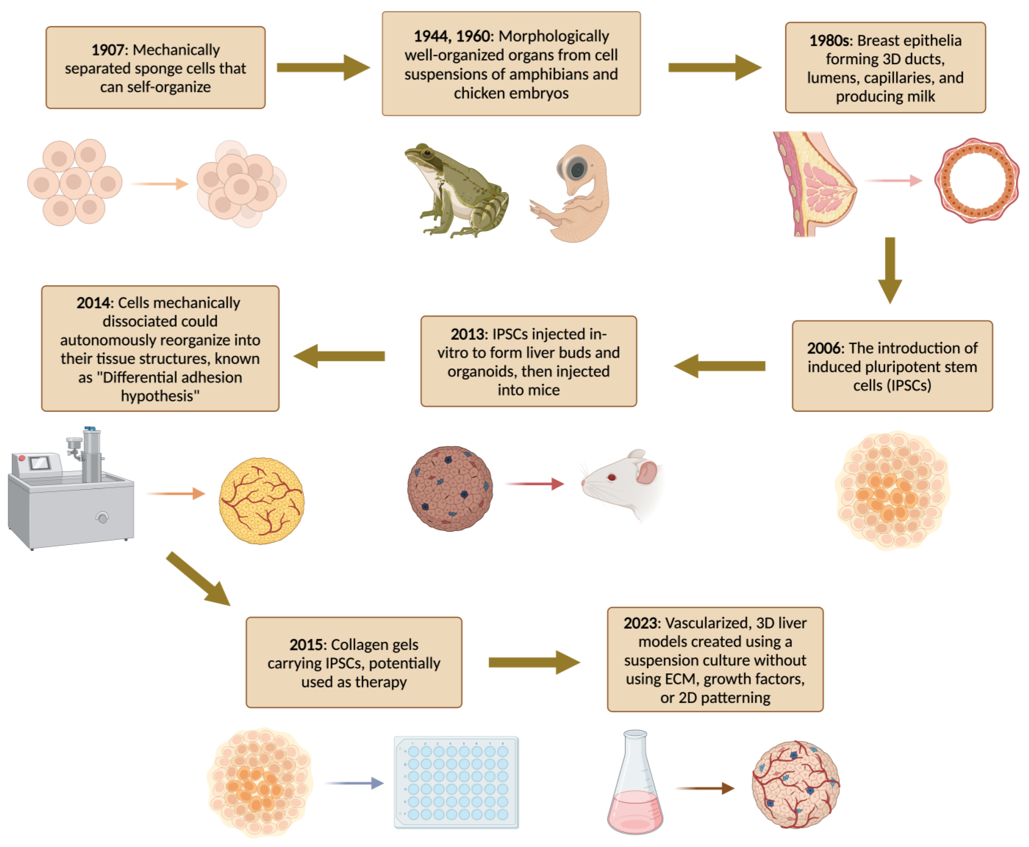
2. A Brief History of Organoids

The development of organoid-like structures dates back to 1907 when Wilson HV first demonstrated how whole sponges may be artificially reared through self-organization of dissociated sponge cells in vitro [52]. Over the past decades, several research groups have applied dissociation–reaggregation experimental strategies to develop miniaturized model systems of different bio-constructs (the reader is referred to more specialized reports (Figure 1) [52,53,54,55,56,57,58,59,60,61,62,63,64,65,66,67,68,69,70,71]). A real shift in the modern field of organoid biology started in 2009 when Sato et al. reported that adult intestinal stem cells expressing G protein-coupled receptor 5 (Lgr5), which contains leucine-rich repeats, generate intestinal organoids in a 3D microenvironment [72]. The authors reported that murine intestine-derived adult stem cells self-organize and differentiate into crypt-villus in Matrigel (a substrate for 3D cell culture). The results of this benchmark study paved the way for further developments in organoid research using cells derived from different source organs. The technique was subsequently refined to generate human intestinal organoids and organoids from various organs in which leucine-rich repeat-containing G protein-coupled receptor 5 (Lgr5+) progenitor cells are present, including the colon, stomach, and liver [63]. The isolation of individual Lgr5+ cells from the livers of adult mice and their cultivation into hepatic organoids unveiled their colony-forming capacity [66]. Extended cultivation confirmed their ability to self-renew indefinitely [72].
In 2013, Takebe and co-workers successfully produced liver buds and liver organoids in vitro by co-culturing human-induced pluripotent stem-cell-derived liver endodermal cells with stromal cells [66]. After forming liver-bud-like structures, the authors injected the engineered organoids into immunodeficient mice. Interestingly, the vessels of the artificial constructs invaded the host vascular network, and the liver bud mimics induced in vitro were nearly identical to their in vivo counterparts. Following transplantation, bioartificial liver buds facilitated the development of functional liver tissue in mice models, offering a promising avenue for regenerative treatments in cases of organ failure. This process faithfully recapitulated the cellular transformations observed during embryonic organ bud development [73]. Subsequently, the same research team developed a large-scale organoid production platform capable of efficiently producing homogeneous small liver bud constructs [74,75]. Hence, a broad spectrum of organoid models derived from both adult primary cells and pluripotent stem cells has emerged, underscoring the remarkable versatility of this technology [76,77,78]. In the wake of these advancements, liver organoids have become invaluable tools in research, facilitating investigations into liver development, diseases, and drug toxicity [79]. Their application has enabled the study of the mechanisms underpinning liver disorders, including liver cancer, viral hepatitis, and non-alcoholic fatty liver disease [80]. Continued progress in organoid technology holds the potential to revolutionize regenerative medicine, with the prospect of developing transplantable organs or seeding bio-artificial liver devices akin to kidney dialysis machines.
Figure 1. Timeline of the history of the generation of organoid cultures [52,53,54,55,56,57,58,59,60,61,62,63,64,65,66,67,68,69,70,71].
Bioengineering 11 00346 g001

Overview of the Organoids Concept

The establishment of liver organoids is based on fundamental biological principles of organogenesis and maturation, in which stem cells differentiate and self-assemble into physiologically active immature and mature tissues and organ structures regulated by various signaling molecules. Mimicking these innate features to develop and optimize stem-cell-derived organoid models requires exogenous cellular and tissue microenvironment stimuli. The cell morphogenesis, physiology, fate determination, and organization that drive organogenesis are directed by the combined use of multiple biochemical signaling factors and biophysical restraints from the surrounding extracellular microenvironment and adjacent cells [81,82].
For example, during the early stages of fertilization and embryogenesis, the embryo reorganizes and immediately divides into different cell types in a constrained neighborhood until an appropriate embryonic cavity is formed. At this stage, the fertilized egg divides into multiple cells with slight changes. Consequently, the cells become more cohesive and compact until blastocyst formation occurs. In addition, cell polarization is facilitated by paracrine signaling pathways and mechanical cues exerted by neighboring cells, which ultimately allow for the formation of germ layers and the organization of tissue structures [83,84]. Researchers in the field of tissue engineering and organoids-based regenerative medicine have succeeded to some extent in mimicking these basic principles of embryology to establish organoid constructs simply by controlling cell assembly and maintaining shape, size, and coordination inputs. This review will shed light on the cell and tissue engineering principles used to create organoid systems and their application in liver research.

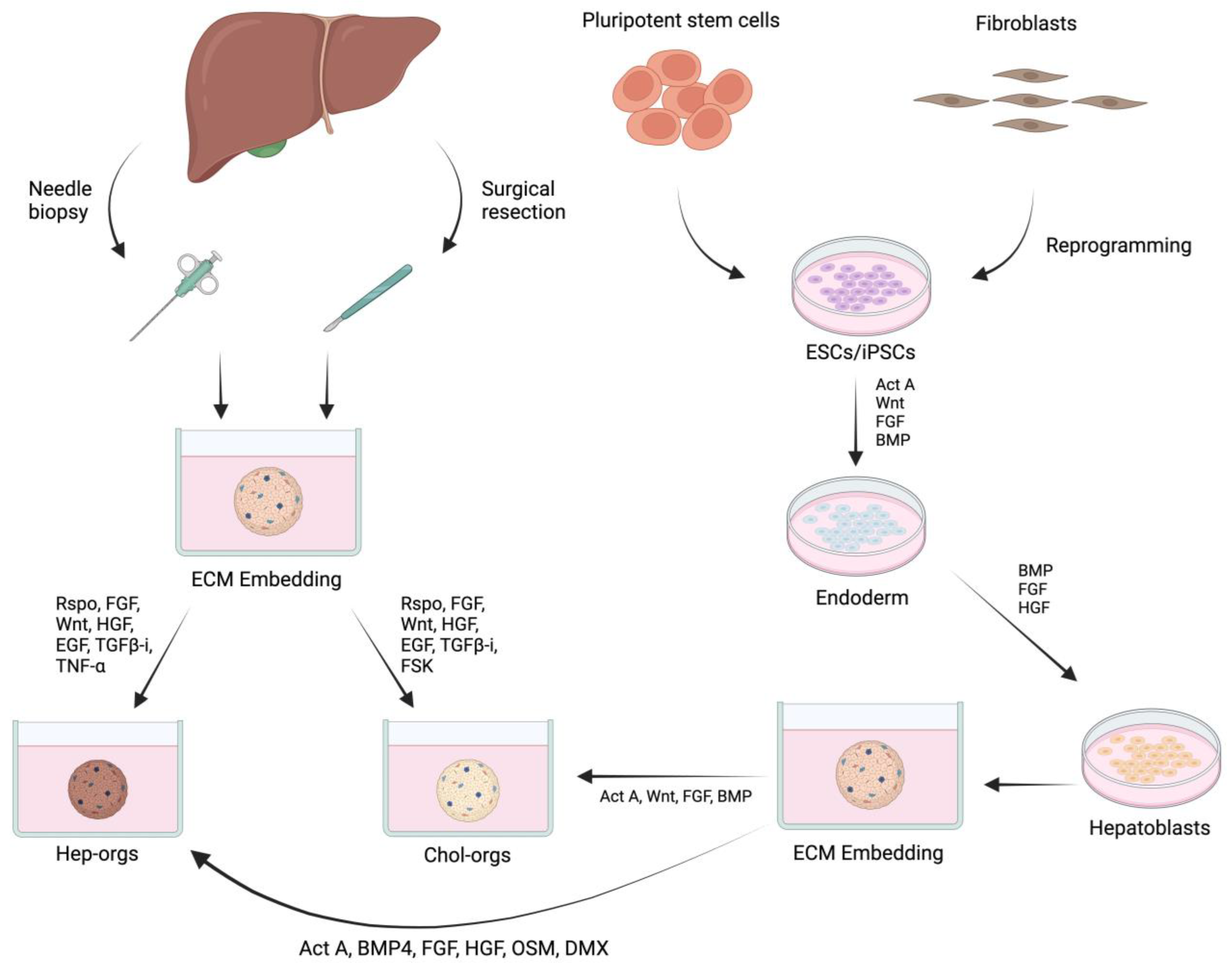
3. Cell Source and Biological Factors for Growing Liver Organoids

Adopting stem cells for organoid growth is one of the most critical advances in tissue engineering and regenerative medicine research. Organoids can be established from pluripotent stem cells (PSCs) or organ-specific adult stem cells (ASCs). Liver progenitors expressing the membrane receptor Leucine-rich repeat-containing G protein-coupled receptor 5 (LGR5) are widely used for generating liver organoids. It has been well documented that LGR5-expressing cells possess the potential for self-renewal and self-assembly and that they can generate organoids of functional cholangiocytes and hepatocytes (Figure 2) [85,86,87].
Published studies have indicated that liver-derived LGR5+ progenitors could be stimulated to generate mini liver tissue or organoids while retaining their “non-stem-cell” characteristics after a long-term 3D culture microenvironment. PSCs can self-renew and differentiate into specific lineages or any cell type in the body, including liver cells. Protocols have been developed for the gradual differentiation of PSCs into hepatic cells, producing liver organoids of hepatocytes or cholangiocytes that have liver-like functions, such as albumin, urea, and bile acid synthesis [66,88]. This approach has proven to be highly effective with both human embryonic stem cells and human-induced pluripotent stem cells, and it has been recently employed for the in vitro modeling of liver diseases [89]. However, the complexity of differentiating human pluripotent stem-cell-derived hepatic cells and the potential formation of teratomas due to any remaining undifferentiated cells in the system present significant challenges for the derivation and application of PSC-derived hepatic cells in cell and organoid-based therapies [90].
In addition, when hepatocytes are cultured in traditional monolayers, they typically undergo dedifferentiation, leading to a loss of phenotype and function [91]. This lack of morphology and function often results in the inability to detect metabolism-mediated hepatotoxicity of drugs in vitro [92]. Nevertheless, the differentiation of pluripotent stem cells, such as embryonic stem cells (ESCs) and induced pluripotent stem cells (iPSCs), into hepatic cells has been a key strategy since organoids’ early development. This method allows for the gradual differentiation of stem cells into liver organoids, mimicking the developmental process observed in natural liver formation [93].
Growth factors play a crucial role in the in vitro growth of organoids by manipulating the regulatory activities of cells related to self-renewal, expansion in a 3D microenvironment, differentiation after exposure to a specific culture medium, and self-organization into stable or static structures. During embryonic development, early liver development begins with the patterning of the endodermal layer, which also gives rise to the endodermal tissues of the pancreas, intestinal tract, and gut tube [94]. Researchers have therefore mimicked the natural phenomenon by using culture media supplemented with several growth factors and biomolecules that play key roles in signaling pathways crucial for proliferation, differentiation, maturation, and migration in the endodermal system. The major biological factors widely used for the growth, maintenance, and genetic manipulation of organoids (including liver) include noggin, R-spongin 1 (Wnt pathway enhancer), epidermal growth factor (EGF), hepatocyte growth factor (HGF), fibroblast growth factor (FGF), transforming growth factor-β (TGF-β), TGF-β inhibitor (A83-01), cAMP pathway agonist forskolin (FSK), and retinoic acid (RA) [95,96,97].

4. Bioengineering Strategies for Growing Liver Organoids

A variety of tissue engineering techniques have been investigated to support cells’ growth, proliferation, and differentiation into organoids of hepatocytes and cholangiocytes. Three-dimensional cultures based on hydrogels or scaffold matrices prepared from biocompatible materials of natural or semi-synthetic origin provide a biomimetic in a three-dimensional microenvironment for cells to differentiate and organize into functional liver organoids over time [98,99]. To effectively model liver functionality in vitro, it is critical not only to create an inbuilt vasculature, but also to tailor the cellular phenotype by modulating structural properties of the matrix with appropriate stiffness and porosity. The hydrous microenvironment and related properties of the extracellular matrix-mimicking culture system are essential for ensuring a constant and efficient permeation of nutrients and oxygen supply, which plays a vital role in supporting the functional integrity of the liver model [100,101]. Natural biomaterials, such as hydrogels, present several advantages, including their capacity to mimic the extracellular matrix (ECM) and sustain a supportive microenvironment, offering mechanical and biochemical signals to cells [79,102,103,104].
Currently, the establishment of 3D hepatic organoids predominantly relies on Matrigel, a natural hydrogel derived from a protein mixture secreted by Engelbreth–Holm–Swarm murine sarcoma cells [105]. Matrigel serves as a supportive substrate that mimics the complex extracellular matrix (ECM) environment of the liver [106]. To guarantee uniformity and repeatability, it is crucial to establish well-defined culture conditions for organoids, especially with regard to their composition, to facilitate future clinical applications [107]. Over the past decade, there has been a notable progression in organoid culture techniques. Initially reliant solely on Matrigel, these techniques have since advanced to incorporate biohybrids and semisynthetic or synthetic hydrogels, offering greater control and standardization in the generation of organoids [108]. While Matrigel provides valuable support for cells and facilitates their physical attachment and self-organization, which is crucial for the development of organoids, it comes with several limitations. These include inconsistencies between batches and variations across species, as well as concerns regarding the transmission of animal pathogens and potential incompatibility or immunogenicity issues in clinical applications due to having unidentified components from its animal origin [100,109]. The development of liver organoid models using chemically defined synthetic polymers eliminates the need for animal components. It also aims to overcome the batch-to-batch variability associated with traditional matrices like EHS and produce highly reproducible organoids, making them more suitable for disease modeling and clinical applications [110,111]. Liver organoids have been successfully generated within the PEG-RGD, (PIC), and laminin-111 hydrogel, and the stiffness of the hydrogel has been manipulated to replicate fibrotic liver disease. Another study demonstrated the generation of human liver organoids using polyisocyanopeptides (PIC) and laminin-111 [111]. Additionally, natural polymer hydrogels, such as fibrin/laminin, have been employed to generate liver organoids due to their ability to provide appropriate biochemical and physical support for organoid formation and expansion [110]. Hence, incorporating biomaterials, whether synthetic or derived from natural sources, has significantly accelerated the advancement of liver organoid development.

Microfabrication Strategies to Control the Assembly of Organoids

The microfluidic-based approach employs microfluidic devices that enable the growth and differentiation of liver cells. These devices provide precise control over the cellular microenvironment, including the composition of the media and the flow of nutrients and waste. Microfluidics refers to a technology that enables the manipulation of small amounts of fluids within tiny channels. This innovative approach has found application in creating microenvironments that closely resemble the natural conditions of organs [112]. This can exert precise control over the microenvironment, enabling the establishment of nutrient and oxygen gradients, as well as facilitating cell–cell interactions [112,113,114]. The aim is to effectively provide nutrients and oxygen to the inner components of the organoids [100]. In their study, Rennert et al. successfully developed a functional 3D human liver model within a microfluidic biochip. The liver organoids in this model receive a continuous supply of nutrients and oxygen through a fluid flow system, which closely mimics the structure of the liver sinusoid. To measure oxygen consumption, a luminescence-based sensor was integrated into the microfluidic chip. The results confirmed that the introduction of microfluidic flow using a perfusion system led to an enhanced expression of liver transporter proteins and promoted the formation of hepatocyte microvilli [114]. Another microfluidic chip was developed by Banaeiyan et al. to culture HepG2 cells and hepatic liver cells, aiming to replicate the liver lobule microenvironment. The chip featured a tissue-like hexagonal design and incorporated micro-channels to simulate the convection–diffusion mechanism of blood circulation [115]. Prodanov and co-workers developed a microfluidic chip with two chambers separated by a porous membrane. They aimed to recreate the liver sinusoid using human cells for 28 days and successfully maintained a 3D model [116]. The application of an efficient digital microfluidic system has been demonstrated for drug screening and evaluation of hepatotoxicity [117]. Esch and his team combined the Liver Chip and Intestine Chip to evaluate toxicity in the digestive system. The liver compartment contained HepG2/C3A cells (which are hepatocellular carcinoma cells) cultured in a silicon chip, while the intestine compartment included a co-culture of Caco-2 (Colon carcinoma) and HT29-MTX (mucus-secreting colon epithelium) cells. Interestingly, linking the intestine compartment to the liver compartment via microfluidic channels exacerbated this effect, resembling the first-pass metabolism despite reducing nanoparticle exposure in the Liver Chip [118]. The utilization of microfluidics in organoid development offers several benefits, including precise control over the microenvironment, efficient mass transport facilitated by fluid flow, and the ability to integrate with different sensors and actuators. However, there are also limitations to consider, such as the challenges of standardization and scalability. Additionally, the operation of microfluidic systems may require external pumps, tubing, connectors, and valves [93]. The approaches mentioned above have emerged as the most widely used and have demonstrated their effectiveness in generating functional liver organoids. They have the potential to revolutionize disease modeling, drug testing, and personalized medicine in the future.

5. Organoids for Liver Cancer Research

Liver cancer organoids may be derived from adult tissue biopsies or by reprogramming patient-derived cells with growth factor cocktails (Figure 3 and Figure 4). Using patient-derived organoid models to investigate mechanisms of chemotherapy resistance and to reverse drug resistance by targeting cancer stem cells and oncogenes is one of the hottest topics in cancer research. Over the years, the tumor microenvironment (TME) has been widely studied and targeted by various drugs, such as PD-L1 inhibitors and anti-angiogenics [119]. Yet, altered microenvironmental elements throughout therapy result in acquired drug resistance, leading to decreased clinical effectiveness [119]. Observing these changes in the TME could aid in developing new drugs for advanced HCC. Organoids have provided an excellent 3D environment in preclinical studies for studying TME resistance and potential new targets. Cancer-associated fibroblasts (CAFs), a major component of the TME, are known to be involved in the progression of cirrhosis and are described as promoting tumor proliferation and invasion. CAFs have been co-cultured with primary liver organoids and revealed to promote resistance to chemotherapeutic drugs, including Sorafenib, Regorafenib, and 5-fluorouracil [73]. Sorafenib is an anti-cancer agent that targets various kinases involved in certain oncogenic pathways [120]. The role of CD 44 positive HCC cells and certain hedgehog signaling pathways in Sorafenib has been described [121,122]. Co-administration of hedgehog signaling pathways with Sorafenib exhibited increased sensitivity to Sorafenib [123]. Martin and co-workers discussed the resistance to trans-arterial chemoembolization (TACE) of HCC, noting that up to 40% of patients do not respond to treatment [124]. PKM2, a crucial enzyme in glucose metabolism, was identified as participating in cancer cell metabolism.
Figure 3. Generation of cancer organoid from human liver biopsies of hepatocellular carcinoma and paired non-tumor liver tissues. (A) Schematic illustration; (B) image of biopsy pieces employed for growing organoids; (C) images of a tumor and paired non-tumor liver tissue organoids captured using a bright-field microscope (Scale bar: 500 μm). Abbreviations: hepatocellular carcinoma (HCC); cholangiocellular carcinoma (CCC); passage (P). Figure 3 is adopted with copyright permission from [125], Elsevier.
Bioengineering 11 00346 g003
Nevertheless, numerous targets for novel anti-cancer agents have been established via organoid research. These would permit the establishment of precision therapy for HCC patients, thus reducing overall side effects in contrast with traditional chemotherapy. Inhibitors of purine metabolism are commonly used to treat various cancer types. However, the significance of purine metabolism in liver carcinogenesis remains uncertain. Chong and co-workers discovered the significance of amplified de novo purine synthesis in HCC using cell lines and organoids. E2F1, a transcriptional factor, was observed to regulate the PI3K pathway, which promotes carcinogenesis by upregulating purine synthesis [126]. Purine metabolism, therefore, could be a potential target for personalized therapy. Preferential deposition of fluorescent nano-diamond particles (FDP) coated with doxorubicin, a chemotherapy agent, allows growth inhibition and cell death selectively in tumor cells, thus lowering adverse effects [127]. Oroyxlin A (transketolase inhibitor) is a novel anti-cancer drug studied using liver organoids. Its impact on inhibiting transketolase, a rate-limiting enzyme in de novo nucleotide synthesis, suppressed growth and cell death [128]. Lim and coworkers employed liver organoids to observe and deduce rational drug combinations for proteosome inhibitors, thus overcoming the limited furtherance in combinational therapy, along with discovering enhanced efficacy and clinical outcomes [129]. Omacetaxine is an FDA-approved global protein synthesis inhibitor used as an anti-cancer agent for chronic myelogenous leukemia [130]. Its effect on six hepatocellular carcinoma patient-derived cell lines was assessed, which revealed early apoptosis, late apoptosis, or both in all six lines [131].
Figure 4. Establishment of patient-derived biliary tract cancer organoids. (A) Experimental strategy for biopsy collection from biliary tract of cancer patients. (B) Pie charts comparing stratification of all original tumors versus stratification of all tumor-derived organoids based on histology (top), tumor-node-metastasis (TNM), and disease stage (bottom). (C) Microscopy images for histology staining (H&E) and bright-field imaging of patient-derived organoids and matched primary tumors from three main histological subtypes (scale bars, 50 μm). (D) Immunohistochemistry analysis of biliary tract cancer markers (CK19 and CK7) in original tumor samples and BTC organoids (scale bars, 50 μm). Abbreviations: biliary tract cancer (BTC); intrahepatic cholangiocarcinoma (ICC); extrahepatic cholangiocarcinoma (ECC); and gallbladder carcinoma (GBC). Figure 4 is adopted with copyright permission from [132], Elsevier.
Bioengineering 11 00346 g004
Liver organoids have been pivotal in exploring the connection between hepatocytes and cholangiocarcinoma (CC) in aiming to unveil CC’s etiology [72]. In an interesting study, Sun et al. utilized human-induced hepatocytes to generate liver organoids and overexpress ten genes enriched in intrahepatic cholangiocarcinoma (ICC). This led to notable changes, including the loss of circular morphology, mucous vacuole development, increased nucleus-to-cytoplasm ratio, nuclear atypia, duct or cavity structures, mucin release, and upregulated NOTCH signaling genes. After being orthotopically implanted in mice, these organoids resulted in a 100% tumor development rate, resembling ICCs due to specific ICC markers (CK19, SOX9). This suggested an important role of the RASG12V gene in hepatocyte-to-ICC conversion [133]. Studies using liver cancer organoids from PPTR mice (Prom1 overexpression, Pten, and TP53 loss) demonstrated strong stem cell properties, potentially leading to invasive and metastatic tumors [134]. Interestingly, some organoids showed features of both hepatocellular carcinoma and cholangiocarcinoma, which can differentiate into mature liver cells, reducing intrahepatic cholangiocarcinoma aggressiveness by blocking the Wnt signaling pathway [135]. These findings highlight the potential for gene or pathway targeting to prevent cholangiocarcinoma. Organoids have been important in investigating mitochondrial function and energy metabolism in cholangiocarcinoma. The Warburg effect underscores glycolysis as a primary energy production mechanism in tumor cells, making mitochondria crucial [136]. Studies found altered mitochondrial shape and reduced metabolism in cholangiocarcinoma organoids with knocked-out mitochondrial fusion genes (OPA1 and MFN1), leading to decreased oxygen consumption and ATP production. Inhibiting apoptosis prevented cholangiocarcinoma organoid development [137]. Another study cultured cholangiocarcinoma organoids without glucose, observing reduced proliferation, altered shape, increased stem cell markers, enhanced AKT phosphorylation, and reduced gemcitabine sensitivity [138]. Inhibition of AKT phosphorylation reversed stem-cell-like features and gemcitabine resistance. This suggests that cholangiocarcinoma cells can adapt to glucose shortage by enhancing stem cell characteristics through AKT phosphorylation.
Hepatitis infects more than 354 million people worldwide and amplifies the risk of HCC [WHO]. A characteristic of the hepatitis B virus (HBV) is the formation of a stable, persistent cDNA episome. This incorporates into the host DNA, rendering it resistant to antiviral drugs [139]. Due to the dysregulation of significant host factors and antiviral targets in 2D culture systems, 3D organoid systems allow for improved research in studying virus–host interactions as well as potential therapeutics [140]. Liver organoids derived from adult tissue have been infected with HBV to study pro-apoptotic drugs that promote cell death via the extrinsic pathway to eliminate hepatocytes with HBV DNA [141]. Hepatitis C virus (HCV) is another less commonly infecting virus leading to liver cirrhosis and HCC. Hepatocytes in vivo exhibit polarity, allowing the HCV virus to enter via certain tight junctions [142] Huh-7.5 hepatoma cells, utilized for conducting in vitro studies, express poorly in 2D but express excellent polarity in 3D organoids, which have allowed for ameliorated understanding of the HCV life cycle [142]. Improved binding of HCV has been observed in Huh-7 3D models, showing polarity over 2D models possessing poor polarity [143].

Current Limitations of Organoid Models

Organoid technology is still constrained by several hurdles and limitations. The fundamental obstacle associated with this technology is that the culture, maintenance, and expansion of organoids are heavily dependent on Matrigel, a 3D culture matrix extract derived from Engelbreth–Holm–Swarm mouse sarcoma cells. While Matrigel serves as a commercially accessible hydrogel substrate, these extracts encounter issues linked to inconsistencies in composition and reproducibility across different batches. Furthermore, these extracts may harbor unknown pathogens and are highly immunogenic, making them unsuitable for transplantation into human patients and severely hampering the clinical application of organoids. A major drawback of adult stem-cell-derived liver organoids is that they are derived from epithelial cells and utilize a simplified extracellular matrix (ECM) that lacks structural and functional compartmentalization [76,144]. Existing organoid culture systems are monocultures and lack innervation, vascularization, and immune interactions, making them inadequate for understanding the full spectrum of hepatocarcinoma. Therefore, there is a strong need to use approaches, such as tailored matrices and co-cultures, to generate organoids with specific cell populations, such as mesenchymal cells, endothelial cells, and immune cells, in addition to hepatocytes [125,132]
Another drawback is the limited effectiveness of organoid models. A study by Nuciforo et al. on organoid models of hepatocellular carcinoma showed that it is difficult to culture hepatocellular carcinoma cells [125]. The success rates for developing hepatocellular carcinoma organoids (HCCOs) from human primary hepatocellular carcinoma have been reported as ~26%. However, for other cancers, the success rate is significantly higher. Success rates for colorectal cancer are approximately 90% [145], and for pancreatic cancer, they are around 75–83% [146]. The variability among organs indicates that the success rate for hepatocellular carcinoma is much lower than for other cancers. A possible reason for the low success rate could be that the generation of hepatocellular carcinoma organoids is limited to a subset of HCCs. Furthermore, only poorly differentiated tumors are able to produce HCC organoids, which, according to Nuciforo et al., is strongly associated with the histopathological features of the tumors [125]. This could be because the highly differentiated, slowly growing tumors do not reach the threshold of cell proliferation rate required for the development of HCC organoids. Similarly, according to Broutier and coworkers, there is a significant correlation between the original tumor’s proliferation index and success rate. In samples taken from tumors with proliferating cells greater than 5%, the success rate of establishing organoid cultures was 100% [147]. Future studies may seek to culture HCCOs from lower-grade tumors to capture the entire disease spectrum.
Developing liver organoid cultures is often costly and requires skilled professionals, specialized techniques, and equipment. For example, laboratories often produce conditioned media to save experimental costs. Unfortunately, this leads to issues of batch-to-batch variability and, in some instances, small amounts of serum (0.5–1%) are present in the final organoid growth medium, jeopardizing the consistency of the organoid model. A typical base medium for mammalian cell culture is advanced DMEM/F12. However, DMEM/F12 cannot be used for the culture of liver organoids prior to the addition of multiple agents [148]. To encourage the advancement of organoid technology and optimize its application in biological research, the availability of appropriately designed and reasonably priced media is essential. Variation among different iPSC lines and their derivatives continues to be a major challenge, especially for iPSC-derived organoids, when iPSCs and their derivatives are used for disease modeling and cell therapy. When generated from different individuals or iPSC core facilities, variation between and within iPSC lines is frequently observed in distinguishing iPSC tumorigenicity, genomic instability, epigenetic status, and maturation status. Successful development of “comparable” iPSCs and their derivatives requires quality characteristics that produce consistent, high-quality iPSCs.
Therefore, in 2018, the Global Alliance for iPSC Therapies in the UK identified the creation of QC guidelines for clinical-grade iPSC production [149]. Identity verification, microbiological sterility, endotoxin, genetic fidelity and stability (karyotyping and residual vector testing), potency assessment, expression of pluripotency markers, and post-thawed viability are among the essential quality characteristics for clinical-grade iPSC creation. However, there may still be variations during the purification process, iPSC differentiation, iPSC expansion, reprogramming, colony selection, culture system selection, and iPSC reprogramming within various iPSC cell banks. Such issues must be resolved through routine and ongoing validation of the iPSCs. Moreover, recent studies on genetic and epigenetic differences in iPSCs have highlighted iPSC safety-related concerns.
Genome instability, single nucleotide variants, choroidal neovascularization (CNV), and loss of heterozygosity are examples of genetic changes found in iPSCs. Through reprogramming and prolonged in vitro culture, these mutations can be introduced and accumulated in iPSCs from their parental cells [150].
The potential for tumorigenicity due to genetic differences in iPSCs is one of the highest safety concerns. Another issue with iPSCs’ genetic and epigenetic heterogeneity is that it may impair the differentiation potential of the cells and lead to unexpected phenotypes [151]. Further research initiatives, such as creating a specialized mutation database for iPSCs and establishing a standardized set of criteria to screen genetic variations, are imperative for assessing genomic stability. This evaluation is crucial as genetic variants within iPSCs could have significant functional and safety implications.

6. Conclusions and Future Perspectives

The liver is an important organ in the human body, and its metabolic, immune, digestive, homeostatic, and detoxification roles depend on efficient crosstalk and hierarchical organization. Organoid technology has received tremendous interest in liver bioengineering to mimic liver-like features and functions for biomedical investigations. Recent advances in organoid research indicate that this technology can be applied to culture and fabricate liver organoids for disease models, cancer research, viral infection models, and translational research [49,50,51,150]. Organoid-based approaches offer alternative mini-liver-like platforms for investigating cancer initiation, development, progression, and therapy response assessment. However, reported organoid-based models mimic only a fraction of the physiological activities and structural patterns of the liver. Therefore, a more classical multicellular-based organoid model system that recapitulates most, if not all, of the cellular architecture and complex signaling pathways of the liver is a supreme requirement for tissue bioengineering and the regenerative medicine field. To design and produce such hierarchical organoid models with proper distribution and patterning of the cells, a better understanding of the biomolecular composition of the liver scaffold matrix and its role in cell-specific support is urgently needed. The development of advanced biomaterials with cell-specific supporting roles of the liver scaffolding matrix might provide the desired physiochemical features and mechanical integrity. Other critical challenges that need to be resolved in order to realize organoid technology for wider application include a proper selection of biomaterial sources, appropriate cell types (parenchymal and nonparenchymal cells), vasculature embedding, and quality control of the cultures. Besides the biomaterials and cells, technological obstacles also need to be addressed.

Author Contributions

Conceptualization, T.A.M.; Writing—original draft preparation, T.A.M., A.J., J.K., B.T., M.A. and A.M.; Writing—review & editing: R.C., A.A., S.K., M.S.M., A.W.A., A.Y., A.M.A., K.A., Y.K.V. and D.C.B.; Methodology and Investigation, M.A. and A.M.; Formal analysis, A.J., J.K. and B.T.; Data Curation and Software, R.C., A.A., S.K., M.S.M. and Y.K.V.; Validation, A.Y.; Resources, A.M.A., K.A. and D.C.B.; Supervision, T.A.M. All authors have read and agreed to the published version of the manuscript.

Funding

This study was supported by the King Faisal Specialist Hospital & Research Centre, Saudi Arabia (Project number; RAC# 2150012 and RAC# 2220008).

Conflicts of Interest

The authors declare no conflict of interest.

References

  1. Sung, H.; Ferlay, J.; Siegel, R.L.; Laversanne, M.; Soerjomataram, I.; Jemal, A.; Bray, F. Global Cancer Statistics 2020: GLOBOCAN Estimates of Incidence and Mortality Worldwide for 36 Cancers in 185 Countries. CA Cancer J. Clin. 2021, 71, 209–249. [Google Scholar] [CrossRef]
  2. Sia, D.; Villanueva, A.; Friedman, S.L.; Llovet, J.M. Liver Cancer Cell of Origin, Molecular Class, and Effects on Patient Prognosis. Gastroenterology 2017, 152, 745–761. [Google Scholar] [CrossRef] [PubMed]
  3. Brindley, P.J.; Bachini, M.; Ilyas, S.I.; Khan, S.A.; Loukas, A.; Sirica, A.E.; Teh, B.T.; Wongkham, S.; Gores, G.J. Cholangiocarcinoma. Nat. Rev. Dis. Primers 2021, 7, 65. [Google Scholar] [CrossRef]
  4. Sripa, B.; Tangkawattana, S.; Brindley, P.J. Update on Pathogenesis of Opisthorchiasis and Cholangiocarcinoma. Adv. Parasitol. 2018, 102, 97–113. [Google Scholar]
  5. Rumgay, H.; Ferlay, J.; de Martel, C.; Georges, D.; Ibrahim, A.S.; Zheng, R.; Wei, W.; Lemmens, V.E.; Soerjomataram, I. Global, Regional and National Burden of Primary Liver Cancer by Subtype. Eur. J. Cancer 2022, 161, 108–118. [Google Scholar] [CrossRef] [PubMed]
  6. Mondaca, S.; Yarmohammadi, H.; Kemeny, N.E. Regional Chemotherapy for Biliary Tract Tumors and Hepatocellular Carcinoma. Surg. Oncol. Clin. 2019, 28, 717–729. [Google Scholar] [CrossRef]
  7. Chinnappan, R.; Mir, T.A.; Alsalameh, S.; Makhzoum, T.; Alzhrani, A.; Al-Kattan, K.; Yaqinuddin, A. Low-Cost Point-of-Care Monitoring of ALT and AST Is Promising for Faster Decision Making and Diagnosis of Acute Liver Injury. Diagnostics 2023, 13, 2967. [Google Scholar] [CrossRef]
  8. Chinnappan, R.; Mir, T.A.; Alsalameh, S.; Makhzoum, T.; Adeeb, S.; Al-Kattan, K.; Yaqinuddin, A. Aptasensors Are Conjectured as Promising ALT and AST Diagnostic Tools for the Early Diagnosis of Acute Liver Injury. Life 2023, 13, 1273. [Google Scholar] [CrossRef] [PubMed]
  9. Kashefi-Kheyrabadi, L.; Mehrgardi, M.A.; Wiechec, E.; Turner, A.P.F.; Tiwari, A. Ultrasensitive Detection of Human Liver Hepatocellular Carcinoma Cells Using a Label-Free Aptasensor. Anal. Chem. 2014, 86, 4956–4960. [Google Scholar] [CrossRef]
  10. Park, S.; Cho, E.; Chueng, S.-T.D.; Yoon, J.-S.; Lee, T.; Lee, J.-H. Aptameric Fluorescent Biosensors for Liver Cancer Diagnosis. Biosensors 2023, 13, 617. [Google Scholar] [CrossRef]
  11. Ohashi, K.; Yokoyama, T.; Yamato, M.; Kuge, H.; Kanehiro, H.; Tsutsumi, M.; Amanuma, T.; Iwata, H.; Yang, J.; Okano, T. Engineering Functional Two-and Three-Dimensional Liver Systems In Vivo Using Hepatic Tissue Sheets. Nat. Med. 2007, 13, 880–885. [Google Scholar] [CrossRef]
  12. Smith, A.J.; Lilley, E. The Role of the Three Rs in Improving the Planning and Reproducibility of Animal Experiments. Animals 2019, 9, 975. [Google Scholar] [CrossRef]
  13. Langer, R.; Vacanti, J.P. Tissue Engineering. Science 1993, 260, 920–926. [Google Scholar] [CrossRef]
  14. Lauschke, V.M.; Hendriks, D.F.; Bell, C.C.; Andersson, T.B.; Ingelman-Sundberg, M. Novel 3D Culture Systems for Studies of Human Liver Function and Assessments of the Hepatotoxicity of Drugs and Drug Candidates. Chem. Res. Toxicol. 2016, 29, 1936–1955. [Google Scholar] [CrossRef] [PubMed]
  15. Mir, T.A.; Nakamura, M. Three-Dimensional Bioprinting: Toward the Era of Manufacturing Human Organs as Spare Parts for Healthcare and Medicine. Tissue Eng. Part B Rev. 2017, 23, 245–256. [Google Scholar] [CrossRef]
  16. Arai, K.; Yoshida, T.; Okabe, M.; Goto, M.; Mir, T.A.; Soko, C.; Tsukamoto, Y.; Akaike, T.; Nikaido, T.; Zhou, K. Fabrication of 3D-culture Platform with Sandwich Architecture for Preserving Liver-specific Functions of Hepatocytes Using 3D Bioprinter. J. Biomed. Mater. Res. Part A 2017, 105, 1583–1592. [Google Scholar] [CrossRef] [PubMed]
  17. Mir, T.A.; Iwanaga, S.; Kurooka, T.; Toda, H.; Sakai, S.; Nakamura, M. Biofabrication Offers Future Hope for Tackling Various Obstacles and Challenges in Tissue Engineering and Regenerative Medicine: A Perspective. Int. J. Bioprinting 2019, 5, 153. [Google Scholar] [CrossRef]
  18. Nair, D.G.; Weiskirchen, R. Recent Advances in Liver Tissue Engineering as an Alternative and Complementary Approach for Liver Transplantation. Curr. Issues Mol. Biol. 2024, 46, 262–278. [Google Scholar] [CrossRef] [PubMed]
  19. Nakamura, M.; Mir, T.A.; Arai, K.; Ito, S.; Yoshida, T.; Iwanaga, S.; Kitano, H.; Obara, C.; Nikaido, T. Bioprinting with Pre-Cultured Cellular Constructs towards Tissue Engineering of Hierarchical Tissues. Int. J. Bioprinting 2015, 1, 39–48. [Google Scholar]
  20. Kim, J.; Kang, W.; Sinn, D.H.; Gwak, G.-Y.; Paik, Y.-H.; Choi, M.S.; Lee, J.H.; Koh, K.C.; Paik, S.W. Substantial Risk of Recurrence Even after 5 Recurrence-Free Years in Early-Stage Hepatocellular Carcinoma Patients. Clin. Mol. Hepatol. 2020, 26, 516. [Google Scholar] [CrossRef]
  21. Kulik, L.; El-Serag, H.B. Epidemiology and Management of Hepatocellular Carcinoma. Gastroenterology 2019, 156, 477–491. [Google Scholar] [CrossRef] [PubMed]
  22. Chernyak, V.; Fowler, K.J.; Kamaya, A.; Kielar, A.Z.; Elsayes, K.M.; Bashir, M.R.; Kono, Y.; Do, R.K.; Mitchell, D.G.; Singal, A.G. Liver Imaging Reporting and Data System (LI-RADS) Version 2018: Imaging of Hepatocellular Carcinoma in at-Risk Patients. Radiology 2018, 289, 816–830. [Google Scholar] [CrossRef]
  23. Faccia, M.; Garcovich, M.; Ainora, M.E.; Riccardi, L.; Pompili, M.; Gasbarrini, A.; Zocco, M.A. Contrast-Enhanced Ultrasound for Monitoring Treatment Response in Different Stages of Hepatocellular Carcinoma. Cancers 2022, 14, 481. [Google Scholar] [CrossRef] [PubMed]
  24. Bartolotta, T.V.; Taibbi, A.; Picone, D.; Anastasi, A.; Midiri, M.; Lagalla, R. Detection of Liver Metastases in Cancer Patients with Geographic Fatty Infiltration of the Liver: The Added Value of Contrast-Enhanced Sonography. Ultrasonography 2017, 36, 160. [Google Scholar] [CrossRef]
  25. Simmons, O.; Fetzer, D.T.; Yokoo, T.; Marrero, J.A.; Yopp, A.; Kono, Y.; Parikh, N.; Browning, T.; Singal, A.G. Predictors of Adequate Ultrasound Quality for Hepatocellular Carcinoma Surveillance in Patients with Cirrhosis. Aliment. Pharmacol. Ther. 2017, 45, 169–177. [Google Scholar] [CrossRef]
  26. Gregory, J.; Burgio, M.D.; Corrias, G.; Vilgrain, V.; Ronot, M. Evaluation of Liver Tumour Response by Imaging. JHEP Rep. 2020, 2, 100100. [Google Scholar] [CrossRef] [PubMed]
  27. Cheng, J.T.; Tan, N.E.; Volk, M.L. Utility of Positron Emission Tomography-Computed Tomography Scan in Detecting Residual Hepatocellular Carcinoma Post Treatment: Series of Case Reports. World J. Gastrointest. Oncol. 2020, 12, 358. [Google Scholar] [CrossRef]
  28. Shao, Y.; Liu, T.; Hsu, C.; Lu, L.; Shen, Y.; Lin, Z.; Cheng, A.; Hsu, C. Early Alpha-foetoprotein Response Associated with Treatment Efficacy of Immune Checkpoint Inhibitors for Advanced Hepatocellular Carcinoma. Liver Int. 2019, 39, 2184–2189. [Google Scholar] [CrossRef]
  29. Piñero, F.; Dirchwolf, M.; Pessôa, M.G. Biomarkers in Hepatocellular Carcinoma: Diagnosis, Prognosis and Treatment Response Assessment. Cells 2020, 9, 1370. [Google Scholar] [CrossRef]
  30. Kim, H.I.; Lim, J.; Shim, J.H. Role of the Alpha-Fetoprotein Response in Immune Checkpoint Inhibitor-Based Treatment of Patients with Hepatocellular Carcinoma. J. Cancer Res. Clin. Oncol. 2022, 148, 2069–2077. [Google Scholar] [CrossRef]
  31. He, C.; Peng, W.; Liu, X.; Li, C.; Li, X.; Wen, T.-F. Post-Treatment Alpha-Fetoprotein Response Predicts Prognosis of Patients with Hepatocellular Carcinoma: A Meta-Analysis. Medicine 2019, 98, e16557. [Google Scholar] [CrossRef]
  32. Wang, T.; Zhang, K.-H. New Blood Biomarkers for the Diagnosis of AFP-Negative Hepatocellular Carcinoma. Front. Oncol. 2020, 10, 1316. [Google Scholar] [CrossRef] [PubMed]
  33. Zhang, G.; Ha, S.-A.; Kim, H.K.; Yoo, J.; Kim, S.; Lee, Y.S.; Hur, S.Y.; Kim, Y.W.; Kim, T.E.; Park, Y.G. Combined Analysis of AFP and HCCR-1 as an Useful Serological Marker for Small Hepatocellular Carcinoma: A Prospective Cohort Study. Dis. Markers 2012, 32, 265–271. [Google Scholar] [CrossRef] [PubMed]
  34. Feng, H.; Li, B.; Li, Z.; Wei, Q.; Ren, L. PIVKA-II Serves as a Potential Biomarker That Complements AFP for the Diagnosis of Hepatocellular Carcinoma. BMC Cancer 2021, 21, 401. [Google Scholar] [CrossRef] [PubMed]
  35. Omar, M.A.; Omran, M.M.; Farid, K.; Tabll, A.A.; Shahein, Y.E.; Emran, T.M.; Petrovic, A.; Lucic, N.R.; Smolic, R.; Kovac, T.; et al. Biomarkers for Hepatocellular Carcinoma: From Origin to Clinical Diagnosis. Biomedicines 2023, 11, 1852. [Google Scholar] [CrossRef]
  36. Zhu, H.-F.; Feng, J.-K.; Xiang, Y.-J.; Wang, K.; Zhou, L.-P.; Liu, Z.-H.; Cheng, Y.-Q.; Shi, J.; Guo, W.-X.; Cheng, S.-Q. Combination of Alpha-Fetoprotein and Neutrophil-to-Lymphocyte Ratio to Predict Treatment Response and Survival Outcomes of Patients with Unresectable Hepatocellular Carcinoma Treated with Immune Checkpoint Inhibitors. BMC Cancer 2023, 23, 547. [Google Scholar] [CrossRef]
  37. Pons, F.; Varela, M.; Llovet, J.M. Staging Systems in Hepatocellular Carcinoma. Hpb 2005, 7, 35–41. [Google Scholar] [CrossRef]
  38. Vogel, A.; Cervantes, A.; Chau, I.; Daniele, B.; Llovet, J.M.; Meyer, T.; Nault, J.-C.; Neumann, U.; Ricke, J.; Sangro, B. Hepatocellular Carcinoma: ESMO Clinical Practice Guidelines for Diagnosis, Treatment and Follow-Up. Ann. Oncol. 2018, 29, iv238–iv255. [Google Scholar] [CrossRef]
  39. Tümen, D.; Heumann, P.; Gülow, K.; Demirci, C.-N.; Cosma, L.-S.; Müller, M.; Kandulski, A. Pathogenesis and Current Treatment Strategies of Hepatocellular Carcinoma. Biomedicines 2022, 10, 3202. [Google Scholar] [CrossRef] [PubMed]
  40. Galle, P.R.; Forner, A.; Llovet, J.M.; Mazzaferro, V.; Piscaglia, F.; Raoul, J.-L.; Schirmacher, P.; Vilgrain, V. EASL Clinical Practice Guidelines: Management of Hepatocellular Carcinoma. J. Hepatol. 2018, 69, 182–236. [Google Scholar] [CrossRef]
  41. Heimbach, J.K.; Kulik, L.M.; Finn, R.S.; Sirlin, C.B.; Abecassis, M.M.; Roberts, L.R.; Zhu, A.X.; Murad, M.H.; Marrero, J.A. AASLD Guidelines for the Treatment of Hepatocellular Carcinoma. Hepatology 2018, 67, 358–380. [Google Scholar] [CrossRef] [PubMed]
  42. Raoul, J.-L.; Forner, A.; Bolondi, L.; Cheung, T.T.; Kloeckner, R.; de Baere, T. Updated Use of TACE for Hepatocellular Carcinoma Treatment: How and When to Use It Based on Clinical Evidence. Cancer Treat. Rev. 2019, 72, 28–36. [Google Scholar] [CrossRef] [PubMed]
  43. O’Leary, C.; Mahler, M.; Soulen, M.C. Liver-Directed Therapy for Hepatocellular Carcinoma. Chin. Clin. Oncol. 2021, 10, 8. [Google Scholar] [CrossRef] [PubMed]
  44. Calderaro, J.; Ziol, M.; Paradis, V.; Zucman-Rossi, J. Molecular and Histological Correlations in Liver Cancer. J. Hepatol. 2019, 71, 616–630. [Google Scholar] [CrossRef] [PubMed]
  45. Finn, R.S.; Qin, S.; Ikeda, M.; Galle, P.R.; Ducreux, M.; Kim, T.-Y.; Kudo, M.; Breder, V.; Merle, P.; Kaseb, A.O. Atezolizumab plus Bevacizumab in Unresectable Hepatocellular Carcinoma. N. Engl. J. Med. 2020, 382, 1894–1905. [Google Scholar] [CrossRef] [PubMed]
  46. Qin, S.; Jiang, J.; Lu, Y.; Nice, E.C.; Huang, C.; Zhang, J.; He, W. Emerging Role of Tumor Cell Plasticity in Modifying Therapeutic Response. Signal Transduct. Target. Ther. 2020, 5, 228. [Google Scholar] [CrossRef] [PubMed]
  47. Roehlen, N.; Muller, M.; Nehme, Z.; Crouchet, E.; Jühling, F.; Del Zompo, F.; Cherradi, S.; Duong, F.H.T.; Almeida, N.; Saviano, A.; et al. Treatment of HCC with Claudin-1-Specific Antibodies Suppresses Carcinogenic Signaling and Reprograms the Tumor Microenvironment. J. Hepatol. 2023, 78, 343–355. [Google Scholar] [CrossRef] [PubMed]
  48. Llovet, J.M.; Kelley, R.K.; Villanueva, A.; Singal, A.G.; Pikarsky, E.; Roayaie, S. Hepatocellular Carcinoma. Nat. Rev. Dis. Primers 2021, 7, 7. [Google Scholar] [CrossRef] [PubMed]
  49. Prior, N.; Inacio, P.; Huch, M. Liver Organoids: From Basic Research to Therapeutic Applications. Gut 2019, 68, 2228–2237. [Google Scholar] [CrossRef]
  50. Chang, M.; Bogacheva, M.S.; Lou, Y.-R. Challenges for the Applications of Human Pluripotent Stem Cell-Derived Liver Organoids. Front. Cell Dev. Biol. 2021, 9, 748576. [Google Scholar] [CrossRef]
  51. Obeid, D.A.; Mir, T.A.; Alzhrani, A.; Altuhami, A.; Shamma, T.; Ahmed, S.; Kazmi, S.; Fujitsuka, I.; Ikhlaq, M.; Shabab, M. Using Liver Organoids as Models to Study the Pathobiology of Rare Liver Diseases. Biomedicines 2024, 12, 446. [Google Scholar] [CrossRef]
  52. Wilson, H. A New Method by Which Sponges May Be Artificially Reared. Science 1907, 25, 912–915. [Google Scholar] [CrossRef] [PubMed]
  53. Corrò, C.; Novellasdemunt, L.; Li, V.S. A Brief History of Organoids. Am. J. Physiol.-Cell Physiol. 2020, 319, C151–C165. [Google Scholar] [CrossRef] [PubMed]
  54. Holtfreter, J. Experimental Studies on the Development of the Pronephros. Rev. Can. Biol. 1943, 3, 220–250. [Google Scholar]
  55. Weiss, P.; Taylor, A. Reconstitution of Complete Organs from Single-Cell Suspensions of Chick Embryos in Advanced Stages of Differentiation. Proc. Natl. Acad. Sci. USA 1960, 46, 1177–1185. [Google Scholar] [CrossRef] [PubMed]
  56. Stefanelli, A.; Zacchei, A.; Ceccherini, V. Retinal Reconstitution in Vitro after Disaggregation of Embryonic Chicken Eyes (Translated from Italian). Acta Embryol. Morphol. Exper. 1961, 4, 47–55. [Google Scholar]
  57. Evans, M. Origin of Mouse Embryonal Carcinoma Cells and the Possibility of Their Direct Isolation into Tissue Culture. Reproduction 1981, 62, 625–631. [Google Scholar] [CrossRef] [PubMed]
  58. Shannon, J.M.; Mason, R.J.; Jennings, S.D. Functional Differentiation of Alveolar Type II Epithelial Cells in Vitro: Effects of Cell Shape, Cell-Matrix Interactions and Cell-Cell Interactions. Biochim. Biophys. Acta (BBA)-Mol. Cell Res. 1987, 931, 143–156. [Google Scholar] [CrossRef]
  59. Thomson, J.A.; Itskovitz-Eldor, J.; Shapiro, S.S.; Waknitz, M.A.; Swiergiel, J.J.; Marshall, V.S.; Jones, J.M. Embryonic Stem Cell Lines Derived from Human Blastocysts. Science 1998, 282, 1145–1147. [Google Scholar] [CrossRef]
  60. Takahashi, K.; Yamanaka, S. Induction of Pluripotent Stem Cells from Mouse Embryonic and Adult Fibroblast Cultures by Defined Factors. Cell 2006, 126, 663–676. [Google Scholar] [CrossRef]
  61. Eiraku, M.; Watanabe, K.; Matsuo-Takasaki, M.; Kawada, M.; Yonemura, S.; Matsumura, M.; Wataya, T.; Nishiyama, A.; Muguruma, K.; Sasai, Y. Self-Organized Formation of Polarized Cortical Tissues from ESCs and Its Active Manipulation by Extrinsic Signals. Cell Stem Cell 2008, 3, 519–532. [Google Scholar] [CrossRef] [PubMed]
  62. Sato, T.; Vries, R.G.; Snippert, H.J.; Van De Wetering, M.; Barker, N.; Stange, D.E.; Van Es, J.H.; Abo, A.; Kujala, P.; Peters, P.J. Single Lgr5 Stem Cells Build Crypt-Villus Structures in Vitro without a Mesenchymal Niche. Nature 2009, 459, 262–265. [Google Scholar] [CrossRef] [PubMed]
  63. Barker, N.; Huch, M.; Kujala, P.; van de Wetering, M.; Snippert, H.J.; van Es, J.H.; Sato, T.; Stange, D.E.; Begthel, H.; van den Born, M. Lgr5+ ve Stem Cells Drive Self-Renewal in the Stomach and Build Long-Lived Gastric Units In Vitro. Cell Stem Cell 2010, 6, 25–36. [Google Scholar] [CrossRef] [PubMed]
  64. Eiraku, M.; Takata, N.; Ishibashi, H.; Kawada, M.; Sakakura, E.; Okuda, S.; Sekiguchi, K.; Adachi, T.; Sasai, Y. Self-Organizing Optic-Cup Morphogenesis in Three-Dimensional Culture. Nature 2011, 472, 51–56. [Google Scholar] [CrossRef]
  65. Nakano, T.; Ando, S.; Takata, N.; Kawada, M.; Muguruma, K.; Sekiguchi, K.; Saito, K.; Yonemura, S.; Eiraku, M.; Sasai, Y. Self-Formation of Optic Cups and Storable Stratified Neural Retina from Human ESCs. Cell Stem Cell 2012, 10, 771–785. [Google Scholar] [CrossRef] [PubMed]
  66. Takebe, T.; Sekine, K.; Enomura, M.; Koike, H.; Kimura, M.; Ogaeri, T.; Zhang, R.-R.; Ueno, Y.; Zheng, Y.-W.; Koike, N. Vascularized and Functional Human Liver from an iPSC-Derived Organ Bud Transplant. Nature 2013, 499, 481–484. [Google Scholar] [CrossRef]
  67. Chua, C.W.; Shibata, M.; Lei, M.; Toivanen, R.; Barlow, L.J.; Bergren, S.K.; Badani, K.K.; McKiernan, J.M.; Benson, M.C.; Hibshoosh, H. Single Luminal Epithelial Progenitors Can Generate Prostate Organoids in Culture. Nat. Cell Biol. 2014, 16, 951–961. [Google Scholar] [CrossRef]
  68. Post, Y.; Puschhof, J.; Beumer, J.; Kerkkamp, H.M.; de Bakker, M.A.; Slagboom, J.; de Barbanson, B.; Wevers, N.R.; Spijkers, X.M.; Olivier, T. Snake Venom Gland Organoids. Cell 2020, 180, 233–247. [Google Scholar] [CrossRef]
  69. Harrison, S.P.; Siller, R.; Tanaka, Y.; Chollet, M.E.; de la Morena-Barrio, M.E.; Xiang, Y.; Patterson, B.; Andersen, E.; Bravo-Pérez, C.; Kempf, H. Scalable Production of Tissue-like Vascularized Liver Organoids from Human PSCs. Exp. Mol. Med. 2023, 55, 2005–2024. [Google Scholar] [CrossRef]
  70. Hendriks, D.; Pagliaro, A.; Andreatta, F.; Ma, Z.; van Giessen, J.; Massalini, S.; López-Iglesias, C.; van Son, G.J.; DeMartino, J.; Damen, J.M.A. Human Fetal Brain Self-Organizes into Long-Term Expanding Organoids. Cell 2024, 187, 712–732. [Google Scholar] [CrossRef]
  71. Maimets, M.; Rocchi, C.; Bron, R.; Pringle, S.; Kuipers, J.; Giepmans, B.N.; Vries, R.G.; Clevers, H.; De Haan, G.; Van Os, R. Long-Term in Vitro Expansion of Salivary Gland Stem Cells Driven by Wnt Signals. Stem Cell Rep. 2016, 6, 150–162. [Google Scholar] [CrossRef]
  72. Huch, M.; Gehart, H.; van Boxtel, R.; Hamer, K.; Blokzijl, F.; Verstegen, M.M.A.; Ellis, E.; van Wenum, M.; Fuchs, S.A.; de Ligt, J.; et al. Long-Term Culture of Genome-Stable Bipotent Stem Cells from Adult Human Liver. Cell 2015, 160, 299–312. [Google Scholar] [CrossRef]
  73. Liu, J.; Li, P.; Wang, L.; Li, M.; Ge, Z.; Noordam, L.; Lieshout, R.; Verstegen, M.M.A.; Ma, B.; Su, J.; et al. Cancer-Associated Fibroblasts Provide a Stromal Niche for Liver Cancer Organoids That Confers Trophic Effects and Therapy Resistance. Cell. Mol. Gastroenterol. Hepatol. 2021, 11, 407–431. [Google Scholar] [CrossRef] [PubMed]
  74. Koike, H.; Iwasawa, K.; Ouchi, R.; Maezawa, M.; Giesbrecht, K.; Saiki, N.; Ferguson, A.; Kimura, M.; Thompson, W.L.; Wells, J.M. Modelling Human Hepato-Biliary-Pancreatic Organogenesis from the Foregut–Midgut Boundary. Nature 2019, 574, 112–116. [Google Scholar] [CrossRef] [PubMed]
  75. Takebe, T.; Sekine, K.; Kimura, M.; Yoshizawa, E.; Ayano, S.; Koido, M.; Funayama, S.; Nakanishi, N.; Hisai, T.; Kobayashi, T. Massive and Reproducible Production of Liver Buds Entirely from Human Pluripotent Stem Cells. Cell Rep. 2017, 21, 2661–2670. [Google Scholar] [CrossRef]
  76. Harrison, S.P.; Baumgarten, S.F.; Verma, R.; Lunov, O.; Dejneka, A.; Sullivan, G.J. Liver Organoids: Recent Developments, Limitations and Potential. Front. Med. 2021, 8, 574047. [Google Scholar] [CrossRef]
  77. Takeishi, K.; de l’Hortet, A.C.; Wang, Y.; Handa, K.; Guzman-Lepe, J.; Matsubara, K.; Morita, K.; Jang, S.; Haep, N.; Florentino, R.M. Assembly and Function of a Bioengineered Human Liver for Transplantation Generated Solely from Induced Pluripotent Stem Cells. Cell Rep. 2020, 31, 107711. [Google Scholar] [CrossRef] [PubMed]
  78. Velazquez, J.J.; LeGraw, R.; Moghadam, F.; Tan, Y.; Kilbourne, J.; Maggiore, J.C.; Hislop, J.; Liu, S.; Cats, D.; de Sousa Lopes, S.M.C. Gene Regulatory Network Analysis and Engineering Directs Development and Vascularization of Multilineage Human Liver Organoids. Cell Syst. 2021, 12, 41–55. [Google Scholar] [CrossRef]
  79. Zhu, X.; Zhang, B.; He, Y.; Bao, J. Liver Organoids: Formation Strategies and Biomedical Applications. Tissue Eng. Regen. Med. 2021, 18, 573–585. [Google Scholar] [CrossRef]
  80. Liu, Q.; Zeng, A.; Liu, Z.; Wu, C.; Song, L. Liver Organoids: From Fabrication to Application in Liver Diseases. Front. Physiol. 2022, 13, 956244. [Google Scholar] [CrossRef]
  81. Rossant, J.; Tam, P.P. Early Human Embryonic Development: Blastocyst Formation to Gastrulation. Dev. Cell 2022, 57, 152–165. [Google Scholar] [CrossRef] [PubMed]
  82. Vogt, N. Human Embryogenesis in a Dish. Nat. Methods 2020, 17, 125. [Google Scholar] [CrossRef] [PubMed]
  83. Fan, T.; Huang, Y. Accessible Chromatin Reveals Regulatory Mechanisms Underlying Cell Fate Decisions during Early Embryogenesis. Sci. Rep. 2021, 11, 7896. [Google Scholar] [CrossRef] [PubMed]
  84. Shahbazi, M.N. Mechanisms of Human Embryo Development: From Cell Fate to Tissue Shape and Back. Development 2020, 147, dev190629. [Google Scholar] [CrossRef]
  85. Huch, M.; Dorrell, C.; Boj, S.F.; Van Es, J.H.; Li, V.S.; Van De Wetering, M.; Sato, T.; Hamer, K.; Sasaki, N.; Finegold, M.J. In Vitro Expansion of Single Lgr5+ Liver Stem Cells Induced by Wnt-Driven Regeneration. Nature 2013, 494, 247–250. [Google Scholar] [CrossRef] [PubMed]
  86. Kruitwagen, H.S.; Oosterhoff, L.A.; Vernooij, I.G.; Schrall, I.M.; van Wolferen, M.E.; Bannink, F.; Roesch, C.; van Uden, L.; Molenaar, M.R.; Helms, J.B. Long-Term Adult Feline Liver Organoid Cultures for Disease Modeling of Hepatic Steatosis. Stem Cell Rep. 2017, 8, 822–830. [Google Scholar] [CrossRef]
  87. Schneeberger, K.; Sánchez-Romero, N.; Ye, S.; van Steenbeek, F.G.; Oosterhoff, L.A.; Pla Palacin, I.; Chen, C.; van Wolferen, M.E.; van Tienderen, G.; Lieshout, R. Large-scale Production of LGR5-positive Bipotential Human Liver Stem Cells. Hepatology 2020, 72, 257–270. [Google Scholar] [CrossRef]
  88. Si-Tayeb, K.; Noto, F.K.; Nagaoka, M.; Li, J.; Battle, M.A.; Duris, C.; North, P.E.; Dalton, S.; Duncan, S.A. Highly Efficient Generation of Human Hepatocyte–like Cells from Induced Pluripotent Stem Cells. Hepatology 2010, 51, 297–305. [Google Scholar] [CrossRef]
  89. Yusa, K.; Rashid, S.T.; Strick-Marchand, H.; Varela, I.; Liu, P.-Q.; Paschon, D.E.; Miranda, E.; Ordóñez, A.; Hannan, N.R.; Rouhani, F.J. Targeted Gene Correction of A1-Antitrypsin Deficiency in Induced Pluripotent Stem Cells. Nature 2011, 478, 391–394. [Google Scholar] [CrossRef] [PubMed]
  90. Feng, S.; Wu, J.; Qiu, W.-L.; Yang, L.; Deng, X.; Zhou, Y.; Chen, Y.; Li, X.; Yu, L.; Li, H. Large-Scale Generation of Functional and Transplantable Hepatocytes and Cholangiocytes from Human Endoderm Stem Cells. Cell Rep. 2020, 33, 108455. [Google Scholar] [CrossRef]
  91. Chen, X.; Jiang, W.; Ahmed, A.; Mahon, C.S.; Müllner, M.; Cao, B.; Xia, T. Engineering Protective Polymer Coatings for Liver Microtissues. Chem. Res. Toxicol. 2018, 32, 49–56. [Google Scholar] [CrossRef] [PubMed]
  92. Bachmann, A.; Moll, M.; Gottwald, E.; Nies, C.; Zantl, R.; Wagner, H.; Burkhardt, B.; Sánchez, J.J.M.; Ladurner, R.; Thasler, W. 3D Cultivation Techniques for Primary Human Hepatocytes. Microarrays 2015, 4, 64–83. [Google Scholar] [CrossRef] [PubMed]
  93. Yu, F.; Hunziker, W.; Choudhury, D. Engineering Microfluidic Organoid-on-a-Chip Platforms. Micromachines 2019, 10, 165. [Google Scholar] [CrossRef]
  94. Zaret, K.S. Genetic Programming of Liver and Pancreas Progenitors: Lessons for Stem-Cell Differentiation. Nat. Rev. Genet. 2008, 9, 329–340. [Google Scholar] [CrossRef] [PubMed]
  95. Broughton, J.P.; Deng, X.; Yu, G.; Fasching, C.L.; Servellita, V.; Singh, J.; Miao, X.; Streithorst, J.A.; Granados, A.; Sotomayor-Gonzalez, A.; et al. CRISPR–Cas12-Based Detection of SARS-CoV-2. Nat. Biotechnol. 2020, 38, 870–874. [Google Scholar] [CrossRef] [PubMed]
  96. Hendriks, D.; Brouwers, J.F.; Hamer, K.; Geurts, M.H.; Luciana, L.; Massalini, S.; López-Iglesias, C.; Peters, P.J.; Rodríguez-Colman, M.J.; Chuva de Sousa Lopes, S. Engineered Human Hepatocyte Organoids Enable CRISPR-Based Target Discovery and Drug Screening for Steatosis. Nat. Biotechnol. 2023, 41, 1567–1581. [Google Scholar] [CrossRef]
  97. Lancaster, M.A.; Huch, M. Disease Modelling in Human Organoids. Dis. Models Mech. 2019, 12, dmm039347. [Google Scholar] [CrossRef] [PubMed]
  98. Calamaio, S.; Serzanti, M.; Boniotti, J.; Fra, A.; Garrafa, E.; Cominelli, M.; Verardi, R.; Poliani, P.L.; Dotti, S.; Villa, R.; et al. Human iPSC-Derived 3D Hepatic Organoids in a Miniaturized Dynamic Culture System. Biomedicines 2023, 11, 2114. [Google Scholar] [CrossRef]
  99. Nie, Y.-Z.; Zheng, Y.-W.; Ogawa, M.; Miyagi, E.; Taniguchi, H. Human Liver Organoids Generated with Single Donor-Derived Multiple Cells Rescue Mice from Acute Liver Failure. Stem Cell Res. Ther. 2018, 9, 5. [Google Scholar] [CrossRef]
  100. Kim, S.K.; Kim, Y.H.; Park, S.; Cho, S.-W. Organoid Engineering with Microfluidics and Biomaterials for Liver, Lung Disease, and Cancer Modeling. Acta Biomater. 2021, 132, 37–51. [Google Scholar] [CrossRef]
  101. Ye, S.; Boeter, J.W.; Penning, L.C.; Spee, B.; Schneeberger, K. Hydrogels for Liver Tissue Engineering. Bioengineering 2019, 6, 59. [Google Scholar] [CrossRef] [PubMed]
  102. Yu, Y.; Alkhawaji, A.; Ding, Y.; Mei, J. Decellularized Scaffolds in Regenerative Medicine. Oncotarget 2016, 7, 58671. [Google Scholar] [CrossRef] [PubMed]
  103. Mir, T.A.; Alzhrani, A.; Nakamura, M.; Iwanaga, S.; Wani, S.I.; Altuhami, A.; Kazmi, S.; Arai, K.; Shamma, T.; Obeid, D.A. Whole Liver Derived Acellular Extracellular Matrix for Bioengineering of Liver Constructs: An Updated Review. Bioengineering 2023, 10, 1126. [Google Scholar] [CrossRef] [PubMed]
  104. Mir, T.A.; Nakamura, M.; Sakai, S.; Iwanaga, S.; Wani, S.I.; Alzhrani, A.; Arai, K.; Mir, B.A.; Kazmi, S.; Assiri, A.M. Mammalian-Specific Decellularized Matrices Derived Bioink for Bioengineering of Liver Tissue Analogues: A Review. Int. J. Bioprinting 2023, 9, 714. [Google Scholar] [CrossRef] [PubMed]
  105. Ramli, M.N.B.; Lim, Y.S.; Koe, C.T.; Demircioglu, D.; Tng, W.; Gonzales, K.A.U.; Tan, C.P.; Szczerbinska, I.; Liang, H.; Soe, E.L. Human Pluripotent Stem Cell-Derived Organoids as Models of Liver Disease. Gastroenterology 2020, 159, 1471–1486. [Google Scholar] [CrossRef] [PubMed]
  106. Poling, H.M.; Wu, D.; Brown, N.; Baker, M.; Hausfeld, T.A.; Huynh, N.; Chaffron, S.; Dunn, J.C.; Hogan, S.P.; Wells, J.M. Mechanically Induced Development and Maturation of Human Intestinal Organoids in Vivo. Nat. Biomed. Eng. 2018, 2, 429–442. [Google Scholar] [CrossRef] [PubMed]
  107. Hoang, P.; Ma, Z. Biomaterial-Guided Stem Cell Organoid Engineering for Modeling Development and Diseases. Acta Biomater. 2021, 132, 23–36. [Google Scholar] [CrossRef]
  108. Magno, V.; Meinhardt, A.; Werner, C. Polymer Hydrogels to Guide Organotypic and Organoid Cultures. Adv. Funct. Mater. 2020, 30, 2000097. [Google Scholar] [CrossRef]
  109. Jian, H.; Li, X.; Dong, Q.; Tian, S.; Bai, S. In Vitro Construction of Liver Organoids with Biomimetic Lobule Structure by a Multicellular 3D Bioprinting Strategy. Cell Prolif. 2023, 56, e13465. [Google Scholar] [CrossRef]
  110. Broguiere, N.; Isenmann, L.; Hirt, C.; Ringel, T.; Placzek, S.; Cavalli, E.; Ringnalda, F.; Villiger, L.; Züllig, R.; Lehmann, R. Growth of Epithelial Organoids in a Defined Hydrogel. Adv. Mater. 2018, 30, 1801621. [Google Scholar] [CrossRef]
  111. Ye, S.; Boeter, J.W.; Mihajlovic, M.; van Steenbeek, F.G.; van Wolferen, M.E.; Oosterhoff, L.A.; Marsee, A.; Caiazzo, M.; van der Laan, L.J.; Penning, L.C. A Chemically Defined Hydrogel for Human Liver Organoid Culture. Adv. Funct. Mater. 2020, 30, 2000893. [Google Scholar] [CrossRef] [PubMed]
  112. Bragheri, F.; Martinez Vazquez, R.; Osellame, R. Chapter 12.3—Microfluidics. In Three-Dimensional Microfabrication Using Two-photon Polymerization; Baldacchini, T., Ed.; William Andrew Publishing: Oxford, UK, 2016; pp. 310–334. ISBN 978-0-323-35321-2. [Google Scholar]
  113. Hofer, M.; Lutolf, M.P. Engineering Organoids. Nat. Rev. Mater. 2021, 6, 402–420. [Google Scholar] [CrossRef] [PubMed]
  114. Rennert, K.; Steinborn, S.; Gröger, M.; Ungerböck, B.; Jank, A.-M.; Ehgartner, J.; Nietzsche, S.; Dinger, J.; Kiehntopf, M.; Funke, H. A Microfluidically Perfused Three Dimensional Human Liver Model. Biomaterials 2015, 71, 119–131. [Google Scholar] [CrossRef] [PubMed]
  115. Banaeiyan, A.A.; Theobald, J.; Paukštyte, J.; Wölfl, S.; Adiels, C.B.; Goksör, M. Design and Fabrication of a Scalable Liver-Lobule-on-a-Chip Microphysiological Platform. Biofabrication 2017, 9, 015014. [Google Scholar] [CrossRef] [PubMed]
  116. Prodanov, L.; Jindal, R.; Bale, S.S.; Hegde, M.; McCarty, W.J.; Golberg, I.; Bhushan, A.; Yarmush, M.L.; Usta, O.B. Long-term Maintenance of a Microfluidic 3D Human Liver Sinusoid. Biotechnol. Bioeng. 2016, 113, 241–246. [Google Scholar] [CrossRef] [PubMed]
  117. Au, S.H.; Chamberlain, M.D.; Mahesh, S.; Sefton, M.V.; Wheeler, A.R. Hepatic Organoids for Microfluidic Drug Screening. Lab Chip 2014, 14, 3290–3299. [Google Scholar] [CrossRef] [PubMed]
  118. Esch, M.B.; Mahler, G.J.; Stokol, T.; Shuler, M.L. Body-on-a-Chip Simulation with Gastrointestinal Tract and Liver Tissues Suggests That Ingested Nanoparticles Have the Potential to Cause Liver Injury. Lab Chip 2014, 14, 3081–3092. [Google Scholar] [CrossRef] [PubMed]
  119. Zhang, J.; Han, H.; Wang, L.; Wang, W.; Yang, M.; Qin, Y. Overcoming the Therapeutic Resistance of Hepatomas by Targeting the Tumor Microenvironment. Front. Oncol. 2022, 12, 988956. [Google Scholar] [CrossRef]
  120. Gong, L.; Giacomini, M.M.; Giacomini, C.; Maitland, M.L.; Altman, R.B.; Klein, T.E. PharmGKB Summary: Sorafenib Pathways. Pharmacogenet. Genom. 2017, 27, 240–246. [Google Scholar] [CrossRef]
  121. Della Corte, C.M.; Viscardi, G.; Papaccio, F.; Esposito, G.; Martini, G.; Ciardiello, D.; Martinelli, E.; Ciardiello, F.; Morgillo, F. Implication of the Hedgehog Pathway in Hepatocellular Carcinoma. World J. Gastroenterol. 2017, 23, 4330–4340. [Google Scholar] [CrossRef]
  122. Rani, B.; Malfettone, A.; Dituri, F.; Soukupova, J.; Lupo, L.; Mancarella, S.; Fabregat, I.; Giannelli, G. Galunisertib Suppresses the Staminal Phenotype in Hepatocellular Carcinoma by Modulating CD44 Expression. Cell Death Dis. 2018, 9, 373. [Google Scholar] [CrossRef] [PubMed]
  123. Wang, S.; Wang, Y.; Xun, X.; Zhang, C.; Xiang, X.; Cheng, Q.; Hu, S.; Li, Z.; Zhu, J. Hedgehog Signaling Promotes Sorafenib Resistance in Hepatocellular Carcinoma Patient-Derived Organoids. J. Exp. Clin. Cancer Res. 2020, 39, 22. [Google Scholar] [CrossRef]
  124. Martin, S.P.; Fako, V.; Dang, H.; Dominguez, D.A.; Khatib, S.; Ma, L.; Wang, H.; Zheng, W.; Wang, X.W. PKM2 Inhibition May Reverse Therapeutic Resistance to Transarterial Chemoembolization in Hepatocellular Carcinoma. J. Exp. Clin. Cancer Res. 2020, 39, 99. [Google Scholar] [CrossRef]
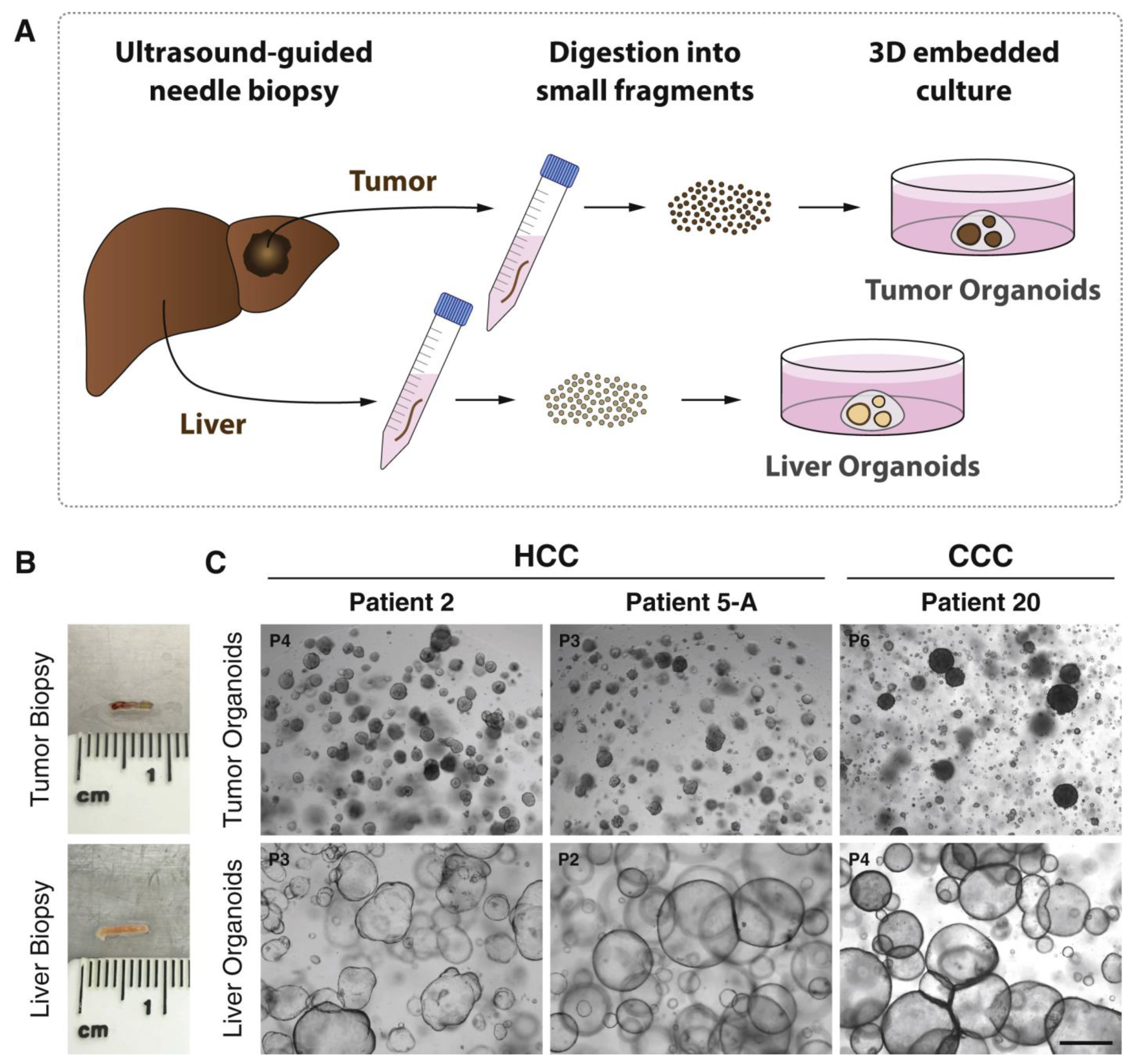
  125. Nuciforo, S.; Fofana, I.; Matter, M.S.; Blumer, T.; Calabrese, D.; Boldanova, T.; Piscuoglio, S.; Wieland, S.; Ringnalda, F.; Schwank, G. Organoid Models of Human Liver Cancers Derived from Tumor Needle Biopsies. Cell Rep. 2018, 24, 1363–1376. [Google Scholar] [CrossRef]
  126. Chong, Y.C.; Toh, T.B.; Chan, Z.; Lin, Q.X.X.; Thng, D.K.H.; Hooi, L.; Ding, Z.; Shuen, T.; Toh, H.C.; Dan, Y.Y.; et al. Targeted Inhibition of Purine Metabolism Is Effective in Suppressing Hepatocellular Carcinoma Progression. Hepatol. Commun. 2020, 4, 1362–1381. [Google Scholar] [CrossRef]
  127. Firestein, R.; Marcinkiewicz, C.; Nie, L.; Chua, H.K.; Velazquez Quesada, I.; Torelli, M.; Sternberg, M.; Gligorijevic, B.; Shenderova, O.; Schirhagl, R.; et al. Pharmacodynamic Studies of Fluorescent Diamond Carriers of Doxorubicin in Liver Cancer Cells and Colorectal Cancer Organoids. Nanotechnol. Sci. Appl. 2021, 14, 139–159. [Google Scholar] [CrossRef] [PubMed]
  128. Jia, D.; Liu, C.; Zhu, Z.; Cao, Y.; Wen, W.; Hong, Z.; Liu, Y.; Liu, E.; Chen, L.; Chen, C.; et al. Novel Transketolase Inhibitor Oroxylin A Suppresses the Non-Oxidative Pentose Phosphate Pathway and Hepatocellular Carcinoma Tumour Growth in Mice and Patient-Derived Organoids. Clin. Transl. Med. 2022, 12, e1095. [Google Scholar] [CrossRef]
  129. Lim, J.J.; Hooi, L.; Dan, Y.Y.; Bonney, G.K.; Zhou, L.; Chow, P.K.-H.; Chee, C.E.; Toh, T.B.; Chow, E.K.-H. Rational Drug Combination Design in Patient-Derived Avatars Reveals Effective Inhibition of Hepatocellular Carcinoma with Proteasome and CDK Inhibitors. J. Exp. Clin. Cancer Res. 2022, 41, 249. [Google Scholar] [CrossRef] [PubMed]
  130. Gandhi, V.; Plunkett, W.; Cortes, J.E. Omacetaxine: A Protein Translation Inhibitor for Treatment of Chronic Myelogenous Leukemia. Clin. Cancer Res. 2014, 20, 1735–1740. [Google Scholar] [CrossRef] [PubMed]
  131. Li, L.; Halpert, G.; Lerner, M.G.; Hu, H.; Dimitrion, P.; Weiss, M.J.; He, J.; Philosophe, B.; Burkhart, R.; Burns, W.R.; et al. Protein Synthesis Inhibitor Omacetaxine Is Effective against Hepatocellular Carcinoma. JCI Insight 2021, 6, e138197. [Google Scholar] [CrossRef]
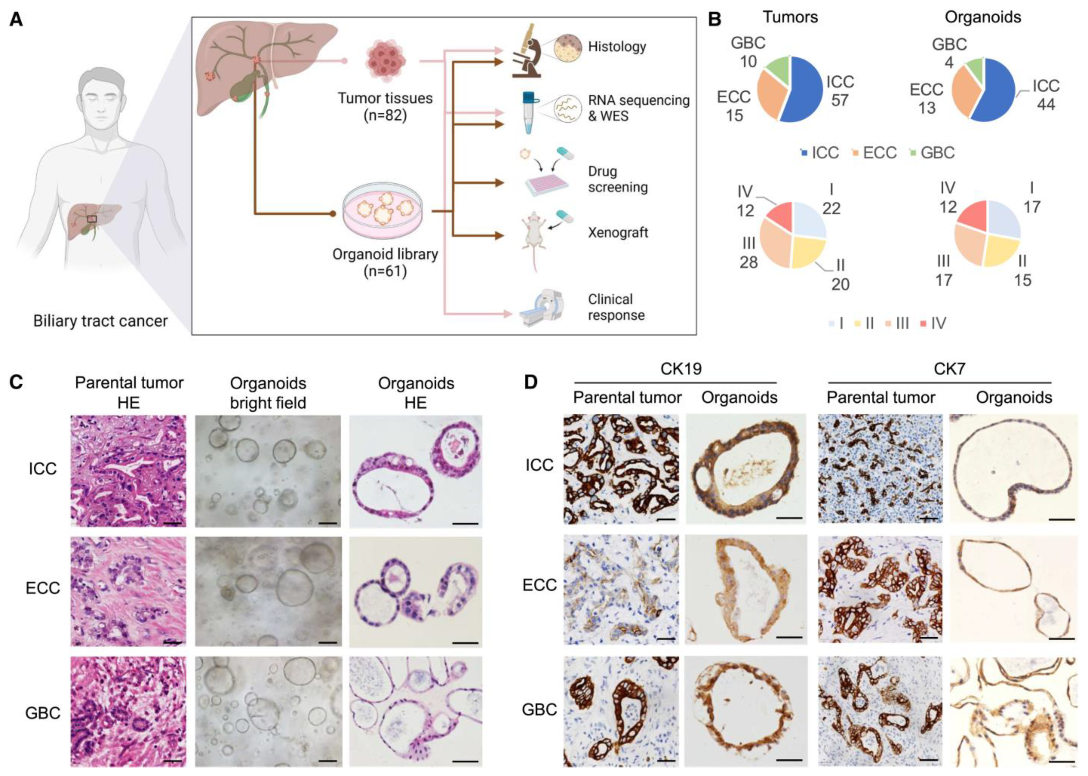
  132. Ren, X.; Huang, M.; Weng, W.; Xie, Y.; Wu, Y.; Zhu, S.; Zhang, Y.; Li, D.; Lai, J.; Shen, S. Personalized Drug Screening in Patient-Derived Organoids of Biliary Tract Cancer and Its Clinical Application. Cell Rep. Med. 2023, 4, 101277. [Google Scholar] [CrossRef] [PubMed]
  133. Sun, L.; Wang, Y.; Cen, J.; Ma, X.; Cui, L.; Qiu, Z.; Zhang, Z.; Li, H.; Yang, R.-Z.; Wang, C.; et al. Modelling Liver Cancer Initiation with Organoids Derived from Directly Reprogrammed Human Hepatocytes. Nat. Cell Biol. 2019, 21, 1015–1026. [Google Scholar] [CrossRef] [PubMed]
  134. Li, L.; Qian, M.; Chen, I.-H.; Finkelstein, D.; Onar-Thomas, A.; Johnson, M.; Calabrese, C.; Bahrami, A.; López-Terrada, D.H.; Yang, J.J.; et al. Acquisition of Cholangiocarcinoma Traits during Advanced Hepatocellular Carcinoma Development in Mice. Am. J. Pathol. 2018, 188, 656–671. [Google Scholar] [CrossRef] [PubMed]
  135. Saito, Y.; Nakaoka, T.; Muramatsu, T.; Ojima, H.; Sukeda, A.; Sugiyama, Y.; Uchida, R.; Furukawa, R.; Kitahara, A.; Sato, T.; et al. Induction of Differentiation of Intrahepatic Cholangiocarcinoma Cells to Functional Hepatocytes Using an Organoid Culture System. Sci. Rep. 2018, 8, 2821. [Google Scholar] [CrossRef] [PubMed]
  136. Porporato, P.E.; Filigheddu, N.; Pedro, J.M.B.-S.; Kroemer, G.; Galluzzi, L. Mitochondrial Metabolism and Cancer. Cell Res. 2018, 28, 265–280. [Google Scholar] [CrossRef] [PubMed]
  137. Li, M.; Wang, L.; Wang, Y.; Zhang, S.; Zhou, G.; Lieshout, R.; Ma, B.; Liu, J.; Qu, C.; Verstegen, M.M.A.; et al. Mitochondrial Fusion Via OPA1 and MFN1 Supports Liver Tumor Cell Metabolism and Growth. Cells 2020, 9, 121. [Google Scholar] [CrossRef]
  138. Yoshikawa, N.; Saito, Y.; Manabe, H.; Nakaoka, T.; Uchida, R.; Furukawa, R.; Muramatsu, T.; Sugiyama, Y.; Kimura, M.; Saito, H. Glucose Depletion Enhances the Stem Cell Phenotype and Gemcitabine Resistance of Cholangiocarcinoma Organoids through AKT Phosphorylation and Reactive Oxygen Species. Cancers 2019, 11, 1993. [Google Scholar] [CrossRef] [PubMed]
  139. Xia, Y.; Guo, H. Hepatitis B Virus cccDNA: Formation, Regulation and Therapeutic Potential. Antivir. Res. 2020, 180, 104824. [Google Scholar] [CrossRef] [PubMed]
  140. Li, P.; Li, Y.; Wang, Y.; Liu, J.; Lavrijsen, M.; Li, Y.; Zhang, R.; Verstegen, M.M.A.; Wang, Y.; Li, T.-C.; et al. Recapitulating Hepatitis E Virus-Host Interactions and Facilitating Antiviral Drug Discovery in Human Liver-Derived Organoids. Sci. Adv. 2022, 8, eabj5908. [Google Scholar] [CrossRef]
  141. Clark, M.P.; Huynh, T.; Rao, S.; Mackiewicz, L.; Mason, H.; Romal, S.; Stutz, M.D.; Ahn, S.H.; Earnest, L.; Sozzi, V.; et al. Clinical Stage Drugs Targeting Inhibitor of Apoptosis Proteins Purge Episomal Hepatitis B Viral Genome in Preclinical Models. Cell Death Dis. 2021, 12, 641. [Google Scholar] [CrossRef]
  142. Baktash, Y.; Madhav, A.; Coller, K.E.; Randall, G. Single Particle Imaging of Polarized Hepatoma Organoids upon Hepatitis C Virus Infection Reveals an Ordered and Sequential Entry Process. Cell Host Microbe 2018, 23, 382–394.e5. [Google Scholar] [CrossRef] [PubMed]
  143. Collett, S.; Torresi, J.; Silveira, L.E.; Truong, V.K.; Christiansen, D.; Tran, B.M.; Vincan, E.; Ramsland, P.A.; Elbourne, A. Investigating Virus-Host Cell Interactions: Comparative Binding Forces between Hepatitis C Virus-like Particles and Host Cell Receptors in 2D and 3D Cell Culture Models. J. Colloid Interface Sci. 2021, 592, 371–384. [Google Scholar] [CrossRef]
  144. Yu, J.H.; Ma, S. Organoids as Research Models for Hepatocellular Carcinoma. Exp. Cell Res. 2022, 411, 112987. [Google Scholar] [CrossRef] [PubMed]
  145. Van de Wetering, M.; Francies, H.E.; Francis, J.M.; Bounova, G.; Iorio, F.; Pronk, A.; van Houdt, W.; van Gorp, J.; Taylor-Weiner, A.; Kester, L. Prospective Derivation of a Living Organoid Biobank of Colorectal Cancer Patients. Cell 2015, 161, 933–945. [Google Scholar] [CrossRef]
  146. Boj, S.F.; Hwang, C.-I.; Baker, L.A.; Chio, I.I.C.; Engle, D.D.; Corbo, V.; Jager, M.; Ponz-Sarvise, M.; Tiriac, H.; Spector, M.S. Organoid Models of Human and Mouse Ductal Pancreatic Cancer. Cell 2015, 160, 324–338. [Google Scholar] [CrossRef]
  147. Broutier, L.; Mastrogiovanni, G.; Verstegen, M.M.; Francies, H.E.; Gavarró, L.M.; Bradshaw, C.R.; Allen, G.E.; Arnes-Benito, R.; Sidorova, O.; Gaspersz, M.P. Human Primary Liver Cancer–Derived Organoid Cultures for Disease Modeling and Drug Screening. Nat. Med. 2017, 23, 1424–1435. [Google Scholar] [CrossRef]
  148. Dong, R.; Zhang, B.; Zhang, X. Liver Organoids: An in Vitro 3D Model for Liver Cancer Study. Cell Biosci. 2022, 12, 1–16. [Google Scholar] [CrossRef]
  149. Sullivan, S.; Stacey, G.N.; Akazawa, C.; Aoyama, N.; Baptista, R.; Bedford, P.; Bennaceur Griscelli, A.; Chandra, A.; Elwood, N.; Girard, M. Quality Control Guidelines for Clinical-Grade Human Induced Pluripotent Stem Cell Lines. Regen. Med. 2018, 13, 859–866. [Google Scholar] [CrossRef]
  150. Yoshihara, M.; Hayashizaki, Y.; Murakawa, Y. Genomic Instability of iPSCs: Challenges towards Their Clinical Applications. Stem Cell Rev. Rep. 2017, 13, 7–16. [Google Scholar] [CrossRef] [PubMed]
  151. Kim, K.; Doi, A.; Wen, B.; Ng, K.; Zhao, R.; Cahan, P.; Kim, J.; Aryee, M.; Ji, H.; Ehrlich, L. Epigenetic Memory in Induced Pluripotent Stem Cells. Nature 2010, 467, 285–290. [Google Scholar] [CrossRef]
Figure 2. Conceptual diagram of the process of preparing cells from various sources to produce liver organoids. Abbreviations: Rspo, R-Spondin; TGFb-I, transforming growth factor beta inhibitor; TNFα, tumor necrosis factor alpha; ESCs, embryonic stem cells; FGF, fibroblast growth factor; FSK, forskolin; HGF, hepatocyte growth factor; iPSCs, induced pluripotent stem cells; Act A, activin A; BMP4, bone morphogenetic protein 4; DMX, dexamethasone; ECM, extracellular matrix; EGF, epidermal growth factor; OSM, oncostatin M; RA, retinoic acid.
Bioengineering 11 00346 g002
Disclaimer/Publisher’s Note: The statements, opinions and data contained in all publications are solely those of the individual author(s) and contributor(s) and not of MDPI and/or the editor(s). MDPI and/or the editor(s) disclaim responsibility for any injury to people or property resulting from any ideas, methods, instructions or products referred to in the content.

