
Three-Dimensional Bioprinting in Soft Tissue Engineering for Plastic and Reconstructive Surgery

Abstract
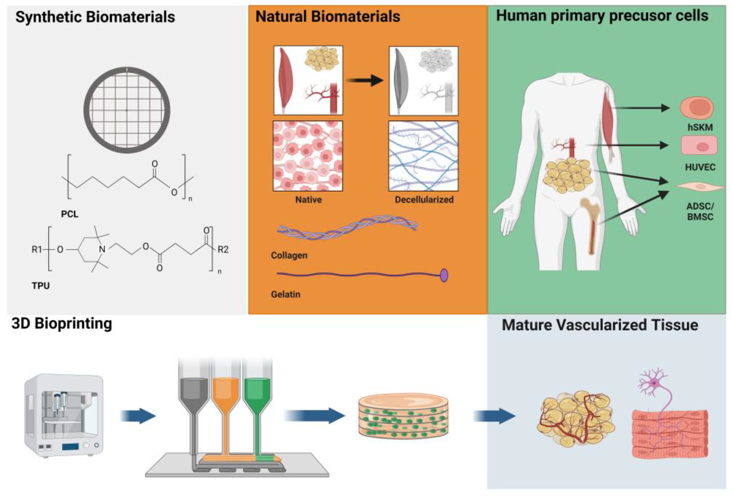
:1. Introduction
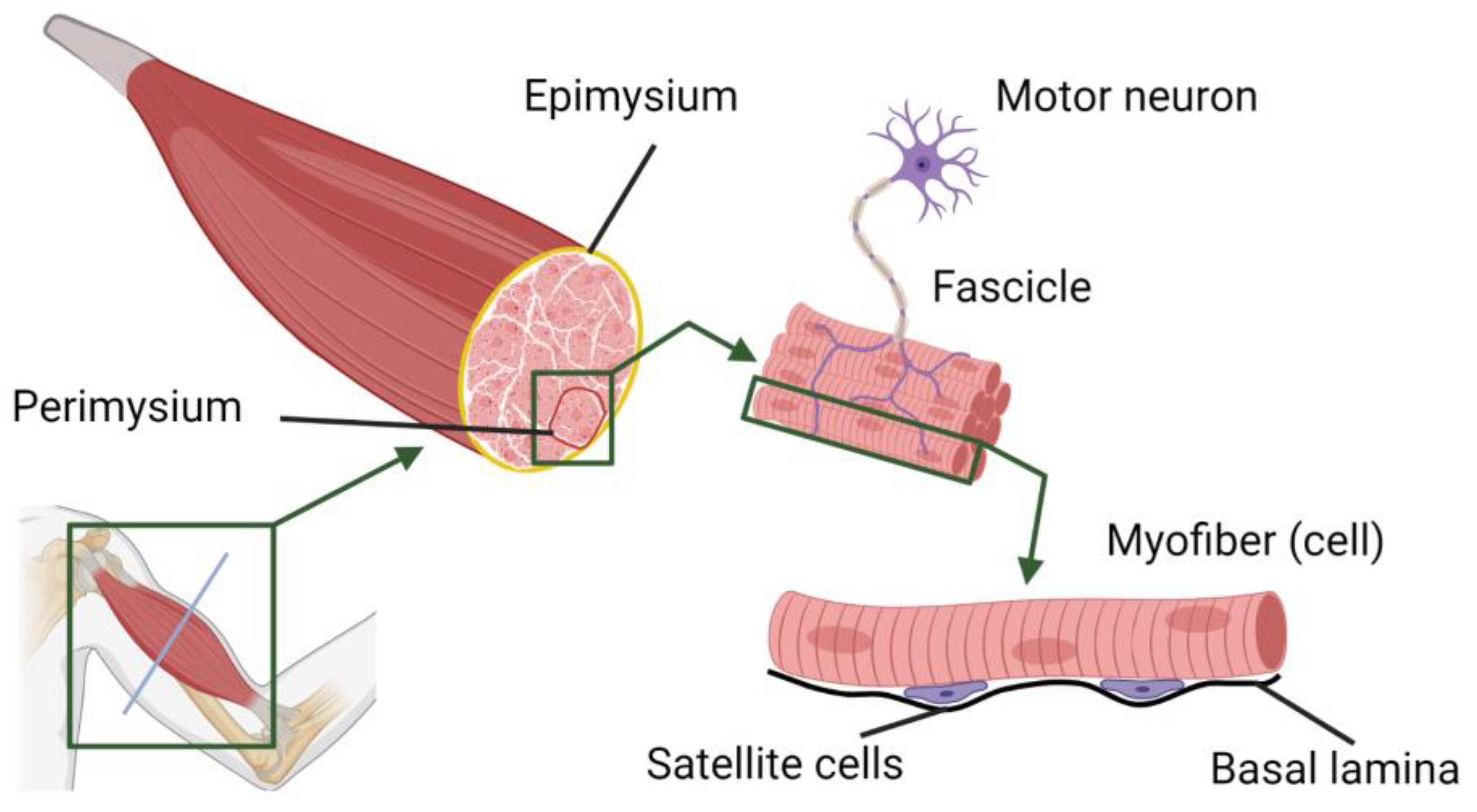
2. Skeletal Muscle Tissue Engineering
2.1. Cellular Aspects of Skeletal Muscle TE
2.2. Three-Dimensional (Bio) Printing and Skeletal Muscle TE
3. Adipose Tissue Engineering

| Paper | Cell Type | Bioink | Experiment Type | Key Findings | Limitations |
|---|---|---|---|---|---|
| Pati et al. (2015) [63] | hADSC | PCL + hdECM | In vitro | Successful cultivation of the cells and differentiation | Mechanical properties do not match adipose tissue |
| In vivo murine VML model | dECM showed proangiogenic effect, printed scaffold superior over non-printed | Small size of construct | |||
| Ahn et al. (2022) [65] | hADSC | hdECM + Alginate | In vitro | In-bath hybrid printing technique superior to 3D printing; culturing of functional adipose tissue | Low dimensions of the tissue, no in vivo application |
| Lee et al. (2021) [64] | - | PCL + mixture of collagen type I and hdECM hydrogels | In vivo murine model | hdECM hydrogel promotes neovascularization and tissue formation | Small size of construct |
| Van Damme et al. (2020) [66] | - | GelMa + PLA (sacrificial) | In silico | Comparison of indirect vs. direct printing technique -> similar results regarding mechanical properties | No biological testing |
| Negrini et al. (2019) [67] | hMSC * | Alginate microbeads (sacrificial) MBA crosslinked gelatin hydrogel | In vitro | Microporous gelatin hydrogels, suitable as scaffolds for AT (porosity, mechanical properties, enzymatic degradability, and hMSC proliferation and differentiation) | In vivo application pending |
| Ex vivo | Perfusable vascular channel in the scaffold | ||||
| Negrini et al. (2020) [70] | hADSC | MBA crosslinked gelatin hydrogel | In vitro | Physical and mechanical properties for use as AT scaffolds Support cell proliferation and differentiation | In vivo application pending |
| Säljö et al. (2022) [62] | Stroma vascular fraction | Alginate and nanocellulose | In vivo murine model | Printability of mechanically purified lipoaspirate and in vivo long-term survival | Control group missing, formation of fibrotic tissue rather than mature adipose tissue |
4. Vascularization
| Paper | Cell Type | Bioink | Experiment Type | Key Findings | Limitations |
|---|---|---|---|---|---|
| Sousa et al. (2021) [76] | HUVEC | Alginate (sacrificial) and photocrosslinkable glycidyl methacrylated xanthan gum (XG-GMA) | In vitro | Layer-by-layer-coated 3D-printed perfusable microchannels embedded in XG-GMA hydrogels | No in vivo investigation, upscaling needed |
| Shao et al. (2020) [77] | HUVEC, MC3T3-E1 (mouse osteoblast cell line) | GelMa, gelatin (sacrificial) | In vitro | Synchronous 3D bioprinting of cell-laden constructs with nutrient networks, construct size up to 3 × 3 × 3 cm3 | Cultivation with osteoblast with no investigation for differentiation, no in vivo application |
| Machour et al. (2022) [71] | Human adipose microvascular endothelial cells + dental pulp stem cells | Recombinant human collagen methacrylate (rhCollMA) hydrogel, PLLA + PLGA | In vivo rat model | Hierarchical vessel network composed of microscale and mesoscale vasculatures, anastomosis with rat femoral artery | Proof of principle of the anastomosis, studies on successful in vivo tissue engineering pending, small size of the construct |
| Szklanny et al. (2021) [79] | Human adipose microvascular endothelial cells + dental pulp stem cells iPS-derived cardiomyocytes | Recombinant human collagen methacrylate (rhCollMA) hydrogel, PLLA + PLGA | In vitro | Supply of nutrients to differentiated and functional cardiomyocytes via the vascular network | |
| In vivo rat model | Anastomosis with rat femoral artery | ||||
| Kolesky et al. (2016) [78] | HUVEC, hMSC * | Pluronic F-127 and thrombin (sacrificial); gelatin and fibrinogen | In vitro | Creation of thick human tissues (>1 cm) replete with an engineered extracellular matrix, embedded vasculature, and multiple cell types | No in vivo investigation, upscaling needed |
| Kreimendahl et al. (2021) [75] | HUVECs + HDFs | Fibrin + hyaluronic acid | In vitro | Use of FRESH printing technique: enables printing of low-viscose natural polymers with high shape stability, formation of a vascular network | No in vivo investigation, upscaling needed |
| Li et al. (2020) [80] | C3A | Alginate + silk fibroin | In vitro | Development of mechanically improved bioink. Scaffold with hierarchical microchannel network | No in vivo investigation, in vitro testing with cell line |
| Erdem et al. (2020) [74] | 3T3 fibroblasts or rat cardiomyocytes | GelMa + CPO | In vitro | Development of a printable, O2 delivering biomaterial, cell viability under hypoxia was similar to normoxic conditions when CPO was added | No in vivo investigation, small size of the construct |
5. Conclusions
Author Contributions
Funding
Institutional Review Board Statement
Informed Consent Statement
Data Availability Statement
Conflicts of Interest
References
- Lin, C.H.; Lin, Y.T.; Yeh, J.T.; Chen, C.T. Free Functioning Muscle Transfer for Lower Extremity Posttraumatic Composite Structure and Functional Defect. Plast. Reconstr. Surg. 2007, 119, 2118–2126. [Google Scholar] [CrossRef] [PubMed]
- Tordjman, D.; d’Utruy, A.; Bauer, B.; Bellemère, P.; Pierrart, J.; Masmejean, E. Tendon Transfer Surgery for Radial Nerve Palsy. Hand Surg. Rehabil. 2022, 41, S90–S97. [Google Scholar] [CrossRef] [PubMed]
- Mughal, M.; Sindali, K.; Man, J.; Roblin, P. ‘Fat Chance’: A Review of Adipose Tissue Engineering and Its Role in Plastic and Reconstructive Surgery. Ann. R. Coll. Surg. Engl. 2021, 103, 245–249. [Google Scholar] [CrossRef] [PubMed]
- O’Brien, F.J. Biomaterials & Scaffolds for Tissue Engineering. Mater. Today 2011, 14, 88–95. [Google Scholar]
- Whitaker, M.J.; Quirk, R.A.; Howdle, S.M.; Shakesheff, K.M. Growth Factor Release from Tissue Engineering Scaffolds. J. Pharm. Pharmacol. 2010, 53, 1427–1437. [Google Scholar] [CrossRef] [PubMed]
- Lee, Y.-B.; Polio, S.; Lee, W.; Dai, G.; Menon, L.; Carroll, R.S.; Yoo, S.-S. Bio-Printing of Collagen and VEGF-Releasing Fibrin Gel Scaffolds for Neural Stem Cell Culture. Exp. Neurol. 2010, 223, 645–652. [Google Scholar] [CrossRef] [PubMed]
- Yang, G.; Mahadik, B.; Choi, J.Y.; Fisher, J.P. Vascularization in Tissue Engineering: Fundamentals and State-of-Art. Prog. Biomed. Eng. 2020, 2, 012002. [Google Scholar] [CrossRef] [PubMed]
- Weigand, A.; Horch, R.E.; Boos, A.M.; Beier, J.P.; Arkudas, A. The Arteriovenous Loop: Engineering of Axially Vascularized Tissue. Eur. Surg. Res. 2018, 59, 286–299. [Google Scholar] [CrossRef]
- Sun, W.; Gregory, D.A.; Tomeh, M.A.; Zhao, X. Silk Fibroin as a Functional Biomaterial for Tissue Engineering. Int. J. Mol. Sci. 2021, 22, 1499. [Google Scholar] [CrossRef]
- Eldeeb, A.E.; Salah, S.; Elkasabgy, N.A. Biomaterials for Tissue Engineering Applications and Current Updates in the Field: A Comprehensive Review. AAPS PharmSciTech 2022, 23, 267. [Google Scholar] [CrossRef]
- Samal, J.; Weinandy, S.; Weinandy, A.; Helmedag, M.; Rongen, L.; Hermanns-Sachweh, B.; Kundu, S.C.; Jockenhoevel, S. Co-Culture of Human Endothelial Cells and Foreskin Fibroblasts on 3D Silk-Fibrin Scaffolds Supports Vascularization. Macromol. Biosci. 2015, 15, 1433–1446. [Google Scholar] [CrossRef]
- El Maachi, I.; Kyriakou, S.; Rütten, S.; Kopp, A.; Köpf, M.; Jockenhoevel, S.; Fernández-Colino, A. Silk Fibroin as Adjuvant in the Fabrication of Mechanically Stable Fibrin Biocomposites. Polymers 2022, 14, 2251. [Google Scholar] [CrossRef] [PubMed]
- Pasquier, E.; Rosendahl, J.; Solberg, A.; Ståhlberg, A.; Håkansson, J.; Chinga-Carrasco, G. Polysaccharides and Structural Proteins as Components in Three-Dimensional Scaffolds for Breast Cancer Tissue Models: A Review. Bioengineering 2023, 10, 682. [Google Scholar] [CrossRef] [PubMed]
- Zhu, W.; Ma, X.; Gou, M.; Mei, D.; Zhang, K.; Chen, S. 3D Printing of Functional Biomaterials for Tissue Engineering. Curr. Opin. Biotechnol. 2016, 40, 103–112. [Google Scholar] [CrossRef] [PubMed]
- Jiang, W.; Mei, H.; Zhao, S. Applications of 3D Bio-Printing in Tissue Engineering and Biomedicine. J. Biomed. Nanotechnol. 2021, 17, 989–1006. [Google Scholar] [CrossRef] [PubMed]
- Cui, X.; Gao, G.; Qiu, Y. Accelerated Myotube Formation Using Bioprinting Technology for Biosensor Applications. Biotechnol. Lett. 2013, 35, 315–321. [Google Scholar] [CrossRef] [PubMed]
- Moroni, L.; Burdick, J.A.; Highley, C.; Lee, S.J.; Morimoto, Y.; Takeuchi, S.; Yoo, J.J. Biofabrication Strategies for 3D in Vitro Models and Regenerative Medicine. Nat. Rev. Mater. 2018, 3, 21–37. [Google Scholar] [CrossRef]
- Hao, Y.; Cao, B.; Deng, L.; Li, J.; Ran, Z.; Wu, J.; Pang, B.; Tan, J.; Luo, D.; Wu, W. The First 3D-Bioprinted Personalized Active Bone to Repair Bone Defects: A Case Report. Int. J. Bioprint. 2022, 9, 70–75. [Google Scholar] [CrossRef]
- Słynarski, K.; de Jong, W.C.; Snow, M.; Hendriks, J.A.A.; Wilson, C.E.; Verdonk, P. Single-Stage Autologous Chondrocyte-Based Treatment for the Repair of Knee Cartilage Lesions: Two-Year Follow-up of a Prospective Single-Arm Multicenter Study. Am. J. Sports Med. 2020, 48, 1327–1337. [Google Scholar] [CrossRef]
- Zhu, J.; Wang, Y.; Zhong, L.; Pan, F.; Wang, J. Advances in Tissue Engineering of Vasculature through Three-dimensional Bioprinting. Dev. Dyn. 2021, 250, 1717–1738. [Google Scholar] [CrossRef]
- Frontera, W.R.; Ochala, J. Skeletal Muscle: A Brief Review of Structure and Function. Calcif. Tissue Int. 2015, 96, 183–195. [Google Scholar] [CrossRef] [PubMed]
- Pien, N.; Krzyslak, H.; Shastry Kallaje, S.; Van Meerssche, J.; Mantovani, D.; De Schauwer, C.; Dubruel, P.; Van Vlierberghe, S.; Pennisi, C.P. Tissue Engineering of Skeletal Muscle, Tendons and Nerves: A Review of Manufacturing Strategies to Meet Structural and Functional Requirements. Appl. Mater. Today 2023, 31, 101737. [Google Scholar] [CrossRef]
- Gotti, C.; Sensini, A.; Fornaia, G.; Gualandi, C.; Zucchelli, A.; Focarete, M.L. Biomimetic Hierarchically Arranged Nanofibrous Structures Resembling the Architecture and the Passive Mechanical Properties of Skeletal Muscles: A Step Forward Toward Artificial Muscle. Front Bioeng. Biotechnol. 2020, 8, 767. [Google Scholar] [CrossRef] [PubMed]
- Samandari, M.; Quint, J.; Rodríguez-delaRosa, A.; Sinha, I.; Pourquié, O.; Tamayol, A. Bioinks and Bioprinting Strategies for Skeletal Muscle Tissue Engineering. Adv. Mater. 2022, 34, 2105883. [Google Scholar] [CrossRef] [PubMed]
- Volpi, M.; Paradiso, A.; Costantini, M.; Świȩszkowski, W. Hydrogel-Based Fiber Biofabrication Techniques for Skeletal Muscle Tissue Engineering. ACS Biomater. Sci. Eng. 2022, 8, 379–405. [Google Scholar] [CrossRef] [PubMed]
- Ostrovidov, S.; Salehi, S.; Costantini, M.; Suthiwanich, K.; Ebrahimi, M.; Sadeghian, R.B.; Fujie, T.; Shi, X.; Cannata, S.; Gargioli, C.; et al. 3D Bioprinting in Skeletal Muscle Tissue Engineering. Small 2019, 15, 1805530. [Google Scholar] [CrossRef] [PubMed]
- Kim, J.H.; Seol, Y.-J.; Ko, I.K.; Kang, H.-W.; Lee, Y.K.; Yoo, J.J.; Atala, A.; Lee, S.J. 3D Bioprinted Human Skeletal Muscle Constructs for Muscle Function Restoration. Sci. Rep. 2018, 8, 12307. [Google Scholar] [CrossRef] [PubMed]
- Fan, T.; Wang, S.; Jiang, Z.; Ji, S.; Cao, W.; Liu, W.; Ji, Y.; Li, Y.; Shyh-Chang, N.; Gu, Q. Controllable Assembly of Skeletal Muscle-like Bundles through 3D Bioprinting. Biofabrication 2022, 14, 015009. [Google Scholar] [CrossRef]
- Ronzoni, F.L.; Aliberti, F.; Scocozza, F.; Benedetti, L.; Auricchio, F.; Sampaolesi, M.; Cusella, G.; Redwan, I.N.; Ceccarelli, G.; Conti, M. Myoblast 3D Bioprinting to Burst in Vitro Skeletal Muscle Differentiation. J. Tissue Eng. Regen. Med. 2022, 16, 484–495. [Google Scholar] [CrossRef]
- Wong, A.; Pomerantz, J.H. The Role of Muscle Stem Cells in Regeneration and Recovery after Denervation: A Review. Plast. Reconstr. Surg. 2019, 143, 779–788. [Google Scholar] [CrossRef]
- Forcina, L.; Miano, C.; Pelosi, L.; Musarò, A. An Overview About the Biology of Skeletal Muscle Satellite Cells. Curr. Genom. 2019, 20, 24–37. [Google Scholar] [CrossRef] [PubMed]
- Le Grand, F.; Rudnicki, M.A. Skeletal Muscle Satellite Cells and Adult Myogenesis. Curr. Opin. Cell Biol. 2007, 19, 628–633. [Google Scholar] [CrossRef] [PubMed]
- Robey, P. “Mesenchymal Stem Cells”: Fact or Fiction, and Implications in Their Therapeutic Use. F1000Research 2017, 6, 524. [Google Scholar] [CrossRef] [PubMed]
- Cai, A.; Hardt, M.; Schneider, P.; Schmid, R.; Lange, C.; Dippold, D.; Schubert, D.W.; Boos, A.M.; Weigand, A.; Arkudas, A.; et al. Myogenic Differentiation of Primary Myoblasts and Mesenchymal Stromal Cells under Serum-Free Conditions on PCL-Collagen I-Nanoscaffolds. BMC Biotechnol. 2018, 18, 75. [Google Scholar] [CrossRef] [PubMed]
- Bajek, A.; Olkowska, J.; Walentowicz-Sadłecka, M.; Sadłecki, P.; Grabiec, M.; Porowińska, D.; Drewa, T.; Roszkowski, K. Human Adipose-Derived and Amniotic Fluid-Derived Stem Cells: A Preliminary in Vitro Study Comparing Myogenic Differentiation Capability. Med. Sci. Monit. 2018, 24, 1733–1741. [Google Scholar] [CrossRef] [PubMed]
- Pittenger, M.F.; Mackay, A.M.; Beck, S.C.; Jaiswal, R.K.; Douglas, R.; Mosca, J.D.; Moorman, M.A.; Simonetti, D.W.; Craig, S.; Marshak, D.R. Multilineage Potential of Adult Human Mesenchymal Stem Cells. Science 1999, 284, 143–147. [Google Scholar] [CrossRef] [PubMed]
- De La Garza-Rodea, A.S.; Van Der Velde-Van Dijke, I.; Boersma, H.; Gonçalves, M.A.F.V.; Van Bekkum, D.W.; De Vries, A.A.F.; Knaän-Shanzer, S. Myogenic Properties of Human Mesenchymal Stem Cells Derived from Three Different Sources. Cell Transplant. 2012, 21, 153–173. [Google Scholar] [CrossRef] [PubMed]
- Stern-Straeter, J.; Bonaterra, G.A.; Juritz, S.; Birk, R.; Goessler, U.R.; Bieback, K.; Bugert, P.; Schultz, J.; Hörmann, K.; Kinscherf, R.; et al. Evaluation of the Effects of Different Culture Media on the Myogenic Differentiation Potential of Adipose Tissue- or Bone Marrow-Derived Human Mesenchymal Stem Cells. Int. J. Mol. Med. 2014, 33, 160–170. [Google Scholar] [CrossRef]
- Lesman, A.; Koffler, J.; Atlas, R.; Blinder, Y.J.; Kam, Z.; Levenberg, S. Engineering Vessel-like Networks within Multicellular Fibrin-Based Constructs. Biomaterials 2011, 32, 7856–7869. [Google Scholar] [CrossRef]
- Lesman, A.; Habib, M.; Caspi, O.; Gepstein, A.; Arbel, G.; Levenberg, S.; Gepstein, L. Transplantation of a Tissue-Engineered Human Vascularized Cardiac Muscle. Tissue Eng. Part A 2010, 16, 115–125. [Google Scholar] [CrossRef]
- Bach, A.D.; Beier, J.P.; Stark, G.B. Expression of Trisk 51, Agrin and Nicotinic-Acetycholine Receptor?-Subunit during Muscle Development in a Novel Three-Dimensional Muscle-Neuronal Co-Culture System. Cell Tissue Res. 2003, 314, 263–274. [Google Scholar] [CrossRef] [PubMed]
- Bitto, F.F.; Klumpp, D.; Lange, C.; Boos, A.M.; Arkudas, A.; Bleiziffer, O.; Horch, R.E.; Kneser, U.; Beier, J.P. Myogenic Differentiation of Mesenchymal Stem Cells in a Newly Developed Neurotised AV-Loop Model. Biomed Res. Int. 2013, 2013, 935046. [Google Scholar] [CrossRef] [PubMed]
- Yaffe, D.; Saxel, O. Serial Passaging and Differentiation of Myogenic Cells Isolated from Dystrophic Mouse Muscle. Nature 1977, 270, 725–727. [Google Scholar] [CrossRef] [PubMed]
- Bukowska, J.; Szóstek-Mioduchowska, A.Z.; Kopcewicz, M.; Walendzik, K.; Machcińska, S.; Gawrońska-Kozak, B. Adipose-Derived Stromal/Stem Cells from Large Animal Models: From Basic to Applied Science. Stem Cell Rev. Rep. 2021, 17, 719–738. [Google Scholar] [CrossRef] [PubMed]
- Gokyer, S.; Yilgor, E.; Yilgor, I.; Berber, E.; Vrana, E.; Orhan, K.; Monsef, Y.A.; Guvener, O.; Zinnuroglu, M.; Oto, C.; et al. 3D Printed Biodegradable Polyurethaneurea Elastomer Recapitulates Skeletal Muscle Structure and Function. ACS Biomater. Sci. Eng. 2021, 7, 5189–5205. [Google Scholar] [CrossRef] [PubMed]
- Pati, F.; Jang, J.; Ha, D.-H.; Won Kim, S.; Rhie, J.-W.; Shim, J.-H.; Kim, D.-H.; Cho, D.-W. Printing Three-Dimensional Tissue Analogues with Decellularized Extracellular Matrix Bioink. Nat. Commun. 2014, 5, 3935. [Google Scholar] [CrossRef] [PubMed]
- Kiratitanaporn, W.; Berry, D.B.; Mudla, A.; Fried, T.; Lao, A.; Yu, C.; Hao, N.; Ward, S.R.; Chen, S. 3D Printing a Biocompatible Elastomer for Modeling Muscle Regeneration after Volumetric Muscle Loss. Biomater. Adv. 2022, 142, 213171. [Google Scholar] [CrossRef]
- Choi, Y.J.; Kim, T.G.; Jeong, J.; Yi, H.G.; Park, J.W.; Hwang, W.; Cho, D.W. 3D Cell Printing of Functional Skeletal Muscle Constructs Using Skeletal Muscle-Derived Bioink. Adv. Healthc. Mater. 2016, 5, 2636–2645. [Google Scholar] [CrossRef]
- Kim, W.J.; Lee, H.; Lee, J.U.; Atala, A.; Yoo, J.J.; Lee, S.J.; Kim, G.H. Efficient Myotube Formation in 3D Bioprinted Tissue Construct by Biochemical and Topographical Cues. Biomaterials 2020, 230, 119632. [Google Scholar] [CrossRef]
- Boso, D.; Maghin, E.; Carraro, E.; Giagante, M.; Pavan, P.; Piccoli, M. Extracellular Matrix-Derived Hydrogels as Biomaterial for Different Skeletal Muscle Tissue Replacements. Materials 2020, 13, 2483. [Google Scholar] [CrossRef]
- Russell, C.S.; Mostafavi, A.; Quint, J.P.; Panayi, A.C.; Baldino, K.; Williams, T.J.; Daubendiek, J.G.; Hugo Sánchez, V.; Bonick, Z.; Trujillo-Miranda, M.; et al. In Situ Printing of Adhesive Hydrogel Scaffolds for the Treatment of Skeletal Muscle Injuries. ACS Appl. Bio Mater. 2020, 3, 1568–1579. [Google Scholar] [CrossRef] [PubMed]
- Juhas, M.; Engelmayr, G.C.; Fontanella, A.N.; Palmer, G.M.; Bursac, N. Biomimetic Engineered Muscle with Capacity for Vascular Integration and Functional Maturation in Vivo. Proc. Natl. Acad. Sci. USA 2014, 111, 5508–5513. [Google Scholar] [CrossRef] [PubMed]
- Jain, R.K.; Au, P.; Tam, J.; Duda, D.G.; Fukumura, D. Engineering Vascularized Tissue. Nat. Biotechnol. 2005, 23, 821–823. [Google Scholar] [CrossRef] [PubMed]
- Levenberg, S.; Rouwkema, J.; Macdonald, M.; Garfein, E.S.; Kohane, D.S.; Darland, D.C.; Marini, R.; Van Blitterswijk, C.A.; Mulligan, R.C.; D’Amore, P.A.; et al. Engineering Vascularized Skeletal Muscle Tissue. Nat. Biotechnol. 2005, 23, 879–884. [Google Scholar] [CrossRef] [PubMed]
- Choi, Y.J.; Jun, Y.J.; Kim, D.Y.; Yi, H.G.; Chae, S.H.; Kang, J.; Lee, J.; Gao, G.; Kong, J.S.; Jang, J.; et al. A 3D Cell Printed Muscle Construct with Tissue-Derived Bioink for the Treatment of Volumetric Muscle Loss. Biomaterials 2019, 206, 160–169. [Google Scholar] [CrossRef] [PubMed]
- Fornetti, E.; De Paolis, F.; Fuoco, C.; Bernardini, S.; Giannitelli, S.M.; Rainer, A.; Seliktar, D.; Magdinier, F.; Baldi, J.; Biagini, R.; et al. A Novel Extrusion-Based 3D Bioprinting System for Skeletal Muscle Tissue Engineering. Biofabrication 2023, 15, 025009. [Google Scholar] [CrossRef] [PubMed]
- Hwangbo, H.; Lee, H.; Jin, E.; Jo, Y.; Son, J.; Woo, H.M.; Ryu, D.; Kim, G.H. Photosynthetic Cyanobacteria Can Clearly Induce Efficient Muscle Tissue Regeneration of Bioprinted Cell-Constructs. Adv. Funct. Mater. 2023, 33, 2209157. [Google Scholar] [CrossRef]
- Wang, Y.; Wu, Y. Assessment of the Clinical Efficacy of Cell-Assisted Lipotransfer and Conventional Fat Graft: A Meta-Analysis Based on Case-Control Studies. J. Orthop. Surg. Res. 2017, 12, 155. [Google Scholar] [CrossRef]
- Toyserkani, N.M.; Quaade, M.L.; Sørensen, J.A. Cell-Assisted Lipotransfer: A Systematic Review of Its Efficacy. Aesthetic Plast. Surg. 2016, 40, 309–318. [Google Scholar] [CrossRef]
- Ghiasloo, M.; Lobato, R.C.; Díaz, J.M.; Singh, K.; Verpaele, A.; Tonnard, P. Expanding Clinical Indications of Mechanically Isolated Stromal Vascular Fraction: A Systematic Review. Aesthet. Surg. J. 2020, 40, NP546–NP560. [Google Scholar] [CrossRef]
- Tanzi, M.C.; Farè, S. Adipose Tissue Engineering: State of the Art, Recent Advances and Innovative Approaches. Expert Rev. Med. Devices 2009, 6, 533–551. [Google Scholar] [CrossRef] [PubMed]
- Säljö, K.; Apelgren, P.; Stridh Orrhult, L.; Li, S.; Amoroso, M.; Gatenholm, P.; Kölby, L. Long-Term in Vivo Survival of 3D-Bioprinted Human Lipoaspirate-Derived Adipose Tissue: Proteomic Signature and Cellular Content. Adipocyte 2022, 11, 34–46. [Google Scholar] [CrossRef]
- Pati, F.; Ha, D.-H.; Jang, J.; Han, H.H.; Rhie, J.-W.; Cho, D.-W. Biomimetic 3D Tissue Printing for Soft Tissue Regeneration. Biomaterials 2015, 62, 164–175. [Google Scholar] [CrossRef] [PubMed]
- Lee, S.; Lee, H.S.; Chung, J.J.; Kim, S.H.; Park, J.W.; Lee, K.; Jung, Y. Enhanced Regeneration of Vascularized Adipose Tissue with Dual 3D-Printed Elastic Polymer/DECM Hydrogel Complex. Int. J. Mol. Sci. 2021, 22, 2886. [Google Scholar] [CrossRef] [PubMed]
- Ahn, M.; Cho, W.; Kim, B.S.; Cho, D. Engineering Densely Packed Adipose Tissue via Environmentally Controlled In-Bath 3D Bioprinting. Adv. Funct. Mater. 2022, 32, 2200203. [Google Scholar] [CrossRef]
- Van Damme, L.; Briant, E.; Blondeel, P.; Van Vlierberghe, S. Indirect versus Direct 3D Printing of Hydrogel Scaffolds for Adipose Tissue Regeneration Lana Van Damme, Emilie Briant, Phillip Blondeel, Sandra Van Vlierberghe. MRS Adv. 2020, 5, 855–864. [Google Scholar] [CrossRef]
- Contessi Negrini, N.; Bonnetier, M.; Giatsidis, G.; Orgill, D.P.; Farè, S.; Marelli, B. Tissue-Mimicking Gelatin Scaffolds by Alginate Sacrificial Templates for Adipose Tissue Engineering. Acta Biomater. 2019, 87, 61–75. [Google Scholar] [CrossRef] [PubMed]
- Peirsman, A.; Nguyen, H.T.; Van Waeyenberge, M.; Ceballos, C.; Bolivar, J.; Kawakita, S.; Vanlauwe, F.; Tirpáková, Z.; Van Dorpe, S.; Van Damme, L.; et al. Vascularized Adipose Tissue Engineering: Moving towards Soft Tissue Reconstruction. Biofabrication 2023, 15, 032003. [Google Scholar] [CrossRef]
- Benmeridja, L.; De Moor, L.; De Maere, E.; Vanlauwe, F.; Ryx, M.; Tytgat, L.; Vercruysse, C.; Dubruel, P.; Van Vlierberghe, S.; Blondeel, P.; et al. High-throughput Fabrication of Vascularized Adipose Microtissues for 3D Bioprinting. J. Tissue Eng. Regen. Med. 2020, 14, 840–854. [Google Scholar] [CrossRef]
- Contessi Negrini, N.; Celikkin, N.; Tarsini, P.; Farè, S.; Święszkowski, W. Three-Dimensional Printing of Chemically Crosslinked Gelatin Hydrogels for Adipose Tissue Engineering. Biofabrication 2020, 12, 025001. [Google Scholar] [CrossRef]
- Machour, M.; Szklanny, A.A.; Levenberg, S. Fabrication of Engineered Vascular Flaps Using 3D Printing Technologies. J. Vis. Exp. 2022, 183, e63920. [Google Scholar] [CrossRef]
- Askari, M.; Afzali Naniz, M.; Kouhi, M.; Saberi, A.; Zolfagharian, A.; Bodaghi, M. Recent Progress in Extrusion 3D Bioprinting of Hydrogel Biomaterials for Tissue Regeneration: A Comprehensive Review with Focus on Advanced Fabrication Techniques. Biomater. Sci. 2021, 9, 535–573. [Google Scholar] [CrossRef] [PubMed]
- Erdem, A.; Haghniaz, R.; Ertas, Y.N.; Sangabathuni, S.K.; Nasr, A.S.; Swieszkowski, W.; Ashammakhi, N. Methods for Fabricating Oxygen Releasing Biomaterials. J. Drug Target. 2022, 30, 188–199. [Google Scholar] [CrossRef] [PubMed]
- Erdem, A.; Darabi, M.A.; Nasiri, R.; Sangabathuni, S.; Ertas, Y.N.; Alem, H.; Hosseini, V.; Shamloo, A.; Nasr, A.S.; Ahadian, S.; et al. 3D Bioprinting of Oxygenated Cell-laden Gelatin Methacryloyl Constructs. Adv. Healthc. Mater. 2020, 9, e1901794. [Google Scholar] [CrossRef] [PubMed]
- Kreimendahl, F.; Kniebs, C.; Tavares Sobreiro, A.M.; Schmitz-Rode, T.; Jockenhoevel, S.; Thiebes, A.L. FRESH Bioprinting Technology for Tissue Engineering—The Influence of Printing Process and Bioink Composition on Cell Behavior and Vascularization. J. Appl. Biomater. Funct. Mater. 2021, 19, 22808000211028810. [Google Scholar] [CrossRef] [PubMed]
- Sousa, C.F.V.; Saraiva, C.A.; Correia, T.R.; Pesqueira, T.; Patrício, S.G.; Rial-Hermida, M.I.; Borges, J.; Mano, J.F. Bioinstructive Layer-by-Layer-Coated Customizable 3D Printed Perfusable Microchannels Embedded in Photocrosslinkable Hydrogels for Vascular Tissue Engineering. Biomolecules 2021, 11, 863. [Google Scholar] [CrossRef] [PubMed]
- Shao, L.; Gao, Q.; Xie, C.; Fu, J.; Xiang, M.; He, Y. Synchronous 3D Bioprinting of Large-Scale Cell-Laden Constructs with Nutrient Networks. Adv. Healthc. Mater. 2020, 9, 1901142. [Google Scholar] [CrossRef] [PubMed]
- Kolesky, D.B.; Homan, K.A.; Skylar-Scott, M.A.; Lewis, J.A. Three-Dimensional Bioprinting of Thick Vascularized Tissues. Proc. Natl. Acad. Sci. USA 2016, 113, 3179–3184. [Google Scholar] [CrossRef] [PubMed]
- Szklanny, A.A.; Machour, M.; Redenski, I.; Chochola, V.; Goldfracht, I.; Kaplan, B.; Epshtein, M.; Simaan Yameen, H.; Merdler, U.; Feinberg, A.; et al. 3D Bioprinting of Engineered Tissue Flaps with Hierarchical Vessel Networks (VesselNet) for Direct Host-to-implant Perfusion. Adv. Mater. 2021, 33, 2170335. [Google Scholar] [CrossRef]
- Li, H.; Li, N.; Zhang, H.; Zhang, Y.; Suo, H.; Wang, L.; Xu, M. Three-Dimensional Bioprinting of Perfusable Hierarchical Microchannels with Alginate and Silk Fibroin Double Cross-Linked Network. 3D Print. Addit. Manuf. 2020, 7, 78–84. [Google Scholar] [CrossRef]
- Paulsen, S.J.; Miller, J.S. Tissue Vascularization through 3D Printing: Will Technology Bring Us Flow? Dev. Dyn. 2015, 244, 629–640. [Google Scholar] [CrossRef]
- Mir, A.; Lee, E.; Shih, W.; Koljaka, S.; Wang, A.; Jorgensen, C.; Hurr, R.; Dave, A.; Sudheendra, K.; Hibino, N. 3D Bioprinting for Vascularization. Bioengineering 2023, 10, 606. [Google Scholar] [CrossRef]


| Paper | Cell Type | Bioink | Experiment Type | Key Findings | Limitations |
|---|---|---|---|---|---|
| Russell et al. (2020) [51] | C2C12 | GelMa | In vitro | Successful differentiation, mechanical properties similar to skeletal muscle | Use of mouse cell line |
| - | In vivo murine VML model | Proof of principle for the handheld printing device | No muscle regeneration in vivo | ||
| Kiratitanaporn (2022) [47] | C2C12 | Poly(glycerol sebacate) acrylate + skmdECM coating | In vitro | Superiority of the scaffold coated with dECM | Secondary seeding of the scaffolds with limited infiltration |
| - | In vivo rat VML model | Coating with dECM increased cellular infiltration, decreased fibrosis | Good cellular infiltration in the dECM-coated scaffold with limited muscle regeneration | ||
| Gokyer et al. (2021) [45] | C2C12 | Thermoplastic polyurethane | In vitro | Development of a biocompatible and biodegradable, elastomeric, segmented TPU | Small size of the construct, vascularization not investigated |
| hADSC | In vivo rat VML model | Comparison of cell-laden construct vs. acellular: more regeneration of muscle tissue in cellular construct in contrast to the acellular scaffold or the control group | |||
| Choi et al. (2016) [48] | C2C12 | skmdECM (porcine) + PCL | In vitro | Successful differentiation and parallel alignment | Use of mouse cell line, no in vivo evaluation |
| Choi et al. (2019) [55] | hSKM + HUVECs | skmdECM (porcine) + vdECM (Aorta descendens, porcine) | In vivo rat VML model | Coaxial nozzle enables the fabrication of a compartmentalized structure; improved de novo muscle fiber formation, vascularization, and innervation, 85% of functional recovery in VML injuries (compared to non-printed constructs) | Small size of construct, upscaling necessary for clinical application |
| Fornetti et al. (2023) [56] | Mabs or hSKM | PolyEthylene Glycol (PEG) fibrinogen | In vitro | Newly developed printing system for the use of PEG Fibrinogen | Printing system not explained. Presented results with mainly fibrotic tissue in vivo |
| In vivo murine VML model | |||||
| Hwangbo et al. (2023) [57] | C2C12 or hADSC | GelMa | In vitro | Symbiotic co-cultivation with cyanobacteria (converting CO2 to O2) to reduce hypoxia, improvement of alignment of the cells through on-time electric stimulation while printing; combination of both led to increased myogenic differentiation in vitro + muscle regeneration in in vivo VML model using hADSC | Bacterial conversion through photosynthesis, light penetration through skin needed; implantation of bacteria in human muscle defects with high regulatory requirements |
| In vivo murine VML model | |||||
| Fan et al. (2022) [28] | C2C12 | Fibrinogen + gelatin | In vitro | Improved differentiation of thinner muscle bundles (0.6 mm vs. 2 + 5 mm) | Use of mouse cell line, innovation missing |
Disclaimer/Publisher’s Note: The statements, opinions and data contained in all publications are solely those of the individual author(s) and contributor(s) and not of MDPI and/or the editor(s). MDPI and/or the editor(s) disclaim responsibility for any injury to people or property resulting from any ideas, methods, instructions or products referred to in the content. |
© 2023 by the authors. Licensee MDPI, Basel, Switzerland. This article is an open access article distributed under the terms and conditions of the Creative Commons Attribution (CC BY) license (https://creativecommons.org/licenses/by/4.0/).
Share and Cite
Bülow, A.; Schäfer, B.; Beier, J.P. Three-Dimensional Bioprinting in Soft Tissue Engineering for Plastic and Reconstructive Surgery. Bioengineering 2023, 10, 1232. https://doi.org/10.3390/bioengineering10101232
Bülow A, Schäfer B, Beier JP. Three-Dimensional Bioprinting in Soft Tissue Engineering for Plastic and Reconstructive Surgery. Bioengineering. 2023; 10(10):1232. https://doi.org/10.3390/bioengineering10101232
Chicago/Turabian StyleBülow, Astrid, Benedikt Schäfer, and Justus P. Beier. 2023. "Three-Dimensional Bioprinting in Soft Tissue Engineering for Plastic and Reconstructive Surgery" Bioengineering 10, no. 10: 1232. https://doi.org/10.3390/bioengineering10101232