Article

Impact of Pb Toxicity on the Freshwater Pearl Mussel, Lamellidens marginalis: Growth Metrics, Hemocyto-Immunology, and Histological Alterations in Gill, Kidney, and Muscle Tissue

by
Mohammad Amzad Hossain
1,*,†,
Toma Chowdhury
1,†,
Gourab Chowdhury
1,†,
Petra Schneider
2,*,
Monayem Hussain
1,
Bipresh Das
1 and
Mohammed Mahbub Iqbal
1,†
1
Department of Fish Biology and Genetics, Sylhet Agricultural University, Sylhet 3100, Bangladesh
2
Department for Water, Environment, Civil Engineering and Safety, Magdeburg-Stendal University of Applied Sciences, Breitscheidstraße 2, 39114 Magdeburg, Germany
*
Authors to whom correspondence should be addressed.
Authors contributed equally to this research.
Toxics 2023, 11(6), 475; https://doi.org/10.3390/toxics11060475
Submission received: 30 April 2023 / Revised: 11 May 2023 / Accepted: 19 May 2023 / Published: 23 May 2023
(This article belongs to the Special Issue Environmental Pollution and Related Aquatic Ecotoxicity)

Abstract

:
Pb is one of the most extensively used harmful heavy metals in Bangladesh, and its occurrence in waters affects aquatic organisms significantly. The tropical pearl mussel, Lamellidens marginalis, was exposed to different concentrations (T1 21.93 mgL−1, T2 43.86 mgL−1, and T3 87.72 mgL−1) of Pb(NO3)2 and was evaluated against a control C 0 mgL−1 of Pb(NO3)2, followed by a 96 h acute toxicity test. The LC50 value was recorded as 219.32 mgL−1. The physicochemical parameters were documented regularly for each treatment unit. The values of % SGR, shell weight, soft tissue wet weight, and weight gain remained statistically higher for the control group in comparison with the treatment. No mortality was noted for control units, while a gradually decreased survival rate was recorded for the different treatment groups. Fulton’s condition factor was recorded as highest in the control and lowest in the T3 unit, while the condition indices did not vary between the control and treatment groups. The hemocyte was accounted as maximum in the control and T1, while minimum in T2 and T3. The serum lysosomal parameters also followed a similar pattern, and a significantly low level of lysosomal membrane stability, and serum lysosome activity was noted for T3 and T2 units in comparison to the control group. The histology of the gill, kidney, and muscle was well structured in the control group, while distinct pathologies were observed in the gill, kidney, and muscle tissue of different treatment groups. The quantitative comparison revealed that the intensity of pathological alteration increased as the dosage of Pb increased. The current study, therefore, indicated that intrusion of Pb(NO3)2 in the living medium significantly alters growth performance and hemocyte counts, and chronic toxicity induces histomorphological abnormalities in vital organs.

1. Introduction

Pollution in aquatic environments with a substantial array of pollutants has drawn serious attention globally [1,2]. The discharge of untreated waste with several chemical pollutants and toxicants into water bodies, which is the consequence of modern agricultural practices, rapid urbanization, and industrialization, is responsible for the degradation of aquatic ecological systems [3,4]. Nowadays, heavy metal contamination or pollution of many aquatic habitats is becoming a matter of serious concern [5,6]. Although heavy metals are natural elements, they are likely non-biodegradable in the environment and congest living tissues all along the tropic chain. Heavy metals also tend to geo-accumulate, particularly in fine-grained sediments [7], and most mussels are likely to live on the sediment bed and filter water for food [8].
Lead (Pb) is one of the most toxic heavy metals in nature and is prevalent due to its high industrial application [9]. Like other polluted water bodies globally, Pb pollution in the freshwater bodies of Bangladesh is also becoming an emerging issue, mostly due to the improper management of industrial emissions [6,10]. Since its biological function is still unknown [11,12], the presence of Pb in living tissues signifies the bioaccumulation of this heavy metal from the surrounding environment [10,13]. In aquatic ecosystems, water and food particles are the major pathways through which Pb enters living creatures and accumulates in vital organs [14,15,16]. Several studies have shown that aquatic species pick up this kind of toxic metal following the concentrations present in the neighboring environment [4,17]. The biological equilibrium becomes highly threatened if the concentration of Pb grows beyond the tolerance limit in an aquatic ecosystem, ultimately resulting in the degradation of the entire ecosystem [14,18]. Thus, the higher the heavy metal discharge into the water, the greater the accumulation of heavy metals in the living tissues and sediments [19]. Higher metal concentration causes cellular toxicity and oxidative stress due to the production of reactive oxygen species (ROS) [5,20]. As a result, organisms suffer a lot due to physiological and metabolic dysfunctions, which ultimately destroy ecological health [21]. In addition, this is undoubtedly a sign of danger for the superior consumers, the humans who consume those organisms as food [18].
The freshwater pearl mussel, Lamellidens marginalis, is one of the most extensively distributed molluscan animals inhabiting the tropical water of the Indian subcontinent [22]. Like other mollusks, L. marginalis have a strong stress tolerance capacity and sensitivity to pollutants, which make them a reliable bio-indicator and sentinel species to identify the pollution level of particular water bodies [23,24]. Moreover, they are considered water cleaners, which can filter a large number of fine particles suspended or dissolved in the water column or bottom sediment, indirectly maintaining the physicochemical properties of aquatic habitats [10,25]. Because of their impressive bioaccumulation capacity, they are being implemented in bio-monitoring programs worldwide to investigate aquatic pollution [23,26].
Nowadays, acute toxicity tests are being implemented as standard tools to measure the effects of toxicants in aquatic habitats [27], where the median lethal concentration (LC50) is an extensively applicable and preferable approach in toxicology studies [28]. Besides, previous studies have reported the adverse effects of heavy metals on survival, growth, and the activity of digestive enzymes in molluscans and other aquatic species [14,16,29]. Studies have shown that toxic exposure impairs the membrane stability of lysosomes in hemocytes and causes significant hydrolysis of serum lysosomes [30,31]. Consistently, organisms exposed to higher concentrations of toxicants were found to exhibit not only poor hemolymph status and declined hemocytes in invertebrates [32,33] but also distinguishable histological changes due to heavy metal toxicity in mussels [34,35]. Lead nitrate is one of the extensively tested representatives of Pb in aquatic organisms [36]. Toxicity trials with Pb(NO3)2 have already been conducted in several species of marine and freshwater mollusks [17,28,37], but very few studies have been conducted on topical freshwater mussels. Therefore, the present study aimed to determine the lethal concentration LC50 of Pb in freshwater mussels, and experiments were designed to assess the impact of chronic sublethal lead exposure on growth, hemocyte count, serum lysosome activities, and histological changes in the vital organs.

2. Materials and Methods

2.1. Collection of Live Animals and Rearing

The present experiment was performed in the wet laboratory of the Department of Fish Biology and Genetics, Sylhet Agricultural University, Sylhet (Bangladesh), from January to February 2022. Mussels were collected from a nearby freshwater body located in Gopalganj, Sylhet, and thereafter transported to the Sylhet Agricultural University in an aerated plastic drum, maintaining all essential safeguards. Then, the mussels were thoroughly rinsed and subjected to 48 h of pre-acclimatization. After that, they were fed dried green algae and thoroughly conditioned in separate aquariums for 14 days before conducting further acute and chronic toxicity assays. Only healthy mussels were considered for both acute and chronic toxicity exposure. A brief overview of activities and laboratory assays in the current research work is outlined in Figure 1.

2.2. Acute Toxicity Test and Preparing Concentrations for Chronic Exposure

The acute toxicity test involved assigning 14 dosages of Pb(NO3)2 (0, 96, 120, 144, 168, 192, 216, 240, 264, 278, 302, 326, and 350 mgL−1) based on previous research on mussels to measure the 96 h LC50 value. Twenty mussels were accommodated randomly in each treatment unit consisting of 60 L tape water, and ample aeration was ensured by using a centrally located air pump. The rate of mortality was monitored every 24 h, and final values were transformed and plotted in the Probit regression line against the log of toxicant concentration following Finney’s methodology [38]. The LC50 value was reported to be 219.32 mgL−1, and sublethal doses were assigned as described in Table 1 for further chronic toxicity tests.
Again, glass aquariums filled with 60 L tape water ensuring proper aeration were used for the chronic toxicity test. The initial weight, length, and depth of the mussels are mentioned in Table 4. The mussels were given dried powdered spirulina (Osaka Green-1, Thailand) as food during the experimental period. They were fed at 2% of their body weight three times a day for the first 14 days; afterward, the feeding rate was steadily lowered to 1.5% to maintain uniform water quality for all units. Unused feed and other waste products were siphoned regularly to maintain a suitable environment for the mussels’ existence. Each aquarium was partially refilled with tape water to prevent water loss via evaporation. All tanks were cleaned and washed, and the dosages of Pb were renewed every 14 days.

2.3. Sampling and Observation of Hydrological Parameters of Rearing Water

The mussels from all treatment units were randomly sampled to measure the growth parameters every 15 days until the end of the trial. Hydrological parameters, such as temperature, pH, salinity, ammonia (NH3), dissolved oxygen (DO), total dissolved solids (TDS), and conductivity, were measured three times during the trial period (1st day, 15th day, and 40th day) using a professional YSI digital multi-Probe meter, Model 58, and a commercial Ammonia Kit (Lifesonic, West Bengal, India).

2.4. Tools for Growth Analysis

Specific growth rate SGR (%) = (lnW2 − lnW1/T2−T1) ×100, where W1 = the initial weight of the mussel at the beginning of the treatment (g), W2 = the final weight of the mussel at the end of the treatment (g), and T2-T1 = duration of the treatment in days. Percentage of weight gain = (Wf − WI/WI) × 100, where Wf = mean final weight of the mussel and WI = mean initial weight of the mussel. Percentages of length gain = (Lf − LI/LI) ×100, where Lf = mean final length of the mussel and LI = mean initial length of the mussel. The widely used condition index (CI) of the mussels was measured following the description of Uddin et al. [39]. After dissection, the shells were dried in ambient sunlight for 2.5 h before being counted in the calculation. Fulton’s condition factors were calculated by inserting the final shell length and final shell weight into an equation from Htun-Han [40].

2.5. Hemocyte Counts, Neutral Red Retention Assay, and Serum Lysosome Activity

At the end of the trial, about 0.5 mL of hemolymph samples were extracted in a centrifuge tube from each animal using a 1 mL syringe and a 0.8 mm needle. The sample hemolymph was then fixed in a similar volume of Alsevier solution (MP Biomedicals, Solon, OH, USA) formulated by putting 3% formaldehyde [41]. The hemocyte concentration was quantified through the hemocytometry approach as described by Munari et al. [42].
Lysosomal membrane stability in hemocytes was evaluated using the Neutral Red Retention Assay (NRRA) [30]. The hemocytes were exposed to 0.01% Neutral Red (Sigma-Aldrich, St. Louis, MO, USA), and the dye retention period for 50% of the lysosome was noted for each of the observations in the microscope. The serum lysosome activity was quantified using the methodology adopted from Chu and Peyre [43] and Capolupo et el. [44]. The method involved the separation of serum from 500 µL of hemolymph by using centrifugation at −4 °C for 10 min. Then the serum was conjugated with Micrococcus lysodeikticus (Sigma-Aldrich) in phosphate buffer and the absorbance was spectrophotometrically read at 450 nm in 1 min intervals. The protein content was quantified using bovine serum antibody (BSA) as a standard.

2.6. Histology of Gill, Kidney, and Muscle

Previously preserved samples in neutral buffered formalin were washed in tap water and processed according to the description of Slaoui and Fiette [45]. The samples were embedded in paraffin and stained with standard hematoxylin and eosin dye. Finally, the slides were observed in a Primo Star (ZEISS, Oberkochen, Germany) light microscope equipped with an Axiocam Camera (ZEISS, Oberkochen, Germany), and pictures were taken in ZEN core v3.0. At least 10 sample slides were studied for each treatment unit, and pathologies were recorded for further analytical purposes. Histopathology was identified by following the description of McElwain and Bullard [46] and Carella et el. [47], all the slides were observed, and pathology found to be below 5% was marked as absent, 6–25% as weak, 26–50% as moderate (**), and above 50% as severe (***).

2.7. Statistical Tools

The data were valued using a one-way analysis of variance at p < 0.05 in SPSS v26. Tukey’s HSD post hoc test was used to compare individual means. The hypotheses of normal distribution and homogeneity were verified before conducting the one-way ANOVA. Line graphs and bar diagrams were drawn in Excel, Office 365, using output data from SPSS v26.

3. Results

3.1. Acute Toxicity Test

The 96 h Pb(NO3)2 LC50 in L. marginalis was analyzed using the Probit regression model and found to be 219.32 mgL−1. A meta table was made to represent the Pb(NO3)2 toxicity in different molluscan animals (Table 2). The intensity of Pb(NO3)2 LC50 varied depending on the species of mollusk used in the trial.

3.2. Water Quality Parameters in Different Trial Units

Table 3 shows the water parameters that were measured during the trial period. Most of them remained statistically consistent (p < 0.05).

3.3. Growth Performance, Mortality Rate, and Condition Indices

The overall growth performance is summarized in Table 4. Compared to the control, soft tissue wet weight (g) was reduced in the treatment groups (T3 < T2 < T1) (p < 0.05). Again, the lowest values of final shell weight (g) and specific growth rate % were reported in T3, followed by T1 and T2 (p < 0.05). Significantly lowest values were noted in T3 for weight gain (−2.49 ± 3.95 g) in contrast to the control unit (8.92 ± 3.52 g) (p < 0.05). The lowest mortality was noted in the control group, whereas individuals in the treatment groups experienced a gradual increase in mortality (Figure 2). The lowest survival rate was observed in T3, followed by T2 and T1, in comparison with the control group (Figure 2A) (p < 0.05). However, condition indices (CI) among the different treatment and control groups stayed statistically steady, while Fulton’s (Fk) showed significant differences (Figure 2B).

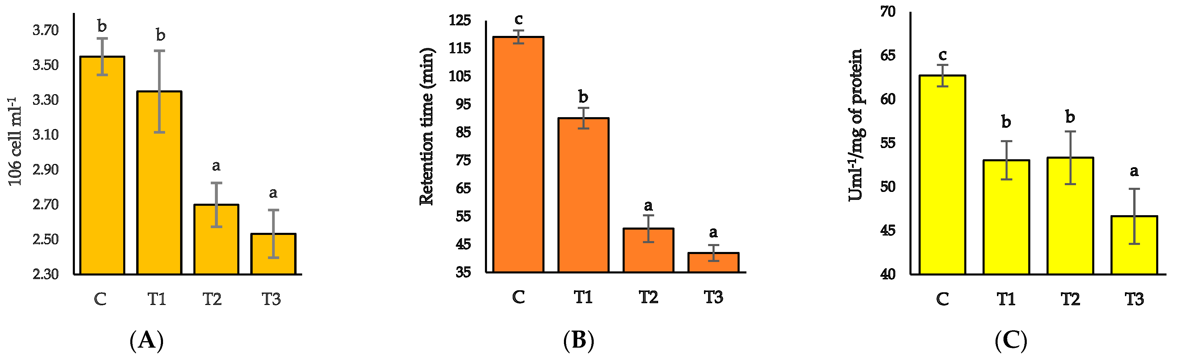
3.4. Hemocytes Count, Lysosomal Membrane Stability (LMS), and Serum Lysosome Activity

Hemocyte counts varied radically between different treatment units, with higher counts in the control and T1 groups and lower in T3 and T2 groups (p > 0.05) (Figure 3A). The retention period of neutral red for LMS and serum lysosome activity significantly differed between the control and treatment groups (Figure 3B,C). All the treatment groups exhibited a quick neutral red retention period and decreased value for serum lysosome activity.

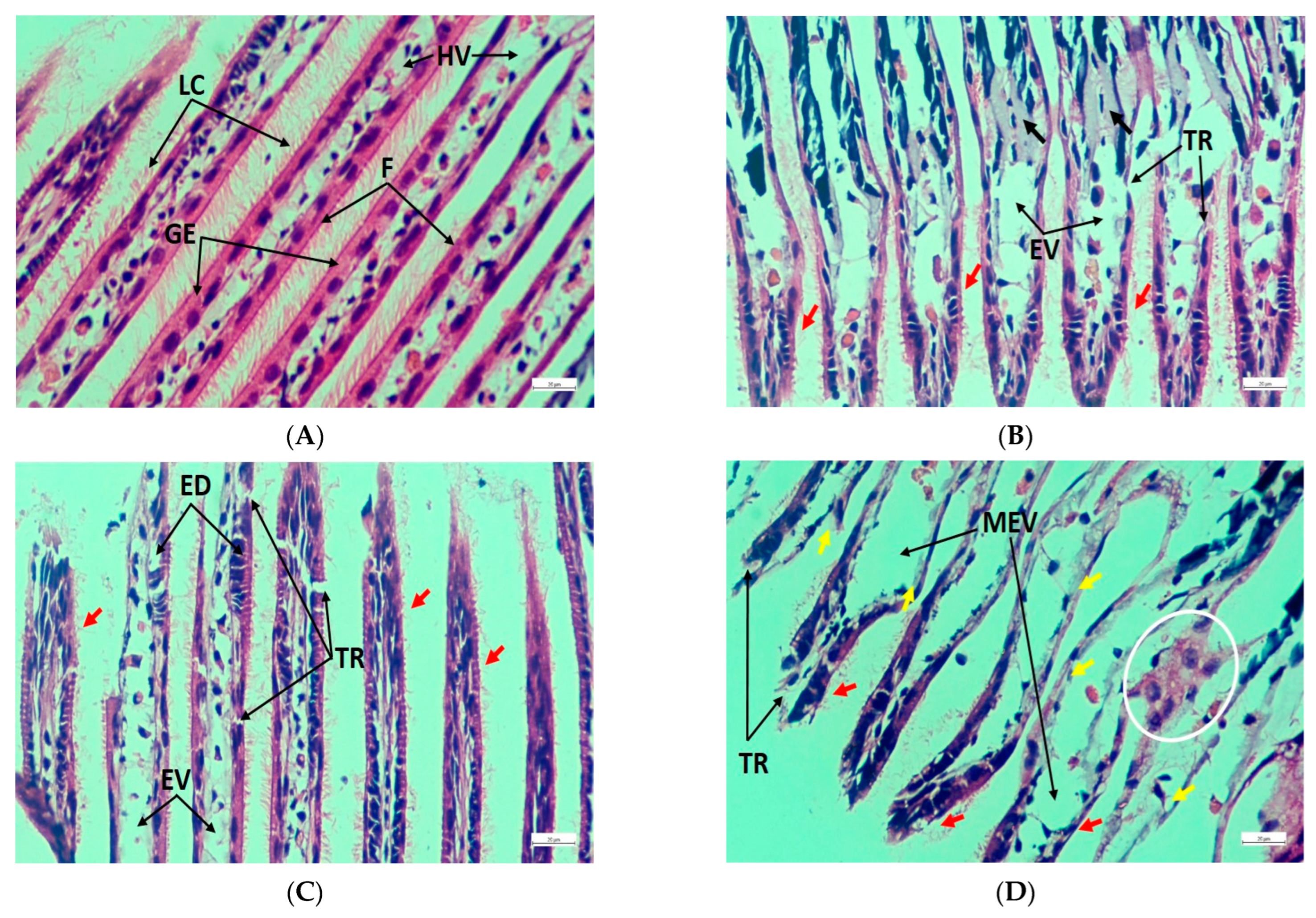
3.5. Histopathology of Gills, Kidneys, and Muscle

Gill filaments from the control group showed a healthy condition consisting of well-structured lateral cilia, gill epithelium, and hemolymph vessel (Figure 4A), whereas filaments from the treated groups revealed tissue rupture, extended vessel, and cilia damage (Figure 4B–D). Furthermore, congestion in filaments and epithelial deformities were identified in T1 and T2, respectively (Figure 4B,C), while filaments from T3 were affected by the massive expansion of hemolymph vessels, severe epithelial damage, and anomalous filament attachment (Figure 4D). Again, healthy renal tissues were detected by regular epithelial vesicles, brown intracellular granules, connective tissue, and nephridial lumen (Figure 5A). On the contrary, increased granules quantity, necrosis, tissue rupture, and extended lumen were noted in the exposed groups (Figure 5B–D). In addition, epithelial lifting was prominent in T2 (Figure 5C), while T3 exhibited damage to connective tissue, large vacuoles due to massive tissue rupture, and the presence of enlarged brown intracellular granules (Figure 5D). Normal muscle structure with horizontal and vertical musculature and irregular fibrous tissues were noted in the control group (Figure 6A); in contrast, the treated groups exhibited cell rupture and necrosis as common pathological disorders (Figure 6B–D). Apart from these alterations, massive vacuoles and fibrous tissue damage also appeared in T2 and T3, respectively (Figure 6C,D). Table 5 summarizes a comparative investigation of histopathological changes in the gills, kidneys, and muscles of L. marginalis detected from the present experiment.

4. Discussion

LC50 is a decisive scientific approach that is widely valid in the study of aquatic toxicology and pollution [5,28,49]. The 96 h LC50 of Pb(NO3)2 obtained from the current study was concomitant with the findings of [48]. The intensity of Pb(NO3)2 LC50 in different studies varied depending on the species, organism size, life stage, and physicochemical properties of the rearing water [17,50]. However, most data suggested features of strong tolerance towards Pb(NO3)2 toxicity in aquatic mollusks. Water quality parameters are vital in evaluating the toxicity of an aquatic environment [2,51]. Significant fluctuations in the physicochemical features of water may stimulate a species to be more sensitive to toxicants [52]. Hence, the physicochemical properties in the current experiment were kept in the optimum range, following the standard requirements [53].
Toxicology studies have not only signified the adverse effect of heavy metals on the growth performance of mussels, but also speculated the growth rate as one of the sensitive bio-indicators [29,54,55]. According to the present findings, the growth and development of L. marginalis were hindered when exposed to Pb(NO3)2, probably due to unprivileged physiological performance resulting in retarded tissue development and shell formation. The present results were comparable to those of Chandurvelan et al. [14] and Hu et al. [56], while Perna canaliculus was tested through exposure to several trace elements, and Mytilus coruscus was treated with toxic nanoparticles, respectively, which eventually resulted in poor physiological conditions and growth performance. Reduced growth metrics have also been consistently observed in several fish species owing to lead toxicity [10,18,57]. In contrast, the effect of heavy metals on growth was insignificant when Lampsilis siliquoidea was exposed to copper [58]. In the current study, the survival rate was also a significant stress indicator that reflected the severity of lead toxicity. As a result, the highest mortality was reported at the top exposure level [59]. Several previous studies have suggested that toxicity causes significant alterations in hemocyte counts in mussels, and it can be considered a potential biomarker to access environmental health [60,61]. Hemocyte counts significantly reduced when Mytilus coruscus was treated with titanium dioxide for 35 days [42], and when Mytius edulis was exposed to cadmium for 7 days [62]. Cellular abnormalities and impaired counts were recorded in the zebra mussel Dreissena polymorpha in response to cadmium toxicant [63]. The notable reduction in hemocyte count for the high-concentration treatment units of the current study remained aligned with the above findings. Serum lysosome activity is one of the widely used biomarkers in the assessment of toxicity compensation changes in the hematocytes of mussels [42,64]. A trend of the quick neutral red retention period and reduced serum lysosome concentration has been noted for Mytilus galloprovincialis exposed to micro- and nanoplastics [31,44].
Heavy metal toxicity causes notable histological changes in the vital organs of invertebrates [35,65,66]. Mussels’ gills are considered to be sensitive to toxicity because of their crucial role in respiration, food absorption, and extended surface area [67,68]. Fahmy and Sayed [33] observed gill filament dilation, epithelial lifting, necrosis, hemocyte congestion, hyperplasia, filament deformities, and cilia damage in bivalve Coelatura aegyptiaca exposed to zinc nanoparticles. Similarly, hemocyte infiltration, epithelial damage, necrotic and ruptured cells, and deformed gill filaments were identified by Vasanthi et al. [34]. These pathological alterations were mostly analogous to the present results. Meanwhile, in contrast to the control, several pathological pieces of evidence were obtained from the exposed units in the current experiment. However, the study of Knowles et al. [69] was consistent with the current findings, while expanded renal tubules, enlarged cytoplasmic vacuoles, and slightly expanded renal cells were spotted in the oysters Crassostrea gigas due to inferior salinity stress. Pathological signs in renal tissues, such as deformed tubules expanded vacuoles with cytoplasm, tubular necrosis, and squeezed glomerulus, were documented in Cyprinus carpio exposed to Pb(NO3)2 [36]. In the present study, histopathological alterations in foot muscle structure were noticeable, thereby indicating the severity of Pb toxicity. Again, data lacking on invertebrate muscle histopathology hindered the interpretation of the results. Current results were comparable with those of Hossain et al. [49], according to whom degenerated muscle fiber, extended vacuoles, cellular rupture, and necrosis were the prominent pathologies in Oreochromis niloticus muscle tissue exposed to chlorpyrifos. Fragmented epithelium and shortened epithelial cells were the major signs of mantle tissue damage in Mytilus galloprovincialis when exposed to ZnCl2 [35]. Therefore, the current experiment suggested that histology of the gill, kidney, and muscle and serum lysosomal activities might be considered significant tools in investigating metallic toxicity in freshwater pearl mussels.

5. Conclusions

Aquatic invertebrates are inevitably impacted by metal contaminants in water, and they act as potential biomarkers for water quality deterioration. Harmful heavy metal contamination in water and bioaccumulation in mussels could lead to public health issues. The results indicated that Pb exposure has a substantial impact on the physical health of L. marginalis. The experimental trial showed an increasing impact correlating with increased exposure. Chronic toxicity represented moderate to severe alterations in internal cellular structure. The current study also revealed the vulnerability status of freshwater ecosystems near large cities and industries due to heavy metal pollution. Therefore, necessary measures and policies should be implemented to protect mollusks from harmful heavy metal exposure.

Author Contributions

Conceptualization, M.A.H., M.M.I. and P.S.; methodology, M.A.H., M.M.I. and P.S.; software, M.A.H. and G.C.; validation, M.A.H., M.M.I. and P.S.; formal analysis, M.A.H. and G.C.; investigation, M.A.H., M.M.I., T.C. and P.S.; resources, M.M.I. and P.S.; data curation, T.C., G.C., M.H. and B.D.; writing—original draft preparation, M.A.H., T.C. and G.C.; writing—review and editing, M.A.H., M.M.I., M.H., B.D. and P.S. visualization, M.A.H., P.S. and G.C.; supervision, M.A.H., P.S. and M.M.I.; project administration, M.M.I.; funding acquisition, M.M.I. All authors have read and agreed to the published version of the manuscript.

Funding

This research is funded by the Sylhet Agricultural University Research System (SAURES-UGC-21-22-6) and University Grants Commission, Bangladesh.

Institutional Review Board Statement

Mussels are not defined as animals based on the current ethical standards. However, designing the research and utilization of animals has been properly approved by the Animals Ethics Committee of Sylhet Agricultural University Research System (SAURES), Bangladesh.

Informed Consent Statement

Not applicable.

Data Availability Statement

Raw and analyzed data in the current research are available from the corresponding author based on reasonable request.

Conflicts of Interest

The authors declare no competing interests.

References

  1. Luo, Y.; Rao, J.; Jia, Q. Heavy Metal Pollution and Environmental Risks in the Water of Rongna River Caused by Natural AMD around Tiegelongnan Copper Deposit, Northern Tibet, China. PLoS ONE 2022, 17, e0266700. [Google Scholar] [CrossRef] [PubMed]
  2. Abdullah, A.H.; Chowdhury, G.; Adikari, D.; Jahan, I.; Andrawina, Y.O.; Hossain, M.A.; Schneider, P.; Iqbal, M.M. Macroplastics Pollution in the Surma River in Bangladesh: A Threat to Fish Diversity and Freshwater Ecosystems. Water 2022, 14, 3263. [Google Scholar] [CrossRef]
  3. Islam, M.S.; Tanaka, M. Impacts of Pollution on Coastal and Marine Ecosystems Including Coastal and Marine Fisheries and Approach for Management: A Review and Synthesis. Mar. Pollut. Bull. 2004, 48, 624–649. [Google Scholar] [CrossRef] [PubMed]
  4. Mokarat, M.; Ruangsakunporn, A.; Chaianunporn, T.; Lomthaisong, K.; Robson, M.G.; Keithmaleesatti, S. Heavy Metal Contamination and Bioaccumulation in Khorat Snail-Eating Turtles (Malayemys khoratensis) from an e-Waste Recycling Activity Site in Thailand. Hum. Ecol. Risk Assess. 2022, 28, 133–153. [Google Scholar] [CrossRef]
  5. Abdel Gawad, S.S. Acute Toxicity of Some Heavy Metals to the Fresh Water Snail, Theodoxus niloticus (Reeve, 1856). Egypt. J. Aquat. Res. 2018, 44, 83–87. [Google Scholar] [CrossRef]
  6. Mahi, T.F.; Chowdhury, G.; Hossain, M.A.; Baishnab, A.K.; Schneider, P.; Iqbal, M.M. Assessment of Lead (Pb) Toxicity in Juvenile Nile Tilapia, Oreochromis niloticus—Growth, Behaviour, Erythrocytes Abnormalities, and Histological Alterations in Vital Organs. Toxics 2022, 10, 793. [Google Scholar] [CrossRef] [PubMed]
  7. Chan, W.S.; Routh, J.; Luo, C.; Dario, M.; Miao, Y.; Luo, D.; Wei, L. Metal Accumulations in Aquatic Organisms and Health Risks in an Acid Mine-Affected Site in South China. Environ. Geochem. Health 2021, 43, 4415–4440. [Google Scholar] [CrossRef]
  8. Inoue, K.; Onitsuka, Y.; Koito, T. Mussel Biology: From the Byssus to Ecology and Physiology, Including Microplastic Ingestion and Deep-Sea Adaptations. Fish. Sci. 2021, 87, 761–771. [Google Scholar] [CrossRef]
  9. Flora, G.; Gupta, D.; Tiwari, A. Toxicity of Lead: A Review with Recent Updates. Interdiscip. Toxicol. 2012, 5, 47–58. [Google Scholar] [CrossRef]
  10. Hussain, M.; Hossain, M.A.; Begum, M.; Roy, N.C. Freshwater Mussel (Lamelliedens marginalis) to Reduce the Lead (Pb) Bioaccumulation in Aquaculture of Stinging Catfish, Heteropneustes fossilis. J. Appl. Aquac. 2022, 1–17. [Google Scholar] [CrossRef]
  11. Usman, K.; Abu-Dieyeh, M.H.; Zouari, N.; Al-Ghouti, M.A. Lead (Pb) Bioaccumulation and Antioxidative Responses in Tetraena qataranse. Sci. Rep. 2020, 10, 17070. [Google Scholar] [CrossRef] [PubMed]
  12. Zalaghi, R.; Safari-Sinegani, A.-A. The Importance of Different Forms of Pb on Diminishing Biological Activities in a Calcareous Soil. Chem. Ecol. 2014, 30, 446–462. [Google Scholar] [CrossRef]
  13. Huang, H.; Li, Y.; Zheng, X.; Wang, Z.; Wang, Z.; Cheng, X. Nutritional Value and Bioaccumulation of Heavy Metals in Nine Commercial Fish Species from Dachen Fishing Ground, East China Sea. Sci. Rep. 2022, 12, 6927. [Google Scholar] [CrossRef]
  14. Chandurvelan, R.; Marsden, I.D.; Glover, C.N.; Gaw, S. Assessment of a Mussel as a Metal Bioindicator of Coastal Contamination: Relationships between Metal Bioaccumulation and Multiple Biomarker Responses. Sci. Total Environ. 2015, 511, 663–675. [Google Scholar] [CrossRef] [PubMed]
  15. Khalesi, M.K.; Abedi, Z.; Behrouzi, S.; Kohestan Eskandari, S. Haematological, Blood Biochemical and Histopathological Effects of Sublethal Cadmium and Lead Concentrations in Common Carp. Bulg. J. Vet. Med. 2017, 20, 141–150. [Google Scholar] [CrossRef]
  16. Abdel-Warith, A.W.A.; Younis, E.S.M.I.; Al-Asgah, N.A.; Rady, A.M.; Allam, H.Y. Bioaccumulation of Lead Nitrate in Tissues and Its Effects on Hematological and Biochemical Parameters of Clarias gariepinus. Saudi J. Biol. Sci. 2020, 27, 840–845. [Google Scholar] [CrossRef]
  17. Shuhaimi-Othman, M.; Nur-Amalina, R.; Nadzifah, Y. Toxicity of Metals to a Freshwater Snail, Melanoides Tuberculata. Sci. World J. 2012, 2012, 1–10. [Google Scholar] [CrossRef]
  18. Kiran, K.; Midhun, S.J.; Vysakh, A.; James, T.J. Antagonistic Effects of Dietary Moringa oleifera on Hemato-biochemical and Oxidative Stress of Lead Nitrate Intoxicated Nile Tilapia, Oreochromis niloticus. Aquac. Res. 2021, 52, 6164–6178. [Google Scholar] [CrossRef]
  19. Saleh, I.; Syamsir, S.; Pramaningsih, V.; Hansen, H. The Use of Green Mussel as Bioindicator of Heavy Metal Pollution in Indonesia: A Review. Environ. Health Toxicol. 2021, 36, e2021026. [Google Scholar] [CrossRef]
  20. Le Saux, A.; David, E.; Betoulle, S.; Bultelle, F.; Rocher, B.; Barjhoux, I.; Cosio, C. New Insights into Cellular Impacts of Metals in Aquatic Animals. Environments 2020, 7, 46. [Google Scholar] [CrossRef]
  21. Arisekar, U.; Jeya, R.; Shalini, R.; Jeyasekaran, G. Human Health Risk Assessment of Heavy Metals in Aquatic Sediments and Freshwater Fish Caught from Thamirabarani River, the Western Ghats of South Tamil Nadu. Mar. Pollut. Bull. 2020, 159, 111496. [Google Scholar] [CrossRef]
  22. Hossain, M.A.; Hussain, M.; Sarker, T.R.; Saha, S.; Iqbal, M.M. Reproductive and Morphometric Traits of Freshwater Mussel Lamellidens marginalis and Associated Hydrology in the Ratargul Freshwater Swamp Forest, Bangladesh. Egypt. J. Aquat. Res. 2022. [Google Scholar] [CrossRef]
  23. Nobles, T.; Zhang, Y. Survival, Growth and Condition of Freshwater Mussels: Effects of Municipal Wastewater Effluent. PLoS ONE 2015, 10, e0128488. [Google Scholar] [CrossRef] [PubMed]
  24. Yasmeen, S.; Pathan, T.S. Behavioural Response in Freshwater Bivalve Mollusk, Lamellidens marginalis Due to Acute Toxicity of Cadmium Chloride. Int. J. Biol. Innov. 2021, 3, 148–153. [Google Scholar] [CrossRef]
  25. Costello, D.M.; Brown, L.M.; Lamberti, G.A. Acute Toxic Effects of Ionic Liquids on Zebra Mussel (Dreissena polymorpha) Survival and Feeding. Green Chem. 2009, 11, 548–555. [Google Scholar] [CrossRef]
  26. Yap, C.K.; Sharifinia, M.; Cheng, W.H.; Al-Shami, S.A.; Wong, K.W.; Al-Mutairi, K.A. A Commentary on the Use of Bivalve Mollusks in Monitoring Metal Pollution Levels. Int. J. Environ. Res. Public Health 2021, 18, 3386. [Google Scholar] [CrossRef]
  27. EPA, U. Guideline No. 497: Defined Approaches on Skin Sensitisation; OECD Guidelines for the Testing of Chemicals, Section 4; OECD: Paris, France, 2021; ISBN 9789264903005. [Google Scholar]
  28. Brahma, N.; Gupta, A. Acute Toxicity of Lead in Fresh Water Bivalves Lamellidens jenkinsianus Obesa and Parreysia (Parreysia) Corrugata with Evaluation of Sublethal Effects on Acetylcholinesterase and Catalase Activity, Lipid Peroxidation, and Behavior. Ecotoxicol. Environ. Saf. 2020, 189, 109939. [Google Scholar] [CrossRef]
  29. Brix, K.V.; Esbaugh, A.J.; Grosell, M. The Toxicity and Physiological Effects of Copper on the Freshwater Pulmonate Snail, Lymnaea stagnalis. Comp. Biochem. Physiol. C Toxicol. Pharmacol. 2011, 154, 261–267. [Google Scholar] [CrossRef]
  30. D’Agata, A.; Cappello, T.; Maisano, M.; Parrino, V.; Giannetto, A.; Brundo, M.V.; Ferrante, M.; Mauceri, A. Cellular Biomarkers in the Mussel Mytilus galloprovincialis (Bivalvia: Mytilidae) from Lake Faro (Sicily, Italy). Ital. J. Zool. 2014, 81, 43–54. [Google Scholar] [CrossRef]
  31. Auguste, M.; Balbi, T.; Ciacci, C.; Canonico, B.; Papa, S.; Borello, A.; Vezzulli, L.; Canesi, L. Shift in Immune Parameters After Repeated Exposure to Nanoplastics in the Marine Bivalve Mytilus. Front. Immunol. 2020, 11, 426. [Google Scholar] [CrossRef]
  32. Kolyuchkina, G.A.; Ismailov, A.D. Morpho-Functional Characteristics of Bivalve Mollusks under the Experimental Environmental Pollution by Heavy Metals. Oceanology 2011, 51, 804–813. [Google Scholar] [CrossRef]
  33. Fahmy, S.R.; Sayed, D.A. Toxicological Perturbations of Zinc Oxide Nanoparticles in the Coelatura aegyptiaca Mussel. Toxicol. Ind. Health 2017, 33, 564–575. [Google Scholar] [CrossRef] [PubMed]
  34. Vasanthi, L.; Revathi, P.; Arulvasu, C.; Munuswamy, N. Biomarkers of Metal Toxicity and Histology of Perna viridis from Ennore Estuary, Chennai, South East Coast of India. Ecotoxicol. Environ. Saf. 2012, 84, 92–98. [Google Scholar] [CrossRef] [PubMed]
  35. Liberatori, G.; Grassi, G.; Guidi, P.; Bernardeschi, M.; Fiorati, A.; Scarcelli, V.; Genovese, M.; Faleri, C.; Protano, G.; Frenzilli, G.; et al. Effect-based Approach to Assess Nanostructured Cellulose Sponge Removal Efficacy of Zinc Ions from Seawater to Prevent Ecological Risks. Nanomaterials 2020, 10, 1283. [Google Scholar] [CrossRef]
  36. Paul, S.; Mandal, A.; Bhattacharjee, P.; Chakraborty, S.; Paul, R.; Kumar Mukhopadhyay, B. Evaluation of Water Quality and Toxicity after Exposure of Lead Nitrate in Fresh Water Fish, Major Source of Water Pollution. Egypt. J. Aquat. Res. 2019, 45, 345–351. [Google Scholar] [CrossRef]
  37. Ramakritinan, C.M.; Chandurvelan, R.; Kumaraguru, A.K. Acute Toxicity of Metals: Cu, Pb, Cd, Hg and Zn on Marine Molluscs, Cerithedia cingulata G., and Modiolus philippinarum H. Indian J. Mar. Sci. 2012, 41, 141–145. [Google Scholar]
  38. Finney, D.J. Probit Analysis, 3rd ed.; Cambridge University Press: New York, NY, USA, 1971. [Google Scholar]
  39. Uddin, M.; Yang, H.-S.; Choi, K.-S.; Kim, H.-J.; Hong, J.-S.; Cho, M. Seasonal Changes in Perkinsus Olseni Infection and Gametogenesis in Manila Clam, Ruditapes philippinarum, from Seonjaedo Island in Incheon, off the West Coast of Korea. J. World Aquac. Soc. 2010, 41, 93–101. [Google Scholar] [CrossRef]
  40. Htun-Han, M. The Reproductive Biology of the Dab Limanda limanda (L.) in the North Sea: Seasonal Changes in the Ovary. J. Fish Biol. 1978, 13, 351–359. [Google Scholar] [CrossRef]
  41. Juhel, G.; Ramsay, R.M.; Davenport, J.; O’Halloran, J.; Culloty, S.C. Effect of the Microcystin-Producing Cyanobacterium, Microcystis aeruginosa, on Immune Functions of the Zebra Mussel Dreissena polymorpha. J. Shellfish Res. 2015, 34, 433–442. [Google Scholar] [CrossRef]
  42. Munari, M.; Matozzo, V.; Benetello, G.; Riedl, V.; Pastore, P.; Badocco, D.; Marin, M.G. Exposure to Decreased PH and Caffeine Affects Hemocyte Parameters in the Mussel Mytilus galloprovincialis. J. Mar. Sci. Eng. 2020, 8, 238. [Google Scholar] [CrossRef]
  43. Chu, F.-L.E.; La Peyre, J.F. Effect of Environmental Factors and Parasitism on Hemolymph Lysozyme and Protein of American Oysters (Crassostrea virginica). J. Invertebr. Pathol. 1989, 54, 224–232. [Google Scholar] [CrossRef]
  44. Capolupo, M.; Valbonesi, P.; Fabbri, E. A Comparative Assessment of the Chronic Effects of Micro-and Nano-Plastics on the Physiology of the Mediterranean Mussel Mytilus galloprovincialis. Nanomaterials 2021, 11, 649. [Google Scholar] [CrossRef] [PubMed]
  45. Slaoui, M.; Fiette, L. Histopathology Procedures: From Tissue Sampling to Histopathological Evaluation. Methods Mol. Biol. 2011, 691, 69–82. [Google Scholar] [CrossRef] [PubMed]
  46. McElwain, A.; Bullard, S.A. Histological Atlas of Freshwater Mussels (Bivalvia, Unionidae): Villosa nebulosa (Ambleminae: Lampsilini), Fusconaia cerina (Ambleminae: Pleurobemini) and Strophitus connasaugaensis (Unioninae: Anodontini). Malacologia 2014, 57, 99–239. [Google Scholar] [CrossRef]
  47. Carella, F.; Villari, G.; Maio, N.; De Vico, G. Disease and Disorders of Freshwater Unionid Mussels: A Brief Overview of Recent Studies. Front. Physiol. 2016, 7, 489. [Google Scholar] [CrossRef]
  48. Brahma, N.; Gupta, A. Effects of Copper and Lead on Freshwater Mollusc Corbicula striatella (Deshayes, 1854). J. Energy Res. Environ. Technol. 2019, 6, 205–208. [Google Scholar]
  49. Hossain, M.A.; Sutradhar, L.; Sarker, T.R.; Saha, S.; Iqbal, M.M. Toxic Effects of Chlorpyrifos on the Growth, Hematology, and Different Organs Histopathology of Nile Tilapia, Oreochromis niloticus. Saudi J. Biol. Sci. 2022, 29, 103316. [Google Scholar] [CrossRef]
  50. Gilroy, È.A.M.; Gillis, P.L.; King, L.E.; Bendo, N.A.; Salerno, J.; Giacomin, M.; de Solla, S.R. The Effects of Pharmaceuticals on a Unionid Mussel (Lampsilis siliquoidea): An Examination of Acute and Chronic Endpoints of Toxicity across Life Stages. Environ. Toxicol. Chem. 2017, 36, 1572–1583. [Google Scholar] [CrossRef]
  51. Milam, C.D.; Farris, J.L.; Dwyer, F.J.; Hardesty, D.K. Acute Toxicity of Six Freshwater Mussel Species (Glochidia) to Six Chemicals: Implications for Daphnids and Utterbackia imbecillis as Surrogates for Protection of Freshwater Mussels (Unionidae). Arch. Environ. Contam. Toxicol. 2005, 48, 166–173. [Google Scholar] [CrossRef]
  52. Wang, N.; Ivey, C.D.; Ingersoll, C.G.; Brumbaugh, W.G.; Alvarez, D.; Hammer, E.J.; Bauer, C.R.; Augspurger, T.; Raimondo, S.; Barnhart, M.C. Acute Sensitivity of a Broad Range of Freshwater Mussels to Chemicals with Different Modes of Toxic Action. Environ. Toxicol. Chem. 2017, 36, 786–796. [Google Scholar] [CrossRef]
  53. Chapman, D.V.; Bradley, C.; Gettel, G.M.; Hatvani, I.G.; Hein, T.; Kovács, J.; Liska, I.; Oliver, D.M.; Tanos, P.; Trásy, B.; et al. Developments in Water Quality Monitoring and Management in Large River Catchments Using the Danube River as an Example. Environ. Sci. Policy 2016, 64, 141–154. [Google Scholar] [CrossRef]
  54. Tedengren, M.; Olsson, B.; Bradley, B.; Zhou, L. Heavy Metal Uptake, Physiological Response and Survival of the Blue Mussel (Mytilus edulis) from Marine and Brackish Waters in Relation to the Induction of Heat-Shock Protein 70. Hydrobiologia 1999, 393, 261–269. [Google Scholar] [CrossRef]
  55. Wang, N.; Ingersoll, C.G.; Greer, I.E.; Hardesty, D.K.; Ivey, C.D.; Kunz, J.L.; Brumbaugh, W.G.; Dwyer, F.J.; Roberts, A.D.; Augspurger, T.; et al. Chronic Toxicity of Copper and Ammonia to Juvenile Freshwater Mussels (Unionidae). Environ. Toxicol. Chem. 2007, 26, 2048–2056. [Google Scholar] [CrossRef] [PubMed]
  56. Hu, M.; Lin, D.; Shang, Y.; Hu, Y.; Lu, W.; Huang, X.; Ning, K.; Chen, Y.; Wang, Y. CO2-Induced PH Reduction Increases Physiological Toxicity of Nano-TiO2 in the Mussel Mytilus coruscus. Sci. Rep. 2017, 7, 40015. [Google Scholar] [CrossRef]
  57. Álvarez-González, C.A.; Martínez-Sánchez, L.; Peña-Marín, E.S.; Guerrero-Zárate, R.; Jesús-Ramírez, F.; Morales-García, V.; Uribe-López, M.; Núñez-Nogueira, G. Effects on the Growth and Digestive Enzyme Activity in Nile Tilapia Fry (Oreochromis niloticus) by Lead Exposure. Water. Air. Soil Pollut. 2020, 231, 1–15. [Google Scholar] [CrossRef]
  58. Jorge, M.B.; Loro, V.L.; Bianchini, A.; Wood, C.M.; Gillis, P.L. Mortality, Bioaccumulation and Physiological Responses in Juvenile Freshwater Mussels (Lampsilis siliquoidea) Chronically Exposed to Copper. Aquat. Toxicol. 2013, 126, 137–147. [Google Scholar] [CrossRef]
  59. Paulino, A.T.; Santos, L.B.; Nozaki, J. Protective Action of Zinc against Lead Poisoning in Tilapia Oreochromis niloticus. Toxicol. Environ. Chem. 2007, 89, 363–370. [Google Scholar] [CrossRef]
  60. Ballina, N.R.; Maresca, F.; Cao, A.; Villalba, A. Bivalve Haemocyte Subpopulations: A Review. Front. Immunol. 2022, 13, 826255. [Google Scholar] [CrossRef]
  61. Hossain, M.A.; Rani Sarker, T.; Sutradhar, L.; Hussain, M.; Mahbub Iqbal, M. Toxic Effects of Chlorpyrifos on the Growth, Hemocytes Counts, and Vital Organ’s Histopathology of Freshwater Mussel, Lamellidens marginalis. J. King Saud Univ. Sci. 2022, 29, 102482. [Google Scholar] [CrossRef]
  62. Coles, J.; Farley, S.; Pipe, R. Alteration of the Immune Response of the Common Marine Mussel Mytilus edulis Resulting from Exposure to Cadmium. Dis. Aquat. Organ. 1995, 22, 59–65. [Google Scholar] [CrossRef]
  63. Evariste, L.; Rioult, D.; Brousseau, P.; Geffard, A.; David, E.; Auffret, M.; Fournier, M.; Betoulle, S. Differential Sensitivity to Cadmium of Immunomarkers Measured in Hemocyte Subpopulations of Zebra Mussel Dreissena polymorpha. Ecotoxicol. Environ. Saf. 2017, 137, 78–85. [Google Scholar] [CrossRef] [PubMed]
  64. Beaudry, A.; Fortier, M.; Masson, S.; Auffret, M.; Brousseau, P.; Fournier, M. Effect of Temperature on Immunocompetence of the Blue Mussel (Mytilus edulis). J. Xenobiotics 2016, 6, 5889. [Google Scholar] [CrossRef]
  65. Alonso, A.; Suárez, P.; Ruiz, Y.; Dobal, V.; San Juan, F. Gonadal Histopathological Disorders in Mytilus Galloprovincialis Male Exposed to Tars Used in Mussel Farms. Front. Mar. Sci. 2019, 6, 577. [Google Scholar] [CrossRef]
  66. Costa, P.M.; Carreira, S.; Costa, M.H.; Caeiro, S. Development of Histopathological Indices in a Commercial Marine Bivalve (Ruditapes decussatus) to Determine Environmental Quality. Aquat. Toxicol. 2013, 126, 442–454. [Google Scholar] [CrossRef] [PubMed]
  67. Pinto, J.; Costa, M.; Leite, C.; Borges, C.; Coppola, F.; Henriques, B.; Monteiro, R.; Russo, T.; Di Cosmo, A.; Soares, A.M.V.M.; et al. Ecotoxicological Effects of Lanthanum in Mytilus galloprovincialis: Biochemical and Histopathological Impacts. Aquat. Toxicol. 2019, 211, 181–192. [Google Scholar] [CrossRef]
  68. Mona, M.H.; El-Khodary, G.M.; Abdel-Halim, K.Y.; Omran, N.E.; Abd El-Aziz, K.K.; El-Saidy, S.A. Histopathological Alterations Induced by Marine Environmental Pollutants on the Bivalve Cerastoderma glaucum (Bruguière 1789) from Temsah Lake, Suez Canal, Egypt. Environ. Sci. Pollut. Res. 2022, 29, 9971–9989. [Google Scholar] [CrossRef]
  69. Knowles, G.; Handlinger, J.; Jones, B.; Moltschaniwskyj, N. Hemolymph Chemistry and Histopathological Changes in Pacific Oysters (Crassostrea gigas) in Response to Low Salinity Stress. J. Invertebr. Pathol. 2014, 121, 78–84. [Google Scholar] [CrossRef]
Figure 1. Flow diagram of the overall laboratory protocols and assays.
Toxics 11 00475 g001
Figure 2. The survival rate and condition indices in the different experimental units. (A) Survival rate, (B) Fulton’s condition factor Fk, and condition index, CI). Different superscripts indicate significant differences at p < 0.05; values are means ± SE.
Toxics 11 00475 g002
Figure 3. Hemocyte count, lysosomal membrane stability (LMS), and serum lysosome activity in different experimental units following exposure to Pb(NO3)2. (A) Hemocyte count, (B) Lysosomal membrane stability (LMS), and (C) Serum lysosome activity). Different superscripts indicate significant differences at p < 0.05; values are means ± SE.
Toxics 11 00475 g003
Figure 4. Gill transverse section photomicrograph of L. marginalis. (A) Control, (B) T1, (C) T2, and (D) T3; LC—Lateral cilia, F—Filaments, GE—Gill epithelium, HV—Hemolymph vessel, TR—Tissue rupture, EV—Extended vessel, MEV—Massive expansion of vessel, ED—Epithelial deformities; Filament congestion—Black arrow, Cilia damage—Red arrow, Epithelial damage—Yellow arrow, Filament attachment, and abnormalities—White circle.
Toxics 11 00475 g004
Figure 5. Renal transverse section photomicrograph of L. marginalis. (A) Control, (B) T1, (C) T2, and (D) T3; EV—Epithelial vesicles, BIG—Brown intracellular granules, CT—Connective tissue, NL—Nephridial lumen, IG—Increased granules, EG—Enormous granules, EL—Extended lumen, DCT—Damage of connective tissue, V—Vacuoles due to massive tissue rupture, EBG—Enlarged brown intracellular granules; Necrosis—Yellow arrow, Tissue rupture—Red arrow, Epithelial lifting—Black arrow.
Toxics 11 00475 g005
Figure 6. Muscle transverse section photomicrograph of L. marginalis. (A) Control, (B) T1, (C) T2, and (D) T3; VM—Vertical musculature, HM—Horizontal musculature, IF—Irregular fibrous tissue, N—Nuclei, CR—Cell rupture, MV—Massive vacuoles due to cell rupture; Necrosis—Yellow arrow, Fibrous tissue damage—Black arrow.
Toxics 11 00475 g006
Table 1. Dose calculation in different treatment groups.
TreatmentConcentration of Pb(NO₃)₂ (mgL−1)Stocking Density (No./Treatment)Replication
C (Control)00.00 (00% of LC50)203
T121.93 (10% of LC50)203
T243.86 (20% of LC50)203
T387.72 (40% of LC50)203
Table 2. 96 h Pb(NO3)2 LC50 in different aquatic mollusks.
SpeciesValue of 96 h LC50 (mgL−1)References
BivalvesLamellidense marginalis219.32Current study
L. jenkinsianus192.14[28]
Corbicula striatella231.01[48]
GastropodsTheodoxus niloticus18[5]
Melanoides tuberculata6.82[17]
Cerithedia cingulate15.50[37]
BivalvesModiolus philippinarum2.86
Table 3. Water quality parameters in the different treatment groups (values are mean ± SE).
TreatmentDay 0Day 15Day 40
TemperatureC20.73 ± 0.2319.56 ± 0.3720.66 ± 0.50
T120.93 ± 0.1120.23 ± 0.4120.66 ± 0.20
T221.00 ± 0.1719.70 ± 0.1720.76 ± 0.15
T321.03 ± 0.2820.13 ± 0.4920.80 ± 0.10
pHC7.96 ± 0.058.20 ± 0.108.23 ± 0.10
T17.83 ± 0.058.19 ± 0.107.96 ± 0.05
T27.70 ± 0.038.16 ± 0.057.76 ± 0.05
T37.60 ± 0.018.06 ± 0.057.53 ± 0.05
SalinityC0.07 ± 00.08 ± 00.10 ± 0
T10.07 ± 00.08 ± 00.11 ± 0
T20.07 ± 00.08 ± 00.11 ± 0
T30.07 ± 00.08 ± 00.09 ± 0
NH3C0.05 ± 00.07 ± 00.07 ± 0
T10.01 ± 00.02 ± 00.02 ± 0
T20.02 ± 00.02 ± 00.02 ± 0
T30.02 ± 00.01 ± 00.02 ± 0
DOC7.26 ± 0.497.48 ± 0.477.39 ± 0.63
T17.58 ± 0.297.58 ± 0.297.60 ± 0.48
T27.54 ± 0.0757.54 ± 0.077.43 ± 0.08
T37.64 ± 0.1047.64 ± 0.107.77 ± 0.08
Table 4. Growth parameters of L. marginalis exposed to different concentrations of Pb(NO3)2.
CT1T2T3
Shell Length Initial (cm)7.23 ± 0.167.16 ± 0.127.26 ± 0.177.22 ± 0.20
Shell Weight Initial (g)41.40 ± 1.841.28 ± 2.0141.06 ± 2.5941.16 ± 2.52
Shell Depth Initial (cm)4.06 ± 0.114.02 ± 0.094.09 ± 0.064.01 ± 0.10
Shell Length Final (cm)7.33 ± 0.097.28 ± 0.107.31 ± 0.107.28 ± 0.13
Shell Weight Final(g)50.32 ± 2.58 b40.69 ± 1.58 a41.41 ± 1.58 a38.67 ± 2.24 a
Shell Depth Final (cm)4.32 ± 0.054.25 ± 0.054.36 ± 0.044.36 ± 0.05
Soft Tissue Wet Weight (g)9.41 ± 0.64 b7.88 ± 0.26 a7.85 ± 0.31 a7.01 ± 0.42 a
Dry Shell Weight (g)16.45 ± 0.9815.05 ± 0.8516.23 ± 1.0715.09 ± 1.50
Length Gain (cm)0.10 ± 0.210.12 ± 0.130.05 ± 0.200.06 ± 0.25
Weight Gain (g)8.92 ± 3.52 b−0.59 ± 2.65 ab0.35 ± 3.29 ab−2.49 ± 3.95 a
Specific Growth Rate %0.48 ± 0.18 b−0.01 ± 0.17 a0.07 ± 0.19 a−0.13 ± 0.25 a
Average Daily Length Gain0.002 ± 0.050.003 ± 0.0030.001 ± 0.0050.001 ± 0.006
Average Daily Weight Gain0.22 ± 0.08 b−0.01 ± 0.06 ab0.01 ± 0.08 ab−0.06 ± 0.09 a
Notes: The columns with different superscripts indicate significant differences at p < 0.05; values are means ± SE.
Table 5. Comparative study of histopathological changes from the present trial in different experimental units.
OrganAbnormalityControlT1T2T3
GillTissue Rupture---****
Extended Vessel---*******
A Massive Expansion of The Vessel---*---**
Epithelial Deformities------**
Filament Congestion---*---*
Cilia Damage---******
Epithelial Damage------****
Filament Attachment And Abnormalities ---------*
KidneyEnormous Granules---*****
Extended Lumen------***
Damage to Connective Tissue---------*
Vacuoles Due to Massive Tissue Rupture------***
Enlarged Brown Intracellular Granules------****
Necrosis---******
Tissue Rupture------****
Epithelial Lifting------*---
MuscleCell Rupture---******
Massive Vacuoles Due to Cell Rupture------***
Necrosis---****
Fibrous Tissue Damage------****
Notes: Absent (---), below 10% as weak (*), 10–50% as moderate (**), and above 50% as severe (***).
Disclaimer/Publisher’s Note: The statements, opinions and data contained in all publications are solely those of the individual author(s) and contributor(s) and not of MDPI and/or the editor(s). MDPI and/or the editor(s) disclaim responsibility for any injury to people or property resulting from any ideas, methods, instructions or products referred to in the content.

