Article

Mango Peels as an Industrial By-Product: A Sustainable Source of Compounds with Antioxidant, Enzymatic, and Antimicrobial Activity

1
Faculty of Chemistry and Chemical Engineering, University of Maribor, Smetanova ulica 17, 2000 Maribor, Slovenia
2
Faculty of Medicine, University of Maribor, Taborska ulica 8, 2000 Maribor, Slovenia
*
Author to whom correspondence should be addressed.
Foods 2024, 13(4), 553; https://doi.org/10.3390/foods13040553
Submission received: 16 January 2024 / Revised: 1 February 2024 / Accepted: 7 February 2024 / Published: 11 February 2024
(This article belongs to the Special Issue Extraction Technology and Characters of Bioactive Substances in Foods)

Abstract

:
Plant waste materials are important sources of bioactive compounds with remarkable health-promoting benefits. In particular, industrial by-products such as mango peels are sustainable sources of bioactive substances, with antioxidant, enzymatic, and antimicrobial activity. Appropriate processing is essential to obtain highly bioactive compounds for further use in generating value-added products for the food industry. The objective of the study was to investigate and compare the biological activity of compounds from fresh and dried mango peels obtained by different conventional methods and unconventional extraction methods using supercritical fluids (SFE). The highest total phenolic content (25.0 mg GAE/g DW) and the total content of eight phenolic compounds (829.92 µg/g DW) determined by LC-MS/MS were detected in dried mango peel extract obtained by the Soxhlet process (SE). SFE gave the highest content of proanthocyanidins (0.4 mg PAC/g DW). The ethanolic ultrasonic process (UAE) provided the highest antioxidant activity of the product (82.4%) using DPPH radical scavenging activity and total protein content (2.95 mg protein/g DW). Overall, the dried mango peels were richer in bioactive compounds (caffeic acid, chlorogenic acid, gallic acid, catechin, and hesperidin/neohesperidin), indicating successful preservation during air drying. Furthermore, outstanding polyphenol oxidase, superoxide dismutase (SOD), and lipase activities were detected in mango peel extracts. This is the first study in which remarkable antibacterial activities against the growth of Gram-negative bacteria (Escherichia coli and Pseudomonas aeruginosa) and Gram-positive bacteria (Bacillus cereus and Staphylococcus aureus) were evaluated by determining the microbial growth inhibition rate after 12 and 24 h incubation periods for mango peel extracts obtained by different methods. Ethanolic SE and UAE extracts from dried mango peels resulted in the lowest minimum inhibitory concentrations (MIC90) for all bacterial species tested. Mango peels are remarkable waste products that could contribute to the sustainable development of exceptional products with high-added value for various applications, especially as dietary supplements.

1. Introduction

Fruit peels account for about 15–60% of waste products in fruit processing and are usually discarded [1,2,3]. A total of around 89 million tons of food waste is produced in the European Union [4,5]. Due to their high biodegradability and fermentability, fruit peels contribute to water and soil pollution, eutrophication, global warming, the greenhouse effect, etc. However, similar to the pulp, the waste parts of the fruit produced during fruit processing are a rich source of phytonutrients and valuable biologically active compounds. By applying appropriate extraction or isolation processes (low operating temperature, no use of toxic solvents) for bioactive ingredients from the waste parts of fruits, especially from peels, it is possible to obtain products with exceptional added value, which can be used mainly in the food, cosmetic, and pharmaceutical industries [6,7].
Mango (Mangifera indica L.) belongs to the Anacardiaceae family. It is one of the most important and popular tropical fruits for its attractive color, excellent taste and aroma, and high nutritional, phytochemical, and medicinal value [8]. The edible part is largely industrially processed into various pulps or juices, with the inedible byproducts discarded. However, mango by-products, especially peels, represent an important source of biologically active compounds that could be used for various applications. Mango by-products account for 35–55% of the total mass. Moreover, their further use could reduce the amount of waste and negative impact on the environment [9,10]. Therefore, a new approach is urgently needed to reduce their accumulation and the resulting pollution and generate value-added products.
Mango peels contain even more specific bioactive compounds than the edible part of the fruit. They are a rich source of various polyphenolic compounds (PCs), dietary fiber, pectin, carotenoids, vitamins (ascorbic acid, tocopherol), minerals (potassium, magnesium, sodium, calcium, copper, iron, manganese, zinc, chromium, phosphorus, chlorine), and enzymes. The main PCs in mango peels are gallic acid, ellagic acid, caffeic acid, ferulic acid, rutin, catechin, quercetin, and kaempferol [11,12]. It is essential to highlight the presence of mangiferin, which belongs to the xanthone class, and is the main PCs of mango. Mangiferin has antioxidant, antidiabetic, anticancer, antimicrobial, and anti-inflammatory effects [13,14]. However, the peels have been found to contain a higher concentration of mangiferin than the pulp and seeds [15,16]. In addition, some enzymes have been shown to be present in active form in mango peels, such as superoxide dismutase (SOD), catalase, peroxidase, protease, and amylase [17]. Different studies also confirmed the good antimicrobial potential of samples obtained from different parts of the mango plant [18,19]. Ethanolic extracts from the peels of different varieties of mango show an excellent inhibitory effect on the growth of various types of Gram-negative bacteria, such as Escherichia coli, Salmonella typhimurium, and Vibrio parahaemolyticus, and Gram-positive bacteria, such as Staphylococcus aureus and Bacillus cereus [20]. In addition, acetonic, ethanolic, and aqueous extracts obtained by the conventional maceration method exhibit efficient antimicrobial activities against various foodborne pathogenic microorganisms [21].
It is important to emphasize that the isolation of bioactive compounds is mainly influenced by the extraction method used, the operating conditions, and the extraction solvent, as different polarities of the solvents directly affect the extraction efficiency [22]. However, the quality and final composition of the extract obtained are mainly influenced by the method of cultivation and location of the plant, weather conditions, plant species, stage of development, the process and temperature of drying and storage, and the final water content of the raw material [23]. Moreover, the antimicrobial activity of the extracted samples is related to the content and synergistic effects of the bioactive substances in the extracts obtained [24].
Some sensitive but important bioactive substances may be degraded during drying and storage. On the other hand, harmful microorganisms may develop if the fresh peels are not processed immediately and when unsuitable drying conditions are used [25,26]. Therefore, the present study focused on the determination of the bioactive compound content in fresh and dried mango peels obtained by different extraction methods. Dried mango peels were obtained by air-drying at room temperature, as this is considered one of the simplest, least demanding, and most economical methods [27]. On the other hand, excessively high temperatures during oven-drying can degrade bioactive compounds [28]. While oven-drying is energy inefficient, it is a cost-effective method [29]. Sun-drying is also a cost-effective method, but it is more susceptible to microbial contamination due to unhygienic conditions. It can also have a negative effect on the plant material by reducing the nutrient content and causing color changes due to direct exposure to UV radiation [30,31]. Although freeze-drying is one of the most suitable methods to dry plant material and successfully preserve valuable substances, it requires very high investment and operating costs, which is very unfavorable [32,33].
Furthermore, the study compares conventional methods (Soxhlet method (SE), ultrasound-assisted method (UAE), homogenization-assisted method (HAE)) and an unconventional technique (supercritical fluid technique (SFE)) to obtain the bioactive compounds from mango peels. The main objective of the study was to investigate the content of bioactive compounds in terms of total phenols (TPC), proanthocyanidins (PACs), certain important PCs, and antioxidant activities. In addition, the total protein (TP) content and activities of specific enzymes in mango peel extracts were determined for the first time. Furthermore, one of the main objectives was also to validate the antimicrobial activity against various Gram-negative and Gram-positive bacterial strains and fungi. To the best of our knowledge, this is the first study to quantitatively evaluate the growth inhibitory properties of mango peel extracts against bacteria, determined as microbial growth inhibition rates (MGIRs).
This study makes a significant contribution to raising awareness of the increasing use of underutilized mango by-products, which, due to their high content of important bioactive compounds, contribute to the production of high-value-added products that can be used in various industries (food, cosmetics, biomedicine, pharmaceuticals), focusing on the use of mango peels dried by a low-cost air-drying process.

2. Materials and Methods

2.1. Chemicals and Reagents

The following chemicals were used in the study: malt extract, potato dextrose broth, Triton X-100, and tryptic soy broth, obtained from Fluka, Buchs, Switzerland. Mueller–Hinton broth (MHB) and potato dextrose agar (PDA) were purchased from Biolife, Milano, Italy. In addition, 4-Aminoantipyrine (4-APP), Coomassie Blue G-250, ethanol (EtOH, ≥99.5%), hydrochloric acid (HCl, 37.0%), meat extract, meat peptone, phosphoric acid (85.0%), potassium dihydrogen phosphate (KH2PO4), sodium chloride (NaCl), sodium dihydrogen phosphate monohydrate (NaH2PO4·H2O), and sodium hydrogen phosphate (Na2HPO4) were obtained from Merck, Darmstadt, Germany. Calcium chloride (CaCl2), D-(+)-glucose anhydrous, and ferrous sulfate heptahydrate (Fe(SO4)·7H2O) were purchased from Kemika, Zagreb, Croatia. Carbobenzoxy-L-Glutaminylglycine (CBZ-Gln-Gly) was obtained from Zedira GmbH, Darmstadt, Germany. Acetic acid (glacial, ≥99.7%), agar, 2,2′-azino-bis(3-ethylbenzothiazoline-6-sulfonic) acid, bovine serum albumin (BSA), caffeic acid (99.0%), casein, (+)-catechin (≥98.0%), chlorogenic acid (≥99.0%), L-3,4-dihydroxyphenylalanine (L-DOPA, ≥98.0%), 3,5-dinitrosalicylic acid (DNS), 2,2-diphenyl-1-picrylhydrazyl (DPPH, ≥97.0%), ellagic acid (≥95.0%), ethylenediaminetetraacetic acid (EDTA, 98.5–101.5%), Folin–Ciocalteu’s phenol reagent, gallic acid (GA, ≥97.5%), glucose assay, hesperidin/neohesperidin (≥97.0%), hydrogen peroxide (H2O2), hydroxylamine hydrochloride (HONH2·HCl), L-glutamic acid (99.0%), L-glutathione reduced (≥98.0%), maltose, mangiferin (≥98.0%), methanol (MeOH, ≥99.9%), yeast extract, peptone from soybean, p-nitrophenyl butyrate (p-NPB, ≥98.0%), potassium sodium tartrate tetrahydrate (KNaC4H4O6·4H2O, 99.0%), pyrogallol (≥99.0%), Sigmacell cellulose, sodium acetate (CH3COONa, ≥99.0%), sodium carbonate (Na₂CO3, ≥99.5%), sodium hydroxide (NaOH, ≥95.0%), starch, trichloroacetic acid (TCA, ≥99.0%), rutin (≥97.0%), and Trizma Base (NH2C(CH2OH)3, ≥99.7%) were purchased from Sigma-Aldrich, St. Louis, MO, USA. Carbon dioxide (CO2, purity 2.5) was obtained from Messer, Ruše, Slovenia. Ciprofloxacin (400 mg/200 mL) was obtained from University Medical Centre Maribor.

2.2. Microorganisms

Selected bacteria (B. cereus (DSM 345), E. coli (DSM 498), Pseudomonas aeruginosa (DSM 1128), S. aureus (DSM 346)) and fungi (black mold Aspergillus brasiliensis (DSM 1988), yeast Candida albicans (DSM 1386)) were purchased from DSMZ-German Collection of Microorganisms and Cell Cultures GmbH from Berlin, Germany.

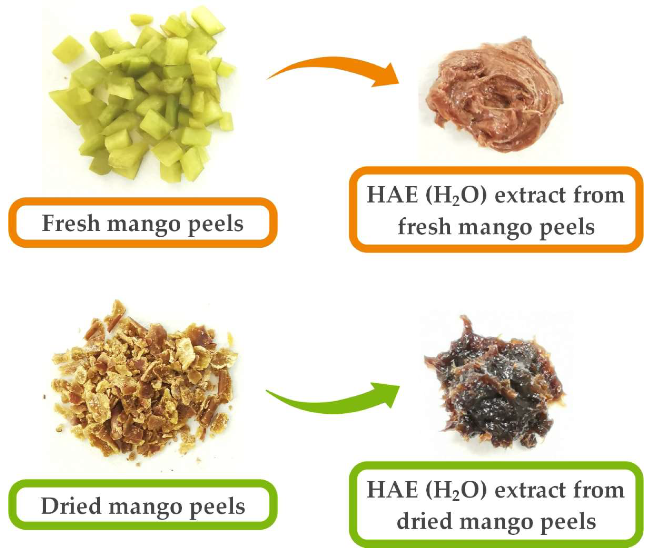
2.3. Preparation of Plant Material

Washed, fully ripe, Keitt-type mango fruits (country of origin: Puerto Rico) were peeled with a fruit peeler. Some of the obtained peels were air-dried without pretreatment at room temperature and protected from direct sunlight to avoid possible degradation of bioactive compounds. Before the extraction process, the completely dried peels were ground to a uniform size. The mean particle size was determined by sieve analysis and amounted to 1.0 mm. The dried peels were then stored at room temperature until further use. The remainder of the fresh peels was used immediately to avoid possible microbial contamination and putrefaction processes. The fresh peels were cut into smaller pieces and then subjected to an extraction process.

2.4. Moisture Content

The moisture content in fresh and dried mango peels was analyzed with the Mettler Toledo HX204 Halogen Moisture Analyzer. The moisture content was determined according to the thermogravimetric principle based on the weight loss of the dried analysis material by heating to 115 °C.

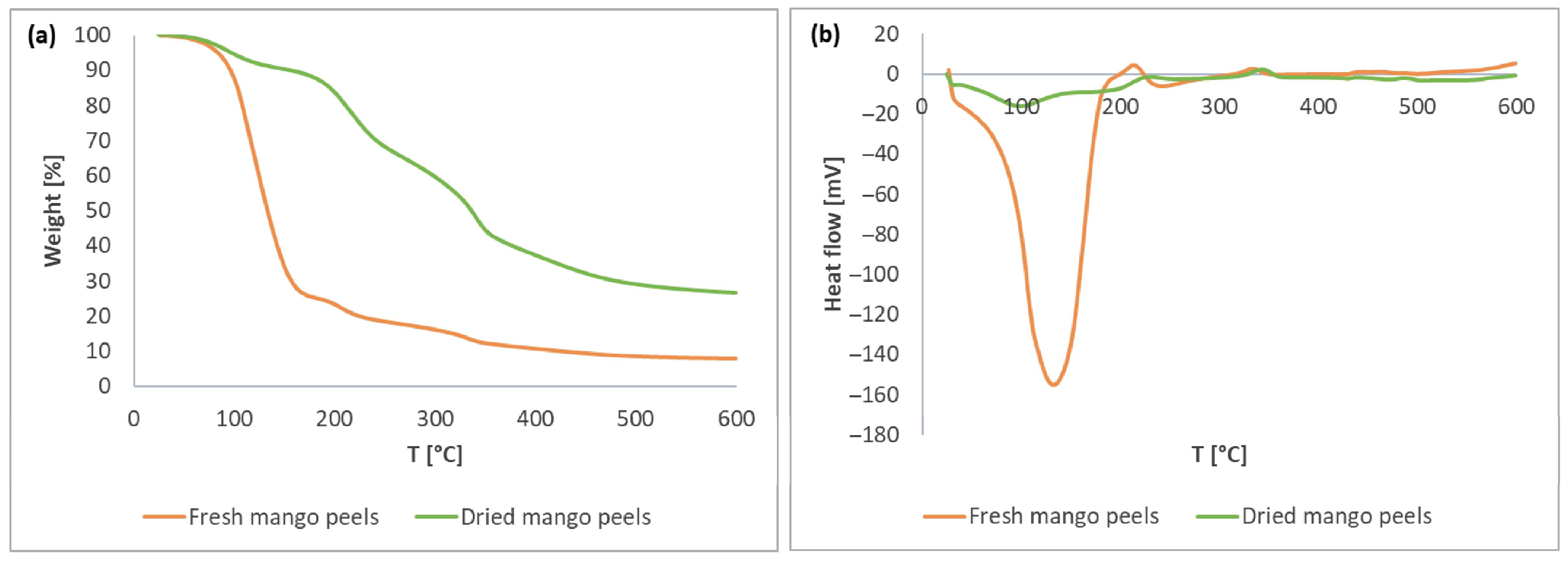
2.5. Thermogravimetric Analysis/Differential Scanning Calorimetry

The mango peels (fresh and dried) were subjected to thermal stability testing, including thermogravimetric analysis/differential scanning calorimetry (TGA/DSC). Both analyses were performed simultaneously on a TGA/DSC instrument (TGA/DSC1, Mettler Toledo AG (MTANA), Zurich, Switzerland) in a nitrogen atmosphere. Samples were weighed into aluminum pans and analyzed at a temperature range from 25 to 600 °C and a rate of 10 °C/min.

2.6. Extraction Procedures

Three different conventional extraction methods, namely SE, UAE, and HAE, were used to extract bioactive compounds from fresh and dried mango peels. Two polar extraction solvents, H2O and EtOH, were used. In addition, unconventional SFE using supercritical carbon dioxide (SC CO2) and EtOH as a co-solvent was also performed. For this method, the use of dry raw material is mandatory. Therefore, only the extraction of dried mango peels was performed. All extraction solvents used are considered GRAS (Generally Recognized as Safe).

2.6.1. Soxhlet Extraction

SE was performed with both fresh and dried mango peels. Peels were transferred into a cellulose thimble and placed in a Soxhlet extraction chamber. The extraction solvent EtOH was added to a round-bottom flask connected to a Soxhlet apparatus with a reflux condenser. The solvent was then heated to reflux. The extraction was carried out until the condensate became colorless (3 refluxes). The ratio of solvent (EtOH) to fresh and dried mango peels was 5:1 and 10:1, respectively.

2.6.2. Ultrasound-Assisted Extraction

UAE was performed on fresh and dried mango peels using EtOH and H2O as extraction solvents, respectively. Fresh or dried mango peels were mixed with an extraction solvent and sonicated at 20 °C and 40 kHz in an ultrasonic bath. Subsequently, filtering was performed through a filter flask and a Buechner funnel to remove solid, insoluble particles. The ratio of solvent (EtOH and H2O) to fresh and dried mango peels was 5:1 and 10:1, respectively.

2.6.3. Homogenization-Assisted Extraction

HAE, using H2O as an extraction solvent, was performed with fresh and dried mango peels. The homogenization process was carried out for 30 min. Then, the supernatant was centrifuged to achieve complete separation of the supernatant from the insoluble material. The ratio of solvent (H2O) to fresh and dried mango peels was 5:1 and 10:1, respectively.

2.6.4. Supercritical Fluid Extraction

SFE was performed in a semi-continuous apparatus using SC CO2 to obtain bioactive compounds from dried mango peels. Dried peels were placed in the autoclave and preheated to an operating temperature of 40 °C in a water bath. The CO2 was compressed to an operating pressure of 200 bar and continuously introduced into the autoclave at a 2–3 mL/min flow rate. At the same time, EtOH was added as a co-solvent to achieve better extraction of the polar bioactive compounds. The ratio of the final consumption of the solvent (EtOH and CO2) to the dried mango peels was 5:1 and 30:1, respectively.
After each extraction procedure, the extraction solvent (EtOH or H2O) was evaporated using a rotary evaporator (Büchi® Rotavapor R-144, Flawil, Switzerland). The extracts were stored in a freezer at −20 °C until further analysis.

2.6.5. Extraction Yield Determination

Each individual extraction was performed three times. The extraction yield was calculated as the ratio between the weight of the extract obtained and the initial weight of the material used. The results are given as mean values.

2.7. Quantitative Determination of Total Phenols and Proanthocyanidins

2.7.1. Determination of Total Phenolic Content (TPC)

The TPC content in the extracts obtained was determined by the Folin–Ciocalteu method. The analysis procedure for TPC is described in detail by Škerget et al. [34]. The results are given as mean values and are expressed as mg of GA equivalents (GAE) per gram of fresh weight (FW) for fresh peels or dry weight (DW) for dried peels.

2.7.2. Determination of Total Proanthocyanidin Content (PAC)

Condensed tannins or PAC were determined by a colorimetric method using HCl and n-butanol, in which acid-catalyzed oxidative depolymerization of PAC leads to the formation of the corresponding anthocyanidin compounds [34]. The results are reported as mean values expressed as mg of PAC per g of FW or DW.

2.8. Determination of Antioxidant Activity

The antioxidant activity of the extracts was determined using the stable free radical 2,2-diphenyl-1-picrylhydrazyl (DPPH) [35]. Antioxidant activity was expressed as a percentage of inhibition regarding the reference solution.

2.9. Liquid Chromatography with Tandem Mass Spectrometry Analysis

Liquid chromatography with mass spectrometry (LC-MS/MS) was used for the identification and quantitative determination of selected phenolic compounds in mango peel extracts, using Agilent 1200 HPLC together with Agilent 6464 QQQ with JetStream technology, as described in detail by Hrnčič et al. [36].

2.10. Determination of Total Protein (TP) Content

The Bradford method [37] was used to determine TP content using bovine serum albumin (BSA) as the standard. All analyses were performed in three replicates and expressed as mean values in mg of protein per g of FW or DW.

2.11. Determination of Enzyme Activities

The enzyme activity of α-amylase [38], cellulase [39], glucoamylase [40], laccase [41], lipase [38], peroxidase [35], polyphenol oxidase (PPO) [42], protease [39], SOD [43], and transglutaminase (TGM) [44] were examined with specific spectrophotometric enzymatic assays for each selected enzyme using a UV spectrophotometer (Varian—CARY® 50 UV–VIS Spectrophotometer, Varian Inc., Middelburg, The Netherlands). Each assay was performed in three replicates. The results are reported as mean values expressed in units (U) per g of protein.

2.12. Determination of Antimicrobial Activity

For the determination of the inhibitory properties of the extracts obtained on the growth of selected microbial cells (Gram-negative bacteria (E. coli and P. aeruginosa), Gram-positive bacteria (B. cereus and S. aureus), and fungi (black mold A. brasiliensis and yeast C. albicans)), the qualitative disk diffusion method and the quantitative microbroth dilution method were used.

2.12.1. Qualitative Disk Diffusion Method

The antimicrobial properties of the mango peel extracts were verified by the qualitative Kirby–Bauer disk diffusion method, which was previously described in detail in the study by Kupnik et al. [45], using 6 mm sterile cellulose discs. Ciprofloxacin was used as a positive control. All tests were performed with three replicates, and the results are given as mean values.

2.12.2. Quantitative Microbroth Dilution Method

The microbroth dilution method was used, which is best suited for the in vitro determination of the susceptibility or resistance of microorganisms [45]. Therefore, the minimum inhibitory concentrations (MICs) and the MGIRs were determined. The method was performed only for those microorganisms that were found to be susceptible when exposed to the mango samples, as determined by the disk diffusion method. All experiments were performed in three replicates and are expressed as mean values.

2.13. Statistical Analysis

All statistical data analyses were performed using IBM® SPSS® Statistics. Statistical data analysis was performed to study the differences between extraction methods. The normality of the distribution of data was tested using Shapiro–Wilk’s test. The homogeneity of variances was determined using Levene’s test. Differences between extractions were determined with one-way analysis of variance (ANOVA) followed by Tukey’s post hoc test (for normally distributed data) and with the nonparametric Kruskal–Wallis H test, followed by pairwise comparison using the Dunn–Bonferroni post hoc method (for abnormally distributed data).

3. Results and Discussion

The present study focused on the influence of various parameters (drying method, extraction method) on obtaining highly active bioactive compounds with good antimicrobial and antioxidant activity.

3.1. Thermal Stability of Mango Peels

For comparison, the moisture content in fresh and dried mango peels was first analyzed. Fresh mango peels contained 77.74% moisture, while completely air-dried peels stored at room temperature in an airtight container still contained 11.06% moisture.
The differences in thermal behavior of fresh and dried mango peels were further investigated using TGA/DSC analysis. Figure 1a shows the thermal degradation of fresh and dried mango peels as determined by thermogravimetric analysis. Due to the high moisture content, fresh mango peels showed a drastic weight loss in the temperature range of 39–202 °C. On the other hand, the weight of dried mango peels gradually decreased throughout the entire temperature range, decreasing by 73.3%. The total weight loss of fresh peels was 92.0%. The two curves are generally comparable after excessive evaporation of moisture in the fresh peels, indicating a similar composition. The residue represents the ash fraction, which contains minerals that cannot be degraded [46]. The residue accounted for 8.0% of the fresh peels and 26.7% of the dried peels, based on the total mass of the studied peel samples.
Figure 1b shows the DSC thermogram for fresh and dried mango peels. An initial melting peak at 99 °C for the dried peels and at 133 °C for the fresh mango peels indicates the evaporation of H2O and the beginning of the degradation of the carbohydrates, indicating the melting point (endothermic peak). For example, the melting point of the different sugars is 126.5 °C for fructose, 159.6 °C for glucose, and 190.4 °C for sucrose [47], which are present in mango fruit [48]. A slight shift in the melting peak in fresh peels is due to the different binding of H2O and sugars compared to the dried peels. Moreover, the two curves of fresh and dried mango peels do not differ further. The second peak (198–254 °C) and the third peak (304–368 °C), shown as exothermic peaks, are due to the degradation of long-chain, high-molecular-weight polymers [46] or various salts, for example, sulfur-containing salts, such as sodium hydrogen sulfite. Sodium hydrogen sulfite, which decomposes at 315 °C, is classified as a food additive [49]. Therefore, it can be applied to the surface of mango peels during post-harvest treatment to preserve their freshness and prevent them from rotting too quickly and experiencing discoloration during long transportation periods.
Since the thermal stability of fresh and dried peels was comparable, we performed further extractions of differently dried peels to compare bioactive compound content and biological activity.
Furthermore, when visually assessing the color of the plant material used (fresh or dried peels), it was found that the dried peels had a darker color due to the lower moisture content. Consequently, the extracts obtained also had a slightly darker color (Figure 2). Therefore, the color of the material may also be related to a change in the content of bioactive compounds.

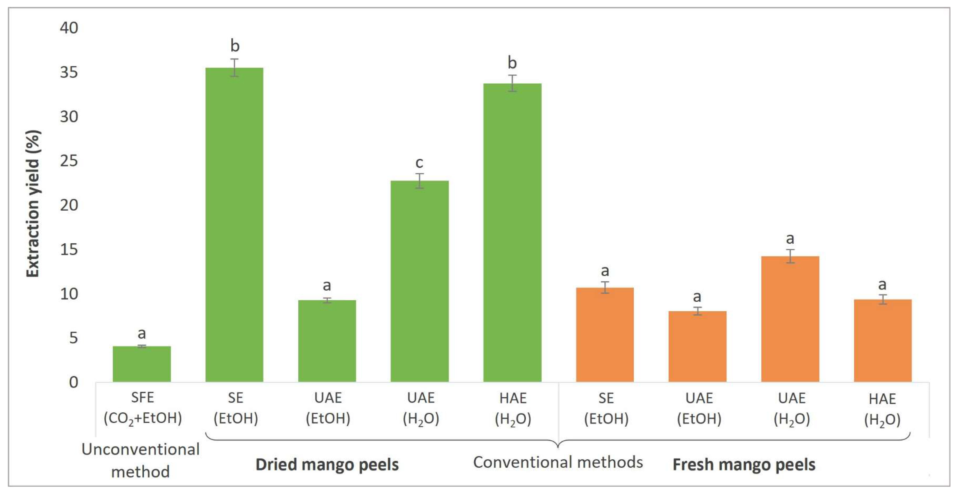
3.2. Extraction Yield Determination

Different extraction methods were performed using fresh or dried mango peels as raw material to compare the effects of the extraction procedure (conventional and unconventional) and the extraction solvent (CO2 + EtOH, EtOH, H2O) used on the profile of bioactive compounds and biological activity. Figure 3 presents the efficiency of each extraction performed, expressed as extraction yield (%).
The highest extraction yield was obtained with conventional extraction techniques (SE (EtOH) from dried peels (35.52%) and HAE (H2O) from dried peels (33.76%)). The explanation lies in the prolonged action of the extraction solvent (EtOH) on the mango peels using SE. During HAE, efficient cell wall disruption was achieved, causing the polar bioactive compounds of the cell wall to dissolve in H2O. However, the operating temperature in SE was above the boiling temperature of EtOH (>78 °C), which could have a negative effect on the extraction yield of sensitive bioactive compounds. In the other extraction techniques performed, the operating temperature was lower (40 °C in the case of SCF, and 22 °C for HAE and UAE).
On the other hand, when comparing UAE with the different extraction solvents used, higher extraction yields were obtained with H2O as the extraction solvent than with EtOH, which can be attributed to the greater polarity of H2O. In general, a higher amount of extract was obtained from the dried peels. This is due to the large amount of moisture in fresh peels. Dorta et al. [50] also obtained higher extraction yields when fresh peels were extracted by microwave-assisted extraction (MAE) compared to dried peels by different drying methods. The extraction yields of air-dried mango peels determined in a study by Souza et al. [51] are in line with our results.
Regarding SFE, since CO2 is a nonpolar solvent, a polar co-solvent such as EtOH should be used to allow the extraction of important polar bioactive compounds. Therefore, the efficiency of SFE was the lowest compared to conventional methods. Sánchez-Camargo et al. [52] optimized the SFE operating conditions for mango peel extraction, with extraction yields ranging from 1.60 to 6.25%. The highest yields were obtained at operating conditions of 300 bar and 60 °C and using EtOH as a co-solvent. This confirms the presence of polar secondary metabolites in the plant material.
A comprehensive comparison with the results of other studies is difficult because the operating conditions of the extractions performed and the extraction solvent used are different. This significantly affects both the extraction yield and the phytochemical and nutraceutical profile of the extracts. Also, usually, the origin, maturity, and growth conditions of the fruit are not the same.

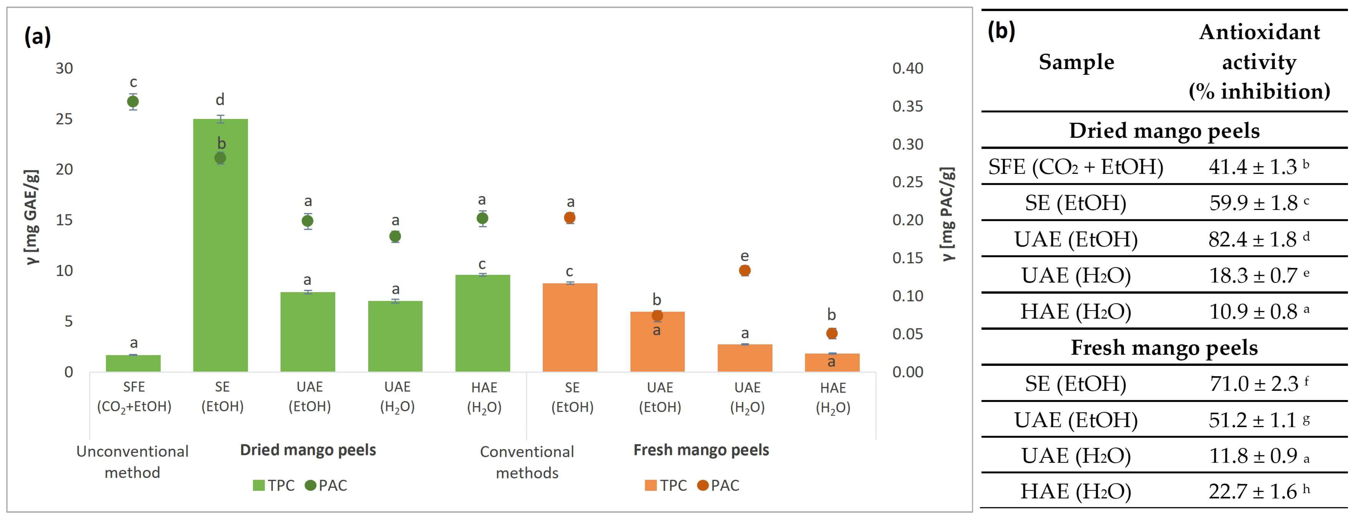
3.3. Content of Secondary Metabolites in Mango Peel Extracts and Their Antioxidant Activity

A quantitative investigation of the content of TPC and PAC of the extracts obtained from fresh and dried mango waste was performed. The results are shown in Figure 4a.
The highest TPC content was achieved in samples obtained by SE (EtOH) (25.0 mg GAE/g DW), as seen in Figure 4a. Since the lowest extraction yield was obtained in the SFE, consequently, the TPC content was also the lowest (1.7 mg GAE/g DW). The extracts obtained from dried mango waste generally had higher TPC content than those obtained from fresh peels using the same extraction procedure. Furthermore, this indicates that the TPC content is maintained regardless of the air drying of the mango peels. Ethanolic extracts, also when SFE (CO2 + EtOH) was used, resulted in higher TPC content compared to aqueous extracts.
The content of extracted TPC during UAE is strongly influenced by operating conditions such as the solid–liquid ratio, temperature, amplitude, and time [53,54,55,56]. Borrás-Enríquez et al. [57] obtained an even higher TPC content (18.14 mg GAE/g DW) using an aqueous solution of EtOH (50%, v/v), as in our study, where separate solvents (EtOH and H2O) were used. The use of EtOH as an extraction solvent proved to be more efficient than the use of MeOH in the extraction of TPC [55]. Moreover, using SFE under the same operating conditions as in the present study, only without a co-solvent, a 2.4-fold lower TPC value was obtained compared to our results. This indicates that EtOH as a co-solvent significantly affects the recovery of bioactive compounds from mango by-products. Overall, the TPC values showed a tendency to decrease with increasing operating temperature and increase with increasing operating pressure [51,58].
Furthermore, the PAC content was determined in the extracts obtained from mango waste peels. PACs are colorless flavonoids that have beneficial effects on human health due to their antioxidant, anticancer, antimicrobial, anti-inflammatory, antidiabetic, neuroprotective, and hypolipidemic properties [59,60].
The results for the PAC content between ethanolic and aqueous extracts varied between 0.2 and 0.4 mg PAC/g DW and between 0.1 and 0.2 mg PAC/g FW. The highest amount of PACs was determined for the SFE extract (0.4 mg PAC/g DW), followed by the SE (EtOH) extract with 0.3 mg PAC/g DW. Thus, the operating conditions used for SFE extraction (temperature, time, pressure, addition of co-solvent) were beneficial.
According to the reviewed literature, different methods can be used to determine the PAC concentration in extracts. However, very few studies report the analysis of the content of PACs in mango peel extracts. The presence of PACs in mango peels is mainly influenced by the geographical area of the species and the extraction process [61]. The content of PACs, expressed as leucoanthocyanidin equivalents (LE), ranged from 1.2 to 5.8 mg LE/g DW, which was mainly influenced by the solvent used, while temperature had a negligible effect [62]. The highest PAC values were obtained with the mango peel extract using MeOH, EtOH:H2O, or Ace:H2O as a solvent. In another study by Dorta et al. [63], MAE yielded 0.0018–0.0048 mg LE/g DW of mango peels. The PAC content in mango peels increases with increasing fruit ripeness in different varieties. However, mango peels contain higher PAC concentrations compared to pulp, as confirmed by different studies [61,64].
The antioxidant activity of the mango peel extracts was determined using the DPPH method. This method is commonly used to evaluate the ability of substances that act as free radical scavengers. These substances are antioxidants that can donate a proton in the presence of the DPPH free radical, which is then converted into a reduced form [65]. The results were expressed as a percentage of inhibition of free radical DDPH and are presented in Figure 4b.
Furthermore, due to the different extraction methods and solvents used, different profiles of the extracted bioactive compounds were obtained. The extracts in which EtOH was used as an extraction solvent (UAE and SE) or as a co-solvent (SFE) exhibited significantly higher DPPH radical scavenging activity. This indicates that EtOH has an important influence on the extraction of bioactive compounds with antioxidant properties. The highest antioxidant activity was determined in UAE (EtOH) extract from dried peels, with 82.4% inhibition, which is 3.6 times higher than in HAE (H2O) extract from fresh peels (22.7%), where the highest % inhibition was achieved among aqueous extracts. On the other hand, no significant differences in the antioxidant potential of fresh and dried raw peels processed by the same method and under the same conditions were detected.
The antioxidant activity of extracts from dried mango peels obtained by UAE reached 87% DPPH inhibition using 50% (v/v) EtOH [57], which is comparable with our results obtained by UAE with 100% EtOH. Moreover, Kaur et al. [56] demonstrated the influence of operating conditions (liquid/solid ratio, temperature, amplitude, time) in the UAE on the determined antioxidant activity of mango peel extracts.
Souza et al. [51] achieved only 25% DPPH inhibition of mango peel extract obtained by SFE without a co-solvent. The present study achieved a 1.7-fold higher inhibition percentage, indicating that the co-solvent EtOH significantly improves the recovery of polar bioactive compounds with antioxidant potential when similar operation conditions are used.

3.4. Content of Important Phenolic Compounds in Mango Peel Extracts

Fruits are an essential source of various PCs with health-promoting properties. Therefore, a quantitative study was performed for eight important PCs, including xanthone (mangiferin), four phenolic acids (caffeic acid, chlorogenic acid, ellagic acid, gallic acid), and three flavonoids (catechin, hesperidin/neohesperidin, rutin) in the obtained mango peel extracts. The results are presented in Table 1.
Numerous studies in the reviewed literature have already shown that all parts of the mango tree, including peels, are a rich source of important bioactive compounds [66]. However, to the best of our knowledge, no study has addressed the effects of the drying process of the starting material on the retention of important bioactive compounds and their contribution to the antimicrobial activity of mango peel extracts obtained by different conventional and unconventional methods.
The results revealed that the obtained mango peel extracts varied in the composition of the analyzed bioactive compounds, considering the extraction procedure, the extraction solvent, and the raw material used (fresh or dried peels).
Mangiferin is one of the most important PCs of the mango tree having health-promoting properties [67]. Most of the analyzed extracts were found to contain mangiferin, with the highest content determined in the aqueous extracts of dried mango peels. In contrast, mangiferin was either not detected in the aqueous extracts of fresh mango peels (UAE extract) or only found in small amounts (HAE extract). The opposite results were obtained with ethanolic extracts, where a higher content of mangiferin was successfully extracted from fresh mango peels compared to dried peels. It can be concluded that air-drying has no significant effect on the possible degradation of mangiferin. Kaur et al. [68] reported that shade-drying at room temperature resulted in higher mangiferin content in the plant material than oven- or sun-drying. On the other hand, the final content of mangiferin is mainly influenced by the choice of extraction method [69] and extraction solvent [70]. In the reviewed literature, the mangiferin content in extracts obtained by different methods and operating conditions ranged from 0.331 to 150 µg/mg DW [57,67,71,72,73]. Furthermore, Marcillo-Parra et al. [74] found that the mangiferin content in mango peel extracts also varied widely depending on the mango variety, ranging from 57.2 to 3140 µg/g DW. Mangiferin is only slightly soluble in EtOH and H2O [75] but has a very low solubility in SC CO2 [76], which is consistent with our results, as the lowest mangiferin concentration was obtained with SFE (0.15 µg/g DW).
Regarding the content of phenolic acids in the extracts of mango waste peels, ellagic acid and gallic acid are the most abundant. The highest composition of ellagic acid was found in UAE (H2O) extract (539.42 µg/g FW) and SE (EtOH) extract (531.98 µg/g FW), while the lowest composition was present in SFE (CO2 + EtOH) extract (37.50 µg/g DW), as ellagic acid is poorly soluble in SC CO2 [77]. Furthermore, all extracts obtained from fresh mango peels had higher ellagic acid content than those obtained from dried peels by the same extraction method and under the same conditions. This indicates that the drying process may have a destructive effect on ellagic acid and its composition [78]. Ellagic acid content has already been determined in mango peel extracts, where its content ranged from 15.995 to 298.69 µg/g DW [73,79,80]. On the contrary, the highest content of gallic acid was determined in UAE (H2O) extract (405.72 µg/g DW), while the lowest content was found in UAE (H2O) extract (8.03 µg/g DW). Gallic acid is soluble in EtOH and H2O [81], whereas in SFE, the addition of a polar co-solvent (EtOH) is needed due to its low solubility in SC CO2 [82]. Extracts from dried peels were found to have a higher gallic acid composition than their fresh counterparts. Drying methods generally do not significantly affect gallic acid and its content in dried samples [83]. Higher gallic acid contents in extracts from dried peels indicate the high moisture content of fresh mango peels used for extraction. In the reviewed literature, different compositions of gallic acid in mango peel extracts have been reported, ranging from 23 to 23,816 µg/g DW [80,84,85,86,87] and 14.5 to 791.5 µg/g FW [88]. A similar phenomenon was observed in the composition of caffeic acid and chlorogenic acid. The content of the aforementioned phenolic acids was higher in extracts from dried peels than in extracts from fresh peels, comparing the same extraction procedure. This indicates that air-drying has no significant effect on caffeic and chlorogenic acids [89,90]. The highest content of chlorogenic acid was determined in SE (EtOH) extract (26.54 µg/g DW), whereas no chlorogenic acid or a very low amount was present in H2O extracts. Furthermore, the extract with the highest caffeic acid composition was the SE (EtOH) extract (6.60 µg/g DW), while no caffeic acid was detected in the UAE (H2O) extract, obtained from fresh peels. This indicates higher chlorogenic and caffeic acid solubility in EtOH than in H2O [91,92]. Compared with the reviewed literature, the content of chlorogenic acid in various mango peel extracts was found to be 760–2280 µg/g DW [93] and 44.05–271.9 µg/g FW [88], while the content of caffeic acid was 16.6–67.3 µg/g DW [93] and 33.03–144.3 µg/g FW [88].
Among the flavonoids, catechin was the most abundant in mango peel extracts. The highest content was observed in the SE (EtOH) extract (109.39 µg/g DW), while the lowest catechin concentration was found in the UAE (H2O) extract, with only 0.38 µg/g FW, since catechin is soluble in H2O and organic solvents, including EtOH [94]. However, in SFE, the solubility of catechin is mainly influenced by the operating conditions and the addition of a co-solvent, such as EtOH [95]. Other studies have also confirmed the presence of catechin in various mango peels, ranging from 45.73 to 115.5 µg/g DW [55,73]. A similar pattern was observed for hesperidin/neohesperidin content, except for the UAE (EtOH) extract, where more hesperidin/neohesperidin was extracted from fresh mango peels. Overall, hesperidin/neohesperidin was found in lower amounts in all extracts. The highest level was observed in the UAE (EtOH) extract (2.67 µg/g FW). Due to its polarity, hesperidin is very poorly soluble in SC CO2 [96]. According to the reviewed literature, to the best of our knowledge, the content of hesperidin/neohesperidin has not been determined or detected in mango peels. However, in a study by Abdel-Aty et al. [97], hesperidin was found to be the major PC in mango seed kernel extract (55.6% of total compounds). Rutin was only detected in the UAE (H2O) extract at a concentration of 0.80 µg/g DW. Other studies have already established that the rutin content in mango peel extracts ranges from 37.15 to 390 µg/g DW, which is mainly influenced by the mango variety [55,80].
The total content of the eight phenolic compounds analyzed was determined in the SE (EtOH) extract (829.92 µg/g DW). Overall, extracts from dried mango peels contained higher total levels of analyzed PCs than extracts from fresh peels obtained by the same extraction procedure and under the same conditions. This is an important fact, because most of the PCs analyzed were retained during the air-drying process.

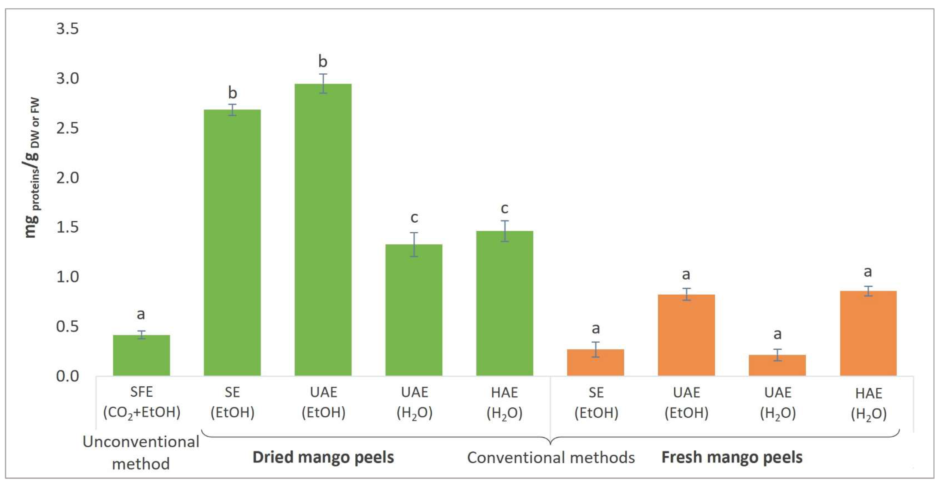
3.5. Total Protein Content and Activities of Certain Enzymes in Mango Peel Extracts

The results of the determination of total proteins in extracts from fresh and dried mango peels are presented in Figure 5.
The highest TP content was observed in the extract obtained from dried peels using UAE (EtOH) (2.95 mg protein/g DW), as presented in Figure 5. The explanation lies in the effective mechanical disintegration of the cell wall of mango peels, causing the release of intracellular proteins. Consequently, since fresh mango peels had a high moisture content (77.74%), a higher protein concentration was observed in extracts from dried peels, indicating successful protein retention during the air-drying process. The lowest TP content was detected in the UAE (H2O) extract, with only 0.21 mg protein/g FW, and the SE (EtOH) extract, with 0.27 mg protein/g FW.
The TP content of fresh mango peels was 1.9 to 2.3%, based on proximate composition analysis [17,98]. Using the Bradford method, the TP content of fresh mango peels processed with acid-washed sand and sodium phosphate buffer using a mortar and pestle was estimated to be 4.0–12.7 mg/g DW. Umbreen et al. [99] determined a TP content of 6.55% in hot air-dried mango peel powder at 50–60 °C. Hot-air-dried raw and ripe mango peel powder yielded 0.16 and 0.18 mg protein/g DW, respectively [100].
However, a comparison with the total protein concentration of extracts from mango peels could not be performed because, to the best of our knowledge, no study has been published using the same extraction procedures as in the present study (SFE, SE, UAE, HAE).
Additionally, the activities of certain enzymes in mango peel extracts have been determined using specific enzymatic assays, as mango peels have been reported to contain various types of enzymes [101]. However, only a few studies are available regarding the enzymatic activities of mango peel extracts. Our main objective was to extract and identify valuable enzymes from mango peels, which could make an important contribution as an exceptional source of enzymes for further applications in various areas of medicine, cosmetics, and nutrition. The determined enzyme activities, defined as U/g protein, are shown in Table 2.
Antioxidant enzymes successfully protect fruits from oxidative stress and reduce the content of reactive oxygen compounds (ROS), as they are involved in the catalytic conversion of ROS and their by-products into stable, non-toxic molecules [102,103]. In the present study, the activities of peroxidase, PPO, and SOD were determined among the antioxidant enzymes. The activity of peroxidase was the lowest compared to the activities of all the enzymes studied. However, in the reviewed literature, peroxidase activity was significantly higher [17,98], though different substrates and methods for activity determination were used. On the contrary, the activity of PPO, responsible for enzymatic browning and melanogenesis [104], was very high in all mango peel extracts. HAE was the most outstanding technique to obtain PPO in a highly active form. The cell wall was successfully disrupted, but the mechanical force did not affect the enzyme. The PPO activities ranged from 14,036.01 to 430,837.99 U/g protein, which is higher than in a study by Tokas et al. [17] (25,800–75,500 U/g protein), where the mango peel extracts were obtained by the maceration of fresh peels. The obtained extracts from mango peels are rich in SOD, since the SOD activity was remarkable in the UAE (EtOH) extracts obtained from fresh and dried peels. The enzyme SOD acts as an excellent therapeutic agent against diseases caused by ROS and provides essential antioxidant protection against oxidative stress [105]. The results of SOD activities in the present study are comparable to the SOD activities in extracts from fresh mango peels obtained using Tris-HCl buffer as the extraction medium [17]. It was found that the activity of SOD was successfully maintained during drying, as the activities of extracts obtained from dried peels by the same extraction were higher than those of fresh peels. In contrast, opposite results were obtained for PPO activity, where the drying process partially affected the decrease in activity.
Furthermore, activities of various digestive enzymes were found in mango peel extracts. α-Amylase and glucoamylase are starch-degrading enzymes and are among the most frequently used biocatalysts in the food industry [106]. Both enzymes were present in their active forms in all extracts. For both enzymes, air-drying generally resulted in a loss of their activities. Further, proteases are protein-degrading enzymes. Protease activity was detected in most extracts obtained from mango peels using different extraction methods, which coincides with the results in the literature [17,98]. Cellulase activity was generally higher in all aqueous extracts, especially those from fresh peels. Regarding ethanolic extracts, cellulase activities were higher in extracts obtained from dried peels, with the highest activity obtained by the UAE (EtOH) extract from dried peels (124.15 U/g protein). Cellulase plays an important role in the degradation of insoluble cellulose to soluble sugars. It shows great potential for various industries, including agriculture, brewing, textiles, pulp and paper, food, and biofuel production [107]. Most digestive enzyme activities decreased with the drying process of mango peels. However, lipase seems to be resistant to drying, as the activities were higher in the extracts obtained from dried peels regardless of the extraction process and solvent used. These enzymes are also important in various industries, including food, biofuels, detergents, and animal feed [108].
All extracts from mango peels also yielded laccase activity, ranging from 30.78 U/g protein (HAE (H2O) extract from dried peels) to 228.88 U/g protein (UAE (H2O) extract from fresh peels). Laccases are multifunctional biocatalysts that play an important role in various applications, including the environmental field, the cosmetics industry, food processing, and the textile industry [109]. TGM was also found in its active form in all extracts, although the activity in the HAE (H2O) extract from fresh mango peels was significantly higher (120.31 U/g protein) than in other extracts. This can be attributed to the extraction process used (HAE), as the cell wall was successfully disrupted, and thus, the extraction of the TGM in its active form was achieved. In general, TGM activity was lower in extracts from dried mango waste. TGMs are also multifunctional enzymes [110]. From a health perspective, TGMs can reduce allergies, control energy intake from food, and act as mediators in wound healing [111].
The ripening of the mango fruit can lead to a progressive decrease in the activity of antioxidant enzymes, while the activity of digestive enzymes increases [17]. In addition, the extraction procedure, solvent, and operating conditions also significantly impact enzyme activities in the final product [98].

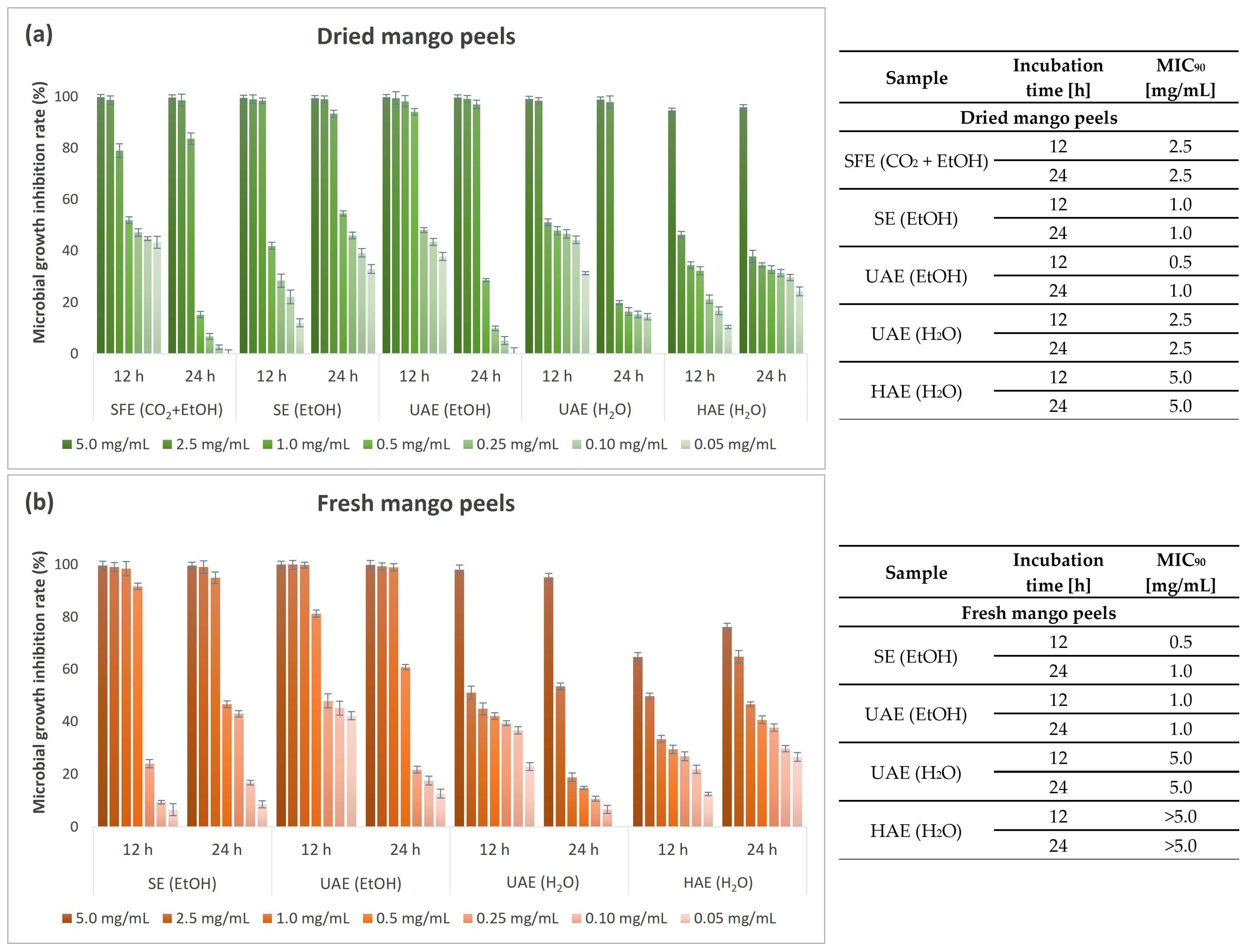
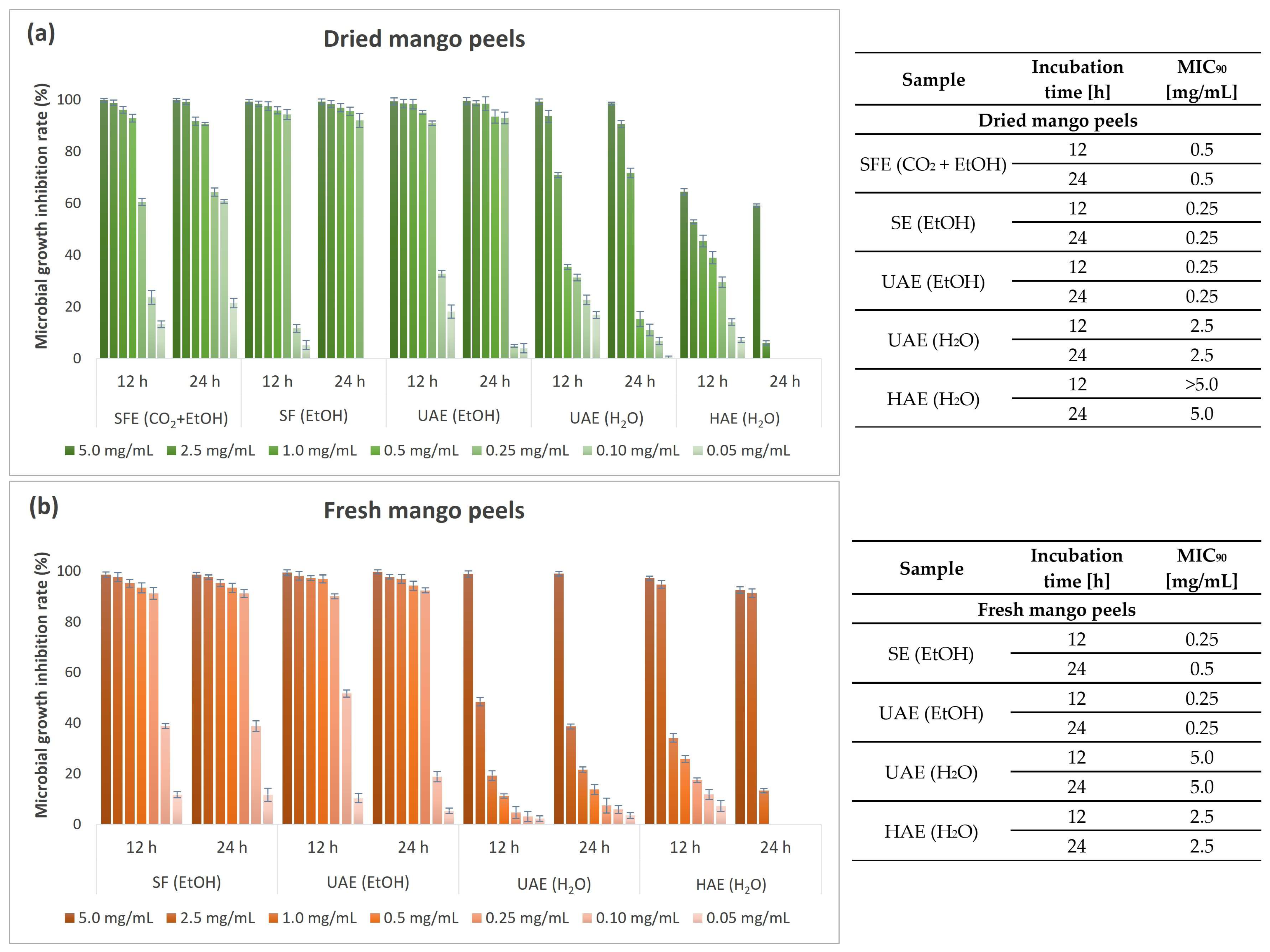
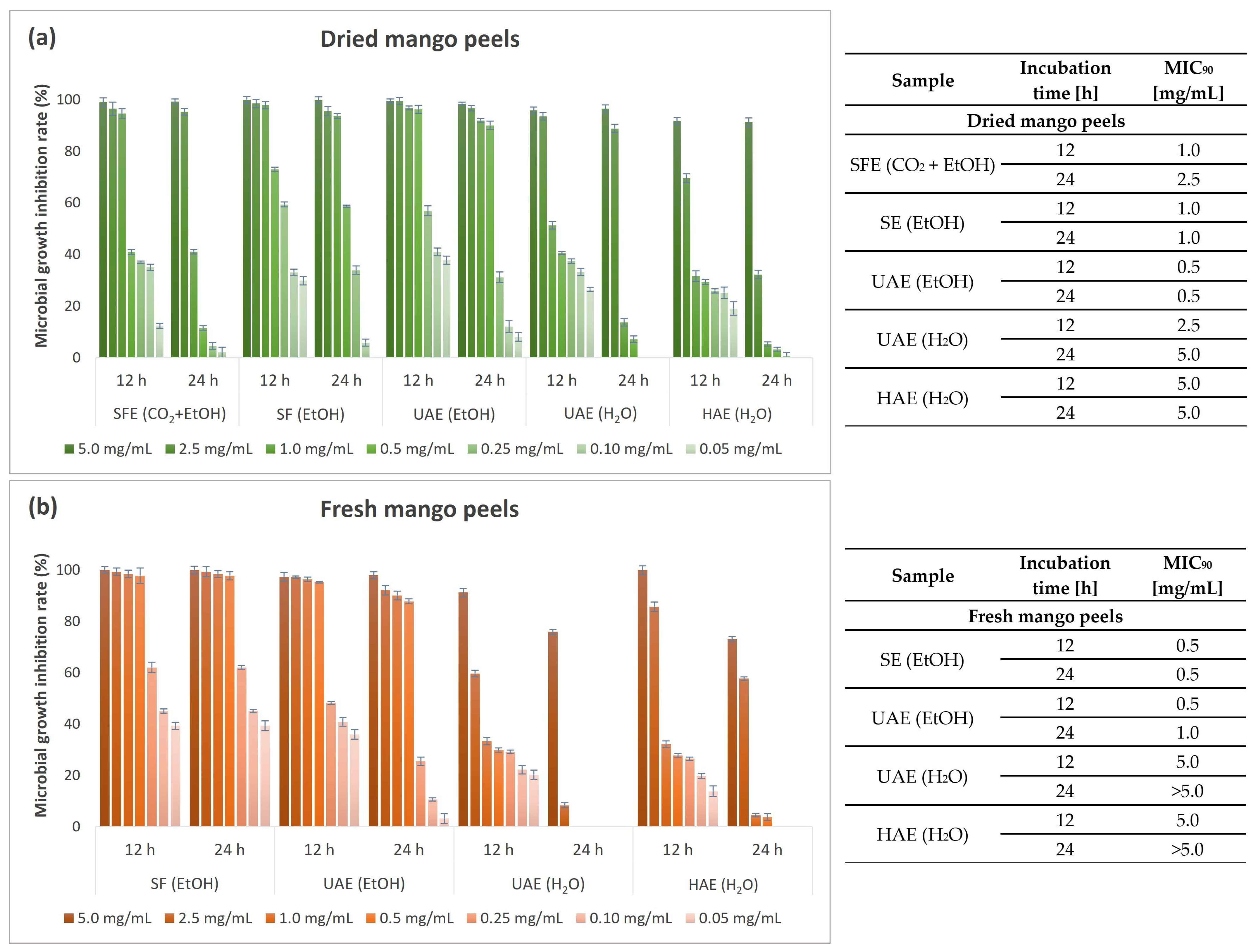
3.6. Antimicrobial Activity of Mango Peel Extracts

Infectious diseases represent a growing global problem, making searching for new potential antimicrobial agents increasingly important. Due to the improper and excessive use of synthetic antimicrobials, various pathogenic microorganisms that cause serious diseases have developed resistance [112,113]. For example, E. coli can cause different illnesses, including diarrhea, urinary tract infections, respiratory illness, and bloodstream infections [114]. P. aeruginosa is associated with causing severe infections and the spread of antimicrobial resistance in vivo [115]. B. cereus is known as a foodborne pathogen that can cause two types of gastrointestinal disease: emetic (vomiting) syndrome and diarrheal syndrome [116]. S. aureus is responsible for various skin infections up to severe invasive infections of the lungs or heart [117]. A. brasiliensis is mostly responsible for allergic reactions and lung infections [118], while C. albicans is the main cause of candidiasis and primary fungal infections [119]. Therefore, natural materials and extracts are receiving increasing attention, as they represent a new source of important biologically active compounds with potential antimicrobial properties [120]. For example, extracts from fruit wastes that contain high levels of bioactive compounds with antimicrobial properties, such as mango peels, could be used as effective, alternative, and safe natural remedies [121]. Thus, a comprehensive and comparative antimicrobial study of the different extracts of fresh and dried mango peels was conducted.

3.6.1. Qualitative Antimicrobial Determination of Mango Peel Extracts

The antimicrobial properties of mango peel extracts were preliminarily evaluated using the qualitative disk diffusion method. The susceptibility of Gram-negative (E. coli, P. aeruginosa) and Gram-positive bacterial species (S. aureus, B. cereus) and fungi (yeast C. albicans, black mold A. brasiliensis) to the addition of mango peel extracts was studied. Table 3 shows the measured diameters of the inhibition zone (in mm) of the individual microorganism tested after exposure to the mango peel extracts and ciprofloxacin as a positive control.
B. cereus appeared to be the most susceptible to all mango peel extracts among the tested bacterial species. The largest inhibition zone was measured during exposure of B. cereus to the EtOH extract obtained by UAE (22 mm) and SF (19 mm) from fresh peels, followed by extracts from dried peels. B. cereus, on the other hand, was the most resistant to the UAE (H2O) extract from dried peels. The second most sensitive bacterial strain to the mango peel extracts tested was E. coli. Exposure of E. coli to the UAE (EtOH) extract from dried and fresh peels resulted in the largest zone of inhibition of all the samples tested. The HAE (H2O) extract showed the lowest inhibitory effect against E. coli. Similar growth inhibition was obtained regarding the antimicrobial efficacy of mango peel extracts on P. aeruginosa and S. aureus growth. Both bacteria were the least susceptible to the addition of the HAE (H2O) extract from fresh and dried peels. Table 3 also shows that ciprofloxacin was effective against all bacterial species tested. The most sensitive strains were P. aeruginosa and B. cereus, as the highest zone of inhibition was determined.
All the extracts obtained from mango peels successfully inhibited the growth of the tested bacterial species, which is an important observation for the possibility of further use as successful natural antibacterial agents. Generally, higher inhibition zones were achieved with extracts obtained by extraction procedures using EtOH as an extraction solvent or co-solvent. The most outstanding sample with the highest microbial sensitivity when exposed to all four bacterial species was the UAE (EtOH) extract from fresh peels. The extraction solvents were almost completely removed before analysis. Therefore, their influence on the antimicrobial potential of the extracts is negligible. In addition, using the same extraction method, slightly higher growth inhibition was obtained using extracts from fresh mango peels than dried peels.
The results are in agreement with the reviewed literature, which also indicates good antibacterial activity of various extracts from mango peels, with the inhibitory potential of EtOH extracts being higher than those of H2O extracts. Methanolic mango peel extract successfully inhibited the growth of S. aureus, Staphylococcus epidermidis, P. aeruginosa, E. coli, and Salmonella sp. [8,122]. The results of the antibacterial activity of ripe and unripe methanolic mango peel extracts show a larger inhibition zone against all tested bacterial species by ripe mango peel extracts [123]. Also, ethanolic mango peel extract showed inhibition properties against E. coli, S. aureus, B. cereus, P. aeruginosa, S. typhimurium, and V. parahaemolyticus [20,124].
The remarkable antibacterial potential of mango peel extract obtained by various methods is consistent with the fact that various phytochemicals and bioactive compounds are responsible for the disruption of the cell membrane of microbial cells, in addition to their ability to scavenge free radicals, activate endothelial cell migration, and inhibit inflammatory and pain pathways [8]. In contrast, no inhibitory properties of mango peel extracts were found against the tested fungal species (yeast C. albicans and black mold A. brasiliensis).

3.6.2. Quantitative Antimicrobial Determination of Mango Peel Extracts

Considering the excellent inhibitory effect on the tested bacterial strains (E. coli, P. aeruginosa, S. aureus, and B. cereus), the antimicrobial activity was additionally determined after exposure to seven different concentrations of the extracts (0.05–5.0 mg/mL) using a more accurate quantitative microbroth dilution method. MGIR values were determined after 12 and 24 h exposure periods of the bacterial strain to different concentrations of the tested mango peel extracts. In addition, MIC90 values were determined based on the MGIR value when at least 90% MGIR was achieved.
Figure 6 shows the results of the antimicrobial study on the growth of Gram-negative bacterium E. coli as performed by the microbroth dilution method.
All mango peel extracts, with the exception of the HAE (H2O) extract from fresh peels, were very successful in inhibiting the growth of Gram-negative bacterium E. coli at the highest concentration tested (5 mg/mL) after 12 h and 24 h of incubation. In general, E. coli was more susceptible to ethanolic than aqueous extracts. A slightly higher susceptibility of E. coli to aqueous extracts from dried peels was observed, indicating successful retention of bioactive compounds regardless of the drying process. With respect to the HAE (H2O) extract from fresh mango peels, the MIC90 value of the tested concentrations could not be determined. The analyses would have to be performed at higher extract concentrations to obtain more reliable results. Nevertheless, a relatively high MIGR (76.26%) was achieved at the highest applied extract concentration after 24 h of incubation.
Figure 7 shows the results of the antimicrobial study on the growth of the second Gram-negative bacterial strain, P. aeruginosa, as performed by the microbroth dilution method.
P. aeruginosa proved to be slightly more resistant to mango peel extracts than E. coli. Overall, P. aeruginosa, like E. coli, was generally more sensitive to ethanolic than aqueous extracts. Among the aqueous extracts, only the UAE (H2O) extract from dried mango peels successfully inhibited the growth of the above bacterium, with an effective inhibition of more than 90% at 5.0 mg/mL. Exceptional results were obtained at the lowest concentration (0.05 mg/mL) of the extracts used, with MGIR values above 35% for seven extracts and above 50% for four of the nine tested extracts, with the highest percentage for the SFE (CO2 + EtOH) extract (63.11%). This is a remarkable result, because P. aeruginosa has become highly resistant to antibiotics. Therefore, new approaches to the treatment of infections are needed. In this regard, mango peel extracts, especially the SFE (CO2 + EtOH) extract, would make an important contribution even at lower concentrations.
Figure 8 shows the results of the antimicrobial study on the growth of the Gram-positive bacterium B. cereus performed by the microbroth dilution method.
Excellent results in terms of inhibitory potential were obtained when Gram-positive bacterium B. cereus was exposed to mango peel extracts. Complete inhibition was achieved at the highest concentration tested for all extracts, except the HAE (H2O) extract from dried peels. Ethanolic extracts, especially extracts obtained by UAE from fresh and dried mango peels and SE from dried peels, are promising for achieving exceptional growth inhibition of B. cereus. Even at a lower added concentration of extracts (0.25 mg/mL), the bacterium was very susceptible to the mentioned extracts, as the MGIR was still above 90%. After 24 h exposure to mango peel extracts, B. cereus proved to be highly or completely resistant at lower concentrations of the extracts tested (0.10 and 0.05 mg/mL). Only the SFE (CO2 + EtOH) extract still strongly inhibited the growth of the bacterium considered, with 60.75% MGIR at 0.10 mg/mL. B. cereus mainly causes intestinal infections but can also cause various eye infections, wounds, skin infections, and respiratory infections. Therefore, mango peel extracts, especially the SFE (CO2 + EtOH) extract, would contribute significantly to maintaining effective antimicrobial activity against B. cereus growth even at lower concentrations.
Figure 9 shows the results of the antimicrobial study on the growth of the second Gram-positive bacterium, S. aureus, performed by the microbroth dilution method.
The second Gram-positive bacterium tested was S. aureus, which was very sensitive to all mango peel extracts after 12 h of incubation. However, after 24 h exposure, the inhibitory potential of the HAE (H2O) extract and UAE (H2O) extract from fresh peels decreased. Moreover, complete resistance of S. aureus was observed at lower added concentrations of the extracts. In contrast, their counterparts, the extracts obtained from dried peels, showed excellent inhibition even after 24 h, although similar complete resistance was obtained at the lowest concentrations. On the other hand, ethanolic extracts were found to possess remarkable antimicrobial properties, with an MGIR above 90% in the case of exposure of S. aureus to the UAE (EtOH) extract from dried peels and the SE (EtOH) extract from fresh peels after 24 h of incubation, resulting in 0.5 mg/mL as MIC90. In the case of the UAE (EtOH) extract from fresh peels and the SFE (CO2 + EtOH) extract, a lower MIC90 was obtained after 12 h than after 24 h. This indicates that S. aureus can adapt to the bioactive compounds with antimicrobial potential in the extracts during prolonged exposure, to such an extent that the MGIR at 1.0 mg/mL decreased from 94.61 to 41.04% when exposed to the SFE (CO2 + EtOH) extract. The antimicrobial properties of plant materials are mainly attributed to the content of various bioactive compounds [125,126,127]. The mechanism of action of PCs is related to bacterial membrane damage, inhibition of virulence factors such as enzymes and toxins, and suppression of bacterial biofilm formation [128]. All phenolic acids investigated in the present study, caffeic acid [129,130], chlorogenic acid [131,132], ellagic acid [133,134], and gallic acid [135,136], were found to have significant antimicrobial activity. In addition, flavonoids have multiple bacteria cellular targets due to their chemical structure. Antibacterial activity is related to their interaction with cell membranes [125]. Flavonoids such as catechin [137,138], hesperidin/neohesperidin [139,140], and rutin [141,142] have antimicrobial potential, according to studies by other authors. Furthermore, mangiferin, an important PC found in all parts of the mango plant, exhibits significant antibacterial activity against both Gram-negative and Gram-positive bacterial strains [19,143].
Thus, we can confirm that the presence of specific PCs (xanthones, phenolic acids, and flavonoids) detected in the extracts obtained contributes significantly to the antimicrobial properties of the samples studied. The SE (EtOH) extracts contained all the PCs tested, with the exception of rutin. Since the content of chlorogenic acid, ellagic acid, caffeic acid, and catechin was very high, it can be confirmed that they synergistically affected the inhibition of the tested bacterial species. The UAE (EtOH) extract of dried peels yielded lower amounts of the tested PCs. However, the synergistic effect of the different PCs was remarkable, as the tested bacterial species were most susceptible to the presence of the UAE (EtOH) extract. On the other hand, the aqueous extracts were rich in certain PCs studied, such as gallic acid, ellagic acid, and catechin. However, chlorogenic acid was only detected in the UAE (H2O) extract from the dried peels. Due to the absence of chlorogenic acid in the aqueous extracts, the synergistic effect was not the same as in the case of the UAE (H2O) extract from dried peels and ethanolic extracts. Therefore, a lower inhibitory effect on bacterial growth of the aqueous extracts was achieved. In the SFE (CO2 + EtOH) extract, all investigated PCs were present except rutin. Since CO2 is a non-polar solvent, moderate amounts of PCs were detected, which were successfully extracted by the addition of the polar co-solvent EtOH. Nevertheless, good antimicrobial efficacy was observed. In this case, the antimicrobial properties can most likely be attributed to the synergistic effect of different polar PCs and non-polar compounds.
Although antimicrobial properties are attributed to the whole mango tree due to its content of numerous important bioactive compounds, there are only a small number of studies covering the quantitative antimicrobial activity of mango peel extracts. Zafra Ciprián et al. [144] determined much higher MIC values for the ethanolic extract and the hydroethanolic extract of mango peel, namely over 100 mg/mL for E. coli and 25 mg/mL and 12.5 mg/mL, respectively, for S. aureus. On the other hand, an MIC of the UAE (EtOH) extract of 0.25 mg/mL was found for E. coli [145]. Riberio et al. [146] determined an MIC value of 2.50 mg/mL for E. coli and 0.30 mg/mL for S. aureus for methanolic mango peel extracts, whereas no inhibitory effect was found for aqueous extracts. Vega-Vega et al. [147] achieved complete inhibition of E. coli and S. aureus with various extracts of mango peels (methanolic polar, methanolic nonpolar, ethanolic polar, and ethanolic nonpolar) at a concentration of 25 mg/mL. In a study by Espinosa-Espinosa et al. [8], the MIC values for methanolic mango peel extract were 4.0 mg/mL for E. coli, S. aureus, and P. aeruginosa. Rakholiya et al. [148] determined MIC values of 1.25 mg/mL for B. cereus, E. coli, P. aeruginosa, and C. albicans with aqueous mango peel extract. Furthermore, mango peel extracts also show improvement in the antimicrobial activity of antibiotics. The addition of ethanolic mango peel extract to three different antibiotics (norfloxacin, erythromycin, and tetracycline) showed a synergistic interaction that resulted in a reduction in S. aureus antibiotic resistance [149].
According to the reviewed literature, the quantitative determination of the microbial growth inhibition rate of mango peels has not yet been performed, especially concerning the comparison of extracts from fresh and dried peels and the preservation of antimicrobial potential during the drying of raw peels.

3.7. The Most Promising Mango Peel Extracts for Various Applications

The study revealed that most of the bioactive compounds studied were successfully preserved, indicating that the air-drying method is a successful and economical method for the pretreatment of mango waste before the extraction process. Overall, extraction methods using EtOH as a solvent (SE, UAE) or a co-solvent (SEF) (Table 4) were found to be more effective compared with methods using H2O (UAE, HAE). In addition, extracts from dried peels generally exhibited a higher profile of bioactive compounds than extracts from fresh mango peels due to the high moisture content in fresh peels (77.74%); the SE (EtOH) extract resulted in the highest TPC content and total content of analyzed PCs. The SFE (CO2 + EtOH) extract was the most abundant in PAC content. Furthermore, the UAE (EtOH) extract exhibited the highest antioxidant activity and TP content. Among the analyzed enzymes, antioxidant enzymes SOD and PPO activities stood out. Furthermore, the extracts were also found to contain lipase in a highly active form.
Regarding antimicrobial potential, mango peel extracts, especially ethanolic extracts, represent a potential natural antimicrobial agent as an alternative to conventional antimicrobial agents, as successful growth inhibition was observed in all bacterial species tested. The most susceptible bacterial strain tested was the Gram-positive bacterium B. cereus, while the most resistant was the Gram-negative bacterium P. aeruginosa.
Besides UAE (EtOH) and SE (EtOH) extracts, the SFE (CO2 + EtOH) extract was a promising extract despite the slightly lower inhibition rates for bacterial growth. SFE is considered a green, sustainable, and environmentally friendly process, in which only small amounts of EtOH can be used as a co-solvent. The use of an unconventional extraction technique is becoming increasingly important, as concerns about the impact of organic solvents on the environment and on extracts have increased in recent years.
The UAE (EtOH), SE (EtOH), and SFE (CO2 + EtOH) extracts are important for providing antibacterial and antioxidant activity and, in addition, represent a good source of certain enzymes in their highly active form, for potential use in various areas of bioengineering.

4. Conclusions

With increasing health and healthy diet awareness, more fruits are being processed, resulting in the generation of enormous amounts of agro-industrial waste from fruit by-products, which has a negative impact on the environment. As they have proven to be a rich source of important bioactive substances with health-promoting benefits, mango peels have been analyzed as a promising source of various bioactive compounds with antioxidant, enzymatic, and antimicrobial activities. Furthermore, a comparison was conducted between extracts from fresh and dried peels to show the effects of an undemanding and inexpensive air-drying method on the maintenance of biological activity. Various conventional (SE, UAE, HAE) and unconventional (SFE) methods were used for the extraction of mango peels. Differences in the extraction yields obtained, bioactive compound content, and biological activity were found due to the different methods, extraction solvents (EtOH, H2O, CO2 with EtOH as co-solvent), operating conditions, and starting material (fresh or dried peels). Of all the extracts tested, the UAE (EtOH), SE (EtOH), and SFE (CO2 + EtOH) extracts from dried mango peels generally stood out as a good source of bioactive compounds in high concentrations. They, therefore, showed good antibacterial, antioxidant, and enzymatic activity.
To the best of our knowledge, there is no comprehensive and detailed study in the literature comparing different extracts of fresh and dried mango peels and their biological activity as obtained by conventional and unconventional methods. Therefore, the present study makes an important contribution to this field. Mango peels thus represent remarkable waste products of the fruit industry that could contribute to the sustainable development of exceptional high-value-added products for various industries, such as pharmaceuticals, cosmetics, and food. For example, bioactive compounds from mango peels are beneficial for the stabilization of cosmetic products due to their antimicrobial and antioxidant activity. They can also provide a soothing effect on the skin. In this case, however, further studies need to be carried out, particularly with regard to the cytotoxicity of the mango peel extracts obtained. Additionally, mango peel extracts can also be used in food packaging to prevent microbial contamination and in the manufacture of food supplements.

Author Contributions

Conceptualization, M.L. and M.P.; methodology, M.L., M.P., P.K. and N.K.; formal analysis, N.K. and P.K.; data curation, N.K.; writing—original draft preparation, N.K.; writing—review, M.L. and M.P.; writing—editing, N.K.; visualization, N.K.; supervision, M.L. and M.P.; funding acquisition, M.L. and Ž.K. All authors have read and agreed to the published version of the manuscript.

Funding

This research was supported by the Slovenian Research Agency (ARIS) within the frame program P2-0046 (Separation Processes and Production Design), project No. J2-3037 (Bionanotechnology as a tool for stabilization and applications of bioactive substances from natural sources), project No. L2-4430 (Production, Isolation and Formulation of Health Beneficial Substances from Helichrysum italicum for Applications in Cosmetic Industry), and young researcher ARIS fellowship contract number No. 1514/FKKT-2023.

Institutional Review Board Statement

Not applicable.

Informed Consent Statement

Not applicable.

Data Availability Statement

Data is contained within the article.

Acknowledgments

The authors acknowledge the use of research equipment for the production of biological substances and their detection, procured within the project “Upgrading national research infrastructures—RIUM”, which was co-financed by the Republic of Slovenia, the Ministry of Higher Education, Science and Innovation, and the European Union from the European Regional Development Fund, and the system of downstream processes for performance of obtaining biological substances (Package 21, ARIS).

Conflicts of Interest

The authors declare no conflicts of interest.

References

  1. Diaconeasa, Z.; Iuhas, C.I.; Ayvaz, H.; Mortas, M.; Farcaş, A.; Mihai, M.; Danciu, C.; Stanilă, A. Anthocyanins from Agro-Industrial Food Waste: Geographical Approach and Methods of Recovery—A Review. Plants 2023, 12, 74. [Google Scholar] [CrossRef] [PubMed]
  2. Leong, Y.K.; Chang, J.-S. Valorization of Fruit Wastes for Circular Bioeconomy: Current Advances, Challenges, and Opportunities. Bioresour. Technol. 2022, 359, 127459. [Google Scholar] [CrossRef]
  3. Rifna, E.J.; Misra, N.N.; Dwivedi, M. Recent Advances in Extraction Technologies for Recovery of Bioactive Compounds Derived from Fruit and Vegetable Waste Peels: A Review. Crit. Rev. Food Sci. Nutr. 2023, 63, 719–752. [Google Scholar] [CrossRef]
  4. Hussain, H.; Mamadalieva, N.Z.; Hussain, A.; Hassan, U.; Rabnawaz, A.; Ahmed, I.; Green, I.R. Fruit Peels: Food Waste as a Valuable Source of Bioactive Natural Products for Drug Discovery. CIMB 2022, 44, 1960–1994. [Google Scholar] [CrossRef] [PubMed]
  5. Kumar, H.; Bhardwaj, K.; Sharma, R.; Nepovimova, E.; Kuča, K.; Dhanjal, D.S.; Verma, R.; Bhardwaj, P.; Sharma, S.; Kumar, D. Fruit and Vegetable Peels: Utilization of High Value Horticultural Waste in Novel Industrial Applications. Molecules 2020, 25, 2812. [Google Scholar] [CrossRef] [PubMed]
  6. Zhu, Y.; Luan, Y.; Zhao, Y.; Liu, J.; Duan, Z.; Ruan, R. Current Technologies and Uses for Fruit and Vegetable Wastes in a Sustainable System: A Review. Foods 2023, 12, 1949. [Google Scholar] [CrossRef] [PubMed]
  7. Ray, A.; Dubey, K.K.; Marathe, S.J.; Singhal, R. Supercritical Fluid Extraction of Bioactives from Fruit Waste and Its Therapeutic Potential. Food Biosci. 2023, 52, 102418. [Google Scholar] [CrossRef]
  8. Espinosa-Espinosa, L.; Garduño-Siciliano, L.; Rodriguez-Canales, M.; Hernandez-Portilla, L.B.; Canales-Martinez, M.M.; Rodriguez-Monroy, M.A. The Wound-Healing Effect of Mango Peel Extract on Incision Wounds in a Murine Model. Molecules 2022, 27, 259. [Google Scholar] [CrossRef] [PubMed]
  9. García-Mahecha, M.; Soto-Valdez, H.; Carvajal-Millan, E.; Madera-Santana, T.J.; Lomelí-Ramírez, M.G.; Colín-Chávez, C. Bioactive Compounds in Extracts from the Agro-Industrial Waste of Mango. Molecules 2023, 28, 458. [Google Scholar] [CrossRef] [PubMed]
  10. Oliver-Simancas, R.; Díaz-Maroto, M.C.; Pérez-Coello, M.S.; Alañón, M.E. Viability of Pre-Treatment Drying Methods on Mango Peel by-Products to Preserve Flavouring Active Compounds for Its Revalorisation. J. Food Eng. 2020, 279, 109953. [Google Scholar] [CrossRef]
  11. Marçal, S.; Pintado, M. Mango Peels as Food Ingredient/Additive: Nutritional Value, Processing, Safety and Applications. Trends Food Sci. Technol. 2021, 114, 472–489. [Google Scholar] [CrossRef]
  12. Adilah, A.N.; Jamilah, B.; Noranizan, M.A.; Hanani, Z.A.N. Utilization of Mango Peel Extracts on the Biodegradable Films for Active Packaging. Food Packag. Shelf Life 2018, 16, 1–7. [Google Scholar] [CrossRef]
  13. Ahmad, R.; Aldholmi, M.; Mostafa, A.; Alqathama, A.; Aldarwish, A.; Abuhassan, A.; Alateeq, L.; Bubshait, S.; Aljaber, M.; Aldossary, S. A Novel Green Extraction and Analysis Technique for the Comprehensive Characterization of Mangiferin in Different Parts of the Fresh Mango Fruit (Mangifera Indica). LWT 2022, 159, 113176. [Google Scholar] [CrossRef]
  14. Maenpuen, S.; Mee-udorn, P.; Pinthong, C.; Athipornchai, A.; Phiwkaow, K.; Watchasit, S.; Pimviriyakul, P.; Rungrotmongkol, T.; Tinikul, R.; Leartsakulpanich, U.; et al. Mangiferin Is a New Potential Antimalarial and Anticancer Drug for Targeting Serine Hydroxymethyltransferase. Arch. Biochem. Biophys. 2023, 745, 109712. [Google Scholar] [CrossRef] [PubMed]
  15. Luo, F.; Lv, Q.; Zhao, Y.; Hu, G.; Huang, G.; Zhang, J.; Sun, C.; Li, X.; Chen, K. Quantification and Purification of Mangiferin from Chinese Mango (Mangifera indica L.) Cultivars and Its Protective Effect on Human Umbilical Vein Endothelial Cells under H2O2-Induced Stress. Int. J. Mol. Sci. 2012, 13, 11260–11274. [Google Scholar] [CrossRef]
  16. Lebaka, V.R.; Wee, Y.-J.; Ye, W.; Korivi, M. Nutritional Composition and Bioactive Compounds in Three Different Parts of Mango Fruit. Int. J. Environ. Res. Public Health 2021, 18, 741. [Google Scholar] [CrossRef]
  17. Tokas, J.; Punia, H.; Baloda, S.; Sheokand, R.N. Mango Peel: A Potential Source of Bioactive Compounds and Phytochemicals. Austin Food Sci. 2020, 5, id1035. [Google Scholar]
  18. Gupta, A.K.; Gurjar, P.S.; Beer, K.; Pongener, A.; Ravi, S.C.; Singh, S.; Verma, A.; Singh, A.; Thakur, M.; Tripathy, S.; et al. A Review on Valorization of Different Byproducts of Mango (Mangifera indica L.) for Functional Food and Human Health. Food Biosci. 2022, 48, 101783. [Google Scholar] [CrossRef]
  19. Alaiya, M.A.; Odeniyi, M.A. Utilisation of Mangifera indica Plant Extracts and Parts in Antimicrobial Formulations and as a Pharmaceutical Excipient: A Review. Future J. Pharm. Sci. 2023, 9, 29. [Google Scholar] [CrossRef]
  20. Huang, C.-Y.; Kuo, C.-H.; Wu, C.-H.; Kuan, A.-W.; Guo, H.-R.; Lin, Y.-H.; Wang, P.-K. Free Radical-Scavenging, Anti-Inflammatory, and Antibacterial Activities of Water and Ethanol Extracts Prepared from Compressional-Puffing Pretreated Mango (Mangifera indica L.) Peels. J. Food Qual. 2018, 2018, e1025387. [Google Scholar] [CrossRef]
  21. Thambi, P.A.; John, S.; Lydia, E.; Iyer, P.; Jane, S. Antimicrobial Efficacy of Mango Peel Powder and Formulation of Recipes Using Mango Peel Powder (Mangifera indica L.). Int. J. Home Sci. 2016, 2, 155–161. [Google Scholar]
  22. Vaou, N.; Stavropoulou, E.; Voidarou, C.C.; Tsakris, Z.; Rozos, G.; Tsigalou, C.; Bezirtzoglou, E. Interactions between Medical Plant-Derived Bioactive Compounds: Focus on Antimicrobial Combination Effects. Antibiotics 2022, 11, 1014. [Google Scholar] [CrossRef] [PubMed]
  23. Krakowska-Sieprawska, A.; Kiełbasa, A.; Rafińska, K.; Ligor, M.; Buszewski, B. Modern Methods of Pre-Treatment of Plant Material for the Extraction of Bioactive Compounds. Molecules 2022, 27, 730. [Google Scholar] [CrossRef] [PubMed]
  24. Bar, M.; Binduga, U.E.; Szychowski, K.A. Methods of Isolation of Active Substances from Garlic (Allium sativum L.) and Its Impact on the Composition and Biological Properties of Garlic Extracts. Antioxidants 2022, 11, 1345. [Google Scholar] [CrossRef] [PubMed]
  25. Grández-Yoplac, D.E.; Mori-Mestanza, D.; Muñóz-Astecker, L.D.; Cayo-Colca, I.S.; Castro-Alayo, E.M. Kinetics Drying of Blackberry Bagasse and Degradation of Anthocyanins and Bioactive Properties. Antioxidants 2021, 10, 548. [Google Scholar] [CrossRef] [PubMed]
  26. El Gamal, R.; Song, C.; Rayan, A.M.; Liu, C.; Al-Rejaie, S.; El Masry, G. Thermal Degradation of Bioactive Compounds during Drying Process of Horticultural and Agronomic Products: A Comprehensive Overview. Agronomy 2023, 13, 1580. [Google Scholar] [CrossRef]
  27. Inyang, U.; Oboh, I.; Etuk, B. Drying and the Different Techniques. Int. J. Food Nutr. Saf. 2017, 8, 45–72. [Google Scholar]
  28. Mbondo, N.N.; Owino, W.O.; Ambuko, J.; Sila, D.N. Effect of Drying Methods on the Retention of Bioactive Compounds in African Eggplant. Food Sci. Nutr. 2018, 6, 814–823. [Google Scholar] [CrossRef]
  29. Hasan, M.U.; Malik, A.U.; Ali, S.; Imtiaz, A.; Munir, A.; Amjad, W.; Anwar, R. Modern Drying Techniques in Fruits and Vegetables to Overcome Postharvest Losses: A Review. J. Food Process. Preserv. 2019, 43, e14280. [Google Scholar] [CrossRef]
  30. Roshanak, S.; Rahimmalek, M.; Goli, S.A.H. Evaluation of Seven Different Drying Treatments in Respect to Total Flavonoid, Phenolic, Vitamin C Content, Chlorophyll, Antioxidant Activity and Color of Green Tea (Camellia sinensis or C. assamica) Leaves. J. Food Sci. Technol. 2016, 53, 721–729. [Google Scholar] [CrossRef]
  31. Guiné, R.P.F. Influence of Drying Method on Some Physical and Chemical Properties of Pears. Int. J. Fruit Sci. 2011, 11, 245–255. [Google Scholar] [CrossRef]
  32. Yao, J.; Chen, W.; Fan, K. Novel Efficient Physical Technologies for Enhancing Freeze Drying of Fruits and Vegetables: A Review. Foods 2023, 12, 4321. [Google Scholar] [CrossRef]
  33. Rybak, K.; Parniakov, O.; Samborska, K.; Wiktor, A.; Witrowa-Rajchert, D.; Nowacka, M. Energy and Quality Aspects of Freeze-Drying Preceded by Traditional and Novel Pre-Treatment Methods as Exemplified by Red Bell Pepper. Sustainability 2021, 13, 2035. [Google Scholar] [CrossRef]
  34. Škerget, M.; Kotnik, P.; Hadolin, M.; Hraš, A.R.; Simonič, M.; Knez, Ž. Phenols, Proanthocyanidins, Flavones and Flavonols in Some Plant Materials and Their Antioxidant Activities. Food Chem. 2005, 89, 191–198. [Google Scholar] [CrossRef]
  35. Leitgeb, M.; Kupnik, K.; Knez, Ž.; Primožič, M. Enzymatic and Antimicrobial Activity of Biologically Active Samples from Aloe arborescens and Aloe barbadensis. Biology 2021, 10, 765. [Google Scholar] [CrossRef] [PubMed]
  36. Hrnčič, M.K.; Cör, D.; Kotnik, P.; Knez, Ž. Extracts of White and Red Grape Skin and Rosehip Fruit: Phenolic Compounds and Their Antioxidative Activity. Acta Chim. Slov. 2019, 66, 751–761. [Google Scholar] [CrossRef]
  37. Bradford, M.M. A Rapid and Sensitive Method for the Quantitation of Microgram Quantities of Protein Utilizing the Principle of Protein-Dye Binding. Anal. Biochem. 1976, 72, 248–254. [Google Scholar] [CrossRef] [PubMed]
  38. Leitgeb, M.; Knez, Ž.; Hojnik Podrepšek, G. Effect of Green Food Processing Technology on the Enzyme Activity in Spelt Flour. Foods 2022, 11, 3832. [Google Scholar] [CrossRef]
  39. Primožič, M.; Čolnik, M.; Knez, Ž.; Leitgeb, M. Advantages and Disadvantages of Using SC CO2 for Enzyme Release from Halophilic Fungi. J. Supercrit. Fluids 2019, 143, 286–293. [Google Scholar] [CrossRef]
  40. Bermeyer, J.; Bergmeyer, E.-C.H.U. Methods of Enzymatic Analysis, Methods of Enzymatic Analysis: Volume 1: Fundamentals; Bergmeyer, H.-U., Ed.; Wiley-Blackwell: Weinheim, Germany, 1983; Volume 1, ISBN 978-3-527-26041-6. [Google Scholar]
  41. Primozic, M.; Vasic, K.; Kravanja, G.; Knez, Z.; Leitgeb, M. Immobilized Laccase for Sustainable Technological Processes. Chem. Eng. Trans. 2019, 76, 91–96. [Google Scholar] [CrossRef]
  42. Hojnik Podrepšek, G.; Knez, Ž.; Leitgeb, M. The Influence of Supercritical Carbon Dioxide on Graham Flour Enzyme Polyphenol Oxidase Activity. Molecules 2020, 25, 5981. [Google Scholar] [CrossRef]
  43. Kupnik, K.; Primožič, M.; Kokol, V.; Knez, Ž.; Leitgeb, M. Enzymatic, Antioxidant, and Antimicrobial Activities of Bioactive Compounds from Avocado (Persea americana L.) Seeds. Plants 2023, 12, 1201. [Google Scholar] [CrossRef]
  44. Gajšek, M.; Jančič, U.; Vasić, K.; Knez, Ž.; Leitgeb, M. Enhanced Activity of Immobilized Transglutaminase for Cleaner Production Technologies. J. Clean. Prod. 2019, 240, 118218. [Google Scholar] [CrossRef]
  45. Kupnik, K.; Primožič, M.; Knez, Ž.; Leitgeb, M. Antimicrobial Efficiency of Aloe arborescens and Aloe barbadensis Natural and Commercial Products. Plants 2021, 10, 92. [Google Scholar] [CrossRef] [PubMed]
  46. Marsiglia-Fuentes, R.; Chiralt, A.; García-Zapateiro, L.A. Investigating the Water Relations in Aqueous Extract Powders of Mango (Mangifera indica) Peel and Seed Waste for Their Use in Food Matrices as a Value-Added By-Product. Foods 2023, 12, 3497. [Google Scholar] [CrossRef] [PubMed]
  47. Verma, P.; Shah, N.G.; Mahajani, S.M. A Novel Technique to Characterize and Quantify Crystalline and Amorphous Matter in Complex Sugar Mixtures. Food Anal. Methods 2020, 13, 2087–2101. [Google Scholar] [CrossRef]
  48. Wongkaew, M.; Kittiwachana, S.; Phuangsaijai, N.; Tinpovong, B.; Tiyayon, C.; Pusadee, T.; Chuttong, B.; Sringarm, K.; Bhat, F.M.; Sommano, S.R.; et al. Fruit Characteristics, Peel Nutritional Compositions, and Their Relationships with Mango Peel Pectin Quality. Plants 2021, 10, 1148. [Google Scholar] [CrossRef] [PubMed]
  49. Martínez-Blay, V.; Taberner, V.; Pérez-Gago, M.B.; Palou, L. Postharvest Treatments with Sulfur-Containing Food Additives to Control Major Fungal Pathogens of Stone Fruits. Foods 2021, 10, 2115. [Google Scholar] [CrossRef] [PubMed]
  50. Dorta, E.; Lobo, M.G.; González, M. Using Drying Treatments to Stabilise Mango Peel and Seed: Effect on Antioxidant Activity. LWT Food Sci. Technol. 2012, 45, 261–268. [Google Scholar] [CrossRef]
  51. Souza, M.; Mezzomo, N.; Corrêa, L.; Lima, M.; Azevêdo, L.; Ferreira, S. Recovery of Antioxidant Compounds from Mango Peel by Green Extraction Processes. Int. Food Res. J. 2019, 26, 1845–1859. [Google Scholar]
  52. Sánchez-Camargo, A.d.P.; Gutiérrez, L.-F.; Vargas, S.M.; Martinez-Correa, H.A.; Parada-Alfonso, F.; Narváez-Cuenca, C.-E. Valorisation of Mango Peel: Proximate Composition, Supercritical Fluid Extraction of Carotenoids, and Application as an Antioxidant Additive for an Edible Oil. J. Supercrit. Fluids 2019, 152, 104574. [Google Scholar] [CrossRef]
  53. Guandalini, B.B.V.; Rodrigues, N.P.; Marczak, L.D.F. Sequential Extraction of Phenolics and Pectin from Mango Peel Assisted by Ultrasound. Food Res. Int. 2019, 119, 455–461. [Google Scholar] [CrossRef]
  54. Jirasuteeruk, C.; Theerakulkait, C. Ultrasound-Assisted Extraction of Phenolic Compounds from Mango (Mangifera indica Cv. Chok Anan) Peel and Its Inhibitory Effect on Enzymatic Browning of Potato Puree. Food Technol. Biotechnol. 2019, 57, 350–357. [Google Scholar] [CrossRef]
  55. Safdar, M.N.; Kausar, T.; Nadeem, M. Comparison of Ultrasound and Maceration Techniques for the Extraction of Polyphenols from the Mango Peel. J. Food Process. Preserv. 2017, 41, e13028. [Google Scholar] [CrossRef]
  56. Kaur, B.; Panesar, P.S.; Anal, A.K. Standardization of Ultrasound Assisted Extraction for the Recovery of Phenolic Compounds from Mango Peels. J. Food Sci. Technol. 2022, 59, 2813–2820. [Google Scholar] [CrossRef] [PubMed]
  57. Borrás-Enríquez, A.J.; Reyes-Ventura, E.; Villanueva-Rodríguez, S.J.; Moreno-Vilet, L. Effect of Ultrasound-Assisted Extraction Parameters on Total Polyphenols and Its Antioxidant Activity from Mango Residues (Mangifera indica L. Var. Manililla). Separations 2021, 8, 94. [Google Scholar] [CrossRef]
  58. Tunchaiyaphum, S.; Eshtiaghi, M.N.; Yoswathana, N. Extraction of Bioactive Compounds from Mango Peels Using Green Technology. IJCEA 2013, 4, 194–198. [Google Scholar] [CrossRef]
  59. Mannino, G.; Chinigò, G.; Serio, G.; Genova, T.; Gentile, C.; Munaron, L.; Bertea, C.M. Proanthocyanidins and Where to Find Them: A Meta-Analytic Approach to Investigate Their Chemistry, Biosynthesis, Distribution, and Effect on Human Health. Antioxidants 2021, 10, 1229. [Google Scholar] [CrossRef] [PubMed]
  60. Rauf, A.; Imran, M.; Abu-Izneid, T.; Iahtisham-Ul-Haq; Patel, S.; Pan, X.; Naz, S.; Sanches Silva, A.; Saeed, F.; Rasul Suleria, H.A. Proanthocyanidins: A Comprehensive Review. Biomed. Pharmacother. 2019, 116, 108999. [Google Scholar] [CrossRef]
  61. Tan, L.; Jin, Z.; Ge, Y.; Nadeem, H.; Cheng, Z.; Azeem, F.; Zhan, R. Comprehensive ESI-Q TRAP-MS/MS Based Characterization of Metabolome of Two Mango (Mangifera indica L.) Cultivars from China. Sci. Rep. 2020, 10, 20017. [Google Scholar] [CrossRef]
  62. Dorta, E.; Lobo, M.G.; Gonzalez, M. Reutilization of Mango Byproducts: Study of the Effect of Extraction Solvent and Temperature on Their Antioxidant Properties. J. Food Sci. 2012, 77, C80–C88. [Google Scholar] [CrossRef] [PubMed]
  63. Dorta, E.; González, M.; Lobo, M.G.; Laich, F. Antifungal Activity of Mango Peel and Seed Extracts against Clinically Pathogenic and Food Spoilage Yeasts. Nat. Prod. Res. 2016, 30, 2598–2604. [Google Scholar] [CrossRef] [PubMed]
  64. Rashmi, M.; Verghese, A.; Shivashankar, S.; Chakravarthy, A.; Sumathi, M.; Kandakoor, S. Does Change in Tannin Content in Mango (Mangifera indica) Fruits Influence the Extent of Fruit Fly (Bactrocera Dorsalis Hendel) Herbivory. J. Entomol. Zool. Stud. 2017, 381, 381–385. [Google Scholar]
  65. Hangun-Balkir, Y.; McKenney, M.L. Determination of Antioxidant Activities of Berries and Resveratrol. Green Chem. Lett. Rev. 2012, 5, 147–153. [Google Scholar] [CrossRef]
  66. Barreto, J.C.; Trevisan, M.T.S.; Hull, W.E.; Erben, G.; de Brito, E.S.; Pfundstein, B.; Würtele, G.; Spiegelhalder, B.; Owen, R.W. Characterization and Quantitation of Polyphenolic Compounds in Bark, Kernel, Leaves, and Peel of Mango (Mangifera indica L.). J. Agric. Food Chem. 2008, 56, 5599–5610. [Google Scholar] [CrossRef]
  67. Thakare, V.B.; Jadeja, G.C.; Desai, M.A. Extraction of Mangiferin and Pectin from Mango Peels Using Process Intensified Tactic: A Step towards Waste Valorization. Chem. Eng. Res. Des. 2023, 192, 280–288. [Google Scholar] [CrossRef]
  68. Kaur, P.; Gupta, R.C.; Dey, A.; Malik, T.; Pandey, D.K. Optimization of Harvest and Extraction Factors by Full Factorial Design for the Improved Yield of C-Glucosyl Xanthone Mangiferin from Swertia Chirata. Sci. Rep. 2021, 11, 16346. [Google Scholar] [CrossRef]
  69. Lerma-Torres, J.M.; Navarro-Ocaña, A.; Calderón-Santoyo, M.; Hernández-Vázquez, L.; Ruiz-Montañez, G.; Ragazzo-Sánchez, J.A. Preparative Scale Extraction of Mangiferin and Lupeol from Mango (Mangifera indica L.) Leaves and Bark by Different Extraction Methods. J. Food Sci. Technol. 2019, 56, 4625–4631. [Google Scholar] [CrossRef] [PubMed]
  70. Tayana, N.; Inthakusol, W.; Duangdee, N.; Chewchinda, S.; Pandith, H.; Kongkiatpaiboon, S. Mangiferin Content in Different Parts of Mango Tree (Mangifera indica L.) in Thailand. Songklanakarin J. Sci. Technol. 2019, 41, 522–528. [Google Scholar]
  71. Ruiz-Montañez, G.; Ragazzo-Sánchez, J.A.; Calderón-Santoyo, M.; Velázquez-de la Cruz, G.; Ramírez de León, J.A.; Navarro-Ocaña, A. Evaluation of Extraction Methods for Preparative Scale Obtention of Mangiferin and Lupeol from Mango Peels (Mangifera indica L.). Food Chem. 2014, 159, 267–272. [Google Scholar] [CrossRef]
  72. Zuin, V.G.; Segatto, M.L.; Zanotti, K. Towards a Green and Sustainable Fruit Waste Valorisation Model in Brazil: Optimisation of Homogenizer-Assisted Extraction of Bioactive Compounds from Mango Waste Using a Response Surface Methodology. Pure Appl. Chem. 2020, 92, 617–629. [Google Scholar] [CrossRef]
  73. Gómez-Caravaca, A.M.; López-Cobo, A.; Verardo, V.; Segura-Carretero, A.; Fernández-Gutiérrez, A. HPLC-DAD-q-TOF-MS as a Powerful Platform for the Determination of Phenolic and Other Polar Compounds in the Edible Part of Mango and Its by-Products (Peel, Seed, and Seed Husk). Electrophoresis 2016, 37, 1072–1084. [Google Scholar] [CrossRef]
  74. Marcillo-Parra, V.; Anaguano, M.; Molina, M.; Tupuna-Yerovi, D.S.; Ruales, J. Characterization and Quantification of Bioactive Compounds and Antioxidant Activity in Three Different Varieties of Mango (Mangifera indica L.) Peel from the Ecuadorian Region Using HPLC-UV/VIS and UPLC-PDA. NFS J. 2021, 23, 1–7. [Google Scholar] [CrossRef]
  75. Ochocka, R.; Hering, A.; Stefanowicz–Hajduk, J.; Cal, K.; Barańska, H. The Effect of Mangiferin on Skin: Penetration, Permeation and Inhibition of ECM Enzymes. PLoS ONE 2017, 12, e0181542. [Google Scholar] [CrossRef]
  76. Montes, A.; Wehner, L.; Pereyra, C.; de la Ossa, E.J.M. Mangiferin Nanoparticles Precipitation by Supercritical Antisolvent Process. J. Supercrit. Fluids 2016, 112, 44–50. [Google Scholar] [CrossRef]
  77. Montes, A.; Wehner, L.; Pereyra, C.; Martínez de la Ossa, E.J. Generation of Microparticles of Ellagic Acid by Supercritical Antisolvent Process. J. Supercrit. Fluids 2016, 116, 101–110. [Google Scholar] [CrossRef]
  78. Liu, X.; Yan, X.; Bi, J.; Wu, X.; Liu, J.; Zhou, M. Identification of Phenolic Compounds and Antioxidant Activity of Guava Dehydrated by Different Drying Methods. Dry. Technol. 2020, 38, 987–1000. [Google Scholar] [CrossRef]
  79. Ojeda, G.A.; Sgroppo, S.C.; Sánchez-Moreno, C.; de Ancos, B. Mango ‘Criollo’ by-Products as a Source of Polyphenols with Antioxidant Capacity. Ultrasound Assisted Extraction Evaluated by Response Surface Methodology and HPLC-ESI-QTOF-MS/MS Characterization. Food Chem. 2022, 396, 133738. [Google Scholar] [CrossRef]
  80. Velderrain-Rodríguez, G.R.; Torres-Moreno, H.; Villegas-Ochoa, M.A.; Ayala-Zavala, J.F.; Robles-Zepeda, R.E.; Wall-Medrano, A.; González-Aguilar, G.A. Gallic Acid Content and an Antioxidant Mechanism Are Responsible for the Antiproliferative Activity of ‘Ataulfo’ Mango Peel on LS180 Cells. Molecules 2018, 23, 695. [Google Scholar] [CrossRef]
  81. Daneshfar, A.; Ghaziaskar, H.S.; Homayoun, N. Solubility of Gallic Acid in Methanol, Ethanol, Water, and Ethyl Acetate. J. Chem. Eng. Data 2008, 53, 776–778. [Google Scholar] [CrossRef]
  82. Cháfer, A.; Fornari, T.; Stateva, R.P.; Berna, A.; García-Reverter, J. Solubility of the Natural Antioxidant Gallic Acid in Supercritical CO2 + Ethanol as a Cosolvent. J. Chem. Eng. Data 2007, 52, 116–121. [Google Scholar] [CrossRef]
  83. Çoklar, H.; Akbulut, M. Effect of Sun, Oven and Freeze-Drying on Anthocyanins, Phenolic Compounds and Antioxidant Activity of Black Grape (Ekşikara) (Vitis vinifera L.). S. Afr. J. Enol. Vitic. 2017, 38, 264–272. [Google Scholar] [CrossRef]
  84. Alañón, M.E.; Pimentel-Moral, S.; Arráez-Román, D.; Segura-Carretero, A. Profiling Phenolic Compounds in Underutilized Mango Peel By-Products from Cultivars Grown in Spanish Subtropical Climate over Maturation Course. Food Res. Int. 2021, 140, 109852. [Google Scholar] [CrossRef] [PubMed]
  85. Aznar-Ramos, M.J.; Razola-Díaz, M.d.C.; Verardo, V.; Gómez-Caravaca, A.M. Comparison between Ultrasonic Bath and Sonotrode Extraction of Phenolic Compounds from Mango Peel By-Products. Horticulturae 2022, 8, 1014. [Google Scholar] [CrossRef]
  86. Peng, D.; Zahid, H.F.; Ajlouni, S.; Dunshea, F.R.; Suleria, H.A.R. LC-ESI-QTOF/MS Profiling of Australian Mango Peel By-Product Polyphenols and Their Potential Antioxidant Activities. Processes 2019, 7, 764. [Google Scholar] [CrossRef]
  87. Suleria, H.A.R.; Barrow, C.J.; Dunshea, F.R. Screening and Characterization of Phenolic Compounds and Their Antioxidant Capacity in Different Fruit Peels. Foods 2020, 9, 1206. [Google Scholar] [CrossRef] [PubMed]
  88. Abbasi, A.M.; Guo, X.; Fu, X.; Zhou, L.; Chen, Y.; Zhu, Y.; Yan, H.; Liu, R.H. Comparative Assessment of Phenolic Content and in Vitro Antioxidant Capacity in the Pulp and Peel of Mango Cultivars. Int. J. Mol. Sci. 2015, 16, 13507–13527. [Google Scholar] [CrossRef]
  89. Gąsecka, M.; Siwulski, M.; Magdziak, Z.; Budzyńska, S.; Stuper-Szablewska, K.; Niedzielski, P.; Mleczek, M. The Effect of Drying Temperature on Bioactive Compounds and Antioxidant Activity of Leccinum scabrum (Bull.) Gray and Hericium erinaceus (Bull.) Pers. J. Food Sci. Technol. 2020, 57, 513–525. [Google Scholar] [CrossRef]
  90. Miletić, N.; Mitrović, O.; Popović, B.; Nedović, V.; Zlatković, B.; Kandić, M. Polyphenolic Content and Antioxidant Capacity in Fruits of Plum (Prunus domestica L.) Cultivars “Valjevka” and “Mildora” as Influenced by Air Drying. J. Food Qual. 2013, 36, 229–237. [Google Scholar] [CrossRef]
  91. Wang, L.; Pan, X.; Jiang, L.; Chu, Y.; Gao, S.; Jiang, X.; Zhang, Y.; Chen, Y.; Luo, S.; Peng, C. The Biological Activity Mechanism of Chlorogenic Acid and Its Applications in Food Industry: A Review. Front. Nutr. 2022, 9, 943911. [Google Scholar] [CrossRef]
  92. Ji, W.; Meng, Q.; Ding, L.; Wang, F.; Dong, J.; Zhou, G.; Wang, B. Measurement and Correlation of the Solubility of Caffeic Acid in Eight Mono and Water + ethanol Mixed Solvents at Temperatures from (293.15 to 333.15) K. J. Mol. Liq. 2016, 224, 1275–1281. [Google Scholar] [CrossRef]
  93. Agatonovic-Kustrin, S.; Kustrin, E.; Morton, D.W. Phenolic Acids Contribution to Antioxidant Activities and Comparative Assessment of Phenolic Content in Mango Pulp and Peel. S. Afr. J. Bot. 2018, 116, 158–163. [Google Scholar] [CrossRef]
  94. Cuevas-Valenzuela, J.; González-Rojas, Á.; Wisniak, J.; Apelblat, A.; Pérez-Correa, J.R. Solubility of (+)-Catechin in Water and Water-Ethanol Mixtures within the Temperature Range 277.6–331.2K: Fundamental Data to Design Polyphenol Extraction Processes. Fluid Ph. Equilibria 2014, 382, 279–285. [Google Scholar] [CrossRef]
  95. Soetaredjo, F.E.; Ismadji, S.; Yuliana Liauw, M.; Angkawijaya, A.E.; Ju, Y.-H. Catechin Sublimation Pressure and Solubility in Supercritical Carbon Dioxide. Fluid Ph. Equilibria 2013, 358, 220–225. [Google Scholar] [CrossRef]
  96. Kawamoto, Y.; Suidou, Y.; Suetsugu, T.; Takamizu, A.; Tanaka, M.; Hoshino, M.; Diono, W.; Kanda, H.; Goto, M. Functional Ingredients Extraction from Citrus Genkou by Supercritical Carbon Dioxide. Asian J. Appl. Sci. 2016, 4, 833–838. [Google Scholar]
  97. Abdel-Aty, A.M.; Salama, W.H.; Hamed, M.B.; Fahmy, A.S.; Mohamed, S.A. Phenolic-Antioxidant Capacity of Mango Seed Kernels: Therapeutic Effect against Viper Venoms. Rev. Bras. Farmacogn. 2018, 28, 594–601. [Google Scholar] [CrossRef]
  98. Ajila, C.M.; Bhat, S.G.; Prasada Rao, U.J.S. Valuable Components of Raw and Ripe Peels from Two Indian Mango Varieties. Food Chem. 2007, 102, 1006–1011. [Google Scholar] [CrossRef]
  99. Umbreen, H.; Arshad, M.U.; Saeed, F.; Bhatty, N.; Hussain, A.I. Probing the Functional Potential of Agro-Industrial Wastes in Dietary Interventions. J. Food Process. Preserv. 2015, 39, 1665–1671. [Google Scholar] [CrossRef]
  100. Abdul Aziz, N.A.; Wong, L.M.; Bhat, R.; Cheng, L.H. Evaluation of Processed Green and Ripe Mango Peel and Pulp Flours (Mangifera indica Var. Chokanan) in Terms of Chemical Composition, Antioxidant Compounds and Functional Properties. J. Sci. Food Agric. 2012, 92, 557–563. [Google Scholar] [CrossRef]
  101. Serna-Cock, L.; García-Gonzales, E.; Torres-León, C. Agro-Industrial Potential of the Mango Peel Based on Its Nutritional and Functional Properties. Food Rev. Int. 2016, 32, 364–376. [Google Scholar] [CrossRef]
  102. Adiletta, G.; Zampella, L.; Coletta, C.; Petriccione, M. Chitosan Coating to Preserve the Qualitative Traits and Improve Antioxidant System in Fresh Figs (Ficus carica L.). Agriculture 2019, 9, 84. [Google Scholar] [CrossRef]
  103. Sáez, G.T.; Están-Capell, N. Antioxidant Enzymes. In Encyclopedia of Cancer; Schwab, M., Ed.; Springer: Berlin/Heidelberg, Germany, 2017; pp. 288–294. ISBN 978-3-662-46875-3. [Google Scholar]
  104. Zolghadri, S.; Bahrami, A.; Hassan Khan, M.T.; Munoz-Munoz, J.; Garcia-Molina, F.; Garcia-Canovas, F.; Saboury, A.A. A Comprehensive Review on Tyrosinase Inhibitors. J. Enzyme Inhib. Med. Chem. 2019, 34, 279–309. [Google Scholar] [CrossRef]
  105. Younus, H. Therapeutic Potentials of Superoxide Dismutase. Int. J. Health Sci. 2018, 12, 88–93. [Google Scholar]
  106. Contesini, F.J.; De Alencar Figueira, J.; Kawaguti, H.Y.; De Barros Fernandes, P.C.; De Oliveira Carvalho, P.; Da Graça Nascimento, M.; Sato, H.H. Potential Applications of Carbohydrases Immobilization in the Food Industry. Int. J. Mol. Sci. 2013, 14, 1335–1369. [Google Scholar] [CrossRef] [PubMed]
  107. Ejaz, U.; Sohail, M.; Ghanemi, A. Cellulases: From Bioactivity to a Variety of Industrial Applications. Biomimetics 2021, 6, 44. [Google Scholar] [CrossRef] [PubMed]
  108. Raveendran, S.; Parameswaran, B.; Ummalyma, S.B.; Abraham, A.; Mathew, A.K.; Madhavan, A.; Rebello, S.; Pandey, A. Applications of Microbial Enzymes in Food Industry. Food Technol. Biotechnol. 2018, 56, 16–30. [Google Scholar] [CrossRef] [PubMed]
  109. Brugnari, T.; Braga, D.M.; dos Santos, C.S.A.; Torres, B.H.C.; Modkovski, T.A.; Haminiuk, C.W.I.; Maciel, G.M. Laccases as Green and Versatile Biocatalysts: From Lab to Enzyme Market—An Overview. Bioresour. Bioprocess. 2021, 8, 131. [Google Scholar] [CrossRef]
  110. Eckert, R.L.; Kaartinen, M.T.; Nurminskaya, M.; Belkin, A.M.; Colak, G.; Johnson, G.V.W.; Mehta, K. Transglutaminase Regulation of Cell Function. Physiol. Rev. 2014, 94, 383–417. [Google Scholar] [CrossRef] [PubMed]
  111. Amirdivani, S.; Khorshidian, N.; Fidelis, M.; Granato, D.; Koushki, M.R.; Mohammadi, M.; Khoshtinat, K.; Mortazavian, A.M. Effects of Transglutaminase on Health Properties of Food Products. Curr. Opin. Food Sci. 2018, 22, 74–80. [Google Scholar] [CrossRef]
  112. Aslam, B.; Wang, W.; Arshad, M.I.; Khurshid, M.; Muzammil, S.; Rasool, M.H.; Nisar, M.A.; Alvi, R.F.; Aslam, M.A.; Qamar, M.U.; et al. Antibiotic Resistance: A Rundown of a Global Crisis. Infect. Drug Resist. 2018, 11, 1645–1658. [Google Scholar] [CrossRef]
  113. Samreen; Ahmad, I.; Malak, H.A.; Abulreesh, H.H. Environmental Antimicrobial Resistance and Its Drivers: A Potential Threat to Public Health. J. Glob. Antimicrob. Resist. 2021, 27, 101–111. [Google Scholar] [CrossRef]
  114. Braz, V.S.; Melchior, K.; Moreira, C.G. Escherichia Coli as a Multifaceted Pathogenic and Versatile Bacterium. Front. Cell. Infect. Microbiol. 2020, 10, 548492. [Google Scholar] [CrossRef]
  115. Wu, X.; Yang, L.; Wu, Y.; Li, H.; Shao, B. Spread of Multidrug-Resistant Pseudomonas Aeruginosa in Animal-Derived Foods in Beijing, China. Int. J. Food Microbiol. 2023, 403, 110296. [Google Scholar] [CrossRef]
  116. Bottone, E.J. Bacillus cereus, a Volatile Human Pathogen. Clin. Microbiol. Rev. 2010, 23, 382–398. [Google Scholar] [CrossRef] [PubMed]
  117. Tong, S.Y.C.; Davis, J.S.; Eichenberger, E.; Holland, T.L.; Fowler, V.G. Staphylococcus aureus Infections: Epidemiology, Pathophysiology, Clinical Manifestations, and Management. Clin. Microbiol. Rev. 2015, 28, 603–661. [Google Scholar] [CrossRef]
  118. Mousavi, B.; Hedayati, M.; Hedayati, N.; Ilkit, M.; Syedmousavi, S. Aspergillus Species in Indoor Environments and Their Possible Occupational and Public Health Hazards. Curr. Med. Mycol. 2016, 2, 36–42. [Google Scholar] [CrossRef]
  119. Jawhara, S. Healthy Diet and Lifestyle Improve the Gut Microbiota and Help Combat Fungal Infection. Microorganisms 2023, 11, 1556. [Google Scholar] [CrossRef] [PubMed]
  120. Allemailem, K.S. Antimicrobial Potential of Naturally Occurring Bioactive Secondary Metabolites. J. Pharm. Bioallied Sci. 2021, 13, 155–162. [Google Scholar] [CrossRef]
  121. Nirmal, N.P.; Khanashyam, A.C.; Mundanat, A.S.; Shah, K.; Babu, K.S.; Thorakkattu, P.; Al-Asmari, F.; Pandiselvam, R. Valorization of Fruit Waste for Bioactive Compounds and Their Applications in the Food Industry. Foods 2023, 12, 556. [Google Scholar] [CrossRef]
  122. Jabin, T.; Kamal, S.; Islam, S.; Hasan Razu, M.; Kumar Paul, G.; Karmaker, P.; Huda, M.; Rahman, M.; Moniruzzaman, M.; Salah Uddin, M.; et al. Effect of Gamma Irradiation on Chemical Composition, Antioxidant Activity, Antibacterial Activity, Shelf Life, and Cytotoxicity in the Peels of Two Mango Varieties Grown in Bangladesh. Arab. J. Chem. 2023, 16, 104708. [Google Scholar] [CrossRef]
  123. Falusi, V.; Adesina, I.; Aladejimokun, A.; Elehinafe, T. Phytochemical Screening and Antibacterial Activity of Methanolic Extracts of Ripe and Unripe Peels of Mango (Mangifera indica L.). JALSI 2017, 14, 1–7. [Google Scholar] [CrossRef]
  124. El-Desoukey, R.M.A.; Aljor, N.M.; Alaotibi, A.D. The Phytochemical and Antimicrobial Effect of Mango (Mangifera indica L.) Peel Extracts on Some Animal Pathogens as Eco-Friendly. Acta Sci. Microbiol. 2020, 3, 34–39. [Google Scholar] [CrossRef]
  125. Lobiuc, A.; Pavăl, N.-E.; Mangalagiu, I.I.; Gheorghiță, R.; Teliban, G.-C.; Amăriucăi-Mantu, D.; Stoleru, V. Future Antimicrobials: Natural and Functionalized Phenolics. Molecules 2023, 28, 1114. [Google Scholar] [CrossRef]
  126. Bouarab-Chibane, L.; Forquet, V.; Lantéri, P.; Clément, Y.; Léonard-Akkari, L.; Oulahal, N.; Degraeve, P.; Bordes, C. Antibacterial Properties of Polyphenols: Characterization and QSAR (Quantitative Structure–Activity Relationship) Models. Front. Microbiol. 2019, 10, 829. [Google Scholar] [CrossRef]
  127. Ecevit, K.; Barros, A.A.; Silva, J.M.; Reis, R.L. Preventing Microbial Infections with Natural Phenolic Compounds. Future Pharmacol. 2022, 2, 460–498. [Google Scholar] [CrossRef]
  128. Miklasińska-Majdanik, M.; Kępa, M.; Wojtyczka, R.D.; Idzik, D.; Wąsik, T.J. Phenolic Compounds Diminish Antibiotic Resistance of Staphylococcus aureus Clinical Strains. Int. J. Environ. Res. Public Health 2018, 15, 2321. [Google Scholar] [CrossRef]
  129. Kępa, M.; Miklasińska-Majdanik, M.; Wojtyczka, R.D.; Idzik, D.; Korzeniowski, K.; Smoleń-Dzirba, J.; Wąsik, T.J. Antimicrobial Potential of Caffeic Acid against Staphylococcus aureus Clinical Strains. BioMed Res. Int. 2018, 2018, e7413504. [Google Scholar] [CrossRef] [PubMed]
  130. Khan, F.; Bamunuarachchi, N.I.; Tabassum, N.; Kim, Y.-M. Caffeic Acid and Its Derivatives: Antimicrobial Drugs toward Microbial Pathogens. J. Agric. Food Chem. 2021, 69, 2979–3004. [Google Scholar] [CrossRef] [PubMed]
  131. Lou, Z.; Wang, H.; Zhu, S.; Ma, C.; Wang, Z. Antibacterial Activity and Mechanism of Action of Chlorogenic Acid. J. Food Sci. 2011, 76, M398–M403. [Google Scholar] [CrossRef] [PubMed]
  132. Kabir, F.; Katayama, S.; Tanji, N.; Nakamura, S. Antimicrobial Effects of Chlorogenic Acid and Related Compounds. J. Korean Soc. Appl. Biol. Chem. 2014, 57, 359–365. [Google Scholar] [CrossRef]
  133. Ghudhaib, K.K.; Hanna, E.R.; Jawad, A.H. Effect of Ellagic Acid on Some Types of Pathogenic Bacteria. JNUS 2010, 13, 79–85. [Google Scholar] [CrossRef]
  134. Hassan, S.; Hamed, S.; Almuhayawi, M.; Hozzein, W.; Selim, S.; AbdElgawad, H. Bioactivity of Ellagic Acid and Velutin: Two Phenolic Compounds Isolated from Marine Algae. Egypt. J. Bot. 2021, 61, 219–231. [Google Scholar] [CrossRef]
  135. Borges, A.; Ferreira, C.; Saavedra, M.J.; Simões, M. Antibacterial Activity and Mode of Action of Ferulic and Gallic Acids Against Pathogenic Bacteria. Microb. Drug Resist. 2013, 19, 256–265. [Google Scholar] [CrossRef] [PubMed]
  136. Keyvani-Ghamsari, S.; Rahimi, M.; Khorsandi, K. An Update on the Potential Mechanism of Gallic Acid as an Antibacterial and Anticancer Agent. Food Sci. Nutr. 2023, 11, 5856–5872. [Google Scholar] [CrossRef] [PubMed]
  137. Wu, M.; Brown, A.C. Applications of Catechins in the Treatment of Bacterial Infections. Pathogens 2021, 10, 546. [Google Scholar] [CrossRef] [PubMed]
  138. Ma, Y.; Ding, S.; Fei, Y.; Liu, G.; Jang, H.; Fang, J. Antimicrobial Activity of Anthocyanins and Catechins against Foodborne Pathogens Escherichia coli and Salmonella. Food Control 2019, 106, 106712. [Google Scholar] [CrossRef]
  139. Iranshahi, M.; Rezaee, R.; Parhiz, H.; Roohbakhsh, A.; Soltani, F. Protective Effects of Flavonoids against Microbes and Toxins: The Cases of Hesperidin and Hesperetin. Life Sci. 2015, 137, 125–132. [Google Scholar] [CrossRef]
  140. Choi, S.-S.; Lee, S.-H.; Lee, K.-A. A Comparative Study of Hesperetin, Hesperidin and Hesperidin Glucoside: Antioxidant, Anti-Inflammatory, and Antibacterial Activities In Vitro. Antioxidants 2022, 11, 1618. [Google Scholar] [CrossRef]
  141. Al-Majmaie, S.; Nahar, L.; Sharples, G.P.; Wadi, K.; Sarker, S.D. Isolation and Antimicrobial Activity of Rutin and Its Derivatives from Ruta chalepensis (Rutaceae) Growing in Iraq. Rec. Nat. Prod. 2019, 13, 64–70. [Google Scholar] [CrossRef]
  142. Singh, M.; Govindarajan, R.; Rawat, A.K.S.; Khare, P.B. Antimicrobial Flavonoid Rutin from Pteris vittata L. Against Pathogenic Gastrointestinal Microflora. Am. Fern J. 2008, 98, 98–103. [Google Scholar] [CrossRef]
  143. Imran, M.; Arshad, M.S.; Butt, M.S.; Kwon, J.-H.; Arshad, M.U.; Sultan, M.T. Mangiferin: A Natural Miracle Bioactive Compound against Lifestyle Related Disorders. Lipids Health Dis. 2017, 16, 84. [Google Scholar] [CrossRef]
  144. Zafra Ciprián, D.I.; Nevárez Moorillón, G.V.; Soto Simental, S.; Guzmán Pantoja, L.E.; López Hernández, L.H.; Santiago Castro, J.T.; Villalobos Delgado, L.H. Ataulfo Mango (Mangifera indica L.) Peel Extract as a Potential Natural Antioxidant in Ground Beef. Processes 2023, 11, 1772. [Google Scholar] [CrossRef]
  145. Parvez, G.M.; Rana, M.M.; Jahan, E.N.; Mosaddik, D.A. Alternation of Antimicrobial Potential of Mango Peel and Pulp after Formalin Treatment against Six Bacteria. J. Pharmacogn. Phytochem. 2016, 5, 158–161. [Google Scholar]
  146. Ribeiro, A.C.B.; Cunha, A.P.; da Silva, L.M.R.; Mattos, A.L.A.; de Brito, E.S.; de Souza Filho, M.d.S.M.; de Azeredo, H.M.C.; Ricardo, N.M.P.S. From Mango By-Product to Food Packaging: Pectin-Phenolic Antioxidant Films from Mango Peels. Int. J. Biol. Macromol. 2021, 193, 1138–1150. [Google Scholar] [CrossRef]
  147. Vega-Vega, V.; Silva-Espinoza, B.A.; Cruz-Valenzuela, M.R.; Bernal-Mercado, A.T.; Gonzalez-Aguilar, G.A.; Ruiz-Cruz, S.; Moctezuma, E.; Siddiqui, M.W.; Ayala-Zavala, J.F. Antimicrobial and Antioxidant Properties of Byproduct Extracts of Mango Fruit. JABFQ 2013, 86, 205–211. [Google Scholar] [CrossRef]
  148. Rakholiya, K.; Kaneria, M.; Desai, D.; Chanda, S. Antimicrobial Activity of Decoction Extracts of Residual Parts (Seed and Peels) of Mangifera indica L. Var. Kesar against Pathogenic and Food Spoilage Microorganism; Formatex Research Center: Torremolinos-Malaga, Spain, 2013; pp. 850–856. [Google Scholar]
  149. Oliveira, S.M.S.D.; Falcão-Silva, V.S.; Siqueira-Junior, J.P.; Costa, M.J.D.C.; Diniz, M.D.F.F.D.M. Modulation of Drug Resistance in Staphylococcus aureus by Extract of Mango (Mangifera indica L., Anacardiaceae) Peel. Rev. Bras. Farmacogn. 2011, 21, 190–193. [Google Scholar] [CrossRef]
Figure 1. TGA (a) and DSC (b) of fresh and air-dried mango peels.
Foods 13 00553 g001
Figure 2. Plant material used (fresh and dried mango peels) and HAE (H2O) extracts obtained from fresh and dried mango peels.
Foods 13 00553 g002
Figure 3. Extraction yields obtained by different extraction procedures (SFE—supercritical fluid extraction; SE—Soxhlet extraction; UAE—ultrasound-assisted extraction; HAE—homogenization-assisted extraction) from fresh or dried mango peels with different solvents (CO2 + EtOH, EtOH, H2O). Different letters indicate significant difference (p < 0.05).
Foods 13 00553 g003
Figure 4. Content of total phenols (TPC) and proanthocyanidins (PAC) (a) and antioxidant activity (b) of mango peel extracts from fresh and dried peels obtained by different extraction procedures (SFE—supercritical fluid extraction; SE—Soxhlet extraction; UAE—ultrasound-assisted extraction; HAE—homogenization-assisted extraction) with different solvents (CO2 + EtOH, EtOH, H2O). Different letters indicate significant difference (p < 0.05).
Foods 13 00553 g004
Figure 5. Total protein (TP) content in mango peel extracts from fresh and dried peels obtained by different extraction procedures (SFE—supercritical fluid extraction; SE—Soxhlet extraction; UAE—ultrasound-assisted extraction; HAE—homogenization-assisted extraction) with different solvents (CO2 + EtOH, EtOH, H2O). TP is expressed as mg protein per g of DW for dried peels or FW for fresh peels. Different letters indicate significant difference (p < 0.05).
Foods 13 00553 g005
Figure 6. Microbial growth inhibition rate (%) and MIC90 values (mg/mL) for different concentrations of mango peel extracts from dried (a) and fresh peels (b) obtained by different extraction procedures (SFE—supercritical fluid extraction; SE—Soxhlet extraction; UAE—ultrasound-assisted extraction; HAE—homogenization-assisted extraction) with different solvents (CO2 + EtOH, EtOH, H2O) against Gram-negative bacterium E. coli.
Foods 13 00553 g006
Figure 7. Microbial growth inhibition rate (%) and MIC90 values (mg/mL) for different concentrations of mango peel extracts from dried (a) and fresh peels (b) obtained by different extraction procedures (SFE—supercritical fluid extraction; SE—Soxhlet extraction; UAE—ultrasound-assisted extraction; HAE—homogenization-assisted extraction) with different solvents (CO2 + EtOH, EtOH, H2O) against Gram-negative bacterium P. aeruginosa.
Foods 13 00553 g007
Figure 8. Microbial growth inhibition rate (%) and MIC90 values (mg/mL) for different concentrations of mango peel extracts from dried (a) and fresh peels (b) obtained by different extraction procedures (SFE—supercritical fluid extraction; SE—Soxhlet extraction; UAE—ultrasound-assisted extraction; HAE—homogenization-assisted extraction) with different solvents (CO2 + EtOH, EtOH, H2O) against Gram-positive bacterium B. cereus.
Foods 13 00553 g008
Figure 9. Microbial growth inhibition rate (%) and MIC90 value (mg/mL) for different concentrations of mango peel extracts from dried (a) and fresh peels (b) obtained by different extraction procedures (SFE—supercritical fluid extraction; SE—Soxhlet extraction; UAE—ultrasound-assisted extraction; HAE—homogenization-assisted extraction) with different solvents (CO2 + EtOH, EtOH, H2O) against Gram-positive bacterium S. aureus.
Foods 13 00553 g009
Table 1. The presence of important phenolic compounds (PCs) in mango peel extracts from fresh and dried peels obtained by different extraction procedures with different solvents (CO2 + EtOH, EtOH, H2O). SFE—supercritical fluid extraction; SE—Soxhlet extraction; UAE—ultrasound-assisted extraction; HAE—homogenization-assisted extraction. Different letters in the same row indicate significant difference (p < 0.05).
Phenolic CompoundContent (µg PC/g DW) Content (µg PC/g FW)
SFE
(CO2 + EtOH)
SE
(EtOH)
UAE
(EtOH)
UAE
(H2O)
HAE
(H2O)
SE
(EtOH)
UAE
(EtOH)
UAE
(H2O)
HAE
(H2O)
Xanthones
Mangiferin0.15 ± 0.01 a1.73 ± 0.03 c0.24 ± 0.01 a39.74 ± 0.54 d60.43 ± 0.72 b 3.86 ± 0.06 c24.87 ± 0.41 d-1.36 ± 0.02 c
Phenolic acids
Caffeic acid2.80 ± 0.12 c6.60 ± 0.24 a2.86 ± 0.01 c5.11 ± 0.12 a1.81 ± 0.03 c 0.56 ± 0.01 d0.25 ± 0.01 d-0.08 ± 0.00 b
Chlorogenic acid2.54 ± 0.06 a26.54 ± 0.17 c4.69 ± 0.04 a10.82 ± 0.09 b- 3.06 ± 0.02 a3.04 ± 0.04 a--
Ellagic acid37.00 ± 0.17 a310.00 ± 1.32 c96.94 ± 0.65 d97.23 ± 1.28 d50.37 ± 0.64 e 531.98 ± 4.09 b268.89 ± 1.41c539.42 ± 3.50 b324.49 ± 3.02 c
Gallic acid58.24 ± 1.23 c373.53 ± 0.42 a71.31 ± 0.53 d405.72 ± 0.75 a370.12 ± 2.72 a 92.75 ± 0.51 d37.59 ± 0.55 c8.03 ± 0.12 b10.68 ± 0.04 b
Flavonoids
Catechin15.35 ± 0.73 c109.39 ± 0.6 a67.59 ± 0.27 d100.13 ± 0.44 a46.59 ± 0.16 d 31.91 ± 0.12 d1.95 ± 0.02 e-0.38 ± 0.01 b
Hesperidin/Neohesperidin0.23 ± 0.01 a2.12 ± 0.04 b0.59 ± 0.01 c0.88 ± 0.02 c2.38 ± 0.03 b 1.45 ± 0.02 d2.67 ± 0.01 b0.62 ± 0.02 c0.69 ± 0.02 c
Rutin---0.80 ± 0.03- ----
Total content of analyzed phenolic acids101.08 ± 1.58716.67 ± 2.14175.80 ± 1.23518.89 ± 2.24422.30 ± 3.38 628.35 ± 4.63309.77 ± 2.00547.45 ± 3.62335.24 ± 3.07
Total content of analyzed flavonoids15.57 ± 0.74111.51 ± 0.4068.18 ± 0.28101.81 ± 0.4948.98 ± 0.19 33.37 ± 0.154.62 ± 0.030.62 ± 0.021.07 ± 0.03
Total content of analyzed phenolic compounds116.80 ± 2.32829.92 ± 2.57244.22 ± 1.52660.43 ± 3.24531.71 ± 4.29 665.58 ± 4.84339.26 ± 2.44548.07 ± 3.36337.67 ± 3.12
Table 2. Activities of certain enzymes in mango peel extracts from fresh and dried peels obtained by different extraction procedures with different solvents (CO2 + EtOH, EtOH, H2O), expressed as U per g of protein obtained in each extract. SFE—supercritical fluid extraction; SE—Soxhlet extraction; UAE—ultrasound-assisted extraction; HAE—homogenization-assisted extraction. Different letters in the same column indicate significant difference (p < 0.05).
Activity (U/g protein ± SD)
Sampleα-AmylaseCellulaseGlucoamylaseLaccaseLipasePeroxidasePPOProteaseSODTGM
Dried mango peels
SFE (CO2 + EtOH)272.78 ± 7.91 d37.93 ± 2.69 a303.04 ± 15.12 a154.23 ± 9.65 b2155.15 ± 34.84 d0.17 ± 0.00 a329,109.98 ± 370.77 d5.43 ± 0.08 a12,400.67 ± 185.61 d1.05 ± 0.00 a
SE (EtOH)66.92 ± 4.85 c72.63 ± 4.23 a206.00 ± 16.3 a36.08 ± 1.74 a5502.79 ± 102.31 b0.01 ± 0.00 a99,992.16 ± 143.32 c6.19 ± 0.31 a9368.46 ± 42.63 b3.88 ± 0.02 d
UAE (EtOH)429.08 ± 12.88 e124.15 ± 9.63d260.77 ± 6.33 a173.23 ± 5.32 b1505.90 ± 34.65 a0.19 ± 0.01 a14,036.01 ± 125.32 a3.08 ± 0.51b106,502.73 ± 421.05 a1.85 ± 0.01 a
UAE (H2O)110.36 ± 5.64 d132.86 ± 9.61d232.99 ± 8.51 a35.05 ± 0.95 a2885.41 ± 38.21 d0.06 ± 0.00 a122,938.17 ± 531.61 c2.42 ± 0.09 b17,302.85 ± 105.92 d1.27 ± 0.04 a
HAE (H2O)9.94 ± 0.52 a122.51 ± 10.32 d592.60 ± 22.16 b30.78 ± 0.75 a18,643.62 ± 86.96 c-407,622.96 ± 540.49 b-47,844.09 ± 103.61 c9.76 ± 0.02 b
Fresh mango peels
SE (EtOH)1372.05 ± 53.65 b22.97 ± 3.51 a1375.13 ± 81.19 c41.41 ± 2.36 a3551.50 ± 58.45 e0.01 ± 0.00 a106,299.10 ± 654.61 c8.05 ± 0.51 c31,349.82 ± 205.15 c10.06 ± 0.08 b
UAE (EtOH)226.39 ± 9.71 d38.98 ± 2.15 a584.46 ± 27.77 b124.74 ± 5.54 b1160.53 ± 44.99 a0.14 ± 0.01 a27,350.05 ± 259.28 a2.56 ± 0.25 b95,360.96 ± 329.58 a1.86 ± 0.03 a
UAE (H2O)414.98 ± 25.91 e238.14 ± 12.19 b606.13 ± 32.84 b228.88 ± 8.13 c1847.06 ± 21.62 a0.91 ± 0.05 b342,415.39 ± 456.32 d1.69 ± 0.23 b7445.10 ± 89.51 b2.99 ± 0.03 d
HAE (H2O)230.26 ± 21.71 d460.42 ± 18.21 c665.78 ± 25.48 b53.24 ± 0.58 a5189.92 ± 44.21 b-430,837.99 ± 653.94 b-30,210.78 ± 213.62 c120.31 ± 0.21 c
Table 3. Antimicrobial activity of mango peel extracts from fresh and dried peels obtained by different extraction procedures with different solvents (CO2 + EtOH, EtOH, H2O) and ciprofloxacin as a positive control, expressed as a zone of inhibition (mm). SFE—supercritical fluid extraction; SE—Soxhlet extraction; UAE—ultrasound-assisted extraction; HAE—homogenization-assisted extraction.
SampleZone of Inhibition (mm ± SD)
Gram-Positive BacteriaGram-Negative Bacteria
E. coliP. aeruginosaB. cereusS. aureus
Dried mango peel
SFE (CO2 + EtOH)17 ± 110 ± 214 ± 110 ± 2
SE (EtOH)18 ± 114 ± 118 ± 015 ± 1
UAE (EtOH)19 ± 114 ± 216 ± 214 ± 1
UAE (H2O)14 ± 27 ± 110 ± 19 ± 1
HAE (H2O)12 ± 19 ± 112 ± 17 ± 1
Fresh mango peel
SE (EtOH)18 ± 216 ± 219 ± 116 ± 1
UAE (EtOH)21 ± 116 ± 122 ± 215 ± 2
UAE (H2O)17 ± 210 ± 115 ± 111 ± 1
HAE (H2O)10 ± 18 ± 113 ± 17 ± 0
Ciprofloxacin43 ± 155 ± 052 ± 141 ± 1
Table 4. The properties and content of the three most promising mango peel extracts suitable for various applications.
SFE (CO2 + EtOH)SE (EtOH)UAE (EtOH)
Total phenolic content (mg GAE/g DW)1.7 ± 0.025.0 ± 0.47.9 ± 0.2
Proanthocyanidin content (mg PAC/g DW)0.4 ± 0.10.3 ± 0.10.2 ± 0.0
Phenolic compounds present in high concentrationsChlorogenic acid, ellagic acid, gallic acid, catechin
Antioxidant activity (% inhibition)41.4 ± 1.359.9 ± 1.882.4 ± 1.8
Antibacterial potential (MIC90 after 24 h)Gram-negative bacteriaE. coli2.51.01.0
P. aeruginosa2.52.52.5
Gram-positive bacteriaB. cereus0.50.250.25
S. aureus2.51.00.5
Total protein content (mg protein/g DW)0.41 ± 0.042.68 ± 0.062.95 ± 0.10
Enzymes present in their highly active formα-Amylase, cellulase, glucoamylase, laccase, lipase, PPO, SOD
Disclaimer/Publisher’s Note: The statements, opinions and data contained in all publications are solely those of the individual author(s) and contributor(s) and not of MDPI and/or the editor(s). MDPI and/or the editor(s) disclaim responsibility for any injury to people or property resulting from any ideas, methods, instructions or products referred to in the content.

