Article

Transcriptome Reveals Regulation of Quorum Sensing of Hafnia alvei H4 on the Coculture System of Hafnia alvei H4 and Pseudomonas fluorescens ATCC13525

1
School of Food Science and Technology, Dalian Polytechnic University, No. 1 Qinggongyuan, Ganjingzi District, Dalian 116034, China
2
Liaoning Key Lab for Aquatic Processing Quality and Safety, No. 1 Qinggongyuan, Ganjingzi District, Dalian 116034, China
*
Author to whom correspondence should be addressed.
Foods 2024, 13(2), 336; https://doi.org/10.3390/foods13020336
Submission received: 27 November 2023 / Revised: 16 January 2024 / Accepted: 19 January 2024 / Published: 21 January 2024
(This article belongs to the Special Issue Recent Advances in Aquatic Food Products Processing)

Abstract

:
In the food industry, foodborne spoilage bacteria often live in mixed species and attach to each other, leading to changes in spoilage characteristics. Quorum sensing (QS) has been reported to be a regulating mechanism for food spoiling by certain kinds of bacteria. Here, the contents of biofilm, extracellular polysaccharides, and biogenic amines in the coculture system of Hafnia alvei H4 and Pseudomonas fluorescens ATCC13525 were significantly reduced when the QS element of H. alvei H4 was deleted, confirming that QS of H. alvei H4 is involved in the dual-species interactions. Then, transcriptomics was used to explore the regulatory mechanism at the mRNA molecular level. The deletion of the QS element decreased the transcript levels of genes related to chemotaxis, flagellar assembly, and the two-component system pathway of H. alvei H4 in the coculture system. Furthermore, a total of 732 DEGs of P. fluorescens ATCC13525 were regulated in the dual species, which were primarily concerned with biofilm formation, ATP-binding cassette transporters, and amino acid metabolism. Taken together, the absence of the QS element of H. alvei H4 weakened the mutual cooperation of the two bacteria in the coculture system, making it a good target for managing infection with H. alvei and P. fluorescens.

1. Introduction

Aquatic foods are highly perishable due to their high protein content [1], in which microorganisms are necessary for food spoilage because of their ability to degrade nutrients [2]. The development and metabolic processes of microorganisms produce sulfides, amines, alcohols, ketones, aldehydes, and organic acids, which have disagreeable and undesirable odors [3]. But only a limited number of microorganisms, known as special spoilage microorganisms (SSOs), can defeat other microbes, grow significantly, and generate the appropriate metabolites [4,5].
Hafnia alvei was identified as the SSO of vacuum-packed spoiled meat [6] and spoiled seafood [7]. Furthermore, research has demonstrated that H. alvei can mediate the expression of various spoilage enzymes [8]. Amino acid decarboxylases, for instance, convert amino acid precursors into corresponding biogenic amines in H. alvei, which accelerates its spoilage [9,10]. Pseudomonas fluorescens is a common parasite in fish [11], poultry [12], dairy [13], and other fresh products. P. fluorescens is well-known for being able to generate dense biofilms [14,15], which contributes to the risk of food contamination with spoilage [16,17]. It has been discovered that the two most common Gram-negative bacteria found in damaged milk and chill-stored proteinaceous raw foods are P. fluorescens and H. alvei [18,19]. This suggests that these two organisms pose a threat to the food industry and food safety, and further research is urgently needed to control contamination from these bacteria to slow down the deterioration of the quality of fresh food. Naturally spoiled food contains numerous bacterial species and strains. The microbial interactions that occur during meat spoilage have a significant impact on the microbiota composition. Spoilage characteristics can be influenced by various relationships, including antagonism, mutualism, and commensalism, among the microbiota members [20]. Therefore, the study of the interaction between bacteria under coculture conditions is more in line with the real natural environment of food.
QS is a density-dependent mechanism that regulates gene expression in bacteria by secreting and detecting small molecules called autoinducers. The N-acyl-1-homoserine lactone (AHL) synthase and AHL reporter are involved in the QS system, which plays an important role in regulating biofilm formation and the expression of spoilage genes. This demonstrates the relationship between the QS system, biofilm formation, and the potential for spoilage [21]. A previous study has indicated that in H. alvei H4, there is a LuxI/R-type QS system present. Additionally, there was no AHL synthesis in the luxI mutant [7], indicating that quorum sensing is disrupted. However, the biological properties and importance of these systems have mainly focused on mono-species cultures with reasonably well-established conditions [22], where most microorganisms live in complex communities [23]. QS signals can be used as a strategy for bacteria to compete with other microbes coresiding in the same environment [24]. The overall role of QS as a regulator of interspecies interactions with mixed species is relatively poorly understood, especially in the context of QS-regulated effectors. H. alvei induces other bacterial species in the same environment to display food-quality-related behaviors [6], but the mechanism is not understood. Therefore, it is particularly necessary to explore the mechanism of interaction and QS between H. alvei H4 and other spoilage bacteria.
In this study, the effect of QS of H. alvei H4 WT on biofilm development, extracellular polysaccharide, and biogenic amine production in both H. alvei H4 and P. fluorescens ATCC13525 was assessed. Subsequently, dual-seq analyses were performed on the cells to uncover the regulatory mechanism of QS in regulating the spoilage ability of the two bacteria.

2. Materials and Methods

2.1. Bacterial Strains and Growth Conditions

The wild-type H. alvei H4 (H. alvei H4 WT) and a luxI mutant (H. alvei H4 ΔluxI), which had been previously constructed in our lab, were used in this study [7]; P. fluorescens ATCC13525 was purchased from the BeNa Culture Collection Centre. Every strain was regularly cultured in Luria Bertani (LB) medium (10 g/L tryptone, 10 g/L sodium chloride, and 5 g/L yeast extract) at 30 °C, and 20 μg/mL chloramphenicol was added for ∆luxI incubation.

2.2. Crystal Violet Biofilm Assay

The biofilms generated by P. fluorescens ATCC13525 and H. alvei H4 WT/ΔluxI single or dual species were compared using a crystal violet (CV) assay, in accordance with the method provided by Hou et al. [25]. The overnight-cultured P. fluorescens ATCC13525 and H. alvei H4 cultures were diluted 100-fold into 96-well plates (about 105 CFU/mL). After 12, 24, 36, and 48 h of incubation at 30 °C under static conditions, the culture medium was carefully collected and cleaned with sterile PBS (pH 7.0). Subsequently, 250 µL of anhydrous methanol was added and incubated for 15 min to preserve the cells. After staining with CV (0.1% w/v) for 15 min, the biofilm cells that clung to the lower surface and sides of each well were removed using deionized water (250 µL per well). The CV was resolubilized with 200 µL of 33% glacial acetic acid and then shaken at a speed of 300 rpm for a duration of 15 min. The optical density of the solution was subsequently measured at OD590 nm using a Spectra M2 spectrophotometer.

2.3. Extracellular Polysaccharides Assay

The procedure described by Harimawan et al. [26] was followed to extract extracellular polysaccharides of biofilm. Briefly, at each time interval (12, 24, 36, and 48 h), 10 mL of cultures was carefully aspirated and then centrifuged at 4 °C (8000× g, 15 min). Then, the precipitate was resuspended in saline, and it was placed in a water bath set at 80 °C for 30 min. After a 30 min centrifugation at 4 °C (12,000× g), the supernatant was passed through a 0.22 µm membrane. The content of exopolysaccharides in the supernatant was measured using the phenol sulfuric acid method.

2.4. Biogenic Amine Measurement Using HPLC

The amount of putrescine or cadaverine was measured using the previously described method [27]. The single colonies of three strains were selected and cultured in the amino acid medium at 30 °C and 150 rpm of shaking until the medium became turbid, and then, it was transferred again. At each time interval (12, 24, 48, and 72 h), 1 mL of cultures was carefully aspirated, 9 mL of 10% trichloroacetic acid (TCA) was added, and it was stored for 2 h at 4 °C. Subsequently, centrifuged at 4 °C (3000× g, 10 min) for 10 min, 200 µL of the liquid above the solid residue was pipetted, and 80 µL NaOH (2M), 120 µL of saturated NaHCO3, and 800 µL of dansyl chloride (10 mg/mL, prepared in acetone) were added in order to complete the derivatization process. Following that, the mixture was placed in a warm bath set at 45 °C for 40 min. Subsequently, 50 µL of ammonia was added, and the mixture was allowed to sit at room temperature for 30 min. Finally, 550 µL of chromatography grade acetonitrile was added and centrifuged at 4 °C for 5 min at 3000× g. The resulting supernatant was filtered three times through a 0.22 µm filter membrane and stored at −80 °C. The same procedure was followed to detect biogenic amines produced by H. alvei H4 and ΔluxI in the presence of P. fluorescens ATCC1355.
The amount of BA was measured using an Agilent 1260 HPLC unit (Agilent Technologies Inc., Santa Clara, CA, USA) with an Agilent Zorbax SB-C 18 column (4.6 × 150 mm). There was a 1.0 mL/min flow rate. The column was eluted using an optimized gradient comprising ultrapure water (Solvent A) and acetonitrile (Solvent B) in a binary solution, following an optimized gradient: 0–10 min, 55% B; 10–15 min, 55–65% B; 15–20 min, 65–80% B; 20–25 min, 80% B; 25–30 min, 80–90% B; 30–33 min, 90% B; 33–35 min, 90–55% B.

2.5. Transcriptome Analysis and Quantitative Reverse Transcription (RT-qPCR)

The method of transcriptome analysis was referenced from Xue et al. [28]. H. alvei H4 and P. fluorescens ATCC13525 in dual species were used to extract RNA using the RNA prep pure Cell/Bacteria Kit (Tiangen Biotech, Beijing, China). Before conducting the experiments, the mRNA was isolated from total RNA by employing specific probes to eliminate rRNA contaminants. The fragmentation process was then carried out by subjecting the mRNA to high temperatures in the presence of divalent cations in the First Strand Synthesis Reaction Buffer (5X). For the generation of the first strand cDNA, a random hexamer primer was utilized along with M-MuLV Reverse Transcriptase (RNase H-). Subsequently, in the DNA polymerase I system, the dUTP was used as a substitute for dTTP in order to synthesize the second strand of cDNA. Finally, to ensure the selection of cDNA fragments in the desired length range of 370~420 bp, the library fragments underwent purification using the AMPure XP system from Beckman Coulter in the USA. PCR was then performed with Phusion High-Fidelity DNA polymerase, Universal PCR primers and Index (X) Primer. At last, the Agilent Bioanalyzer 2100 System was used to evaluate the library quality after the PCR products had been purified using the AMPure XP System.
RT-qPCR experiments with SYBR Green were performed to validate genes of interest and specific genes associated with the spoilage factors or viability of H. alvei H4 and P. fluorescens ATCC13525. Threshold cycles for the endogenous control (16S rRNA) were used to standardize the data. The cycle number at which the intensity of the fluorescence reached a predefined threshold was identified as the threshold cycle or Ct. Each gene was quantitated using the −2−ΔΔCt method.

2.6. Statistical Analysis

Every experiment was repeated in triplicate. The statistical software IBM SPSS (v19.0, IBM SPSS Inc., Armonk, NY, USA) was used to perform a one-way analysis of variance (ANOVA) on the data, which were reported as mean ± standard deviation (SD). A portion of the data was examined on the online platform of NovoMagic Cloud Platform https://magic.novogene.com (accessed on 16 August 2023).

2.7. Accession Numbers

The raw and processed transcriptome data of H. alvei H4 and P. fluorescens ATCC13525 in dual RNA seq were deposited at the Gene Expression Omnibus (GEO) database under the accession number GSE245811.

3. Results and Discussion

3.1. Characteristics under Coculture Conditions

3.1.1. Biofilm Formation

In the food industry, bacteria can adhere to solid surfaces to form biofilms, which are extremely difficult to detect and eradicate [29]. In order to investigate whether biofilm formation is influenced by interspecies interactions between H. alvei H4 and P. fluorescens ATCC1355, crystal violet staining was used to monitor the biofilm formation of H. alvei H4 and P. fluorescens ATCC1355 in a mono-inoculated or co-inoculated system in LB medium. First, there was a synergistic interaction between H. alvei H4 and P. fluorescens ATCC1355, where an increase in optical density was observed for the dual-species biofilms compared with biofilms formed by a single-culture strain (Figure 1A). Consistent with previous reports, mixed species always showed a high potential for biofilm formation, both under natural conditions and in the laboratory environment [30]. The hypothesis is that the synergistic effect in dual-species biofilms could be attributed to QS of H. alvei H4 WT. In particular, reductions in dual-species biofilm formation were observed when the luxI gene was deleted. These findings suggest that QS of H. alvei H4 WT plays a favorable role in the development of biofilms involving two species (Figure 1A). It may be that the destruction of QS led to the weakening of other metabolic functions in H. alvei H4 WT, which, in turn, affected the activities of H. alvei H4 WT and the cocultured P. fluorescens ATCC13525.

3.1.2. Exopolysaccharide Assay

Polysaccharides are also major components of EPS in the P. fluorescens biofilm matrix [14]. The amount of polysaccharides in mono- and dual-species biofilms was quantified, as presented in Figure 1B. Similar results to the optical density biofilm assay were obtained for the exopolysaccharide secretion. All strains demonstrated a strong exopolysaccharide-forming ability at 30 °C, especially the wild-type coculture system, whose exopolysaccharide biomass was significantly higher than that of the single species, indicating that the interactions between H. alvei H4 WT and P. fluorescens ATCC13525 promoted the capacity of the biofilm. However, the EPS secretion viability of P. fluorescens ATCC13525 cocultured with H. alvei ΔluxI was significantly decreased, suggesting the critical role of QS in the regulation of the interaction between H. alvei H4 WT and P. fluorescens ACTT13525.

3.1.3. Biogenic Amine Production

The formation of BAs is a result of amino acid decarboxylation, which is associated with the activities of microorganisms [31]. Putrescine (PUT) and cadaverine (CAD) are two common BAs found in spoiled seafood, with putrescine being synthesized through ornithine decarboxylation or agmatine deamination and cadaverine originating from lysine decarboxylation [32]. The content of PUT and CAD in various culture systems was measured to investigate the impact of QS on these biogenic amines (Figure 1C,D). It was demonstrated that at each period, the amount of PUT and CAD produced by H. alvei H4 WT was higher than P. fluorescens ACTT13525, which was almost undetectable. The amount of H. alvei H4 ΔluxI biogenic amines was substantially reduced (p < 0.05) compared with the WT strain, and when they combined with P. fluorescens ACTT13525, the wild-type coculture system was still at an advantage in terms of yield. The higher levels of biogenic amines found in the dual species could be attributed to the influence of QS on the production of amino acid decarboxylase.

3.2. Transcriptome Analysis under Coculture Conditions

3.2.1. Transcriptional Responses in H. alvei H4 and P. fluorescens ATCC13525

The cocultures formed by duo species (H. alvei H4 WT + P. fluorescens ACTT13525) and duo species (H. alvei H4 ΔluxI + P. fluorescens ACTT13525) underwent RNA-seq. Sequences read from two different transcriptomes were mapped to the reference genomes of two bacteria (H. alvei and P. fluorescens), respectively, using bioinformatic methods (Figure 2A). Among these wild-type coculture system biological replicates, an average of 55.38% clean reads were mapped to the H. alvei reference genome (data set WP-H), and 43.93% clean reads were mapped to the P. fluorescens ATCC13525 reference genome (data set WP-P). Among these deletion-type coculture systematic biological replicates, an average of 50.52% clean reads were mapped to the H. alvei reference genome (data set IP-H), and 48.73% clean reads were mapped to the P. fluorescens ATCC13525 reference genome (data set IP-P).
In the differential gene expression analysis of RNA-seq data, H. alvei H4 and P. fluorescens ATCC13525 in the wild-type coculture system were used as controls. In H. alvei H4 ΔluxI, 1428 of the identified genes were significantly differentially expressed (DEGs), of which 675 were upregulated, and 753 were downregulated by comparing IP-H and WP-H. Similarly, a total of 792 DEGs were detected in P. fluorescens ACTT13525 in IP-P, including 418 upregulated and 374 downregulated DEGs. Volcano plots were used to visualize the distinct transcriptional profiles of DEGs in the two comparisons (Figure 2). To obtain a more profound understanding of the potential functional differences resulting from DEGs, a comprehensive functional grouping analysis based on the GO and KEGG database mapping was carried out (Figure 3). The enrichment genes in the three GO categories are summarized in Figure 3; all DEGs were categorized into three distinct groups: biological processes, cellular components, and molecular functions. The most enriched terms in these three categories in H. alvei H4 were the peptide metabolic process, organelles, and the structural constituents of ribosomes, respectively (Figure 3A). In P. fluorescens ATCC13525, after being cocultured with H. alvei H4 ΔluxI, the oxidation–reduction process, ribosome, and oxidoreductase activity were the most enriched terms in these three categories, respectively (Figure 3C). The KEGG database was used to further analyze the biological functions related to DEGs, and the results reveal 20 significantly changed metabolic pathways (Figure 3).
Many differential genes are involved in (but not limited to) flagellar assembly, bacterial chemotaxis, transcriptional regulators, the signal transduction system, and amino acids metabolism (Table 1 and Table 2). The following is a detailed discussion of the KEGG enrichment pathways that are engaged in the reaction network.

3.2.2. Analysis of Genes Associated with Biofilm Formation

Flagellar Assembly and Bacterial Chemotaxis

Bacterial attachment to the surface is an essential initial step in biofilm formation, which is facilitated by flagella. It has been suggested that the flagellum is responsible for motility in Gram-negative bacteria [33]. The primary components of flagellar are the matrix, hook, and filaments. As shown in Table 1, gene downregulation caused by QS disruption was mainly focused on flagellar structural genes (flgBCDE, flgFGHIJKL, fliCfliDfliS, fliJK, fliMN, and flhA), two transcriptional regulatory protein genes (flgM and fliA), and several genes encoding chemotaxis proteins (motA and cheW) in H. alvei H4 ΔluxI (Table 1). The flagellar basal-body protein and lateral flagellar protein are encoded by the flgC and flgL genes. FlgD and FliN are important components of the flagellar hook and the flagellar cytoplasmic C-ring (the mechanical core of the flagella), respectively, while MotA is a motility protein present on the surface of the bacteria that provides the power for normal flagellar motility [34,35,36]. The downregulation of the above genes may reduce the fixation ability and driving ability of the substrate, ultimately leading to a decrease in bacterial adhesion.
In line with these observations, the downregulated genes of P. fluorescens ATCC13525 in response to QS of H. alvei H4 WT were two genes encoding transcription regulators and RNA polymerase sigma factor, including flgM, encoding the anti-σ-28 factor, which plays a crucial role in regulating the flagellar number and flagellar assembly rate, and fliA (as a σ factor regulating bacterial flagella gene expression) [37]. These results indicate that the downregulation of flgM and fliA in the IP-P group was due to the absence of the QS element in H. alvei H4 WT and further indicated that QS of H. alvei H4 WT exerted a positive effect on the flagellar synthesis transcriptional regulators of P. fluorescens ATCC13525, rather than directly on the basic structure of the flagellum.
Bacterial chemotaxis is a crucial mechanism in various biological processes, such as the formation of biofilms and the regulation of quorum sensing, etc. It relies on the mobility of bacteria, wherein their movement is triggered by chemical stimuli [38]. The entire process of bacterial chemotaxis is governed by a complex interplay among different proteins, including methyl-accepting chemotactic proteins (MCPs) and cytoplasmic proteins (Ches). MCPs are responsible for sensing signals, while cytoplasmic proteins process sensory signals and then transmit control signals to flagellar motors for chemotactic movement [39]. MCPs are able to cross the cell membrane, detect chemical changes in the surrounding environment, and then send out signals by themselves to induce bacteria to adapt to growth. The downregulation of MCPs leads to the decreased ability of bacteria to sense signals in the environment [40]. In addition, a previous study showed that the phosphorylated form of cytoplasmic proteins cheY binds to FliM and FliN, switching the rotation direction of the rotation of flagellar motors [41]. In this study, the expression of tsr (encoding MCPs) was downregulated in the IP-H group, and the genes encoding FliM and FliN were also significantly downregulated (Table 1). Therefore, the loss of the QS element can lead to the inhibition of the expression of chemotactic-related genes in H. alvei H4 WT during coculture, further affecting the content of biofilms. In P. fluorescens ATCC13525, the chemotactic adaptation protein CheW, which interacts with MCPs and CheA and is necessary for the MCP-mediated control of CheA activity, was likewise downregulated [42]. This suggests that QS in H. alvei H4 WT influences the chemotactic expression of P. fluorescens ATCC13525 in a coculture system. The expression of these chemotactic genes in H. alvei WT and P. fluorescens is also regulated by the AHL-mediated QS system, which is consistent with the case in Aeromonas [43].

Signal Transduction System

Bacteria have multiple signal transduction systems, and two-component systems (TCSs) are significant signaling pathways that control chemotaxis, biofilm formation, and responses to environmental stimuli [44]. The primary constituents of the TCS are histidine protein kinases (HKs) and response regulators (RRs). The EnvZ/OmpR system is a common two-component system, which creates a viable way for bacteria to survive in adverse conditions [45]. Briefly, OmpR is activated by EnvZ and binds to the target gene promoter, activating the expression of corresponding genes such as ompF and ompC. Thereby, it senses external signal stimuli and modulates the motility of microbial cells, thus affecting biofilm formation [46]. As shown in Table 2, the downregulation of these signal transduction genes for envZ (encoding HKs) and ompR (encoding RRs) further proves that QS of H. alvei H4 WT may influence biofilm formation by modulating its own signaling in the coculture, which is similar to the result that DEGs enriched in TCSs may sense external signal stimuli and modulate the motility of microbial cells, thus influencing the formation of biofilms [46]. We found that in response to the destruction of QS, most of the DEGs related to signal transduction were involved in the two-component system of H. alvei H4 WT.
However, the genes for ABC transporters in P. fluorescens ATCC13525 were among the most strongly downregulated genes when cocultured with H. alvei H4 ΔluxI. ABC transporters play a crucial role in facilitating the transportation of essential nutrients and the removal of harmful substances across the membrane in microorganisms. This effectively enhances the microorganisms’ ability to resist stressful conditions [47]. Additionally, these transporters are responsible for carrying a diverse range of substrates, such as ions and macromolecules, as well as materials required for biofilm formation [48]. RbsB is a ribose-binding protein, a subset of which is also the primary chemoreceptor for chemotaxis. The research of Zhang et al. [49] suggests that RbsB may play an important role in QS systems, and Armbruster et al. [50] also demonstrate that the deletion of RbsB significantly affects the absorption of AI-2 and biofilm formation. In this study, the expression of RbsB in the IP-P group was downregulated (Table 2), which may explain the decrease in biofilm formation during the coculture of P. fluorescens ATCC13525 and H. alvei H4 ΔluxI (Figure 1A).
Bacteria can cooperate with each other to form dense biofilms on the surfaces of various food production equipment, which are difficult to remove and cause food spoilage. The above results demonstrate that disrupting QS of H. alvei H4 WT can downregulate genes related to biofilm formation in cocultures, thereby reducing biofilm formation (Figure 1), and is an effective way to inhibit food spoilage.

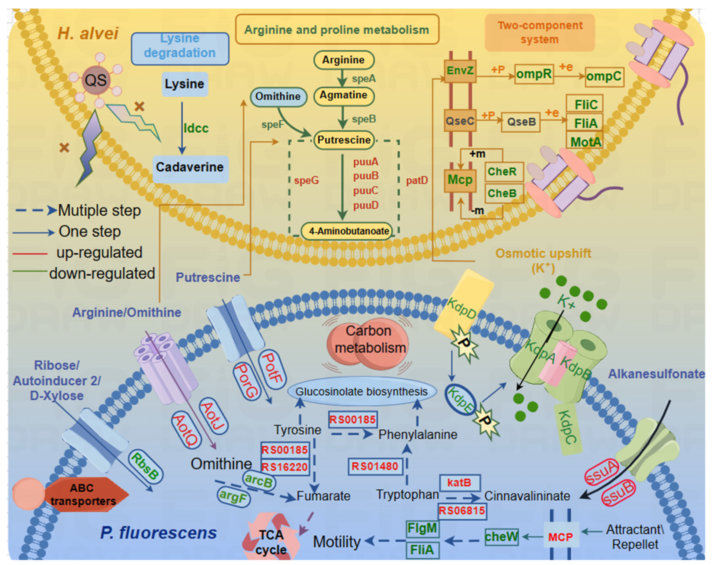
3.2.3. Amino Acid Metabolism

BAs are organic compounds with biological activity that result from the decarboxylation of precursor amino acids caused by microorganisms’ amino acid decarboxylases [51]. Therefore, the contents of precursor amino acids are highly likely to affect the production of biogenic amines by microorganisms. According to our previous results, CAD and PUT are the main BAs found in H. alvei H4 WT. CAD originates from lysine decarboxylation. ldcc is a key gene (encoding lysine decarboxylase) in the decarboxylation of lysine for the synthesis of CAD. According to Table 1, ldcc was downregulated in the IP-H group compared to the WP-H group for H. alvei H4, while it was not activated in P. fluorescens ATCC13525 of the IP-P group compared to the WP-P group, which may inhibit the decarboxylation of lysine to produce CAD. The synthesis of PUT involves a relatively complex multi-step process [52], which is synthesized via ornithine decarboxylation or agmatine deamination. In the IP-H group, there was a notable decrease in the expression of three genes involved in the PUT synthesis pathway: speA, speB, and speF. Specifically, speA encodes arginine decarboxylase, speB encodes agmatinase, and speF encodes ornithine decarboxylase (Table 1), whereas no change occurred in P. fluorescens ATCC 13525 of the IP-P group compared to the WP-P group. The downregulation of these genes suggests a hindered process in the synthesis of PUT. On the contrary, the expression of puuABCD in the putrescine degradation pathway was upregulated in H. alvei H4 ΔluxI (Table 1), which accelerated the consumption rate of PUT. In addition, several pot genes encoding PUT transporter protein were downregulated in Table 1. The decrease in putrescine content was attributed to a faster rate of putrescine consumption due to increased expression of puuABCD and a lower rate of putrescine synthesis due to the downregulation of speABF. The content of cadaverine in the later stage was higher than that of putrescine (Figure 2), possibly because the anabolic pathway of cadaverine was relatively simple.
In addition, a high increase in amino acid metabolism may enable bacterial cells to adapt the structure and function of biofilm in response to environmental changes. [46]. It has also been found that amino acid biosynthesis is essential for biofilm formation [53]. In the transcription results for P. fluorescens ACTT13525 in the IP-P group, CPH89_RS06815 and katB, which are responsible for encoding peroxidase HPII involved in tryptophan metabolism, were upregulated (Table 2). It is suspected that the metabolism of tryptophan is accelerated, leading to a decrease in biofilm content in the deletion coculture system. Meanwhile, the metabolisms of other amino acids, including threonine and D-amino-acid were dysregulated when cocultured with H. alvei H4 ΔluxI. The genes CPH89_RS00185, encoding aspartate/tyrosine/aromatic aminotransferase, and CPH89_RS08005, encoding D-amino acid dehydrogenase, were upregulated. In contrast, CPH89_RS21450, encoding an aspartate aminotransferase family protein involved in the biosynthesis of arginine, an essential source of protein and nucleic acid biosynthesis, is downregulated in the IP-P group. This suggests that when the QS system of H. alvei H4 WT in the coculture system was frustrated, an imbalanced state and disturbance of the amino acid metabolism in the cells were formed. Combined with the above considerations, QS of H. alvei H4 WT in the coculture affected the amino acid metabolism of P. fluorescens ACTT13525 at the transcriptional level.
When food is contaminated by microorganisms, the microorganisms will metabolize amino acids in proteins to produce biogenic amines. Appropriate measures need to be taken to control the content of biogenic amines in food. And the absence of QS in a coculture inhibits the production of biogenic amines in mixed bacteria by regulating amino acid metabolism, which provides ideas to inhibit the production of biogenic amines in real food processing environments.

3.3. Real-Time Quantitative PCR Validation (RT-qPCR)

In order to confirm the validity of the expression of several genes associated with biofilm formation and spoilage obtained by RNA-seq, a total of 13 genes, including fliC, motA, tsr, puuB, puuC, speB, speF, and ldcc from H. alvei H4, and 5 genes belonging to various metabolic pathways (cheW, katB, CPH89-RS06815, CPH89-RS10590, and RbsB) from P. fluorescens ACTT13525 were selected for transcription polymerase chain reaction analysis (Figure 4). In H. alvei H4 ΔluxI, the gene belonging to the flagellar component (fliC) and bacterial chemotaxis-related genes, including methyl-accepting chemotaxis protein (tsr) and flagellar motor protein (motA), were significantly downregulated in the dual species. And in P. fluorescens ACTT13525 cocultured with H. alvei H4 ΔluxI, the cheW (related to bacterial chemotaxis) and RbsB (related to ABC transporters) were also downregulated. These results indicate that flagella and chemotactic-related genes are, indeed, downregulated after QS of H. alvei H4 WT is destroyed, thus affecting the formation of biofilm in the coculture system. In addition, three genes (speA, speB, and speF) within the PUT synthesis pathway also exhibited downregulation, while the expressions of puuB and puuC involved in the putrescine degradation pathway of H. alvei H4 were upregulated in the IP-H group compared to the WP-H group. This proves that when QS of H. alvei H4 WT is disrupted, the gene expression of the PUT synthesis pathway is inhibited, while the gene expression of the degradation pathway is promoted. Furthermore, the expression levels of CPH89_RS06815 and katB involved in the tryptophan metabolism of P. fluorescens ACTT13525 were upregulated, while the gene CPH89_RS10590 (related to amino acid metabolism) was downregulated, which suggests that QS of H. alvei induces metabolic disorders in P. fluorescens. Moreover, the results of the study indicate that the patterns of gene expression obtained from both dual RNA-seq and RT-qPCR were generally consistent, which shows that the RNA-Seq data are credible and reproducible. Additionally, a strong correlation was observed between the two data sets for each species (H. alvei H4 and P. fluorescens ATCC13525), with Pearson’s correlation coefficients of 0.9086 (Figure 4C) and 0.9861 (Figure 4D), respectively, (p < 0.02). This further supports the validity of the RNA-Seq data.

4. Conclusions

Overall, all the results show that QS of H. alvei not only regulated its own genes but also promoted microbial communication and spoilage by influencing gene expression in cocultured strains. That is, the effects of coexistence are “two-way”, where the presence of H. alvei affects the metabolism and behavior of P. fluorescens and vice versa. It was further established that QS of H. alvei H4 WT plays an important role in a coculture system. Based on phenotyping experiments (Figure 1), we mainly analyzed flagellar assembly, bacterial chemotaxis and the two-component system, the ABC transporter system and amino acid metabolism pathways. The schematic is shown in Figure 5. All of them are directly or indirectly related to biofilm and biogenic amine formation [54]. Specifically, the disruption of QS in H. alvei H4 WT inhibits the flagellar composition of H. alvei and the expression of the flagellar transcriptional regulators of P. fluorescens. At the same time, the expression of chemotactic genes in the two strains is also inhibited, thereby reducing biofilm formation. In addition, the regulation of the signaling systems is also a contributing factor to the decrease in biofilms. This includes the regulation of QS of H. alvei H4 WT through its two-component system, as well as the regulation of the ABC transporter of P. fluorescens. The reduction in the accumulation of biogenic amines is primarily caused by the downregulation of certain amino acid decarboxylase enzymes. In addition, QS of H. alvei H4 WT will result in an imbalance of amino acids and metabolic disorders in P. fluorescens, consequently impacting phenotypic changes in a coculture. This means that H. alvei H4 and P. fluorescens ATCC13525 undergo transcriptional changes in their interactions, which depend on the production of QS of H. alvei H4 WT. Disrupting QS of H. alvei H4 WT will inevitably reduce the capacity for spoilage caused by both H. alvei H4 WT and P. fluorescens ATCC13525. It indicated that QS of H. alvei H4 WT might offer a new target to reduce the food spoilage development caused by H. alvei and P. fluorescens. It is possible to regulate the expression of spoilage indicators such as biofilm and biogenic amines in the entire coculture system by interfering with QS. More rigorous studies are necessary to further explore the role of H. alvei quorum sensing in coculture regulation at the protein level.

Author Contributions

Conceptualization, Y.W.; Methodology Y.W., X.L. and G.Z.; Investigation, Y.W., X.L. and J.B.; Data curation, H.H.; Writing—original draft preparation, Y.W.; Writing—review and editing, H.H.; Supervision, H.H.; Project administration, H.H.; Funding acquisition, H.H. All authors have read and agreed to the published version of the manuscript.

Funding

This work was supported by the National Key Research and Development Program project (No. 2022YFD2100500); the APC was funded by H.H.

Institutional Review Board Statement

Not applicable.

Informed Consent Statement

Not applicable.

Data Availability Statement

The data presented in this study are available on request from the corresponding author. The data are not publicly available due to the need for the first author the apply for a Master’s degree.

Conflicts of Interest

The authors declare no conflicts of interest. The funders had no role in the design of the study; in the collection, analyses, or interpretation of data; in the writing of the manuscript; or in the decision to publish the results.

References

  1. Özogul, F.; Polat, A.; Özogul, Y. The effects of modified atmosphere packaging and vacuum packaging on chemical, sensory and microbiological changes of sardines (Sardina pilchardus). Food Chem. 2004, 85, 49–57. [Google Scholar] [CrossRef]
  2. Wang, Y.; Feng, L.; Lu, H.; Zhu, J.; Kumar, V.; Liu, X. Transcriptomic analysis of the food spoilers Pseudomonas fluorescens reveals the antibiofilm of carvacrol by interference with intracellular signaling processes. Food Control 2021, 127, 108115. [Google Scholar] [CrossRef]
  3. Robson, A.A.; Kelly, M.S.; Latchford, J.W. Effect of temperature on the spoilage rate of whole, unprocessed crabs: Carcinus maenas, Necora puber and Cancer pagurus. Food Microbiol. 2007, 24, 419–424. [Google Scholar] [CrossRef] [PubMed]
  4. Xu, Y.F.; Lin, H.; Sui, J.X.; Cao, L.M. Production and Characterization of Egg Yolk Antibodies (Igy) against Two Specific Spoilage Organisms (SSO) in Aquatic Products. Adv. Mater. Res. 2011, 343–344, 519–529. [Google Scholar] [CrossRef]
  5. Han, F.; Li, M.; Lin, H.; Wang, J.; Cao, L.; Khan, M.N. The novel Shewanella putrefaciens-infecting bacteriophage Spp001: Genome sequence and lytic enzymes. J. Ind. Microbiol. Biotechnol. 2014, 41, 1017–1026. [Google Scholar] [CrossRef]
  6. Bruhn, J.B.; Christensen, A.B.; Flodgaard, L.R.; Nielsen, K.F.; Larsen, T.O.; Givskov, M.; Gram, L. Presence of Acylated Homoserine Lactones (AHLs) and AHL-Producing Bacteria in Meat and Potential Role of AHL in Spoilage of Meat. Appl. Environ. Microbiol. 2004, 70, 4293–4302. [Google Scholar] [CrossRef]
  7. Zhu, Y.L.; Hou, H.M.; Zhang, G.L.; Wang, Y.F.; Hao, H.S. AHLs Regulate Biofilm Formation and Swimming Motility of Hafnia alvei H4. Front. Microbiol. 2019, 10, 1330. [Google Scholar] [CrossRef]
  8. Shen, Y.; Cui, F.C.; Wang, D.F.; Li, T.T.; Li, J.R. Quorum Quenching Enzyme (PF-1240) Capable to Degrade AHLs as a Candidate for Inhibiting Quorum Sensing in Food Spoilage Bacterium Hafnia alvei. Foods 2021, 10, 2700. [Google Scholar] [CrossRef]
  9. Hwang, C.C.; Lee, Y.C.; Huang, Y.R.; Lin, C.M.; Shiau, C.Y.; Hwang, D.F.; Tsai, Y.H. Biogenic amines content, histamine-forming bacteria and adulteration of bonito in tuna candy products. Food Control 2010, 21, 845–850. [Google Scholar] [CrossRef]
  10. Kanki, M.; Yoda, T.; Ishibashi, M.; Tsukamoto, T. Photobacterium phosphoreum caused a histamine fish poisoning incident. Int. J. Food Microbiol. 2004, 92, 79–87. [Google Scholar] [CrossRef]
  11. Ge, Y.; Zhu, J.; Ye, X.; Yang, Y. Spoilage potential characterization of Shewanella and Pseudomonas isolated from spoiled large yellow croaker (Pseudosciaena crocea). Lett. Appl. Microbiol. 2017, 64, 86–93. [Google Scholar] [CrossRef] [PubMed]
  12. Wang, H.; Cai, L.; Li, Y.; Xu, X.; Zhou, G. Biofilm formation by meat-borne Pseudomonas fluorescens on stainless steel and its resistance to disinfectants. Food Control 2018, 91, 397–403. [Google Scholar] [CrossRef]
  13. Zarei, M.; Yousefvand, A.; Maktabi, S.; Pourmahdi Borujeni, M.; Mohammadpour, H. Identification, phylogenetic characterisation and proteolytic activity quantification of high biofilm-forming Pseudomonas fluorescens group bacterial strains isolated from cold raw milk. Int. Dairy J. 2020, 109, 104787. [Google Scholar] [CrossRef]
  14. Mann, E.E.; Wozniak, D.J. Pseudomonas biofilm matrix composition and niche biology. FEMS Microbiol. Rev. 2012, 36, 893–916. [Google Scholar] [CrossRef] [PubMed]
  15. Delille, A.; Quilès, F.; Humbert, F. In Situ Monitoring of the Nascent Pseudomonas fluorescens Biofilm Response to Variations in the Dissolved Organic Carbon Level in Low-Nutrient Water by Attenuated Total Reflectance-Fourier Transform Infrared Spectroscopy. Appl. Environ. Microbiol. 2007, 73, 5782–5788. [Google Scholar] [CrossRef]
  16. Puga, C.H.; Orgaz, B.; SanJose, C. Listeria monocytogenes Impact on Mature or Old Pseudomonas fluorescens Biofilms during Growth at 4 and 20 °C. Front. Microbiol. 2016, 7, 00134. [Google Scholar] [CrossRef]
  17. Li, T.; Yang, B.; Li, X.; Li, J.; Zhao, G.; Kan, J. Quorum sensing system and influence on food spoilage in Pseudomonas fluorescens from turbot. J. Food Sci. Technol. 2018, 55, 3016–3025. [Google Scholar] [CrossRef]
  18. Chen, T.R.; Wei, Q.K.; Chen, Y.J. Pseudomonas spp and Hafnia alvei growth in UHT milk at cold storage. Food Control 2011, 22, 697–701. [Google Scholar] [CrossRef]
  19. Liu, M.; Gray, J.M.; Griffiths, M.W. Occurrence of Proteolytic Activity and N-Acyl-Homoserine Lactone Signals in the Spoilage of Aerobically Chill-Stored Proteinaceous Raw Foods. J. Food Prot. 2006, 69, 2729–2737. [Google Scholar] [CrossRef]
  20. Nychas, G.-J.E.; Skandamis, P.N.; Tassou, C.C.; Koutsoumanis, K.P. Meat spoilage during distribution. Meat Sci. 2008, 78, 77–89. [Google Scholar] [CrossRef]
  21. Fu, L.; Wang, C.; Liu, N.; Ma, A.; Wang, Y. Quorum sensing system-regulated genes affect the spoilage potential of Shewanella baltica. Food Res. Int. 2018, 107, 1–9. [Google Scholar] [CrossRef] [PubMed]
  22. Whiteley, M.; Diggle, S.P.; Greenberg, E.P. Progress in and promise of bacterial quorum sensing research. Nature 2017, 551, 313–320. [Google Scholar] [CrossRef] [PubMed]
  23. Kaspar, J.R.; Lee, K.; Richard, B.; Walker, A.R.; Burne, R.A. Direct interactions with commensal streptococci modify intercellular communication behaviors of Streptococcus mutans. ISME J. 2020, 15, 473–488. [Google Scholar] [CrossRef] [PubMed]
  24. Sikdar, R.; Elias, M. Quorum quenching enzymes and their effects on virulence, biofilm, and microbiomes: A review of recent advances. Expert Rev. Anti-Infect. Ther. 2020, 18, 1221–1233. [Google Scholar] [CrossRef]
  25. Hou, H.M.; Zhu, Y.L.; Wang, J.Y.; Jiang, F.; Qu, W.Y.; Zhang, G.L.; Hao, H.-S. Characteristics of N-Acylhomoserine Lactones Produced by Hafnia alvei H4 Isolated from Spoiled Instant Sea Cucumber. Sensors 2017, 17, 772. [Google Scholar] [CrossRef]
  26. Harimawan, A.; Ting, Y.-P. Investigation of extracellular polymeric substances (EPS) properties of P. aeruginosa and B. subtilis and their role in bacterial adhesion. Colloids Surf. B: Biointerfaces 2016, 146, 459–467. [Google Scholar] [CrossRef]
  27. Sang, X.; Ma, X.; Hao, H.; Bi, J.; Zhang, G.; Hou, H. Evaluation of biogenic amines and microbial composition in the Chinese traditional fermented food grasshopper sub shrimp paste. Lwt 2020, 134, 109979. [Google Scholar] [CrossRef]
  28. Xue, Z.P.; Cu, X.; Xu, K.; Peng, J.H.; Liu, H.R.; Zhao, R.T.; Wang, Z.; Wang, T.; Xu, Z.S. The effect of glutathione biosynthesis of Streptococcus thermophilus ST-1 on cocultured Lactobacillus delbrueckii ssp. bulgaricus ATCC11842. J. Dairy Sci. 2023, 106, 884–896. [Google Scholar] [CrossRef]
  29. Winkelströter, L.K.; Teixeira, F.B.d.R.; Silva, E.P.; Alves, V.F.; De Martinis, E.C.P. Unraveling Microbial Biofilms of Importance for Food Microbiology. Microb. Ecol. 2013, 68, 35–46. [Google Scholar] [CrossRef]
  30. Jahid, I.K.; Ha, S.D. The Paradox of Mixed-Species Biofilms in the Context of Food Safety. Compr. Rev. Food Sci. Food Saf. 2014, 13, 990–1011. [Google Scholar] [CrossRef]
  31. Zhang, C.; Zhu, F.; Jatt, A.N.; Liu, H.; Niu, L.; Zhang, L.; Liu, Y. Characterization of co-culture of Aeromonas and Pseudomonas bacterial biofilm and spoilage potential on refrigerated grass carp (Ctenopharyngodon idellus). Lett. Appl. Microbiol. 2020, 71, 337–344. [Google Scholar] [PubMed]
  32. Linares, D.M.; Martín, M.; Ladero, V.; Alvarez, M.A.; Fernández, M. Biogenic Amines in Dairy Products. Crit. Rev. Food Sci. Nutr. 2011, 51, 691–703. [Google Scholar] [CrossRef] [PubMed]
  33. Rossi, P.; Xing, Q.; Bini, E.; Portaliou, A.G.; Clay, M.C.; Warren, E.M.; Khanra, N.K.; Economou, A.; Kalodimos, C.G. Chaperone Recycling in Late-Stage Flagellar Assembly. J. Mol. Biol. 2023, 435, 167954. [Google Scholar] [CrossRef] [PubMed]
  34. Saijo-Hamano, Y.; Uchida, N.; Namba, K.; Oosawa, K. In Vitro Characterization of FlgB, FlgC, FlgF, FlgG, and FliE, Flagellar Basal Body Proteins of Salmonella. J. Mol. Biol. 2004, 339, 423–435. [Google Scholar] [CrossRef] [PubMed]
  35. Altarriba, M.; Merino, S.; Gavín, R.; Canals, R.o.; Rabaan, A.; Shaw, J.G.; Tomás, J.M. A polar flagella operon (flg) of Aeromonas hydrophila contains genes required for lateral flagella expression. Microb. Pathog. 2003, 34, 249–259. [Google Scholar] [CrossRef]
  36. Deme, J.C.; Johnson, S.; Vickery, O.; Aron, A.; Monkhouse, H.; Griffiths, T.; James, R.H.; Berks, B.C.; Coulton, J.W.; Stansfeld, P.J.; et al. Structures of the stator complex that drives rotation of the bacterial flagellum. Nat. Microbiol. 2020, 5, 1553–1564. [Google Scholar] [CrossRef] [PubMed]
  37. Wilkinson, D.A.; Chacko, S.J.; Vénien-Bryan, C.; Wadhams, G.H.; Armitage, J.P. Regulation of Flagellum Number by FliA and FlgM and Role in Biofilm Formation by Rhodobacter sphaeroides. J. Bacteriol. 2011, 193, 4010–4014. [Google Scholar] [CrossRef]
  38. Hazelbauer, G.L. Bacterial Chemotaxis: The Early Years of Molecular Studies. Annu. Rev. Microbiol. 2012, 66, 285–303. [Google Scholar] [CrossRef]
  39. Karmakar, R. State of the art of bacterial chemotaxis. J. Basic Microbiol. 2021, 61, 366–379. [Google Scholar] [CrossRef]
  40. Porter, S.L.; Wadhams, G.H.; Armitage, J.P. Signal processing in complex chemotaxis pathways. Nat. Rev. Microbiol. 2011, 9, 153–165. [Google Scholar] [CrossRef]
  41. Minamino, T.; Kinoshita, M.; Namba, K. Directional Switching Mechanism of the Bacterial Flagellar Motor. Comput. Struct. Biotechnol. J. 2019, 17, 1075–1081. [Google Scholar] [CrossRef]
  42. Kentner, D.; Sourjik, V. Spatial organization of the bacterial chemotaxis system. Curr. Opin. Microbiol. 2006, 9, 619–624. [Google Scholar] [CrossRef] [PubMed]
  43. Li, Y.; Wang, Y.; Han, S.; Qin, M.; Wu, X.; Niu, W.; Gao, C.; Wang, H. N-acyl-homoserine lactones-mediated quorum sensing promotes intestinal colonization of Aeromonas veronii through facilitating cheA gene expression. Aquaculture 2024, 579, 740189. [Google Scholar] [CrossRef]
  44. Sieuwerts, S.; Molenaar, D.; van Hijum, S.A.F.T.; Beerthuyzen, M.; Stevens, M.J.A.; Janssen, P.W.M.; Ingham, C.J.; de Bok, F.A.M.; de Vos, W.M.; van Hylckama Vlieg, J.E.T. Mixed-Culture Transcriptome Analysis Reveals the Molecular Basis of Mixed-Culture Growth in Streptococcus thermophilus and Lactobacillus bulgaricus. Appl. Environ. Microbiol. 2010, 76, 7775–7784. [Google Scholar] [CrossRef] [PubMed]
  45. Wang, L.C.; Morgan, L.K.; Godakumbura, P.; Kenney, L.J.; Anand, G.S. The inner membrane histidine kinase EnvZ senses osmolality via helix-coil transitions in the cytoplasm. EMBO J. 2012, 31, 2648–2659. [Google Scholar] [CrossRef] [PubMed]
  46. Yan, J.; Yang, Z.; Xie, J. Comparative Transcriptome Analysis of Shewanella putrefaciens WS13 Biofilms Under Cold Stress. Front. Cell. Infect. Microbiol. 2022, 12, 851521. [Google Scholar] [CrossRef]
  47. Zhang, B.Z.; Jiang, C.S.; Cao, H.; Zeng, W.; Ren, J.P.; Hu, Y.F.; Li, W.T.; He, Q.G. Transcriptome analysis of heat resistance regulated by quorum sensing system in Glaesserella parasuis. Front. Microbiol. 2022, 13, 968460. [Google Scholar] [CrossRef]
  48. Xie, Z.; Jian, H.; Jin, Z.; Xiao, X.; Kelly, R.M. Enhancing the Adaptability of the Deep-Sea Bacterium Shewanella piezotolerans WP3 to High Pressure and Low Temperature by Experimental Evolution under H2O2 Stress. Appl. Environ. Microbiol. 2018, 84, e02342-17. [Google Scholar] [CrossRef]
  49. Zhang, P.Y.; Xu, P.P.; Xia, Z.J.; Wang, J.; Xiong, J.; Li, Y.Z. Combined treatment with the antibiotics kanamycin and streptomycin promotes the conjugation of Escherichia coli. FEMS Microbiol. Lett. 2013, 348, 149–156. [Google Scholar] [CrossRef]
  50. Armbruster, C.E.; Pang, B.; Murrah, K.; Juneau, R.A.; Perez, A.C.; Weimer, K.E.D.; Swords, W.E. RbsB (NTHI_0632) mediates quorum signal uptake in nontypeable Haemophilus influenzae strain 86-028NP. Mol. Microbiol. 2011, 82, 836–850. [Google Scholar] [CrossRef]
  51. Li, J.; Zhou, L.; Feng, W.; Cheng, H.; Muhammad, A.I.; Ye, X.; Zhi, Z. Comparison of Biogenic Amines in Chinese Commercial Soy Sauces. Molecules 2019, 24, 1522. [Google Scholar] [CrossRef] [PubMed]
  52. Sun, Z.; Zhang, Y.; Lin, X.; Zhang, S.; Chen, Y.; Ji, C. Inhibition Mechanism of Lactiplantibacillus plantarum on the Growth and Biogenic Amine Production in Morganella morganii. Foods 2023, 12, 3625. [Google Scholar] [CrossRef] [PubMed]
  53. Jijakli, K.; Jensen, P.A.; Whiteson, K.L. Metabolic Modeling of Streptococcus mutans Reveals Complex Nutrient Requirements of an Oral Pathogen. mSystems 2019, 4, 419507. [Google Scholar] [CrossRef] [PubMed]
  54. Zhang, X.; Zheng, L.; Lu, Z.; Zhou, L.; Meng, F.; Shi, C.; Bie, X. Biochemical and molecular regulatory mechanism of the pgpH gene on biofilm formation in Listeria monocytogenes. J. Appl. Microbiol. 2023, 134, lxac086. [Google Scholar] [CrossRef]
Figure 1. Changes in spoilage phenotypes in monoculture and coculture systems at different times. (A) Biofilm biomass; (B) polysaccharide; (C) putrescine; (D) cadaverine. WT: Hafnia alvei H4 wild type; PF: Pseudomonas fluorescens ATCC13525; ΔluxI: Hafnia alvei H4 luxI mutant strain; PF + WT: P. fluorescens ATCC13525 cocultured with H. alvei H4; PF + ΔluxI: P. fluorescens ATCC13525 cocultured with H. alvei H4 ΔluxI a, b, c, d and e represent significant differences between groups.
Foods 13 00336 g001
Figure 2. Experimental setup and total analysis of differentially expressed genes in Hafnia alvei H4 and Pseudomonas fluorescens ATCC13525. (A) Experimental setup for culture preparation and RNA extraction. (B) Volcano plot displaying differential regulation of H. alvei H4 genes in IP-H vs. WP-H; (C) Volcano plot displaying differential regulation of P. fluorescens ATCC13525 genes in IP-P vs. WP-P. Green and red dots represent down- and upregulated genes, respectively, and blue dots represent genes whose expressions were not altered significantly.
Foods 13 00336 g002
Figure 3. Statistical enrichment of DEGs in GO and KEGG pathways (MF: molecular function, BP: biological process, CC: cellular component). (A) GO terms of DEGs in Hafnia alvei H4 of IP-H vs. WP-H; (B) Scatter diagram of the top 20 KEGG enrichment pathways for DEGs in H. alvei H4 of IP-H vs. WP-H; (C) GO terms of DEGs in Pseudomonas fluorescens ATCC13525 of IP-P vs. WP-P; (D) Scatter diagram of the top 20 KEGG enrichment pathways for DEGs in P. fluorescens ATCC13525 of IP-P vs. WP-P.
Foods 13 00336 g003
Figure 4. Validation of the selected DEG expression of Hafnia alvei H4 and Pseudomonas fluorescens ATCC13525 in dual species. (A) Quantitative reverse transcription PCR (RT-qPCR) of differentially expressed genes of H. alvei H4. (B) RT-qPCR of differentially expressed genes of P. fluorescens ATCC13525. (C) The regression line of RNA-Seq data (X-axis) plotted against the RT-qPCR data (Y-axis) of H. alvei H4. (D) The regression line of RNA-Seq data (X-axis) plotted against the RT-qPCR data (Y-axis) of P. fluorescens ATCC13525.
Foods 13 00336 g004
Figure 5. Schematic representation of certain biological pathways in H. alvei H4 and P. fluorescens affected by QS of H. alvei in a coculture system. This model represents the transcriptional response of genes with significant differences in transcription levels (flagellar assembly, bacterial chemotaxis, lysine degradation, arginine and proline metabolism, and two-component system) of H. alvei H4 and (ABC transporters, bacterial chemotaxis, phenylalanine, tyrosine, and tryptophan biosynthesis) of P. fluorescens ATCC13525 after the luxI gene knockout from H. alvei H4 in dual species. The yellow gradient background area represents H. alvei H4, and the blue gradient background area represents P. fluorescens ATCC13525. Most DEGs were integrated and are indicated in red (upregulated), and green (downregulated), respectively.
Foods 13 00336 g005
Table 1. Validation of selected DEGs related to flagellar assembly, bacterial chemotaxis, BA metabolism, and two-component system in H. alvei H4 (IP-H vs. WP-H).
Gene Namelog2 RadioGene Description
flagellar assembly
fliL−4.32515228flagellar basal body-associated protein FliL
fliN−5.766718961flagellar motor switch protein FliN
fliM−2.837097495flagellar motor switch protein FliM
fliJ−2.408896556flagellar export protein FliJ
ERL64_RS17280−1.339639572flagellar hook-length control protein FliK
fliS−2.016760592flagellar export chaperone FliS
motA−2.425518042flagellar motor stator protein MotA
flgB−3.035717278flagellar basal body rod protein FlgB
flgC−3.475398667flagellar basal body rod protein FlgC
flgD−3.258609823flagellar hook assembly protein FlgD
flgE−3.074676572flagellar hook protein FlgE
ERL64_RS17360−3.436893106flagellar basal body rod protein FlgF
flgG−3.336673997flagellar basal body rod protein FlgG
flgK−1.888983693flagellar hook-associated protein FlgK
flgL−2.226392467flagellar hook-associated protein FlgL
ERL64_RS17345−1.831507544flagellar basal body P-ring protein FlgI
bacterial chemotaxis
ERL64_RS14790−2.265739966methyl-accepting chemotaxis protein
Tsr−2.265395717methyl-accepting chemotaxis protein
ERL64_RS14800−2.424604914chemotaxis response regulator protein
cheR−2.098189509protein-glutamate O-methyltransferase CheR
ERL64_RS14800−2.424604914chemotaxis response regulator protein-glutamate methylesterase
two-component system
envZ−0.966750293two-component system sensor histidine kinase EnvZ
ompR−0.459688448two-component system response regulator OmpR
phoR−1.146705289phosphate regulon sensor histidine kinase PhoR
phoQ0.940798155two-component system sensor histidine kinase PhoQ
phoP1.035332663two-component system response regulator PhoP
rpoN−1.164441481RNA polymerase factor sigma-54
BA metabolism
ldcc−3.212727378lysine decarboxylase
speF−0.860138669ornithine decarboxylase SpeF
speA−0.82353036biosynthetic arginine decarboxylase
speB−0.669157809agmatinase
puuA3.06324221glutamine synthetase family protein
puuB2.140552542FAD-binding oxidoreductase
puuC2.239399155aldehyde dehydrogenase PuuC
puuD2.663951099gamma-glutamyl-gamma-aminobutyrate hydrolase
patD1.429802723amino butyraldehyde dehydrogenase
ERL64_RS212803.401803121amma-amino butyraldehyde dehydrogenase
ERL64_RS023253.06324221glutamine synthetase family protein
speG0.419542916spermidine N1-acetyltransferase
potF−0.987904791spermidine/putrescine ABC transporter substrate-binding protein PotF
potI−2.362303979putrescine ABC transporter permease PotI
potH−2.568324997putrescine ABC transporter permease PotH
potG−0.987904791putrescine ABC transporter ATP-binding subunit PotG
Table 2. Selected DEGs related to biofilm formation, amino acid metabolism, and ABC transport in P. fluorescens ATCC13525 (IP-P vs. WP-P).
Gene Namelog2 RadioGene Description
biofilm formation
fliL−0.925171985flagellar basal body-associated protein FliL
flgM−0.699499627flagellar biosynthesis anti-sigma factor FlgM
fliA−0.523949137RNA polymerase sigma factor FliA
CPH89_RS16845−1.029022096OmpA family protein
cheW−0.506899146chemotaxis protein CheW
CPH89_RS23015−0.547698862methyl-accepting chemotaxis protein
tssC−1.370116898type VI secretion system contractile sheath large subunit
tssG2.543096874type VI secretion system baseplate subunit TssG
tssH0.939045148type VI secretion system ATPase TssH
CPH89_RS095051.808730303type VI secretion system tip protein VgrG
amino acid metabolism
CPH89_RS22860−1.373666561methionine gamma-lyase
CPH89_RS03900−1.088510925L-serine ammonia-lyase
CPH89_RS10590−0.592860749homoserine kinase
mmsB−1.5241515883-hydroxyisobutyrate dehydrogenase
CPH89_RS068150.574998597catalase
katB1.644147633catalase KatB
CPH89_RS080052.317725314D-amino acid dehydrogenase
CPH89_RS001850.475801482aspartate/tyrosine/aromatic aminotransferase
CPH89_RS214501.152619749aspartate aminotransferase family protein
CPH89_RS168500.716263227argininosuccinate synthase
CPH89_RS213500.422007256glutamine synthetase family protein
gltB0.609812179glutamate synthase large subunit
CPH89_RS002000.920911912aldehyde dehydrogenase family protein
CPH89_RS278251.897595222CoA-acylating methylmalonate-semialdehyde dehydrogenase
ABC transport
RbsB−0.923885631sugar ABC transporter substrate-binding protein
CPH89_RS28860−1.034779256ABC transporter ATP-binding protein
CPH89_RS10545−0.845831876MetQ/NlpA family ABC transporter substrate-binding protein
CPH89_RS24050−2.801510174ABC transporter permease
CPH89_RS19375−1.480685108ABC transporter ATP-binding protein
CPH89_RS16095−1.459658475amino acid ABC transporter permease
CPH89_RS236902.876670285carbohydrate ABC transporter permease
urtD2.872150064urea ABC transporter ATP-binding protein UrtD
CPH89_RS086651.314199994sulfonate ABC transporter substrate-binding protein
ssuB0.972531529aliphatic sulfonates ABC transporter ATP-binding protein
CPH89_RS151850.63533128glycine betaine ABC transporter substrate-binding protein
Disclaimer/Publisher’s Note: The statements, opinions and data contained in all publications are solely those of the individual author(s) and contributor(s) and not of MDPI and/or the editor(s). MDPI and/or the editor(s) disclaim responsibility for any injury to people or property resulting from any ideas, methods, instructions or products referred to in the content.

