Article

Bioactivity of Microencapsulated Cell-Free Supernatant of Streptococcus thermophilus in Combination with Thyme Extract on Food-Related Bacteria

1
Department of Seafood Processing Technology, Faculty of Fisheries, Cukurova University, 01330 Adana, Turkey
2
Department of Gastronomy and Culinary Arts, Faculty of Tourism, Yozgat Bozok University, 66900 Yozgat, Turkey
3
Department of Food Hygiene and Technology, Faculty of Ceyhan Veterinary Medicine, Cukurova University, 01960 Adana, Turkey
*
Author to whom correspondence should be addressed.
Foods 2024, 13(2), 329; https://doi.org/10.3390/foods13020329
Submission received: 14 December 2023 / Revised: 18 January 2024 / Accepted: 18 January 2024 / Published: 20 January 2024
(This article belongs to the Section Food Microbiology)

Abstract

:
The bioactive properties of the combination of microencapsulated cell-free supernatant (CFS) from Streptococcus thermophilus and thyme extract on food-related bacteria (Photobacterium damselae, Proteus mirabilis, Vibrio vulnificus, Staphylococcus aureus ATCC29213, Enterococcus faecalis ATCC29212, and Salmonella Paratyphi A NCTC13) were investigated. The microencapsulated CFS of S. thermophilus, in combination with ethanolic thyme extract, had a particle size in the range of 1.11 to 11.39 µm. The microencapsulated CFS of S. thermophilus had a wrinkled, spherical form. In the supernatant, especially at 2% (v/w), the thyme extract additive caused a decrease in the wrinkled form and a completely spherical structure. A total of 11 compounds were determined in the cell-free supernatant of S. thermophilus, and acetic acid (39.64%) and methyl-d3 1-dideuterio-2-propenyl ether (10.87%) were the main components. Thyme extract contained seven components, the main component being carvacrol at 67.96% and 1,2,3-propanetriol at 25.77%. Significant differences (p < 0.05) were observed in the inhibition zones of the extracts on bacteria. The inhibitory effect of thyme extract on bacteria varied between 25.00 (P. damselae) and 41.67 mm (V. vulnificus). Less antibacterial activity was shown by the microencapsulated CFS from S. thermophilus compared to their pure form. (p < 0.05). As a result, it was found that microencapsulated forms of CFS from S. thermophilus, especially those prepared in combination with 2% (v/w) thyme extract, generally showed higher bioactive effects on bacteria.

1. Introduction

Fish meat has a high nutritional content and contains essential macro- and micronutrients such as protein, fats, vitamins, and minerals. In addition, since fish muscles have a high post-mortem pH (>6), a high concentration of low-molecular-weight compounds, and high water activity, microbial activity is the primary cause of raw fish deterioration [1,2]. Raw seafood products can easily spoil and also get infected by pathogens, including bacteria, naturally or via cross-contamination. Staphylococcus aureus, Salmonella, E. coli, Vibrio parahaemolyticus, Aeromonas hydrophila, Clostridium botulinum, Listeria monocytogenes, Yersinia spp., and Enterococcus faecalis are common pathogens linked to human foodborne diseases through the consumption of contaminated seafood products [3,4,5,6]. Various synthetic antimicrobials are used to control pathogen growth in food products, but these antimicrobials cause consumer concern due to their harmful effects. For this reason, studies on the use of natural alternative antimicrobials of plant, animal, or bacterial origin are gaining importance.
Streptococcus thermophilus is a member of the lactic acid bacteria that are generally recognised as safe (GRAS) in the United States and as having a “qualified presumption of safety” (QPS) in the European Union because of their long history of safe usage in foodstuff [7]. S. thermophilus is naturally found in fermented milk, cheese, other dairy products, and some plants [8]. Its main feature in fermentation is the formation of lactic acid, which results in rapid acidification and the suppression of other microorganisms. The secondary metabolites generated by S. thermophilus, acetaldehyde and exopolysaccharides, have a significant impact on the product’s flavour and texture. [9]. Along with lactose, S. thermophilus also has the ability to ferment sucrose, glucose, galactose, and sugar. Due to their capacity to rapidly ferment lactose, S. thermophilus is frequently utilised symbiotically with other LAB members as starter cultures in fermented milk products. S. thermophilus has been known to possess probiotic potential due to its immunomodulatory, antibacterial, antioxidant, and anti-inflammatory properties [10,11]. This member is among the most important industrial starter cultures after Lactococcus lactis in dairy products [12]. Some members of S. thermophilus can produce thermophilins, which are short peptides that have inhibitory effects on Gram-positive pathogenic Enterococcus faecalis, Clostridium botulinum, Staphylococcus aureus, and Listeria monocytogenes [8].
A member of the Lamiaceae family of plants, thyme is used in lotions, perfumes, and other cosmetic items, as well as food and beverage and confectionery products as a flavour enhancer. Thyme is well-liked as a medicinal herb and food preservative because of its antiseptic, bronchiolytic, antispasmodic, and antibacterial qualities [13]. These plants’ essential oils contain high concentrations of thymol, carvacrol, p-cymene, and -terpinene [14]. According to studies conducted in vitro, essential oils and the chemical components they contain, such as thymol and carvacrol, have antibacterial and antioxidant effects [15,16]. Besides these, the extracts from thyme may be used as antibacterial and anticarcinogenic agents to combat bacterial pathogens that cause food poisoning and nosocomial infections. [17]. The thyme extract demonstrated inhibitory activity on microbial growth, as well as bactericidal efficacy against Listeria monocytogenes, E. faecalis, Bacillus cereus, E. coli, Salmonella enterica, S. aureus, and Yersinia enterocolitica, at relatively low concentrations [18].
The food industry uses the encapsulation method to extend the shelf life of various food products by including bioactive ingredients and shielding them from unfavourable environmental factors [19]. In addition, the application of microencapsulation in the food business offers a number of benefits. Among them are lowering the main ingredient’s reactivity to environmental factors, slowing the rate at which an external component is absorbed into the main ingredient’s material, facilitating easier handling through encapsulation, regulating the main ingredient’s material’s release, covering up the taste of the main ingredient, and diluting the main ingredient material when used in very small amounts [20,21]. The purpose of the encapsulation approach is to guarantee that the active components are kept in the coating material at nanometric, micrometric, or millimetric dimensions [22]. The most popular encapsulation techniques are spray drying, freeze drying, extrusion, coating with an air suspension, applying liposomes, and crystallisation, with capsule sizes ranging from a few micrometres to a few centimetres [23,24].
It has been noted that spray drying significantly reduces the antioxidant activity after microencapsulation because the major phenolic components of thyme extract are impacted by high temperatures in the microencapsulation process [25]. Furthermore, Ozyurt et al. [26] observed that the amount of essential amino acids and gamma-linoleic fatty acids in the product was retained when maltodextrin was used in increasing amounts as a coating material during spray drying. Therefore, for each component that is intended to be coated, it is crucial to establish the proper microencapsulation settings, including device intake and outlet temperature, flow feed rate, wall material selection, and ratio.
In comparison to probiotic cells, supernatants have better antipathogenic effects and are more stable during storage, according to Saadatzadeh et al. [27]. This is why lactic acid bacteria supernatants are encapsulated, which opens up new antibacterial possibilities. The lactic acid bacteria’s cell-free supernatant (CFS) can be prepared in liquid form and is rich in bioactive compounds. The conversion of the obtained product into microencapsulated form will give the product a longer shelf life and make it simpler to apply in foods by preserving the product from external conditions, in addition to the inclusion of thyme extract, in order to boost the bioactivity of these supernatants. This study aimed to determine the antimicrobial properties of CFS from S. thermophilus and thyme (Origanum vulgare) extract combinations before and after microencapsulation.

2. Materials and Methods

2.1. Thyme Extract and Bacterial Strains

Ethanolic thyme (Origanum vulgare) extract (20 g) was obtained from Biomesi Bioagro Technology R&D (Adana, Turkey). Streptococcus thermophilus [28] was obtained from Kahramanmaraş Sütçü İmam University. Photobacterium damselae, Proteus mirabilis, and Vibrio vulnificus were isolated from spoiled anchovy, mackerel, and sardine meat [29]. Gram-positive Staphylococcus aureus (ATCC29213), Enterococcus faecalis (ATCC29212), and Gram-negative Salmonella Paratyphi A (NCTC13) were purchased from the National Collection of Type Cultures in London, United Kingdom, and the American Type Culture Collection in Rockville, Maryland, in the United States.

2.2. Methods

2.2.1. Preparation of CFS from S. thermophilus

A modification of Lin and Yen’s method [30] was used to create lactic acid bacteria supernatant. M17 broth (56156, Sigma-Aldrich Chemie GmbH, Steinheim, Germany) was used to culture S. thermophilus for 48 h at 37 °C. After the supernatant was transferred to 15 mL falcon tubes, the supernatant was centrifuged at 8000 RPM for 10 min at 4 °C. The obtained supernatant was put through a membrane filter, exposed to UV light for 20 min, and kept at 4 °C until analysis.

2.2.2. Microencapsulation Process

A modification of Marcela et al.’s [31] method was followed for microencapsulation. CFSs obtained from S. thermophilus were microencapsulated individually. Other groups were microencapsulated together with the CFS of S. thermophilus containing 1 and 2% thyme extract (v/w). All groups were coated with 25% maltodextrin (DE: 18–20) Alfasol, Turkey) before drying. The mixture was mixed with Ultra-Turrax for 10 min. A 250 mL measure of the sample from each group was dried using a mini spray dryer (Buchi-290, Flawil, Switzerland). Spray drying conditions were an inlet temperature of 130 °C, an outlet temperature of 75 °C, an aspiration rate of 30 m3/hour, and a feed rate of 20 mL/min. After spray drying, all samples were placed in light-proof plastic bottles and stored at 4 °C until the day of analysis.

2.2.3. Microencapsulation Morphology

The morphology of the particles obtained through spray drying was characterized using Quanta 650 field emission scanning electron microscopy (SEM, FEI Company, Hillsborn, OR, USA) at Cukurova University Central Research Laboratory (CUMERLAB, Adana, Turkey). SEM images were obtained at room temperature and 20 kV voltage, and the results were visually recorded at 10,000 and 20,000 magnification after gold coating with a Q150R ES Coater (Quorum Technologies, Lewes, UK).

2.2.4. GC–MS Analysis of Samples

The chemical composition of the thyme extract and CFS of S. thermophilus was determined using an Agilent 7890A gas chromatograph (GC) incorporating a mass spectrometer (MS, Agilent, Palo Alto, CA, USA). A non-polar (5% -phenyl)-methylpolysiloxane (30 m × 250 μm × 0.25 μm, Agilent 1901S-433HP-5MS) was used as column. The flow rate of helium benefited as the carrier gas was 1.5 mL/min. GC–MS conditions were set to 50 °C initial and 240 °C final oven temperature, 1 µL injection volume, and 105 min run time. The detection of peaks found in GC–MS was obtained through comparison with those in the commercial library of NIST, EPA, and NIH version 2.0.

2.2.5. Antimicrobial Activity Assay of Extracts

Agar Well Diffusion Method

Mueller–Hinton Agar (MHA, Merck 1.05437, Darmstadt, Germany) was used to measure the in vitro antimicrobial activity level of non-encapsulated (pure) and encapsulated CFS from a combination of S. thermophilus and thyme extract (1%, v/w), in accordance with the well diffusion method of Hwanhlem et al. [32]. For the antimicrobial activity test, microencapsulated samples were dissolved in distilled water at a ratio of 1:2. Food-related bacteria (Photobacterium damselae, Proteus mirabilis, Vibrio vulnificus, Staphylococcus aureus, Enterococcus faecalis, and Salmonella Paratyphi A) were grown in Nutrient broth (Merck 1.05443, Darmstadt, Germany) at 37 °C for 24 h and standardised to 0.5 McFarland cell density (108 cfu/mL). Each bacterial cell culture (100 µL) was inoculated into a Petri dish containing 20 mL of Mueller–Hilton agar. Five 5 mm wells were formed in a solid medium. A 50 µL measure of pure or microencapsulated extract was inoculated into 4 wells. Distilled water, or maltodextrin, was transferred to the other well as a control. Petri dishes were then incubated at 37 °C for 24 h. After incubation, the inhibition zones formed around each well were measured in mm using callipers.

Minimum Inhibitory (MIC) and Bactericidal Concentration (MBC)

Minimum inhibitory concentration (MIC) and minimum bactericidal concentration (MBC) were determined according to the microdilution method of CLSI [33]. Test microorganisms incubated at 37 °C for 24 h were standardised to 0.5 MacFarland cell density. Mueller–Hinton Broth (MHB, Oxoid, CM0405, Basingstoke, UK) was used as the medium. A 50 mg/mL stock solution prepared from pure and microencapsulated extracts was diluted to 0.19 μg/mL in sterile tubes. The tube containing only stock solution or pure culture was considered as the control, and the other tubes containing MHB contained test microorganisms and diluted stock solutions. The test tubes were prepared repeatedly and incubated at 37 °C for 24 h. Bacterial growth in the test tubes was compared with the control tubes, and the tubes with the lowest inhibition of bacterial growth were recorded as MIC. In line with the MIC results, the tubes without bacterial growth were inoculated onto the Mueller–Hinton Agar surface, the Petri dishes were incubated at 37 °C for 24 h, and MBK values were recorded.

2.2.6. Statistical Analysis

One-way analysis of variance (ANOVA) and Duncan’s Multiple Comparison Test (SPSS 22, Chicago, IL, USA) were used for the statistical analysis. A significant difference between the groups was shown by the value of p < 0.05.

3. Results and Discussions

3.1. Morphology of Encapsules

Microencapsulations are defined as microparticles between 0.2 and 5000 μm in size [34]. In this study, the microencapsulated CFS from S. thermophilus had a particle size of 1.15–14.08 µm, the microencapsulated CFS from S. thermophilus combined with 1% (v/w) ethanolic thyme extract had a particle size of 1.11–11.50 µm, and the microencapsulated CFS of S. thermophilus combined with 2% (v/w) ethanolic thyme extract had a particle size of 2.71–11.39 µm (Figure 1). The particle sizes obtained through spray drying varied based on the type of coating, the quantity and consistency of the substance included, and the drying environment [35].
Microencapsulated CFS of S. thermophilus had a wrinkled, spherical form. Thyme supplementation in the supernatant caused a decrease in the wrinkled form. In particular, the microencapsulated CFS from S. thermophilus with a 2% (v/w) thyme additive was completely spherical (Figure 2). Differences in spray drying conditions can affect the spherical form of particles [36]. Unlike in this study, microencapsulated CFS from Lactiplantibacillus plantarum exhibited a more wrinkled form in the presence of propolis extract in [37]. The collapses occurring in microencapsules may be due to the possibility of moisture transport during the drying period [25,38].

3.2. Chemical Composition of CFS from S. thermophilus

The chemical composition of the CFS from S. thermophilus is displayed in Table 1. CFS from S. thermophilus included 11 different substances, but the predominant constituents were acetic acid (39.64%), methyl-d3 1-dideuterio-2-propenyl ether (10.87%), and 7-octen-2-ol, 2-methyl-6-methylene (10.46%) (Figure 2). Propane, 2-fluoro-2-methyl- (8.92%), imidodicarbonic diamide (7.64%), tetrahydro-1,3-oxazine-2-thione (7.21%), 1,2,3-propanetriol (5.8%), and 2-methyl-1,3-oxathiolany-propionic acid ethyl ester (5.42%) were other important components found in the CFS from S. thermophilus.
Organic acids (such as lactic acid, acetic acid and propionic acid), fatty acids, hydrogen peroxide, diacetyl, and bacteriocins are among the bioactive compounds produced by LAB that exhibit antimicrobial activity against pathogens and spoilage organisms [39]. The antimicrobial activity of the CFS of lactic acid bacteria is mainly due to lactic acid and acetic acid. Acetic acid, a product of carbon metabolism, shows broad-spectrum antimicrobial activity against bacteria, moulds, and yeasts due to its higher pKa value (4.87) compared to other acids [40,41]. Some LAB strains were able to degrade lactic acid into acetic acid. It has been reported that each mole of lactic acid was converted into approximately 0.5 mol of acetic acid, 0.5 mol of 1,2-propanediol, and traces of ethanol by certain LAB strains [42,43]. This may be the reason for the high proportion of acetic acid (39.64%) in the chemical composition of CFS from S. thermophilus. Lactiplantibacillus plantarum and Lactobacillus acidophilus produced 0.031–6.491 mg/mL lactic acid, 0.372–0.863 mg/mL protein, 0.009–0.029 mg/mL hydrogen peroxide, and 0.450–0.662 mg/mL diacetyl [44]. Bioactive substances produced by lactic acid bacteria may vary according to the cultural conditions and physiological characteristics of bacterial members [44].
Table 2 gives the chemical content of thyme extract. Thyme extract contained seven components, and the main components were phenol, 2-methyl-5-(1-methylethyl) (carvacrol) with 67.96%, and 1,2,3-propanetriol (glycerin) with 25.77%. Additionally, the extract of thyme had 3.97% of propane, 2-fluoro-2-methyl-methyl, and contained less than 0.5% of other components. According to Saleem et al. [45], carvacrol and thymol are the two primary constituents of fresh and dried plants, respectively. Carvacrol (52.32%), linalool L (15.96%), thymol (9.59%), 1-methylpyrroline (3.53%), and trans-caryophyllene (3.43%) were the major constituents of thyme (Zataria multiflora) extract [46]. Mehrabi et al. [47] reported that the main components in thyme were thymol (25.30%), δ-2-carene (8.825%), and carvacrol (8.43%). Mohammadigholami [48] reported that 45 components were found in thyme essential oil, and the main ones were carvacrol (52.4%), γ-trpynene (12.1%), and thymol (10.4%). Thymol (37.54%), p-cymene (14.49%), terpinene (11.15%), linalool (4.71%), and carvacrol (4.62%) were listed as the primary compounds of thyme essential oil by Fadil et al. [49]. The dominant compounds in thyme plants such as Thymus migricus, Thymus eriocalyx, Thymus serpylum, Zataria multiflora, and Thymus kotschyanus were linalool (41.8%), geraniol (61.8%), para cymene (23.8%), carvacrol (57.7%), and uulegone (37.2%) [50]. Thymol (45.16 %), phytol isomer (7.17 %), carvacrol (5.2 %), 9-octadecenal (4.84 %), caryophyllene oxide (4.27%), and trans-β-caryophyllene (2.86 %) were reported as components of thyme extract obtained through supercritical-flow CO2 extraction [51]. The main components identified in the ethanolic extract obtained from the leaves and branches of thyme (Zataria multiflora) were carvacrol (52.32%), linalool (15.96%), thymol (9.59%), 1-methylpyrroline (3.53%), and trans-caryophyllene (3.43%) [42]. Phytochemical compounds of Thymus linearis leaf extract determined using GC-MS were ethyl (9z, 12z)-9, 12-octadecadienoate (22.58%), palmitic acid (11.95%), ethyl palmitate (9.89%), (5.03%), stigmast-5-En-3-Ol, (3. Beta.)- (4.54%), (Z, Z)-6, 9-Cis-3, 4-epoxy-nonadecadiene (3.60%), carvacrol (3.59%), cryptomeridiol (3.22%), heptadecanoic acid, ethyl ester (2.03%), and naphthalene, decahydro-(1.28%) [52]. Various factors, such as geographical conditions, cultural differences, plant development stage, drying methods, and extraction methods, affect the qualitative and quantitative properties of bioactive components in a plant [46,47,53]. These variables are the cause of the detection of various components in thyme extract.

3.3. Antimicrobial Activity Analysis of Samples

3.3.1. Inhibition Diameter Zone of the Samples

The inhibitory zones of the CFS from S. thermophilus on food-related bacteria before and after microencapsulation with the addition of thyme extract at various ratios are shown in Table 3. Significant differences (p < 0.05) were observed in terms of the inhibition zones of extracts on bacteria. The highest inhibition effect was observed in the group where the extract was directly applied (p < 0.05). The inhibitory effect of thyme extract on bacterial strains varied between 25.00 mm (P. damselae) and 41.67 mm (V. vulnificus). The inhibition zone of thyme extract on Salmonella Paratyphi A growth was 27.33 mm. Although thyme essential oil showed strong antibacterial activity against foodborne pathogenic bacteria such as S. aureus ATCC 9144, its extracts did not exhibit any antimicrobial activity [16]. Sabzikar et al. [46] reported that an ethanolic extract of thyme was highly active against the growth inhibition of S. aureus and Candida albicans and showed good antibacterial and antifungal activity. The highest level inhibitory zone diameters were observed in all concentrations of the ethanolic and acetone thyme extracts against Salmonella Typhi and E. coli, respectively. Conversely, the lowest level inhibitory zone diameters were observed in these extracts against Bacillus cereus and Escherichia coli [54].
With an inhibitory zone of 7.33 mm, Photobacterium damselae was the bacterium with the highest resistance to S. thermophilus extract. The most sensitive species to S. thermophilus extract was V. vulnificus (17.33 mm), as in thyme extract. Mehrabi et al. [47] reported that free cell extracts obtained from Lactobacillus acidophilus KMP, Lactiplantibacillus plantarum KMP, and Pediococcus pentosaceus KMP inhibited the proliferation of pathogenic E. coli in a time-dependent manner. The inhibition zones of L. plantarum KMP towards E. coli after 8, 10, 12, 14, and 16 h of incubation were 26.6, 24.9, 22.5, 20.3, and 17.9 mm, respectively. According to another investigation, Micrococcus luteus was inhibited by Streptococcus macedonicus free cell supernatant in a 13.5 mm inhibitory zone [55].
Microencapsulated S. thermophilus extracts were more ineffective against bacterial growth than non-microencapsulated forms (p < 0.05). Before or after microencapsulation, cell-free extract from S. thermophilus displayed the strongest inhibitory impact against Vibrio vulnificus and the lowest inhibition effect against Photobacterium damselae. Kuley et al. [37] found that Proteus mirabilis was most sensitive to the antimicrobial action of crude and microencapsulated CFS obtained from Lactiplantibacillus plantarum, with an inhibition zone between 8.33 and 8.00 mm.

3.3.2. MIC and MBC of the Samples

Table 4 shows the MIC and MBC of non-microencapsulated and microencapsulated samples. The CFS from S. thermophilus exhibited a low inhibition concentration (12.5 and 25 mg/mL, respectively) against V. vulnificus and Proteus mirabilis. The antimicrobial activity of the free cell extract is due to lactic acid, acetic acid, long-chain fatty acids and esters, and proteinaceous compounds [41]. The CFS from L. plantarum has been reported to have a MIC value of 50 mg/mL against fish spoilage bacteria [37]. In the presence of CFS obtained from Streptococcus salivarius M18 bacteria, the proliferation of P. aeruginosa ATCC 27853 was reported to be reduced by approximately 60% after 6 h of incubation at 37 °C and almost entirely inhibited after 24 h of incubation [56].
The lowest MIC value of thyme extract was observed in P. damselae and V. vulnificus at 25 mg/mL. The bacteriostatic concentration of thyme extract on other bacteria was 50 mg/mL. The MBC values of S. thermophilus extract were >100 mg/mL, except for P. damselae and V. vulnificus bacteria. The bactericidal concentration of thyme extract was 50 mg/mL for Vibrio vulnificus, Enterococus faecalis, and Salmonella Paratyphi A, and 100 mg/mL for other bacteria.
Thyme extract derived from various extraction conditions had the greatest efficiency in suppressing the growth of E. faecalis (MIC—0.313 mg/mL) and showed the same MIC value for S. aureus and Y. enterocolitica (1.25 mg/Ml) [18]. The hexanic extract of thyme had the strongest suppressive action against Salmonella Typhimurium, E. faecalis, and E. coli, with an MIC of 0.25 mg/disc, but strains of Methicillin-resistant S. aureus and S. aureus were less sensitive, recording 0.50 mg/disc [18]. Variations in the antibacterial activity of the extracts may be connected to the extraction methods and various concentrations of their major and minor components, as well as to the additive effect of all the components and the various microorganisms that were examined [57]. Thyme essential oil exhibited greater bacteriostatic and bactericidal properties against Gram-positive pathogens in comparison to Gram-negative ones [58]. In this study, non-encapsulated thyme extract played an effective role on all Gram-negative and -positive bacteria, although their inhibition dose varied depending on the bacterial strains. Thyme essential oil contains high concentrations of p-cymene, thymol, and γ-terpinene, exerting the ability to obstruct the growth of Bacillus cereus by breaking down membranes, changing the shape of cells, and lowering the amount of ATP that is present inside cells [59]. In the current study, the antibacterial action of thyme extract might be attributed to the carvacrol and 2-fluoro-2-methyl- propane it contains.
The MBC values of S. thermophilus extract were > 100 mg/mL, except for those of P. damselae and V. vulnificus bacteria. Similar to thyme extract, the microencapsulation of the CFS from S. thermophilus showed a strong effect compared to the crude form, and the MIC value was more than 100 mg/mL against all bacteria except Vibrio vulnificus (100 mg/mL). Spray drying was expected to significantly impair the antioxidative activity following microencapsulation because the high temperatures during the microencapsulation procedure altered the major phenolic components of thyme extract [25]. This may explain why microencapsulated samples have lower antimicrobial activity than the non-encapsulated form. Taking into consideration the loss of bioactivity due to the thyme microencapsulation via the spray dryer, it was determined that a concentration of 2% and higher should be employed in order to improve the activity of CFS from S. thermophilus. The microencapsulated CFS of S. thermophilus containing 1% and 2% thyme extract additives generally had a bacteriostatic concentration of 100 mg/mL against bacteria. However, the microencapsulated CFS of S. thermophilus containing a 2% thyme extract additive had a low inhibition concentration against V. vulnificus, Proteus mirabilis, S. aureus, and S. Paratyphi A (50 mg/mL). The bactericidal value of microencapsulated samples against all examined bacteria was > 100 mg/mL, with the exception of the microencapsulated CFS from S. thermophilus with a 2% (v/w) thyme extract additive. Similar to this study, Gedikoğlu et al. [16] reported that the inhibition effect of the extract against Staphylococcus aureus and Candida albicans increased by increasing the thyme extract concentration from 10% to 40%. When EOs and bacteriocins are used together, they can create membrane pores that change the permeability of the membrane, the proton motive force, the amino acid efflux, and the pH gradient of the bacterium. Nisin and thyme essential oils together demonstrated synergistic effects against S. Typhimurium, although individual thyme essential oil was ineffective in the growth of this bacterium [60]. In this study, combinations of bacterial CFS and 2% (v/w) thyme extract showed the strongest synergistic impact in preventing bacterial growth in microencapsulated samples.

4. Conclusions

The study’s findings showed that microencapsulated CFS from S. thermophilus together with thyme extract had a strong antibacterial impact, though their effects on food-related bacteria varied depending on the species of bacteria. In microencapsulation, the combined use of thyme extract with CFS from S. thermophilus showed a potent antimicrobial effect on food-related bacteria. Therefore, these microencapsulated extracts have the potential to be employed as alternative antimicrobials in foods. In the microencapsulation of CFS from lactic acid bacteria, the thyme extract additive showed an increasing effect on the antimicrobial properties of CFS. However, considering the results obtained in the study and the losses in bioactive substances in the microencapsulation of thyme extract with a spray dryer, it is recommended that 2% (v/w) and higher concentrations be studied in further studies to obtain better results regarding their bioactivity.

Author Contributions

E.K.: conceptualization, methodology, formal analysis, writing—original draft preparation, review and editing; N.K.: investigation, methodology, formal analysis, Y.S.: methodology, formal analysis, H.Y.; formal analysis, review and editing, E.B.; formal analysis, review and editing, G.Ö.: methodology, review and editing. Y.O.: methodology, review and editing. All authors have read and agreed to the published version of the manuscript.

Funding

The authors would like to thank the Scientific Research Projects Unit of Cukurova University (FBA-2021-13869, FYL-2021-14027) for their financial support.

Institutional Review Board Statement

Not applicable.

Informed Consent Statement

Not applicable.

Data Availability Statement

Data are contained within the article.

Acknowledgments

For providing the bacterial member, we are grateful to Yekta Gezginc, an academic member in the Department of Food Engineering at Kahramanmaras Sutçu Imam University.

Conflicts of Interest

The authors declare no conflict of interest.

References

  1. Mikš-Krajnik, M.; Yoon, Y.J.; Ukuku, D.O.; Yuk, H.G. Volatile chemical spoilage indexes of raw Atlantic salmon (Salmo salar) stored under aerobic condition in relation to microbiological and sensory shelf lives. Food Microbiol. 2016, 53, 182–191. [Google Scholar] [CrossRef] [PubMed]
  2. Ozogul, Y.; Boga, E.K.; Akyol, I.; Durmus, M.; Ucar, Y.; Regenstein, J.M.; Köşker, A.R. Antimicrobial activity of thyme essential oil nanoemulsions on spoilage bacteria of fish and food-borne pathogens. Food Biosci. 2020, 36, 100635. [Google Scholar] [CrossRef]
  3. Niamah, A.K. Detected of aero gene in Aeromonas hydrophila isolates from shrimp and peeled shrimp samples in local markets. J. Microbiol. Biotech. Food Sci. 2012, 2, 634. [Google Scholar]
  4. Khan, N.; Ullah, K. Food-borne bacteria associated with contaminated fishes. J. Microbiol. Mol. Gen. 2021, 2, 1–13. [Google Scholar] [CrossRef]
  5. Yamaki, S.; Koji, Y. Food-Borne Pathogens Related to Seafood Products. In Seafood Safety and Quality; Bari, M.L., Yamazaki, K., Eds.; CRC Press: Boca Raton, FL, USA, 2018; pp. 53–71. [Google Scholar]
  6. Wang, D.; Flint, S.H.; Palmer, J.S.; Gagic, D.; Fletcher, G.C.; On, S.L. Global expansion of Vibrio parahaemolyticus threatens the seafood industry: Perspective on controlling its biofilm formation. LWT 2022, 158, 113182. [Google Scholar] [CrossRef]
  7. Martinović, A.; Cocuzzi, R.; Arioli, S.; Mora, D. Streptococcus thermophilus: To survive, or not to survive the gastrointestinal tract, that is the question! Nutrients 2020, 12, 2175. [Google Scholar] [CrossRef]
  8. Huang, Y.Y.; Lu, Y.H.; Liu, X.T.; Wu, W.T.; Li, W.Q.; Lai, S.Q.; Aadil, R.M.; Riaz Rajoka, M.S.; Wang, L.H.; Zeng, X.A. Metabolic properties, functional characteristics, and practical application of Streptococcus thermophilus. Food Rev. Int. 2023, 1–22. [Google Scholar] [CrossRef]
  9. Markakiou, S.; Gaspar, P.; Johansen, E.; Zeidan, A.A.; Neves, A.R. Harnessing the metabolic potential of Streptococcus thermophilus for new bio-technological applications. Curr. Opin. Biotechnol. 2020, 61, 142–152. [Google Scholar] [CrossRef]
  10. Uriot, O.; Denis, S.; Junjua, M.; Roussel, Y.; Dary-Mourot, A.; Blanquet-Diot, S. Streptococcus thermophilus: From yogurt starter to a new promising probiotic candidate? J. Funct. Foods 2017, 37, 74–89. [Google Scholar] [CrossRef]
  11. Dargahi, N.; Johnson, J.C.; Apostolopoulos, V. Immune modulatory effects of probiotic Streptococcus thermophilus on human monocytes. Biologics 2021, 1, 396–415. [Google Scholar] [CrossRef]
  12. Xu, Y.Q.; Hu, J.S.; Liu, D.M.; Tang, J.; Liang, M.H.; Wu, J.J.; Xiong, J. Assessment of the safety and metabolism characteristics of Streptococcus thermophilus DMST-H2 based on complete genome and phenotype analysis. LWT 2023, 184, 114907. [Google Scholar] [CrossRef]
  13. Nzeako, B.C.; Al-Kharousi, Z.S.; Al-Mahrooqui, Z. Antimicrobial activities of clove and thyme extracts. Sultan Qaboos Univ. Med. J. 2006, 6, 33–39. [Google Scholar]
  14. Soleimani, M.; Arzani, A.; Arzani, V.; Roberts, T.H. Phenolic compounds and antimicrobial properties of mint and thyme. J. Herb. Med. 2022, 36, 1–11. [Google Scholar] [CrossRef]
  15. Uysal, B.; Gencer, A.; Oksal, B. Comparative antimicrobial, chemical and morphological study of essential oils of Thymbra spicata var. spicata leaves by solvent-free microwave extraction and hydro-distillation. Int. J. Food Prop. 2015, 18, 2349–2359. [Google Scholar] [CrossRef]
  16. Gedikoğlu, A.; Sökmen, M.; Çivit, A. Evaluation of Thymus vulgaris and Thymbra spicata essential oils and plant extracts for chemical composition, antioxidant, and antimicrobial properties. Food Sci. Nutr. 2019, 7, 1704–1714. [Google Scholar] [CrossRef]
  17. Drosou, C.G.; Krokida, M.K.; Biliaderis, C.G. Encapsulation of bioactive compounds through electrospinning/electrospraying and spray drying: A comparative assessment of food-related applications. Dry. Technol. 2017, 35, 139–162. [Google Scholar] [CrossRef]
  18. Yassin, M.T.; Mostafa, A.A.F.; Al-Askar, A.A.; Sayed, S.R. In vitro antimicrobial activity of Thymus vulgaris extracts against some nosocomial and food poisoning bacterial strains. Process Biochem. 2022, 115, 152–159. [Google Scholar] [CrossRef]
  19. Jovanović, A.A.; Djordjević, V.B.; Petrović, P.M.; Pljevljakušić, D.S.; Zdunić, G.M.; Šavikin, K.P.; Bugarski, B.M. The influence of different extraction conditions on polyphenol content, antioxidant and antimicrobial activities of wild thyme. J. Appl. Res. Med. Aromat. Plants 2021, 25, 100328. [Google Scholar] [CrossRef]
  20. Shahidia, F.; Han, X.Q. Encapsulation of food ingredients. Crit. Rev. Food Sci. Nutr. 1993, 33, 501–547. [Google Scholar] [CrossRef]
  21. Durmus, M.; Özogul, Y.; Ozyurt, G.; Ucar, Y.; Kosker, A.R.; Yazgan, H.; Ibrahim, S.A.; Özogul, F. Effects of citrus essential oils on the oxidative stability of microencapsulated fish oil by spray-drying. Front. Nutr. 2023, 9, 978130. [Google Scholar] [CrossRef] [PubMed]
  22. Rodrigues, F.J.; Cedran, M.F.; Garcia, S. Influence of linseed mucilage incorporated into an alginate-base edible coating containing probiotic bacteria on shelf-life of fresh-cut yacon (Smallanthus sonchifolius). Food Biopro. Tech. 2018, 11, 1605–1614. [Google Scholar] [CrossRef]
  23. Gharsallaoui, A.; Roudaut, G.; Chambin, O.; Voilley, A.; Saurel, R. Applications of spray-drying in microencapsulation of food ingredients. Food Res. Int. 2007, 40, 1107–1121. [Google Scholar] [CrossRef]
  24. Cavalheiro, C.P.; Ruiz-Capillas, C.; Herrero, A.M.; Jiménez-Colmenero, F.; Pintado, T.; de Menezes, C.R.; Fries, L.L.M. Effect of different strategies of Lactobacillus plantarum incorporation in chorizo sausages. J. Sci. Food Agric. 2019, 99, 6706–6712. [Google Scholar] [CrossRef]
  25. Yeşilsu, A.F.; Özyurt, G. Oxidative stability of microencapsulated fish oil with rosemary, thyme and laurel extracts: A kinetic assessment. J. Food Eng. 2019, 240, 171–182. [Google Scholar] [CrossRef]
  26. Özyurt, G.; Uslu, L.; Durmuş, M.; Sakarya, Y.; Uzlaşir, T.; Küley, E. Chemical and physical characterization of microencapsulated Spirulina fermented with Lactobacillus plantarum. Algal Res. 2023, 73, 103149. [Google Scholar] [CrossRef]
  27. Saadatzadeh, A.; Fazeli, M.R.; Jamalifar, H.; Dınarvand, R. Probiotic properties of lyophilized cell free extract of Lactobacillus casei. Jundishapur J. Nat. Pharm. Prod. 2013, 8, 131–137. [Google Scholar] [CrossRef]
  28. Gezginc, Y.; Topcal, F.; Comertpay, S.; Akyol, I. Quantitative analysis of the lactic acid and acetaldehyde produced by Streptococcus thermophilus and Lactobacillus bulgaricus strains isolated from traditional Turkish yogurts using HPLC. J. Dairy Sci. 2015, 98, 1426–1434. [Google Scholar] [CrossRef]
  29. Kuley, E.; Durmus, M.; Balikci, E.; Ucar, Y.; Regenstein, J.M.; Özoğul, F. Fish spoilage bacterial growth and their biogenic amine accumulation: Inhibitory effects of olive by-products. Int. J. Food Prop. 2017, 20, 1029–1043. [Google Scholar] [CrossRef]
  30. Lin, M.Y.; Yen, C.L. Antioxidative ability of lactic acid bacteria. J. Agric. Food Chem. 1999, 47, 1460–1466. [Google Scholar] [CrossRef] [PubMed]
  31. Marcela, F.; Lucía, C.; Esther, F.; Elena, M. Microencapsulation of L-ascorbic acid by spray drying using sodium alginate as wall material. J. Encapsulation Adsorpt. Sci. 2016, 6, 1–8. [Google Scholar] [CrossRef]
  32. Hwanhlem, N.; Ivanova, T.; Haertle, T.; Jaffres, E.; Dousset, X. Inhibition of food-spoilage and foodborne pathogenic bacteria by a nisin z-producing Lactococcus lactis subsp. lactis KT2W2L. LWT 2017, 82, 170–175. [Google Scholar] [CrossRef]
  33. Clinical and Laboratory Standards Institute. Methods for Dilution Antimicrobial Susceptibility Tests for Bacteria that Grow Aerobically; CLSI: Wayne, PA, USA, 2008. [Google Scholar]
  34. Silva, P.T.D.; Fries, L.L.M.; Menezes, C.R.D.; Holkem, A.T.; Schwan, C.L.; Wigmann, É.F.; Bastos, J.D.O.; Silva, C.D.B.D. Microencapsulation: Concepts, mechanisms, methods and some applications in food technology. Ciênc. Rural 2014, 44, 1304–1311. [Google Scholar] [CrossRef]
  35. Wyspiańska, D.; Kucharska, A.Z.; Sokół-Łętowska, A.; Kolniak-Ostek, J. Effect of microencapsulation on concentration of isoflavones during simulated in vitro digestion of isotonic drink. Food Sci. Nutr. 2019, 7, 805–816. [Google Scholar] [CrossRef] [PubMed]
  36. Piñón-Balderrama, C.I.; Leyva-Porras, C.; Terán-Figueroa, Y.; Espinosa-Solís, V.; Álvarez-Salas, C.; Saavedra-Leos, M.Z. Encapsulation of active ingredients in food industry by spray-drying and nano spray-drying technologies. Processes 2020, 8, 889. [Google Scholar] [CrossRef]
  37. Kuley, E.; Kuscu, M.M.; Durmus, M.; Ucar, Y. Inhibitory activity of Co-microencapsulation of cell free supernatant from Lactobacillus plantarum with propolis extracts towards fish spoilage bacteria. LWT 2021, 146, 111433. [Google Scholar] [CrossRef]
  38. Mis Solval, K.; Bankston, J.D.; Bechtel, P.J.; Sathivel, S. Physicochemical properties of microencapsulated ω-3 salmon oil with egg white powder. J. Food Sci. 2016, 81, E600–E609. [Google Scholar] [CrossRef] [PubMed]
  39. Valerio, F.; Lavermicocca, P.; Pascale, M.; Visconti, A. Production of phenyllactic acid by lactic acid bacteria: An approach to the selection of strains contributing to food quality and preservation. FEMS Microbiol. Lett. 2004, 233, 289–295. [Google Scholar] [CrossRef]
  40. Ouwehand, A.; Vesterlund, S. Lactic Acid Bacteria: Classification and Physiology. In Lactic Acid Bacteria, Microbiological and Functional Aspects, 3rd ed.; Salminen, S., von Wright, A., Ouwehand, A., Eds.; Marcel Dekker, Inc.: New York, USA, 2004; pp. 375–396. [Google Scholar]
  41. Mani-Lopez, E.; Arrioja-Bretón, D.; López-Malo, A. The impacts of antimicrobial and antifungal activity of cell-free supernatants from lactic acid bacteria in vitro and foods. Compr. Rev. Food Sci. Food Saf. 2022, 21, 604–641. [Google Scholar] [CrossRef]
  42. Oude Elferink, S.J.; Krooneman, J.; Gottschal, J.C.; Spoelstra, S.F.; Faber, F.; Driehuis, F. Anaerobic conversion of lactic acid to acetic acid and 1, 2-propanediol by Lactobacillus buchneri. App. Environ. Microbiol. 2001, 67, 125–132. [Google Scholar] [CrossRef]
  43. Tao, Y.M.; Bu, C.Y.; Zou, L.H.; Hu, Y.L.; Zheng, Z.J.; Ouyang, J. A comprehensive review on microbial production of 1, 2-propanediol: Micro-organisms, metabolic pathways, and metabolic engineering. Biotechnol. Biofuel. 2021, 14, 216. [Google Scholar] [CrossRef]
  44. George-Okafor, U.; Ozoani, U.; Tasie, F.; Mba-Omeje, K. The efficacy of cell-free supernatants from Lactobacillus plantarum Cs and Lactobacillus acidophilus ATCC 314 for the preservation of home-processed tomato-paste. Sci. Afr. 2020, 8, e00395. [Google Scholar] [CrossRef]
  45. Saleem, M.; Nazli, R.; Afza, N.; Sami, A.; Shaiq Ali, M. Biological significance of essential oil of Zataria multiflora Boiss. Nat. Prod. Res. 2004, 18, 493–497. [Google Scholar] [CrossRef] [PubMed]
  46. Sabzikar, A.; Hosseinihashemi, S.K.; Shirmohammadli, Y.; Jalaligoldeh, A. Chemical composition and antimicrobial activity of extracts from thyme and rosemary against Staphylococcus aureus and Candida albicans. BioResources 2020, 15, 9656. [Google Scholar] [CrossRef]
  47. Mehrabi, A.; Mahmoudi, R.; Khedmati Morasa, H.; Mosavi, S.; Kazeminia, M.; Attaran Rezaei, F.; Shahsavari, S.; Vahidi, R. Study of chemical composition, antibacterial and antioxidant activity of thyme leaves and stems essential oil. J. Med. Plants Byprod. 2021, 2, 253–263. [Google Scholar]
  48. Mohammadigholami, A. Study of antifungal properties and chemical composition of essential oil of Thymus kotscuyanus Boiss. & Hohen. Iran J. Plant Physiol. Biochem. 2016, 1, 52–62. [Google Scholar]
  49. Fadil, M.; Fikri-Benbrahim, K.; Rachiq, S.; Ihssane, B.; Lebrazi, S.; Chraibi, M.; Haloui, T.; Farah, A. Combined treatment of Thymus vulgaris L., Rosmarinus officinalis L. and Myrtus communis L. essential oils against Salmonella Typhimurium: Optimization of antibacterial activity by mixture design methodology. Eur. J. Pharm. Biopharm. 2018, 126, 211–220. [Google Scholar] [CrossRef] [PubMed]
  50. Mehran, M.; Hosseini, H.; Hatami, A.R.; Taghizade, M.; Safaei, A.R. Investigation of seven species of essential oils of thyme and comparison their antioxidant properties. J. Med. Plants 2016, 15, 134–140. [Google Scholar]
  51. Morsy, N.F. Production of thymol rich extracts from ajwain (Carum copticum L.) and thyme (Thymus vulgaris L.) using supercritical CO2. Ind. Crops Product 2020, 145, 112072. [Google Scholar] [CrossRef]
  52. Shah, S.; Hashmi, M.S.; Qazi, I.M.; Durrani, Y.; Sarkhosh, A.; Hussain, I.; Brecht, J.K. Pre-storage chitosan-thyme oil coating control anthracnose in mango fruit. Sci. Hortic. 2021, 284, 110139. [Google Scholar] [CrossRef]
  53. Fournomiti, M.; Kimbaris, A.; Mantzourani, I.; Plessas, S.; Theodoridou, I.; Papaemmanouil, V.; Kapsiotis, I.; Panopoulou, M.; Stavropoulou, E.; Bezirtzoglou, E.E.; et al. Antimicrobial activity of essential oils of cultivated oregano (Origanum vulgare), sage (Salvia officinalis), and thyme (Thymus vulgaris) against clinical isolates of Escherichia coli, Klebsiella oxytoca, and Klebsiella pneumoniae. Microb. Ecol. Health Dis. 2015, 26, 23289. [Google Scholar] [CrossRef]
  54. Kader, W.S.; Sakr, A.A.; Taha, K.M.; Abozid, M.M. Evaluation the antimicrobial activity of thyme and rosemary extracts against some food related bacteria. Menoufia J. Agric. Biotechnol. 2021, 6, 29–40. [Google Scholar] [CrossRef]
  55. Musnadi, A.A.; Khayyira, A.S.; Malik, A. Bioactive fractions from Streptococcus Macedonicus MBF 10-2 produced in an optimized plant-based peptone medium. Indones. J. Pharm. 2021, 32, 52–63. [Google Scholar] [CrossRef]
  56. Tunçer, S.; Karaçam, S. Cell-free supernatant of Streptococcus salivarius M18 impairs the pathogenic properties of Pseudomonas aeruginosa and Klebsiella pneumonia. Arch. Microbiol. 2020, 202, 2825–2840. [Google Scholar] [CrossRef] [PubMed]
  57. Özogul, Y.; El Abed, N.; Özogul, F. Antimicrobial effect of laurel essential oil nanoemulsion on food-borne pathogens and fish spoilage bacteria. Food Chem. 2022, 368, 130831. [Google Scholar] [CrossRef]
  58. Sateriale, D.; Forgione, G.; De Cristofaro, G.A.; Pagliuca, C.; Colicchio, R.; Salvatore, P.; Pagliarulo, C. Antibacterial and antibiofilm efficacy of thyme (Thymus vulgaris L.) essential oil against foodborne illness pathogens, Salmonella enterica subsp. enterica serovar Typhimurium and Bacillus cereus. Antibiotics 2023, 12, 485. [Google Scholar] [CrossRef]
  59. Kang, J.; Liu, L.; Wu, X.; Sun, Y.; Liu, Z. Effect of thyme essential oil against Bacillus cereus planktonic growth and biofilm formation. Appl. Microbiol. Biotechnol. 2018, 102, 10209–10218. [Google Scholar] [CrossRef]
  60. Turgis, M.; Vu, K.D.; Dupont, C.; Lacroix, M. Combined antimicrobial effect of essential oils and bacteriocins against foodborne pathogens and food spoilage bacteria. Int. Food Res. J. 2012, 48, 696–702. [Google Scholar] [CrossRef]
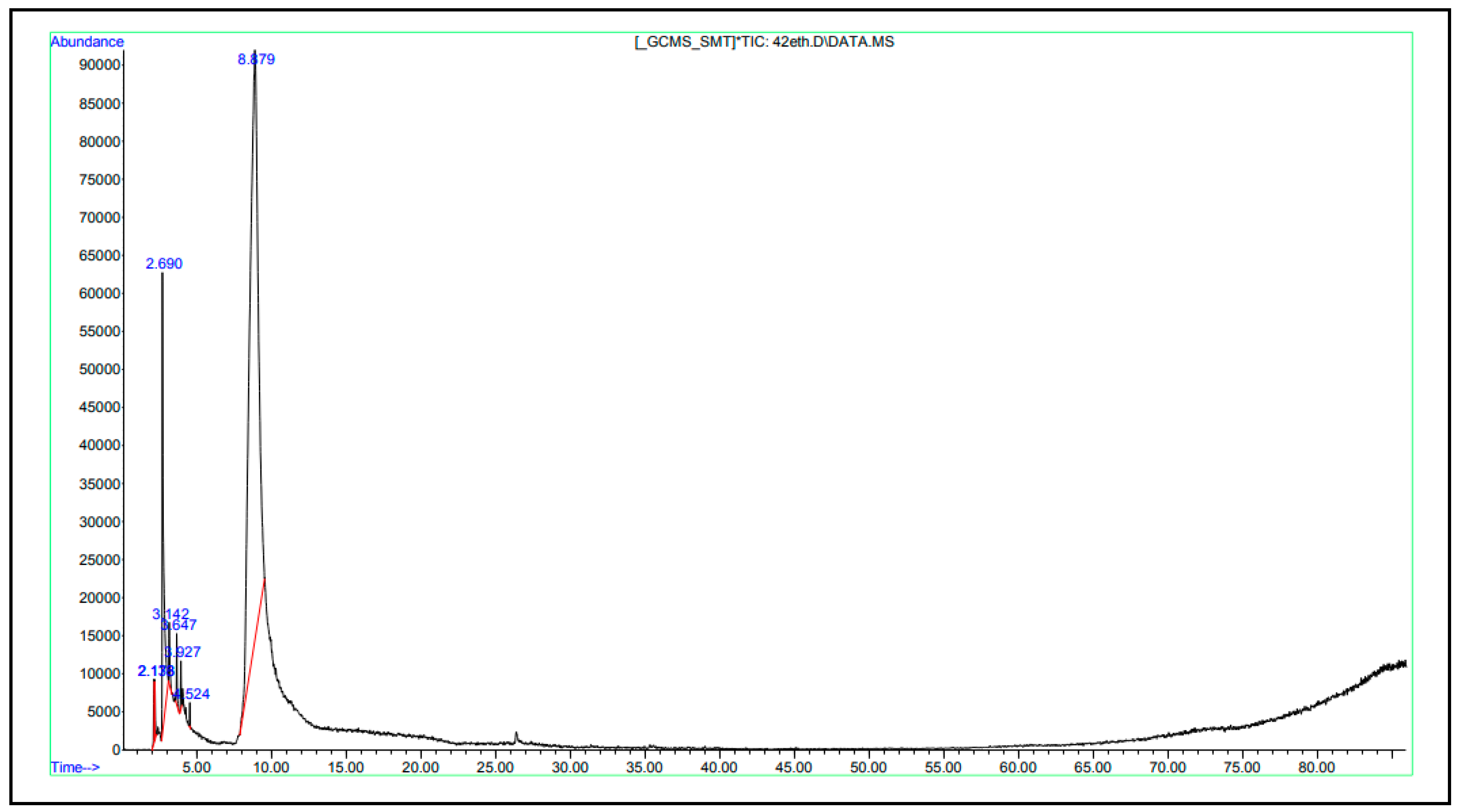
Figure 1. SEM analysis of encapsules. SEM analysis of cell-free supernatant (CFS) of S. thermophilus encapsules. (a) Microencapsulated CFS from S. thermophilus (b) microencapsulated CFS from S. thermophilus combined with thyme extract (1%, v/w), (c) microencapsulated CFS from S. thermophilus combined with thyme extract (2%, v/w).
Foods 13 00329 g001
Figure 2. GC-MS spectra of volatile composition of CFS from S. thermophilus.
Foods 13 00329 g002
Table 1. Chemical content of the CFS from Streptococcus thermophilus.
CompoundsRT%
Methyl-d3 1-dideuterio-2-propenyl ether2.12510.87
7-Octen-2-ol, 2-methyl-6-methylene2.17610.46
Acetic acid2.68639.64
Imidodicarbonic diamide3.1437.64
Tetrahydro-1,3-oxazine-2-thione3.6477.21
2-methyl-1,3-oxathiolany-propionicacid ethyl ester3.9275.42
Butanoic acid, 3-methyl- (isovaleric acid)4.5220.28
(R*,S*)-2-(1′-Nitroethyl)-2-methyl- 1,3-oxathiolane7.9780.94
Methanamine, N-methoxy-8.4702.82
Propane, 2-fluoro-2-methyl-8.6368.92
1,2,3-Propanetriol8.7625.8
RT: retention time.
Table 2. Chemical content of thyme extract.
CompoundsRT%
Ethane, 1,1-diethoxy-3.1490.04
1,1-Diethoxy-2-methylpropan-2-ol7.7490.07
1,2,3,4-Tetrahydroxybutane8.3100.04
1,2,3-Propanetriol (Glycerin)8.36225.77
Propane, 2-fluoro-2-methyl-8.4993.97
Thymol19.4110.05
Phenol, 2-methyl-5-(1-methylethyl) (carvacrol)19.72667.96
Unidentified-2.10
RT: retention time.
Table 3. Inhibition zones (mm) of extracts towards food-related bacteria.
Microencapsulated Groups
CFS from S. thermophilus (CFS)Thyme Extract (TE)CFSCFS + 1%TECFS + 2%TE
Photobacterium damselae7.33 ± 0.58 *b25.00 ± 1.41 a4.50 ± 0.71 c6.00 ± 0.00 bc6.00 ± 0.00 bc
Proteus mirabilis15.25 ± 0.71 b32.00 ± 1.41 a7.50 ± 0.71 d7.50 ± 0.71 d10.50 ± 0.71 c
Vibrio vulnificus17.33 ± 0.58 b41.67 ± 0.58 a8.25 ± 1.06 e11.00 ± 1.41 d13.50 ± 0.71 c
Enterococus faecalis14.00 ± 1.00 b25.33 ± 1.53 a7.00 ± 0.00 cd7.50 ± 0.71 cd9.50 ± 0.71 c
Staphylococcus aureus13.25 ± 0.87 b25.50 ± 1.53 a6.50 ± 0.50 d7.25 ± 0.35 cd8.50 ± 0.71 c
Salmonella Paratyphi A13.50 ± 0.50 b27.33 ± 1.53 a6.25 ± 0.25 d7.00 ± 0.00 d9.38 ± 0.13 c
* Mean value ± Standard deviation (n = 3). There is a significant difference (p < 0.05) between the groups for the values indicated by different letters (a–e) in the same row. CFS + 1%TE: combination of CFS from S. thermophilus and thyme extract (1%, v/w) microencapsules, CFS + 2%TE: combination of S. thermophilus and thyme extract (2%, v/w) microencapsules.
Table 4. Minimum inhibitory (MIC) and bactericidal concentration (MBC) of extracts samples against food-related bacteria.
Microencapsulated Groups
CFS from S. thermophilus (CFS)Thyme Extract (TE)CFSCFS +1%TECFS + 2%TE
MICMBCMICMBCMICMBCMICMBCMICMBC
Photobacterium damselae100>10025100>100>100100>100100>100
Proteus mirabilis2510050100>100>100100>10050>100
Vibrio vulnificus12.51002550100>100100>10050100
Enterococus faecalis100>1005050>100>100100>10050100
Staphylococcus aureus100>10050100>100>100100>100100>100
Salmonella Paratyphi A100>1005050>100>100100>10050100
Mean value ± Standard deviation (n = 3). CFS + 1%TE: combination of CFS from S. thermophilus and thyme extract (1%, v/w) microencapsules, CFS +2%TE: combination of S. thermophilus and thyme extract (2%, v/w) microencapsules.
Disclaimer/Publisher’s Note: The statements, opinions and data contained in all publications are solely those of the individual author(s) and contributor(s) and not of MDPI and/or the editor(s). MDPI and/or the editor(s) disclaim responsibility for any injury to people or property resulting from any ideas, methods, instructions or products referred to in the content.

