Article

Characterization and Antibiotic Resistance of Listeria monocytogenes Strains Isolated from Greek Myzithra Soft Whey Cheese and Related Food Processing Surfaces over Two-and-a-Half Years of Safety Monitoring in a Cheese Processing Facility

by
Nikolaos D. Andritsos
1 and
Marios Mataragas
2,*
1
Department of Food Science and Technology, School of Agricultural Sciences, University of Patras, 2 G. Seferi Str., GR-301 00 Agrinio, Greece
2
Department of Dairy Research, Institute of Technology of Agricultural Products, Hellenic Agricultural Organization “DIMITRA”, 3 Ethnikis Antistaseos Str., GR-452 21 Ioannina, Greece
*
Author to whom correspondence should be addressed.
Foods 2023, 12(6), 1200; https://doi.org/10.3390/foods12061200
Submission received: 7 February 2023 / Revised: 27 February 2023 / Accepted: 10 March 2023 / Published: 12 March 2023

Abstract

:
Listeriosis is a serious infectious disease with one of the highest case fatality rates (ca. 20%) among the diseases manifested from bacterial foodborne pathogens in humans, while dairy products are often implicated as sources of human infection with Listeria monocytogenes. In this study, we characterized phenotypically and genetically by whole-genome sequencing (WGS) 54 L. monocytogenes strains isolated from Myzithra, a traditional Greek soft whey cheese (48 isolates), and swabs collected from surfaces of a cheese processing plant (six isolates) in the Epirus region of Greece. All but one strain of L. monocytogenes belonged to the polymerase chain reaction (PCR) serogroups IIa (16.7%) and IIb (81.5%), corresponding to serotypes 1/2a, 3a and 1/2b, 3b, 7, respectively. The latter was identified as a PCR-serogroup IVb strain (1.8%) of serotypes 4b, 4d, 4e. Bioinformatics analysis revealed the presence of five sequence types (STs) and clonal complexes (CCs); ST1, ST3, ST121, ST 155, ST398 and CC1, CC3, CC121, CC155, CC398 were thus detected in 1.9, 83.3, 11.0, 1.9, and 1.9% of the L. monocytogenes isolates, respectively. Antibiograms of the pathogen against a panel of seven selected antibiotics (erythromycin, tetracycline, benzylpenicillin, trimethoprim-sulfamethoxazole, ampicillin, ciprofloxacin, and meropenem) showed that 50 strains (92.6%), the six surface isolates also included, were intermediately resistant to ciprofloxacin and susceptible to the rest of the six antimicrobial agents tested, whereas strong resistance against the use of a single from three implicated antibiotics was recorded to four strains (7.4%) of the pathogen isolated from Myzithra cheese samples. Thence, the minimum inhibitory concentrations (MICs) were determined for erythromycin (MIC = 0.19 μg/mL), ciprofloxacin (MIC ≥ 0.19 μg/mL), and meropenem (MIC = 0.64 μg/mL), and finally, just one strain was deemed resistant to the latter antibiotic. The phylogenetic positions of the L. monocytogenes strains and their genetic variability were determined through WGS, whilst also stress response and virulence gene analysis for the isolates was conducted. Findings of this work should be useful as they could be utilized for epidemiological investigations of L. monocytogenes in the food processing environment, revealing possible contamination scenarios, and acquired antimicrobial resistance along the food production chain.

1. Introduction

Listeria monocytogenes is the etiological agent of listeriosis disease in both humans and animals. The ubiquitous character of the bacterium makes it widespread in both the natural environment and the food processing environment, thus easily contaminating fresh produce (e.g., fruits and vegetables) [1,2,3,4,5] and foods of animal origin (e.g., meat and dairy products) [6,7,8,9]. Listeriosis is a serious infectious disease with one of the highest case fatality rates in humans (ca. 20%) among the diseases manifesting from bacterial pathogens encountered in foods [10,11,12]. Contrary to the non-invasive form of febrile gastroenteritis (intestinal listeriosis), symptoms of the severe invasive form of the disease occur when the pathogen has spread beyond the intestines of the infected person. They include fever, muscle pain, septicemia, meningitis, and meningoencephalitis, and they usually start within two weeks of eating food contaminated with L. monocytogenes [13,14]. Invasive listeriosis affects the most vulnerable segment of the population; YOPI individuals (i.e., young, old, pregnant, and immunocompromised), such as pregnant women and their newborns, infants, elderly people (above 65 years old) and people with a weakened immune system (e.g., patients undergoing treatment for hemodialysis, cancer, or AIDS). In contrast, febrile gastroenteritis is a mild form of listeriosis that normally ends up as a self-limiting infection, affecting mainly otherwise healthy people after ingesting a rather large number of L. monocytogenes bacteria from contaminated food products [13,14]. In this case, symptoms usually start within a few days (mostly within 24 h) after the consumption of contaminated food with the pathogen and include diarrhea, fever, headache, and muscle pain [14].
Control of L. monocytogenes in the food processing environment is quite difficult due to the pathogen’s ability to tolerate extreme environmental conditions (e.g., acid resistance, heat resistance, and high salt concentrations) [15,16,17,18,19,20,21]; adapt to a wide range of physical/physicochemical stresses (e.g., pH, water activity, and temperature fluctuations) [16,17,19,20]; cope with sublethal stresses induced from treatments with antimicrobials and disinfectants (e.g., plant essential oils or quaternary ammonium compounds (QACs)) [20,21,22,23]; and form biofilms as means of self-preservation [20,24,25]. These factors eventually lead to the persistence of L. monocytogenes in food equipment and premises [20,26,27,28].
Animal-originated foods, such as meat, milk, and dairy products, are excellent substrates for microbial proliferation. Most dairy products (e.g., cheese and yogurt) are the result of microbial activity (fermentation) of the native microbiota contained in milk, albeit in the food and cheese production process in particular, microbial pathogens may gain access to the final product and represent a threat to the consumer [29], given their growth potential and survivability in cheese and its products thereof [30,31,32,33,34,35]. L. monocytogenes cannot grow in the traditional Greek soft acid-curd cheeses, such as Galotyri and Katiki [35,36], or hard ripened cheeses like traditional Greek Graviera cheese [32,37], although effective control of post-processing listerial cross-contamination is needed in Greek Myzithra, Anthotyros, and Manouri soft whey cheeses [38,39,40]. Thankfully, to the best of the authors’ knowledge, the last documented survey conducted in the Greek retail market, although published a decade ago, indicated a low prevalence of Listeria spp. and the absence of positive samples for L. monocytogenes in soft cheeses [41]. In strict alignment with those findings, a recent systematic review and meta-analysis conducted with data from the European Food Safety Authority (EFSA) and the scientific literature emphasized the lowest mean prevalence (ca. 0.8%) of L. monocytogenes in fresh cheeses, like Myzithra, among all other European cheeses (i.e., ripened, veined, smear, and brined) [42].
The vast majority of human listeriosis cases are caused by L. monocytogenes serotypes 1/2a (lineage II), 1/2b and 4b (lineage I) [43,44,45]. Nevertheless, more than 50% of human listeriosis outbreaks are associated with lineage I and to be precise most of these outbreaks are attributed to serotype 4b (polymerase chain reaction serogroup IVb; PCR-serogroup IVb), whereas serotype 1/2a (PCR-serogroup IIa) and 1/2b (PCR-serogroup IIb) strains of the pathogen are mostly isolated from foods [45,46] (pp. 50–51). This could be attributed not only to the inherent genetic characteristics [45] and the competitive growth advantage between the strains (PCR-serogroups IIa, IIb and IIc vs. PCR-serogroup IVb) [47] but also to the selective enrichment protocol used for the L. monocytogenes detection [48], justifying in this way the overrepresentation of certain serotypes of the pathogen in foods.
Epidemiological studies aim to link the pathogen responsible for the advent of an outbreak, isolated from human clinical samples, with the implicated food commodity in case of a foodborne disease investigation. Many times, food and environmental sampling are established towards the investigation of the above linkage, leading to the deciphering of relevant contamination routes for the pathogen under study [49]. Serotyping and serogrouping of strains are of low discriminatory capacity and offer very little to the epidemiological surveillance of the pathogens, such as L. monocytogenes [43]. In this context, laboratory methods with increased discriminatory power, such as pulsed-field gel electrophoresis (PFGE), multilocus sequence typing (MLST) and repetitive element palindromic PCR (rep-PCR) in combination with random amplified polymorphic DNA PCR (RAPD-PCR) analysis, have been utilized for the subtyping of L. monocytogenes isolated from foods and the food processing environment [50,51,52]. However, in recent years, whole-genome sequencing (WGS) is gaining more and more ground and is tending to replace PFGE, MLST and rep-PCR with RAPD-PCR fingerprinting of pathogen isolates [53,54], highlighting the population diversity and persistence of L. monocytogenes in the environment of the food production facility [28,55,56,57]. Therefore, the objective of this work was to carry out the phenotypic and genomic characterization, as well as the antibiotic resistance profile of L. monocytogenes strains isolated from both the cheese processing environment and samples of an end-product (Myzithra cheese). The genetic variability of the strains, along with their phylogenetic positions, were determined by means of WGS. In parallel, a stress response and virulence gene analysis for the isolates was conducted, allowing for a better understanding of L. monocytogenes’ persistence in the cheese production environment and thus contributing towards improved strategies for the elimination of the pathogen.

2. Materials and Methods

2.1. Sampling of Cheese and Related Food Processing Surfaces

Environmental and end-product sampling was performed during the production process in a cheese processing facility located in the Epirus region of northwestern Greece. Related cheese processing surfaces and Myzithra, a traditional Greek soft whey cheese manufactured from sheep and/or goat milk, were sampled to detect L. monocytogenes. Screening for the pathogen’s presence took place over a period of more than two-and-a-half years of microbiological monitoring of L. monocytogenes in the facility (November 2016–July 2019).
Samples of fresh and dried Myzithra end-product comprised of ca. 150 g of cheese. Surface sampling was performed according to the protocol of the International Organization for Standardization (ISO 18593) [58,59] by using pre-moistened in half concentration-Fraser broth (½-FB; Biokar Diagnostics, Pantin, France), sterile cotton applicators (AnQing Jiaxin Medical Technology Co., Ltd., Anqing, China) and swabbing 100 cm2 (10 cm × 10 cm), with a swabbing movement executed in both horizontal and vertical directions onto the surface and by rotating the applicator between fingers, then placing the swab under aseptic conditions in 10 mL of ½-FB contained in Sterilin ™ plastic round-based tubes with screw cap (Sarstedt, Nümbrecht, Germany). Cheese and surface samples were transported into isothermal boxes with ice packs and were analyzed upon arrival at the laboratory.

2.2. L. monocytogenes Isolation

The ISO 11290-1 protocol was used for the detection of L. monocytogenes [60,61] from cheese and surface samples with some slight modifications. Briefly, 25 g of Myzithra end-product were transferred aseptically in a stomacher bag and homogenized with 225 mL of ½-FB in a Stomacher blender (BagMixer® 400 W, Interscience, Saint Nom, France). The homogenate was incubated at 30 °C for 24 h. In the case of surface samples, Sterilin™ tubes containing the swabs in 10 mL of ½-FB were placed for incubation at the aforementioned conditions immediately upon arrival at the laboratory. Following the primary enrichment of samples, 0.1 mL of the initial suspension was transferred aseptically to a tube containing 10 mL of full concentration-FB (FB; Biokar Diagnostics) as a secondary enrichment medium. The inoculated FB was incubated then at 37 °C for 24–48 h. Primary and secondary enrichment broths (½-FB and FB) were streaked on duplicate plates of COMPASS® Listeria agar (Biokar Diagnostics), its formulation of which is in accordance with the agar Listeria according to Ottaviani and Agosti (ALOA) referred in the ISO detection method (ISO 11290-1) [60,61].
ALOA plates were examined visually after incubation at 37 °C for 48 h for the presence of the pathogen indicated by well-isolated blue-green colonies surrounded by an opaque halo. Presumptive L. monocytogenes was confirmed through biochemical testing of the isolates after subculturing the selected colonies from ALOA on tryptone soya agar containing 0.6% yeast extract (TSAYE) and incubating at 37 °C for 18 to 24 h. Biochemical tests were carried out on colonies from pure cultures grown on TSAYE plates and comprised of oxidase test, catalase reaction, motility test at 25 °C, hemolysis on sheep blood agar, CAMP test, L-rhamnose and D-xylose utilization. Confirmed L. monocytogenes isolates were maintained at −80 °C in brain heart infusion (BHI; LabM, Lancashire, UK) broth with 20% glycerol (Biolife, Milan, Italy). The strains were deposited in the microbial culture collection of Eurofins Athens Analysis Laboratories S.A. (AAL; Microbiology Laboratory, Metamorfosi, Attica, Greece) and received an accession number. Before further use of the isolates, the frozen stock of each strain of the pathogen (Supplementary Table S1) was subcultured twice in BHI broth incubated each time at 37 °C for 24 h and then streaked on TSAYE which was incubated at 37 °C for 18 to 24 h.

2.3. Antibiotic Resistance Profiles of L. monocytogenes Strains

Antimicrobial susceptibility and resistance to antibiotics of L. monocytogenes cheese and surface isolates were determined using the standardized disk diffusion method as recommended by the European Committee on Antimicrobial Susceptibility Testing (EUCAST) [62]. In brief, 3–4 colonies picked after activation of the pathogen on the TSAYE plate were suspended in 3 mL of 0.85% peptone salt solution or maximum recovery diluent (MRD; Merck, Darmstadt, Germany), and turbidity was adjusted to a 0.5 McFarland standard. The suspension was used to inoculate ready-to-use 9 cm Mueller-Hinton agar plates supplemented with 5% defibrinated horse blood and 20 mg/l β-NAD (MH-F; Bioprepare, Keratea, Attica, Greece). Inoculation of MH–F blood agar with the pathogen was achieved by means of microbial lawn formed for each L. monocytogenes strain, using sterile cotton applicators (AnQing Jiaxin Medical Technology Co., Ltd.) moistened in the MRD suspension and applied on the surface of MH–F agar with swabbing movement in both horizontal and vertical directions and by rotating the applicator between fingers, covering in that manner the whole agar surface. Antibiotic disks of seven antimicrobial agents were transferred on the MH–F agar surface with the help of forceps. All disks were supplied by Oxoid (Basingstoke, UK): Erythromycin (E; 15 μg), tetracycline (TE; 30 μg), benzylpenicillin (P; 1 IU), trimethoprim-sulfamethoxazole 1:19 (SXT; 25 μg), ampicillin (AMP; 2 μg), ciprofloxacin (CIP; 5 μg) and meropenem (MEM; 10 μg). After 18 h of incubation at 35 °C, the diameters of inhibition zones around the disks were measured to the nearest integral number (in mm) by using a Vernier caliper with the least count of 0.1 mm. Results for inhibition zone diameters per antibiotic and strain of the pathogen were interpreted as susceptible, intermediate, or resistant L. monocytogenes isolates to the respective antimicrobial agent based on the criteria provided by EUCAST [62]. Missing zone diameter breakpoints for L. monocytogenes resistance against E and CIP were obtained from those recommended by EUCAST for Gram-positive Staphylococcus aureus [62].
Profiles of antibiotic resistance for L. monocytogenes strains isolated from cheese and related cheese processing surfaces were complemented with the determination of minimum inhibitory concentrations (MICs) by using the E-test method for those antimicrobial agents inducing resistance of the pathogen, screened through the antimicrobial susceptibility testing (AST) as described above. Following inoculation of MH–F agar as previously described, plastic strips with a gradient concentration of the antibiotic of interest to which the strain showed resistance during AST were placed onto the surface of the agar medium, allowing for the diffusion of the antimicrobial agent into the agar and thus providing, after overnight incubation at 37 °C, an inhibition ellipse where the MIC corresponds to the value at the point of intersection of the growth and inhibition zone with the extremity of the strip. The E-test MICs (in μg/mL or ppm) can be read directly from the upper of the Petri dish [63] (pp. 323–324).

2.4. Whole-Genome Sequencing of Bacterial Isolates

2.4.1. DNA Extraction and Sequencing

Total DNA was extracted from the bacterial cells of L. monocytogenes strains, cultured in BHI broth and incubated overnight at 37 °C after a purity check on TSAYE plates. The genomic DNA was extracted and sequenced by Novogene Genomics Service (Novogene Co., Ltd., Cambridge, UK), and all procedures involving library preparation, genome assembly, and quality control were performed as described by Syrokou et al. [64]. To this end, the complete genomes of the corresponding reference genomes used were downloaded from the NCBI website (https://www.ncbi.nlm.nih.gov; accessed on 30 August 2022). Genome quality was assessed through a re-implementation of an algorithm from an online tool (CheckM) [65] to ensure that genomes had acceptable completeness (≥95%) and contamination (≤5%), while any potential bacterial misassemblies were evaluated with the help of a web app (SkewIT) [66].

2.4.2. Bioinformatics Analyses

All L. monocytogenes isolates were phenotypically characterized in silico from the obtained nucleotide sequences by using the microbial trait analyzer Traitar (GitHub Inc., San Francisco, CA, USA; https://github.com/aweimann/traitar, accessed on 6 February 2023) [67]. Additionally, all strains were molecularly confirmed through PCR-serogrouping and serotyping [68] performed with the Bionumerics platform version 8.1.1 (bioMérieux, Marcy l’ Etoile, France; https://www.applied-maths.com/bionumerics, accessed on 6 February 2023). The core/whole-genome MLST (cg/wg MLST) analyses, along with the assessment of acquired resistance, virulence factors, and screening for phage sequences, were also performed with Bionumerics version 8.1.1 (bioMérieux) for the in silico subtyping of the strains.

3. Results

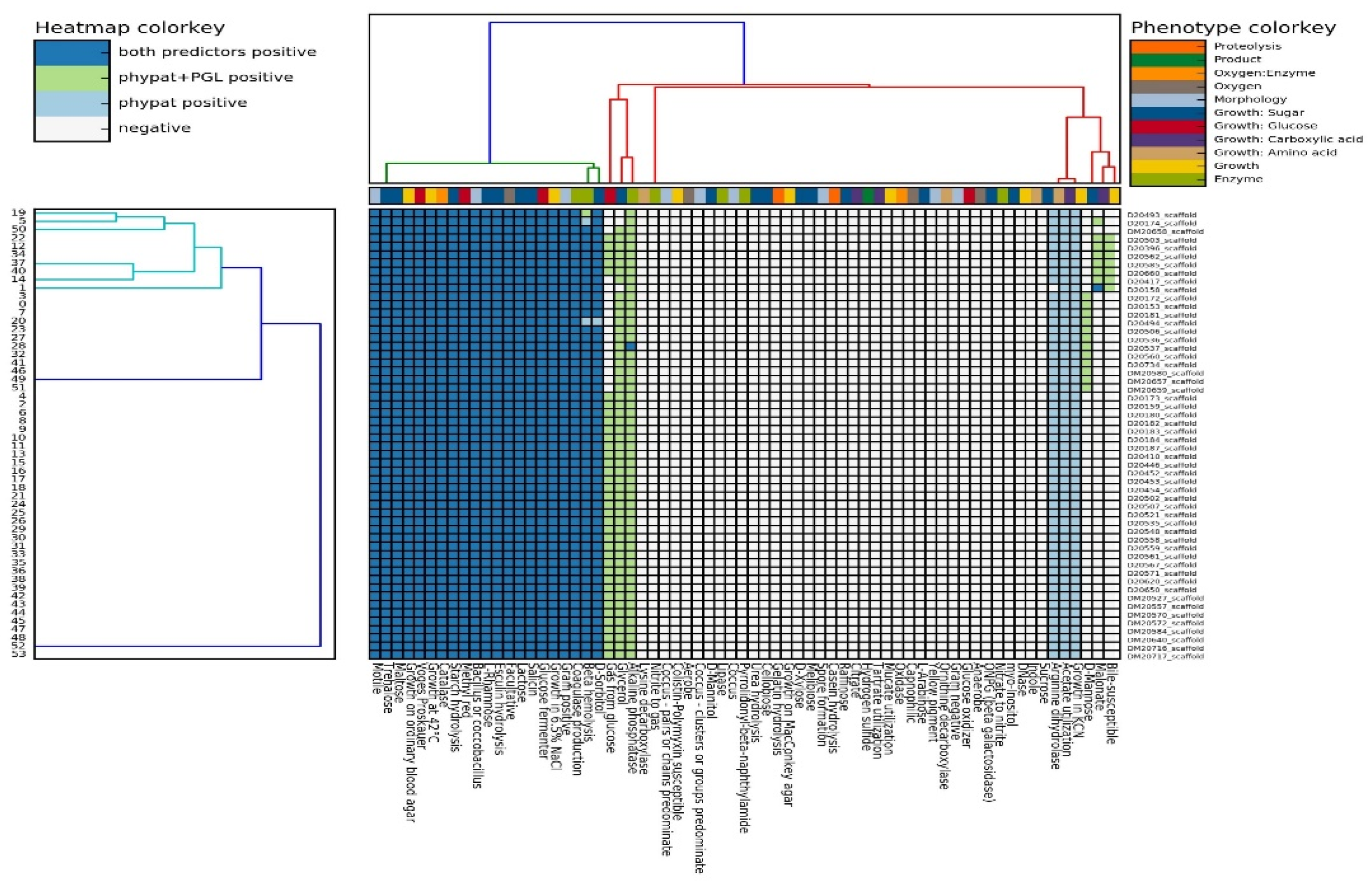
3.1. Phenotypic Characterization of L. monocytogenes Isolates

Fifty-four L. monocytogenes strains were isolated from Myzithra cheese (48 isolates) and related cheese processing surfaces (six isolates). Biochemical testing of the isolates (Supplementary Table S1) was consistent with the predicted phenotype for each L. monocytogenes strain, and all the phenotypic characteristics of the strains were mapped (Figure 1).

3.2. Antibiograms of L. monocytogenes Cheese and Surface Isolates

Seven antibiotics belonging to the classes of macrolides (E), tetracyclines (TE), penicillins (P, AMP), sulfonamides (SXT), fluoroquinolones (CIP), and carbapenems (MEM) were tested for the resistance of L. monocytogenes cheese and surface isolates against them. The antibiotic resistance profiles (antibiograms) of L. monocytogenes strains are presented in Figure 2. All isolates were not clearly susceptible to CIP and presented mostly intermediate (52/54 strains, 96.3% of isolates) to strong (2/54, 3.7%) resistance against the specific antimicrobial agent (Figure 2). CIP is not the drug of choice for the treatment of listeriosis and is not included in the clinical breakpoint tables for L. monocytogenes published by EUCAST [62]. Apart from CIP, therefore, all surface isolates were susceptible to the remaining six antibiotics tested, while 46 out of 48 (95.8%) of the cheese isolates were found to be sensitive during AST to those six antibiotics (Figure 2). None of the strains showed any multidrug resistance. In total, the resistance of four L. monocytogenes strains isolated from Myzithra soft cheese (4/54, 7.4%) was recorded against E, MEM and CIP (Figure 2). Three of the resistant isolates belonged to PCR-serogroup IIb (strains AAL 20153, AAL 20184, AAL 20187) and were classified as ST3 (CC3) strains, while the fourth resistant isolate belonged to PCR-serogroup IIa (strain AAL 20158) and was classified as an ST155 (CC155) strain (see Section 3.4). Furthermore, MIC values for the L. monocytogenes cheese isolates against E, CIP, CIP, and MEM were estimated at 0.19, 0.19, 0.50, and 0.64 μg/mL for strains AAL 20153, AAL 20184, AAL 20158, and AAL 20187, respectively. Following AST and MIC determination, strain AAL 20187 was finally designated as resistant to MEM.
Antibiotic resistance genes were identified in strain AAL 20158 and included fosX, mdrL, and mdrM genes conferring resistance to fosfomycin, QACs/macrolides and multidrug resistance, respectively. However, no resistance genes were identified to the rest of the isolates that exhibited antimicrobial resistance during AST.

3.3. Genetic Diversity of L. monocytogenes Isolates

PCR-serogrouping and serotyping performed for all the isolates revealed the presence of three major serogroups for the pathogen with their respective serotypes (i.e., PCR-serogroups IIa: ser. 1/2a, 3a; IIb: ser. 1/2b, 3b, 7; IVb: 4b, 4d, 4e) (Figure 3).
A total of five different sequence types (STs) and clonal complexes (CCs) were identified in the 54 isolates of L. monocytogenes through the MLST (Figure 4) and cg/wg MLST analyses (Figure 5); ST3 and CC3 (each containing n = 45 strains, 83.3% of isolates), ST121 and CC121 (each n = 6, 11.0%), ST1 and CC1 (each n = 1, 1.9%), ST155 and CC155 (each n = 1, 1.9%), ST398 and CC398 (each n = 1, 1.9%). ST3 and CC3 strains belonged both to PCR-serogroups IIa and IIb with serotypes 1/2a, 3a and 1/2b, 3b, and 7 assigned to them, respectively. Strains of ST121 and CC121, ST155 and CC155, ST398 and CC398 all belonged to PCR-serogroup IIa and were assigned the serotypes 1/2a, 3a, whereas the ST1 and CC1 single strain belonged to PCR-serogroup IVb, which is comprised of serotypes 4b, 4d, 4e. The minimum spanning tree shows that the number of allelic differences between neighboring STs (CCs) ranged from 4 to 6 (Figure 4), but cg/wg MLST reveals genetic diversity between and within STs (CCs) (Figure 5).

3.4. Analysis of Resistance, Virulence and Persistence of L. monocytogenes Isolates

The analysis performed with the Bionumerics software led us to focus on 12 important Listeria genes (Figure 6). Seven genes were related to acquired virulence potential (i.e., inlA, inlB, inlC, inlF, inlH, inlJ, and inlK), three corresponded to pathogenicity islands found in the bacterium (i.e., LIPI-1, LIPI-3, and SSI-1) and two genes were related with acquired resistance mechanisms (i.e., qacH and mdrL) against quaternary ammonium compounds (QACs) commonly used as disinfectants in the food industry.
All strains of PCR-serogroup IIa (with the notable exception of strain AAL 20174, which did not possess the operon LIPI-3) had the internalin A (inlA) gene truncated (Figure 6), something that confers to the reduced virulence potential of those strains. Moreover, the PCR-serogroup IIa strains belonging to ST121 (CC121) possessed the qacH and mdrL (Figure 6) genes indicating acquired resistance to QACs.
ST3 (CC3) and ST121 (CC121) L. monocytogenes strains showed persistence in the food processing environment as they were continuously isolated (even for over 2.5 years) from end-product cheeses and cheese-making surfaces, while they also exhibited mobile phage elements inserted and incorporated into their DNA (prophages) (i.e., from 2 to 8 elements; Figure 6), which are considered to promote increased resistance and adaptation of the strains to the adverse environmental conditions encountered in the food production plant. In contrast, the PCR-serogroup IVb strain belonging to ST1 (CC1) did not exhibit any such prophages at all and was more or less sporadic, indicative of a rather transient than persistent human contamination (Figure 6).

4. Discussion

A total of 54 L. monocytogenes strains isolated from Myzithra soft cheese and swabs collected from food contact surfaces in a cheese processing plant were characterized phenotypically and genetically using a WGS approach. The phenotypic characterization of the isolates in silico was consistent with the biochemical testing performed on the presumptive colonies of the pathogen recovered from ALOA, regarding at least some basic traits utilized for the L. monocytogenes identification (Figure 1). This confirms the constant recovery of the pathogen from end-product and surface samples during routine microbiological monitoring for L. monocytogenes presence in the facility, which in turn indicates potential colonization of the cheese processing environment with different listerial STs [52].
The majority of isolates were classified as PCR-serogroup IIb strains (44/54, 81.5%; ser. 1/2b, 3b, 7), followed by PCR-serogroup IIa (9/54, 16.7%; ser. 1/2a, 3a) and PCR-serogroup IVb strains (1/54, 1.8%; ser. 4b, 4d, 4e). Hence, lineage I and II strains accounted for 83.3 and 16.7% of the isolates, respectively. Many studies have highlighted the dominance of PCR-serogroup IIa (lineage II) strains of L. monocytogenes over a variety of food matrices as compared to other serogroups [6,7,8,50,69,70,71,72,73]. Nevertheless, the aforementioned distribution of serogroups (with the predominance of molecular serogroup IIb; lineage I) is not uncommon and has been reported elsewhere [74,75,76,77]. Especially when it comes to meat and dairy products, PCR-serogroup IIb is most prevalent among the L. monocytogenes isolates [69,74,76].
Antibiotic resistance profiles for L. monocytogenes isolates from raw and/or pasteurized/fresh milk and milk products (e.g., cheeses) were revealed in previous research works against most of the antimicrobial agents tested in our study (i.e., TE, P, E, and SXT) [76,78,79,80]. Likewise, resistance to macrolides (E) emerged for strain AAL 20153 (Figure 2), although no resistance was recorded against tetracyclines, penicillins and sulfonamides in the present work. Furthermore, the MIC for E was found to range between 0.05 to 0.2 ppm in eight strains of L. monocytogenes tested for their antimicrobial resistance [81], something which agrees with our estimate of 0.19 ppm for the lethal concentration of the antibiotic in strain AAL 20153. The surface isolates belonged to two STs (ST3 and ST121; Figure 4) instead of the five STs identified for the cheese isolates (ST1, ST3, ST121, ST155, and ST398). However, no signs of antimicrobial resistance to any of the antibiotics used in the study were inferred for the surface isolates. All L. monocytogenes isolates showed no multidrug resistance, while the vast majority of them (50/54, 92.6%) were susceptible to all the antibiotics, except for CIP, to which generally all strains presented intermediate to strong resistance (Figure 2). The level of tolerance to the fluoroquinolone antibiotic CIP could be the result of cross-resistance developed in L. monocytogenes, when cells of the pathogen are exposed to sublethal concentrations of residual QACs, widely used as biocides in the food industry, leading to the formation of QAC-adapted subpopulations of L. monocytogenes with increased survival against CIP [82,83,84]. Interestingly, the resistance phenotype correlated with genotype in just one (strain AAL 20158) of the four L. monocytogenes strains initially screened as resistant to the single use of one of the three implicated antibiotics. On the other hand, no genes known to encode resistance against either E, CIP, or MEM were detected originally in the rest of the three resistant strains (strains AAL 20153, AAL 20184, and AAL 20187). Zhang et al. [85] described a similar situation where the authors could not detect by WGS any genes encoding resistance to MEM, against which antibiotic two clinical L. monocytogenes isolates showed resistance, suggesting that more research is needed on the matter. We hypothesize that this could be due to the possible fragmentation or mutation of the implicated resistance genes, and thus the necessity of using other search tools for gene alignment is highlighted in this case. For instance, L. monocytogenes genomes were studied with GWAS methodology (genome-wide association studies using pyseer) for the MEM-resistant phenotype, and the results are summarized in Figure 7. The top gene hit ranked by the −log10 (p-value), and the number of k-mer hits (cds-NP_463988.1; upper left) was related to a helix-turn-helix domain-containing protein with an average effect size of ca. 0.4 (0.370). MarR or Multiple antibiotics resistance-type regulators are characterized by winged helix-turn-helix (HTH) DNA-binding motifs and control genes that confer resistance to antibiotics, organic solvents, detergents, etc. This implies that this gene might have a significant role in assisting L. monocytogenes AAL 20187 resistance to MEM. However, other L. monocytogenes isolates may also possess this gene. The k-mer methodology showed regions in the sequence that are overrepresented in AAL 20187 compared to the rest of the strains.
The overwhelming majority of L. monocytogenes strains (51/54, 94.4%) belonged to CC3 and CC121 (Figure 4 and Figure 5), with those CCs commonly found among the food isolates [71,73,86,87]. All CC3 strains except one (strain AAL 20174) together with CC1 carried the hypervirulence pathogenicity island LIPI-3 in addition to LIPI-1 (Figure 6). On the contrary, LIPI-3 was absent in CC121, CC155 and CC398, strains of which CCs present a hypovirulent potential mainly because of the absence of the aforementioned major virulence factor LIPI-3 [88,89]. This hypovirulence is further expressed in CC121 strains of the pathogen due to the lack of internalins (i.e., inlF, inlH, inlI) noticed in the internalin gene family of the strains (Figure 6). Besides, the mutation in the inlA gene that leads to premature stop codons (PMSCs) has been found for CC121, CC155, and CC398 (Figure 6). It is now known that hypervirulent clones of L. monocytogenes, particularly CC1, are strongly associated with dairy products [87], whereas isolates of the pathogen carrying a virulence-attenuated mutation in inlA gene are frequently recovered from food processing environments [90]. Alarmingly, the isolation of the hypervirulent CC1 and CC3 strains from fresh and dry Myzithra as well as cheese processing surfaces, could represent a major public health issue, especially due to the ST1 strain’s close relation to dairy cattle farms [91]. Indeed, it is generally accepted that hypervirulent clones of L. monocytogenes colonize better the intestinal tract and more easily infect humans than the hypovirulent ones [87]. However, it is worthy of comment that hypo- or hypervirulence does not necessarily imply that the strain is capable of adhering to the human colon or effectively invading the host since foodborne diseases, such as listeriosis, are the result of the interaction between the host, the pathogen and the food commodity where the microorganism resides [92]. In that essence, many times, even hypovirulent clones of L. monocytogenes are able to cause infection in YOPI individuals.
Stress survival islet 1 (SSI-1) is comprised of a five-gene islet which contributes to the growth of L. monocytogenes under unfavorable conditions and allows for the pathogen’s survival in food processing environments [93]. SSI-1 was present only in the hypervirulent CC3 strains and the hypovirulent strain AAL 20158 (CC155), which is indicative of the good adaptive response these strains showed to the environment of the cheese processing plant, reflecting the constant recovery and, in absolute numbers, the higher persistence of CC3 strains in the facility (Figure 6). The persistence of L. monocytogenes in the facility was also evident for the hypovirulent strains of CC121 containing the QAC-resistant qacH and mdrL genes and harboring highly conserved plasmids and prophages [94]. CC121 is widely suspected of benzalkonium chloride (BC) tolerance [70,87,95], and strains of this cluster are normally equipped with the transposon Tn6188, which encodes, among others, the qacH gene conferring potential BC resistance [96]. In CC121, the mobile phage elements were at least six and reached up to eight elements for most of the strains as compared to any other CC (Figure 6). The Listeria phages detected in our study were among those used in a comparative genomic analysis of prophages conducted by Vu et al. [97].
Bioinformatics analysis revealed the presence of the competence transcription factor comK for all CC3 strains. L. monocytogenes strains carrying the SSI-1 cluster of genes as well as the comK prophage are more likely to possess a comparatively higher biofilm-forming ability and present enhanced persistence than strains of the pathogen lacking those genes [95,98], something that still has to be elucidated for our strain collection in terms of cell density and biofilm production for the CC3 strains. In addition, future research is to be focused on the single-nucleotide polymorphism (SNP) differences between the isolates in order to determine the genetic evolution of L. monocytogenes over the years in the cheese processing facility through the observed SNP differences caused by the limited single-nucleotide mutations but rapid diversification of prophages [99]. Facility-specific molecular markers utilized from the allelic variations detected across the whole genome of the pathogen’s isolates may be unique for specific processing plants and could be used as signatures for tracking L. monocytogenes throughout the food chain [100]. This has yet to be clarified in this instance. Incorporating cgMLST fingerprinting data obtained by WGS under the umbrella of a single tool used for the comparison of the whole genome of L. monocytogenes across distinct geographical regions is quite challenging. Recently a web-based platform has been proposed for tracking the pathogen worldwide to help with public health surveillance and resolution of foodborne disease outbreak investigations [101].
Notably, CC155 is mostly identified among L. monocytogenes isolates of clinical origin [85,87]. However, a WGS analysis showed that the specific CC was most prevalent among the pathogen’s isolates recovered from food and food production environments in Poland [102]. As far as CC398 is concerned, the only strain contained in this group (strain AAL 20493) warrants further investigation due to the limited information provided in the literature regarding this CC.
The present study provides insights into the pathogenic potential and persistence of the L. monocytogenes isolates recovered from a cheese processing facility. The findings of this work should be useful as they could be utilized for epidemiological investigations of L. monocytogenes in the food processing environment, revealing possible contamination scenarios, acquired antimicrobial resistance along the food production chain and providing critical information for post-outbreak management in the facility [103,104,105]. These findings also suggest that further research is needed for definitive genome mapping of the isolates.

Supplementary Materials

The following supporting information can be downloaded at: https://www.mdpi.com/article/10.3390/foods12061200/s1, Table S1: Listeria monocytogenes strains used in the study.

Author Contributions

Conceptualization, N.D.A. and M.M.; methodology, N.D.A. and M.M; software, M.M.; validation, N.D.A. and M.M.; formal analysis, M.M.; investigation, N.D.A.; resources, N.D.A. and M.M.; data curation, M.M.; writing—original draft preparation, N.D.A. and M.M.; writing—review and editing, N.D.A. and M.M.; visualization, N.D.A. and M.M.; supervision, M.M.; project administration, M.M.; funding acquisition, N.D.A. and M.M. All authors have read and agreed to the published version of the manuscript.

Funding

This research was funded by the Research Committee of the University of Patras, grant number 81974, through the funding program “MEDICUS” (“Microbiological control of the safety of foods of animal origin”).

Data Availability Statement

This Whole Genome Shotgun project (BioProject number PRJNA858978) has been deposited at DDBJ/ENA/GenBank under the accession numbers JANLLB000000000 to JANLLZ000000000, JANLMA000000000 to JANLMZ000000000, and JANLNA000000000 to JANLNC000000000. The version described in this paper is version JANLLB010000000 to JANLLZ010000000, JANLMA010000000 to JANLMZ010000000, and JANLNA010000000 to JANLNC010000000.

Acknowledgments

The authors would like to thank Eurofins Athens Analysis Laboratories S.A. for donating the materials used for the L. monocytogenes isolation and offering the deep-freezing storage of strains.

Conflicts of Interest

The funders had no role in the design of the study; in the collection, analyses, or interpretation of data; in the writing of the manuscript; or in the decision to publish the results. The authors declare no conflict of interest.

References

  1. Alegbeleye, O.O.; Sant’Ana, A.S. Survival and growth behaviour of Listeria monocytogenes in ready-to-eat vegetable salads. Food Control 2022, 138, 109023. [Google Scholar] [CrossRef]
  2. Alegbeleye, O.O.; Singleton, I.; Sant’Ana, A.S. Sources and contamination routes of microbial pathogens to fresh produce during field cultivation: A review. Food Microbiol. 2018, 73, 177–208. [Google Scholar] [CrossRef]
  3. Buchanan, R.L.; Gorris, L.G.M.; Hayman, M.M.; Jackson, T.C.; Whiting, R.C. A review of Listeria monocytogenes: An update on outbreaks, virulence, dose-response, ecology, and risk assessments. Food Control 2017, 75, 1–13. [Google Scholar] [CrossRef]
  4. Huang, J.; Luo, Y.; Zhou, B.; Zheng, J.; Nou, X. Growth and survival of Salmonella enterica and Listeria monocytogenes on fresh-cut produce and their juice extracts: Impacts and interactions of food matrices and temperature abuse conditions. Food Control 2019, 100, 300–304. [Google Scholar] [CrossRef]
  5. Townsend, A.; Strawn, L.K.; Chapman, B.J.; Dunn, L.L. A systematic review of Listeria species and Listeria monocytogenes prevalence, persistence, and diversity throughout the fresh produce supply chain. Foods 2021, 10, 1427. [Google Scholar] [CrossRef] [PubMed]
  6. Acciari, V.A.; Ruolo, A.; Torresi, M.; Ricci, L.; Pompei, A.; Marfoglia, C.; Valente, F.M.; Gentorola, G.; Conte, A.; Salini, R.; et al. Genetic diversity of Listeria monocytogenes strains contaminating food and food producing environment as single based sample in Italy (retrospective study). Int. J. Food Microbiol. 2022, 366, 109562. [Google Scholar] [CrossRef]
  7. Manios, S.G.; Grivokostopoulos, N.C.; Bikouli, V.C.; Doultsos, D.A.; Zilelidou, E.A.; Gialitaki, M.A.; Skandamis, P.N. A 3-year hygiene and safety monitoring of a meat processing plant which uses materials of global origin. Int. J. Food Microbiol. 2015, 209, 60–69. [Google Scholar] [CrossRef] [PubMed]
  8. Pérez-Baltar, A.; Pérez-Boto, D.; Medina, M.; Montiel, R. Genomic diversity and characterization of Listeria monocytogenes from dry-cured ham processing plants. Food Microbiol. 2021, 99, 103779. [Google Scholar] [CrossRef] [PubMed]
  9. Zacharski, K.A.; Southern, M.; Ryan, A.; Adley, C.C. Evaluation of an environmental monitoring program for the microbial safety of air and surfaces in a dairy plant environment. J. Food Prot. 2018, 81, 1108–1116. [Google Scholar] [CrossRef]
  10. EFSA (European Food Safety Authortiy); ECDC (European Centre for Disease Prevention and Control). The European Union one health zoonoses report. EFSA J. 2021, 19, e06971. [Google Scholar] [CrossRef]
  11. Scallan, E.; Hoekstra, R.M.; Angulo, F.J.; Tauxe, R.V.; Widdowson, M.-A.; Roy, S.L.; Jones, J.L.; Griffin, P.M. Foodborne illness acquired in the United States–Major pathogens. Emerg. Infect. Dis. 2011, 17, 7–15. [Google Scholar] [CrossRef] [PubMed]
  12. Vallejo, P.; Cilla, G.; López-Olaizola, M.; Vicente, D.; Marimón, J.M. Epidemiology and clinical features of listeriosis in Gipuzkoa, Spain, 2010-2020. Front. Microbiol. 2022, 13, 894334. [Google Scholar] [CrossRef] [PubMed]
  13. CDC (Centers for Disease Control and Prevention). Listeria (Listeriosis): Symptoms. Available online: https://www.cdc.gov/listeria/symptoms.html (accessed on 26 July 2022).
  14. WHO (World Health Organization). Listeriosis. Available online: https://www.who.int/news-room/fact-sheets/detail/listeriosis (accessed on 26 July 2022).
  15. Adrião, A.; Vieira, M.; Fernandes, I.; Barbosa, M.; Sol, M.; Tenreiro, R.P.; Chambel, M.; Barata, B.; Zilhao, I.; Shama, G.; et al. Marked intra-strain variation in response of Listeria monocytogenes dairy isolates to acid or salt stress and the effect of acid or salt adaptation on adherence to abiotic surfaces. Int. J. Food Microbiol. 2008, 123, 142–150. [Google Scholar] [CrossRef]
  16. Beales, N. Adaptation of microorganisms to cold temperatures, weak acid preservatives, low pH, and osmotic stress: A review. Compr. Rev. Food Sci. Food Saf. 2004, 3, 1–20. [Google Scholar] [CrossRef] [PubMed]
  17. Gandhi, M.; Chikindas, M.L. Listeria: A foodborne pathogen that knows how to survive. Int. J. Food Microbiol. 2007, 113, 1–15. [Google Scholar] [CrossRef]
  18. Lakicevic, B.Z.; den Besten, H.M.W.; De Blase, D. Landscape of stress response and virulence genes among Listeria monocytogenes strains. Front. Microbiol. 2022, 12, 738470. [Google Scholar] [CrossRef]
  19. Myintzaw, P.; Pennone, V.; McAuliffe, O.; Begley, M.; Callanan, M. Correlation of organic acid tolerance and genotypic characteristics of Listeria monocytogenes food and clinical isolates. Food Microbiol. 2022, 104, 104004. [Google Scholar] [CrossRef]
  20. Osek, J.; Lachtara, B.; Wieczorek, K. Listeria monocytogenes—How this pathogen survives in food-production environments? Front. Microbiol. 2022, 13, 866462. [Google Scholar] [CrossRef] [PubMed]
  21. Skandamis, P.N.; Yoon, Y.; Stopforth, J.D.; Kendall, P.A.; Sofos, J.N. Heat and acid tolerance of Listeria monocytogenes after exposure to single and multiple sublethal stresses. Food Microbiol. 2008, 25, 294–303. [Google Scholar] [CrossRef] [PubMed]
  22. Kokkoni, E.-A.; Andritsos, N.; Sakarikou, C.; Michailidou, S.; Argiriou, A.; Giaouris, E. Investigating transcriptomic induction of resistance and/or virulence in Listeria monocytogenes cells surviving sublethal antimicrobial exposure. Foods 2021, 10, 2382. [Google Scholar] [CrossRef]
  23. Pateiro, M.; Munekata, P.E.S.; Sant’Ana, A.S.; Domínguez, R.; Rodríguez-Lazaro, D.; Lorenzo, J.M. Application of essential oils as antimicrobial agents against spoilage and pathogenic microorganisms in meat products. Int. J. Food Microbiol. 2021, 337, 108966. [Google Scholar] [CrossRef] [PubMed]
  24. Bai, X.; Nakatsu, C.H.; Bhunia, A.K. Bacterial biofilms and their implications in pathogenesis and food safety. Foods 2021, 10, 2117. [Google Scholar] [CrossRef]
  25. Poimenidou, S.V.; Chrysadakou, M.; Tzakoniati, A.; Bikouli, V.C.; Nychas, G.-J.; Skandamis, P.N. Variability of Listeria monocytogenes strains in biofilm formation on stainless steel and polystyrene materials and resistance to peracetic acid and quaternary ammonium compounds. Int. J. Food Microbiol. 2016, 237, 164–171. [Google Scholar] [CrossRef] [PubMed]
  26. Carpentier, B.; Cerf, O. Review—Persistence of Listeria monocytogenes in food industry equipment and premises. Int. J. Food Microbiol. 2011, 145, 1–8. [Google Scholar] [CrossRef] [PubMed]
  27. Ferreira, V.; Wiedmann, M.; Teixeira, P.; Stasiewicz, M.J. Listeria monocytogenes persistence in food-associated environments: Epidemiology, strain characteristics, and implications for public health. J. Food Prot. 2014, 77, 150–170. [Google Scholar] [CrossRef]
  28. Toro, M.; Williams-Vergara, J.; Solar, C.; Quesille-Villalobos, A.M.; Kwon, H.J.; Navarrete, P.; Meng, J.; Chen, Y.; Reyes-Jara, A. Evaluation of the persistence and characterization of Listeria monocytogenes in foodservice operations. Foods 2022, 11, 886. [Google Scholar] [CrossRef] [PubMed]
  29. Kousta, M.; Mataragas, M.; Skandamis, P.; Drosinos, E.H. Prevalence and sources of cheese contamination with pathogens at farm and processing levels. Food Control 2010, 21, 805–815. [Google Scholar] [CrossRef]
  30. Andritsos, N.D.; Kallitsis, T.; Roukas, D. Growth potential of Listeria monocytogenes in ready-to-eat Feta cheese-based sauce stored at 4 °C. J. Food Saf. 2019, 39, e12599. [Google Scholar] [CrossRef]
  31. Angelidis, A.S.; Boutsiouki, P.; Papageorgiou, D.K. Loss of viability of Listeria monocytogenes in contaminated processed cheese during storage at 4, 12 and 22 °C. Food Microbiol. 2010, 27, 809–818. [Google Scholar] [CrossRef]
  32. Giannou, E.; Kakouri, A.; Matijašic, B.B.; Rogelj, I.; Samelis, J. Fate of Listeria monocytogenes on fully ripened Greek Graviera cheese stored at 4, 12, or 25 °C in air or vacuum packages: In situ PCR detection of a cocktail of bacteriocins potentially contributing to pathogen inhibition. J. Food. Prot. 2009, 72, 531–538. [Google Scholar] [CrossRef]
  33. Jesus, A.L.T.; Fernandes, M.S.; Kamimura, B.A.; Prado-Silva, L.; Silva, R.; Esmerino, E.A.; Cruz, A.G.; Sant’Ana, A.S. Growth potential of Listeria monocytogenes in probiotic cottage cheese formulations with reduced sodium content. Food Res. Int. 2016, 81, 180–187. [Google Scholar] [CrossRef]
  34. Lahou, E.; Uyttendaele, M. Growth potential of Listeria monocytogenes in soft, semi-soft and semi-hard artisanal cheeses after post-processing contamination in deli retail establishments. Food Control 2017, 76, 13–23. [Google Scholar] [CrossRef]
  35. Mataragas, M.; Stergiou, V.; Nychas, G.-J.E. Modeling survival of Listeria monocytogenes in the traditional Greek soft cheese Katiki. J. Food Prot. 2008, 71, 1835–1845. [Google Scholar] [CrossRef] [PubMed]
  36. Rogga, K.J.; Samelis, J.; Kakouri, A.; Katsiari, M.C.; Savvaidis, I.N.; Kontominas, M.G. Survival of Listeria monocytogenes in Galotyri, a traditional Greek soft acid-curd cheese, stored aerobically at 4 °C and 12 °C. Int. Dairy J. 2005, 15, 59–67. [Google Scholar] [CrossRef]
  37. Samelis, J.; Giannou, E.; Lianou, A. Assuring growth inhibition of listerial contamination during processing and storage of traditional Greek Graviera cheese: Compliance with the new European Union regulatory criteria for Listeria monocytogenes. J. Food Prot. 2009, 72, 2264–2271. [Google Scholar] [CrossRef]
  38. Papageorgiou, D.K.; Bori, M.; Mantis, A. Growth of Listeria monocytogenes in the whey cheeses Myzithra, Anthotyros, and Manouri during storage at 5, 12, and 22 °C. J. Food Prot. 1996, 59, 1193–1199. [Google Scholar] [CrossRef]
  39. Sameli, N.; Samelis, J. Growth and biocontrol of Listeria monocytogenes in Greek Anthotyros whey cheese without or with a crude enterocin A-B-P extract: Interactive effects of the native spoilage microbiota during vacuum-packed storage at 4 °C. Foods 2022, 11, 334. [Google Scholar] [CrossRef]
  40. Vasileiadi, N.; Lappa, A.; Koukouvinos, C.; Tsironi, T.; Mandilara, G. Challenge test for assessing the growth potential of Listeria monocytogenes in Greek soft cheese (Anthotyros). Appl. Sci. 2022, 12, 12349. [Google Scholar] [CrossRef]
  41. Angelidis, A.S.; Georgiadou, S.S.; Zafeiropoulou, V.; Velonakis, E.N.; Papageorgiou, D.K.; Vatopoulos, A. A survey of soft cheeses in Greek retail outlets highlights a low prevalence of Listeria spp. Dairy Sci. Technol. 2012, 92, 189–201. [Google Scholar] [CrossRef] [Green Version]
  42. Martinez-Rios, V.; Dalgaard, P. Prevalence of Listeria monocytogenes in European cheeses: A systematic review and meta-analysis. Food Control 2018, 84, 205–214. [Google Scholar] [CrossRef] [Green Version]
  43. Datta, A.R.; Burall, L.S. Serotype to genotype: The changing landscape of listeriosis outbreak investigations. Food Microbiol. 2018, 75, 18–27. [Google Scholar] [CrossRef] [PubMed]
  44. Kathariou, S. Listeria monocytogenes virulence and pathogenicity, a food safety perspective. J. Food Prot. 2002, 65, 1811–1829. [Google Scholar] [CrossRef] [PubMed]
  45. Orsi, R.H.; den Bakker, H.C.; Wiedmann, M. Listeria monocytogenes lineages: Genomics, evolution, ecology, and phenotypic characteristics. Int. J. Med. Microbiol. 2011, 301, 79–96. [Google Scholar] [CrossRef] [PubMed]
  46. Liu, D. Epidemiology. Handbook of Listeria monocytogenes; Liu, D., Ed.; CRC Press, Taylor and Francis Group: Boca Raton, FL, USA, 2006; pp. 27–60. [Google Scholar]
  47. Andritsos, N.D.; Paramithiotis, S.; Mataragas, M.; Drosinos, E.H. Listeria monocytogenes serogroup 1/2 strains have a competitive growth advantage over serotype 4b during refrigerated storage of an artificially contaminated ready-to-eat pork meat product. Appl. Sci. 2021, 11, 6096. [Google Scholar] [CrossRef]
  48. Zilelidou, E.; Karmiri, C.-V.; Zoumpopoulou, G.; Mavrogonatou, E.; Kletsas, D.; Tsakalidou, E.; Papadimitriou, K.; Drosinos, E.; Skandamis, P. Listeria monocytogenes strains underrepresented during selective enrichment with an ISO method might dominate during passage through simulated gastric fluid and in vitro infection of Caco-2 cells. Appl. Environ. Microbiol. 2016, 82, 6846–6858. [Google Scholar] [CrossRef] [Green Version]
  49. Muhterem-Uyar, M.; Dalmasso, M.; Bolocan, A.S.; Hernandez, M.; Kapetanakou, A.E.; Kuchta, T.; Manios, S.G.; Melero, B.; Minarovičová, J.; Nicolau, A.I.; et al. Environmental sampling for Listeria monocytogenes control in food processing facilities reveals three contamination scenarios. Food Control 2015, 51, 94–107. [Google Scholar] [CrossRef]
  50. Hadjilouka, A.; Andritsos, N.D.; Paramithiotis, S.; Mataragas, M.; Drosinos, E.H. Listeria monocytogenes serotype prevalence and biodiversity in diverse food products. J. Food Prot. 2014, 77, 2115–2120. [Google Scholar] [CrossRef]
  51. Henri, C.; Félix, B.; Guiller, L.; Leekitcharoenphon, P.; Michelon, D.; Mariet, J.-F.; Aarestrup, F.M.; Mistou, M.-Y.; Hendriksen, R.S.; Roussel, S. Population genetic structure of Listeria monocytogenes strains as determined by pulsed-field gel electrophoresis and multilocus sequence typing. Appl. Environ. Microbiol. 2016, 82, 5720–5728. [Google Scholar] [CrossRef] [Green Version]
  52. Stessl, B.; Fricker, M.; Fox, E.; Karpiskova, R.; Demnerova, K.; Jordan, K.; Ehling-Schulz, M.; Wagner, M. Collaborative survey of the colonization of different types of cheese-processing facilities with Listeria monocytogenes. Foodborne Pathog. Dis. 2014, 11, 8–14. [Google Scholar] [CrossRef] [PubMed]
  53. Brown, E.; Dessai, U.; McGarry, S.; Gerner-Smidt, P. Use of whole-genome sequencing for food safety and public health in the United States. Foodborne Pathog. Dis. 2019, 16, 441–450. [Google Scholar] [CrossRef] [Green Version]
  54. Ribot, E.M.; Freeman, M.; Hise, K.B.; Gerner-Smidt, P. Pulse-Net: Entering the age of next-generation sequencing. Foodborne Pathog. Dis. 2019, 16, 451–456. [Google Scholar] [CrossRef] [Green Version]
  55. Alvarez-Molina, A.; Cobo-Díaz, J.F.; López, M.; Prieto, M.; de Toro, M.; Alvarez-Ordóñez, A. Unraveling the emergence and population diversity of Listeria monocytogenes in a newly built meat facility through whole genome sequencing. Int. J. Food Microbiol. 2021, 340, 109043. [Google Scholar] [CrossRef]
  56. Palaiodimou, L.; Fanning, S.; Fox, E.M. Genomic insights into persistence of Listeria monocytogenes in the food processing environment. J. Appl. Microbiol. 2021, 131, 2082–2094. [Google Scholar] [CrossRef] [PubMed]
  57. Unrath, N.; McCabe, E.; Macori, G.; Fanning, S. Application of whole genome sequencing to aid in deciphering the persistence potential of Listeria monocytogenes in food production environments. Microorganisms 2021, 9, 1856. [Google Scholar] [CrossRef]
  58. ISO 18593:2004; Microbiology of Food and Animal Feeding Stuffs–Horizontal Methods for Sampling Techniques from Surfaces Using Contact Plates and Swabs. ISO (International Organization for Standardization): Geneva, Switzerland, 2004.
  59. ISO 18593:2018; Microbiology of the Food Chain–Horizontal Methods for Surface Sampling. ISO (International Organization for Standardization): Geneva, Switzerland, 2018.
  60. ISO 11290-1:1996/Amd. 1:2004; Microbiology of Food and Animal Feeding Stuffs–Horizontal Method for the Detection and Enumeration of Listeria monocytogenes—Part 1: Detection Method. Amendment 1: Modification of the Isolation Media and the Haemolysis Test, and Inclusion of Precision Data. ISO (International Organization for Standardization): Geneva, Switzerland, 2004.
  61. ISO 11290-1:2017; Microbiology of the Food Chain–Horizontal Method for the Detection and Enumeration of Listeria monocytogenes and of Listeria spp.—Part 1: Detection Method. ISO (International Organization for Standardization): Geneva, Switzerland, 2017.
  62. EUCAST (European Committee on Antimicrobial Susceptibility Testing). Breakpoint Tables for Interpretation of MICs and Zone Diameters. Version 12.0. 2022. Available online: https://www.eucast.org (accessed on 26 July 2022).
  63. Marroki, A.; Bousmaha-Marroki, L. Antibiotic resistance diagnostic methods for pathogenic bacteria. In Encyclopedia of Infection and Immunity, 1st ed.; Editor Rezaei, N., Ed.; Elsevier Science Publishing Co. Inc.: New York, NY, USA, 2022; Volume 4, pp. 320–341. [Google Scholar]
  64. Syrokou, M.K.; Paramithiotis, S.; Skandamis, P.N.; Drosinos, E.H.; Bosnea, L.; Mataragas, M. High-quality draft genome sequence data of six Lactiplantibacillus plantarum subsp. argentoratensis strains isolated from various Greek wheat sourdoughs. Data Brief 2021, 37, 107172. [Google Scholar] [CrossRef]
  65. Parks, D.H.; Imelfort, M.; Skennerton, C.T.; Hugenholtz, P.; Tyson, G.W. CheckM: Assessing the quality of microbial genomes recovered from isolates, single cells, and metagenomes. Genome Res. 2015, 25, 1043–1055. [Google Scholar] [CrossRef] [PubMed] [Green Version]
  66. Lu, J.; Salzberg, S.L. SkewIT: The Skew Index Test for large-scale GC Skew analysis of bacterial genomes. PLoS Comput. Biol. 2020, 16, e1008439. [Google Scholar] [CrossRef]
  67. Weimann, A.; Mooren, K.; Frank, J.; Pope, P.B.; Bremges, A.; McHardy, A.C. From genomes to phenotypes: Traitar, the microbial trait analyzer. mSystems 2016, 1, e00101-16. [Google Scholar] [CrossRef] [PubMed] [Green Version]
  68. Doumith, M.; Buchrieser, C.; Glaser, P.; Jacquet, C.; Martin, P. Differentiation of the major Listeria monocytogenes serovars by multiplex PCR. J. Clin. Microbiol. 2004, 42, 3819–3822. [Google Scholar] [CrossRef] [PubMed] [Green Version]
  69. Chen, Y.; Chen, Y.; Pouillot, R.; Dennis, S.; Xian, Z.; Luchansky, J.B.; Porto-Fett, A.C.S.; Lindsay, J.A.; Hammack, T.S.; Allard, M.; et al. Genetic diversity and profiles of genes associated with virulence and stress resistance among isolates from the 2010-2013 interagency Listeria monocytogenes market basket survey. PLoS ONE 2020, 15, e0231393. [Google Scholar] [CrossRef] [PubMed]
  70. Mafuna, T.; Matle, I.; Magwedere, K.; Pierneef, R.E.; Reva, O.N. Whole genome-based characterization of Listeria monocytogenes isolates recovered from the food chain in South Africa. Front. Microbiol. 2021, 12, 669287. [Google Scholar] [CrossRef] [PubMed]
  71. Pyz-Łukasik, R.; Paszkiewicz, W.; Kiełbus, M.; Ziomek, M.; Gondek, M.; Domaradzki, P.; Michalak, K.; Pietras-Ożga, D. Genetic diversity and potential virulence of L. monocytogenes isolates originating from Polish artisanal cheeses. Foods 2022, 11, 2805. [Google Scholar] [CrossRef] [PubMed]
  72. Shen, J.; Zhang, G.; Yang, J.; Zhao, L.; Jiang, Y.; Guo, D.; Wang, X.; Zhi, S.; Xu, X.; Dong, Q.; et al. Prevalence, antibiotic resistance, and molecular epidemiology of Listeria monocytogenes isolated from imported foods in China during 2018 to 2020. Int. J. Food Microbiol. 2022, 382, 109916. [Google Scholar] [CrossRef] [PubMed]
  73. Wang, Y.; Ji, Q.; Li, S.; Liu, M. Prevalence and genetic diversity of Listeria monocytogenes isolated from retail pork in Wuhan, China. Front. Microbiol. 2021, 12, 620482. [Google Scholar] [CrossRef]
  74. Burnett, E.; Kucerova, Z.; Freeman, M.; Kathariou, S.; Chen, J.; Smikle, M. Whole-genome sequencing reveals multiple subpopulations of dominant and persistent lineage I isolates of Listeria monocytogenes in two meat processing facilities during 2011–2015. Microorganisms 2022, 10, 1070. [Google Scholar] [CrossRef]
  75. Figueroa-López, A.M.; Maldonado-Mendoza, I.E.; López-Cervantes, J.; Verdugo-Fuentes, A.A.; Ruiz-Vega, D.A.; Cantú-Soto, E. Prevalence and characterization of Listeria monocytogenes isolated from pork meat and on inert surfaces. Braz. J. Microbiol. 2019, 50, 817–824. [Google Scholar] [CrossRef]
  76. Kayode, A.J.; Okoh, A.I. Assessment of multidrug-resistant Listeria monocytogenes in milk and milk product and One Health perspective. PLoS ONE 2022, 17, e0270993. [Google Scholar] [CrossRef] [PubMed]
  77. Luo, L.; Zhang, Z.; Wang, H.; Wang, P.; Lan, R.; Deng, J.; Miao, Y.; Wang, Y.; Wang, Y.; Xu, J.; et al. A 12-month longitudinal study of Listeria monocytogenes contamination and persistence in pork retail markets in China. Food Control 2017, 76, 66–73. [Google Scholar] [CrossRef]
  78. Gulel, G.T.; Gucukoglu, A.; Cadirci, O.; Saka, E.; Alisarli, M. Serotyping and antibiotic resistance of Listeria monocytogenes isolated from raw water buffalo milk and milk products. J. Food Sci. 2020, 85, 2889–2895. [Google Scholar] [CrossRef]
  79. Kevenk, T.H.; Gulel, G.T. Prevalence, antimicrobial resistance and serotype distribution of Listeria monocytogenes isolated from raw milk and dairy products. J. Food Saf. 2016, 36, 11–18. [Google Scholar] [CrossRef]
  80. Olaimat, A.N.; Al-Holy, M.A.; Shahbaz, H.M.; Al-Nabulsi, A.A.; Ghoush, M.H.A.; Osaili, T.M.; Ayyash, M.M.; Holley, R.A. Emergence of antibiotic resistance in Listeria monocytogenes isolated from food products: A comprehensive review. Compr. Rev. Food Sci. Food Saf. 2018, 17, 1277–1292. [Google Scholar] [CrossRef] [Green Version]
  81. Rodríguez-Melcón, C.; Alonso-Calleja, C.; García-Fernández, C.; Carballo, J.; Capita, R. Minimum Inhibitory Concentration (MIC) and Minimum Bactericidal Concentration (MBC) for twelve antimicrobials (biocides and antibiotics) in wight strains of Listeria monocytogenes. Biology 2022, 11, 46. [Google Scholar] [CrossRef] [PubMed]
  82. Bland, R.; Waite-Cusic, J.; Weisberg, A.J.; Riutta, E.R.; Chang, J.H.; Kovacevic, J. Adaptation to a commercial quaternary ammonium compound sanitizer leads to cross-resistance to select antibiotics in Listeria monocytogenes isolated from fresh produce environments. Front. Microbiol. 2022, 12, 782920. [Google Scholar] [CrossRef]
  83. Guérin, A.; Bridier, A.; Le Grandois, P.; Sévellec, Y.; Palma, F.; Félix, B.; LISTADAPT Study Group; Roussel, S.; Soumet, C. Exposure to quaternary ammonium compounds selects resistance to ciprofloxacin in Listeria monocytogenes. Pathogens 2021, 10, 220. [Google Scholar] [CrossRef] [PubMed]
  84. Kode, D.; Nannapaneni, R.; Bansal, M.; Chang, S.; Cheng, W.-H.; Sharma, C.S.; Kiess, A. Low-level tolerance to fluoroquinolone antibiotic ciprofloxacin in QAC-adapted subpopulations of Listeria monocytogenes. Microorganisms 2021, 9, 1052. [Google Scholar] [CrossRef]
  85. Zhang, X.; Liu, Y.; Zhang, P.; Niu, Y.; Chen, Q.; Ma, X. Genomic characterization of clinical Listeria monocytogenes isolates in Beijing, China. Front. Microbiol. 2021, 12, 751003. [Google Scholar] [CrossRef]
  86. Kurpas, M.; Osek, J.; Moura, A.; Leclercq, A.; Lecuit, M.; Wieczorek, K. Genomic characterization of Listeria monocytogenes isolated from ready-to-eat meat and meat processing environments in Poland. Front. Microbiol. 2020, 11, 1412. [Google Scholar] [CrossRef]
  87. Maury, M.M.; Bracq-Dieye, H.; Huang, L.; Vales, G.; Lavina, M.; Thouvenot, P.; Disson, O.; Leclercq, A.; Brisse, S.; Lecuit, M. Hypervirulent Listeria monocytogenes clones’ adaptation to mammalian gut accounts for their association with dairy products. Nat. Commun. 2019, 10, 2488. [Google Scholar] [CrossRef] [Green Version]
  88. Cotter, P.D.; Draper, L.A.; Lawton, E.M.; Daly, K.M.; Groeger, D.S.; Casey, P.G.; Ross, R.P.; Collin, H. Listeriolysin S, a novel peptide haemolysin associated with a subset of lineage I Listeria monocytogenes. PLoS Pathog. 2008, 4, e1000144. [Google Scholar] [CrossRef] [PubMed] [Green Version]
  89. Quereda, J.J.; Morón-García, A.; Palacios-Gorba, C.; Dessaux, C.; García-del Portillo, F.; Pucciarelli, M.G.; Ortega, A.D. Pathogenicity and virulence of Listeria monocytogenes: A trip from environmental to medical microbiology. Virulence 2021, 12, 2509–2545. [Google Scholar] [CrossRef]
  90. Van Stelten, A.; Roberts, A.R.; Manuel, C.S.; Nightingale, K.K. Listeria monocytogenes isolates carrying virulence-attenuated mutations in internalin A are commonly isolated from ready-to-eat food processing plant and retail environments. J. Food Prot. 2016, 79, 1733–1740. [Google Scholar] [CrossRef]
  91. Varsaki, A.; Ortiz, S.; Santorum, P.; López, P.; López-Alonso, V.; Hernández, M.; Abad, D.; Rodríguez-Grande, J.; Ocampo-Sosa, A.A.; Marínez-Suárez, J.V. Prevalence and population diversity of Listeria monocytogenes isolated from dairy cattle farms in the Cantabria region of Spain. Animals 2022, 12, 2477. [Google Scholar] [CrossRef] [PubMed]
  92. Schiavano, G.F.; Ateba, C.N.; Petruzzelli, A.; Mele, V.; Amagliani, G.; Guidi, F.; De Santi, M.; Pomilio, F.; Blasi, G.; Gattuso, A.; et al. Whole-genome sequencing characterization of virulence profiles of Listeria monocytogenes food and human isolates and in vitro adhesion/invasion assessment. Microorganisms 2022, 10, 62. [Google Scholar] [CrossRef] [PubMed]
  93. Ryan, S.; Begley, M.; Hill, C.; Gahan, C.G.M. A five-gene stress survival islet (SSI-1) that contributes to the growth of Listeria monocytogenes in suboptimal conditions. J. Appl. Microbiol. 2010, 109, 984–995. [Google Scholar] [CrossRef]
  94. Schmitz-Esser, S.; Müller, A.; Stessl, B.; Wagner, M. Genomes of sequence type 121 Listeria monocytogenes strains harbor highly conserved plasmids and prophages. Front. Microbiol. 2015, 6, 380. [Google Scholar] [CrossRef] [PubMed] [Green Version]
  95. Guidi, F.; Orsini, M.; Chiaverini, A.; Torresi, M.; Centorame, P.; Acciari, V.A.; Salini, R.; Palombo, B.; Brandi, G.; Amagliani, G.; et al. Hypo- and hyper-virulent Listeria monocytogenes clones persisting in two different food processing plants of central Italy. Microorganisms 2021, 9, 376. [Google Scholar] [CrossRef]
  96. Hurley, D.; Luque-Sastre, L.; Parker, C.T.; Huynh, S.; Eshwar, A.K.; Nguyen, S.V.; Andrews, N.; Moura, A.; Fox, E.M.; Jordan, K.; et al. Whole-genome sequencing-based characterization of 100 Listeria monocytogenes isolates collected from food processing environments over a four-year period. mSphere 2019, 4, e00252-19. [Google Scholar] [CrossRef] [PubMed] [Green Version]
  97. Vu, H.T.K.; Stasiewicz, M.J.; Benjakul, S.; Vongkamjan, K. Genomic analysis of prophages recovered from Listeria monocytogenes lysogens found in seafood and seafood-related environment. Microorganisms 2021, 9, 1354. [Google Scholar] [CrossRef]
  98. Verghese, B.; Lok, M.; Wen, J.; Alessandria, V.; Chen, Y.; Kathariou, S.; Knabel, S. comK prophage junction fragments as markers for Listeria monocytogenes genotypes unique to individual meat and poultry processing plants and a model for rapid niche-specific adaptation, biofilm formation, and persistence. Appl. Environ. Microbiol. 2011, 77, 3279–3292. [Google Scholar] [CrossRef] [Green Version]
  99. Harrand, A.S.; Jagadeesan, B.; Baert, L.; Wiedmann, M.; Orsi, R.H. Evolution of Listeria monocytogenes in a food processing plant involves limited single-nucleotide substitutions but considerable diversification by gain and loss of prophages. Appl. Environ. Microbiol. 2020, 86, e02493-19. [Google Scholar] [CrossRef]
  100. Brown, P.; Chen, Y.; Siletzky, R.; Parsons, C.; Jaykus, L.-A.; Eifert, J.; Ryser, E.; Logue, C.M.; Stam, C.; Brown, E.; et al. Harnessing whole genome sequence data for facility-specific signatures for Listeria monocytogenes: A case study with turkey processing plants in the United States. Front. Sustain. Food Syst. 2021, 5, 742353. [Google Scholar] [CrossRef]
  101. Liu, Y.-Y.; Chen, C.-C.; Yang, C.-H.; Hsieh, H.-Y.; He, J.-X.; Lin, H.-H.; Lee, C.-C. LmTraceMap: A Listeria monocytogenes fast-tracing platform for global surveillance. PLoS ONE 2022, 17, e0267972. [Google Scholar] [CrossRef]
  102. Lachtara, B.; Wieczorek, K.; Osek, J. Genetic diversity and relationships of Listeria monocytogenes serogroup IIa isolated in Poland. Microorganisms 2022, 10, 532. [Google Scholar] [CrossRef] [PubMed]
  103. Bolocan, A.S.; Oniciuc, E.A.; Alvarez-Ordóñez, A.; Wagner, M.; Rychli, K.; Jordan, K.; Nicolau, A.I. Putative cross-contamination routes of Listeria monocytogenes in a meat processing facility in Romania. J. Food Prot. 2015, 78, 1664–1674. [Google Scholar] [CrossRef] [PubMed]
  104. Stessl, B.; Ruppitsch, W.; Wagner, M. Listeria monocytogenes post-outbreak management—When could a food production be considered under control again? Int. J. Food Microbiol. 2022, 379, 109844. [Google Scholar] [CrossRef] [PubMed]
  105. Sullivan, G.; Orsi, R.H.; Estrada, E.; Strawn, L.; Wiedmann, M. Whole-genome sequencing-based characterization of Listeria isolates from produce packinghouses and fresh-cut facilities suggests both persistence and reintroduction of fully virulent L. monocytogenes. Appl. Environ. Microbiol. 2022, 88, e0117722. [Google Scholar] [CrossRef] [PubMed]
Figure 1. Phenotyping of L. monocytogenes with Traitar. Heatmap of phenotypic characteristics for the pathogen’s strains isolated from Myzithra soft whey cheese and related food processing surfaces. Key phenotypic characteristics for L. monocytogenes identification are grouped on the left side of the heatmap (dark blue entries). The color of the heatmap entries is determined by the origin of the phenotype’s prediction (Traitar phypat and/or phypat + PGL classifier). The colors of the dendrogram indicate similar phenotype distributions across strains, as determined by hierarchical clustering with SciPy (http://docs.scipy.org/doc/scipy/reference/cluster.hierarchy.html, accessed on 6 February 2023).
Foods 12 01200 g001
Figure 2. Antibiotic resistance profiles (antibiograms) of L. monocytogenes strains isolated from Myzithra soft whey cheese and related food processing surfaces. The number and color in the antibiogram for each strain designate its resistance to the selected antibiotic: (1) Green: Sensitive strain. (2) Yellow: Intermediate resistant strain. (3) Red: Resistant strain. The first eleven strains are a subsample from the microorganisms’ pool which displayed exactly the same antibiogram profile for better resolution of the dendrogram.
Foods 12 01200 g002
Figure 3. Distribution of PCR-serogroups and serotypes (ser.) of L. monocytogenes isolates recovered during microbiological monitoring of surfaces and end-products in a cheese processing facility. Distribution comprises of PCR-serogoups IIa: ser. 1/2a, 3a; IIb: ser. 1/2b, 3b, 7; IVb: ser. 4b, 4d, 4e, with the total number of strains and recovery percentages for each serogroup embedded in the chart.
Foods 12 01200 g003
Figure 4. Minimum spanning tree (MST) based on MLST allelic profiles of L. monocytogenes isolates. The MST illustrates the phylogenetic relationship of the isolates, which are depicted in circles (each circle representing an ST and its homologous CC) with different colors (whether it is a surface or end-product isolate), the size of the circle being proportional to the number of strains belonging to the ST (CC). The circle fragments correspond to the different isolates. Links between the circles represent the number of allelic mismatches between STs (CCs).
Foods 12 01200 g004
Figure 5. Minimum spanning tree (MST) based on cg/wgMLST (Core Pasteur Scheme) of L. monocytogenes isolates. This MST offers better resolution between similar strains compared to the MST presented in Figure 4.
Foods 12 01200 g005
Figure 6. Dendrogram depicting the clustering of strains and clustering analysis of genes in L. monocytogenes isolates from Myzithra cheese and food processing surfaces. The heatmap shows the presence (color) or absence (no color) of the selected gene among the isolates. The black color indicates the presence of a truncated inlA gene. Column 1: Sequence type (ST), 2: Strain ID (AAL code), 3: Clonal complex (CC), 4: Isolation source (end-product or surface), 5: PCR-serogroup, 6: Year of strain isolation, 7: Detected genes and persistence trait, 8: Detected phages. Abbreviations for detected genes are given in the text.
Foods 12 01200 g006
Figure 7. Summary plot obtained from GWAS analysis on the significant k-mers related to MEM-resistance of L. monocytogenes AAL 20187 strain.
Foods 12 01200 g007
Disclaimer/Publisher’s Note: The statements, opinions and data contained in all publications are solely those of the individual author(s) and contributor(s) and not of MDPI and/or the editor(s). MDPI and/or the editor(s) disclaim responsibility for any injury to people or property resulting from any ideas, methods, instructions or products referred to in the content.

