
Antimicrobial Effects and Antioxidant Activity of Myrtus communis L. Essential Oil in Beef Stored under Different Packaging Conditions
 , and
, and 
Abstract
:1. Introduction
2. Materials and Methods
2.1. Experimental Design
2.1.1. EO Extraction
2.1.2. EO Volatile Profile
2.1.3. Antioxidant Activity of EO
2.1.4. EO Antibacterial Activity
2.1.5. Microbial Analysis
2.2. Data Analysis
3. Results
4. Discussion
5. Conclusions
Supplementary Materials
Author Contributions
Funding
Data Availability Statement
Acknowledgments
Conflicts of Interest
References
- Mith, H.; Duré, R.; Delcenserie, V.; Zhiri, A.; Daube, G.; Clinquart, A. Antimicrobial activities of commercial essential oils and their components against food-borne pathogens and food spoilage bacteria. Food Sci. Nutr. 2014, 2, 403–416. [Google Scholar] [CrossRef] [PubMed]
- Gould, G.W. Preservation: Past, present and future. Br. Med. Bull. 2000, 56, 84–96. [Google Scholar] [CrossRef]
- Rajkovic, A.; Smigic, N.; Devlieghere, F. Growth of Escherichia coli O157:H7 and Listeria monocytogenes with prior resistance to intense pulsed light and lactic acid. Food Microbiol. 2011, 28, 869–872. [Google Scholar] [CrossRef] [PubMed]
- Maurya, A.; Prasad, J.; Das, S.; Dwivedy, A.K. Essential Oils and Their Application in Food Safety. Front. Sustain. Food Syst. 2021, 5, 653420. [Google Scholar] [CrossRef]
- Jayasena, D.D.; Jo, C. Essential oils as potential antimicrobial agents in meat and meat products: A review. Trends Food Sci. Technol. 2013, 34, 96–108. [Google Scholar] [CrossRef]
- Bakkali, F.; Averbeck, S.; Averbeck, D.; Idaomar, M. Biological effects of essential oils—A review. Food Chem. Toxicol. 2008, 46, 446–475. [Google Scholar] [CrossRef]
- Holley, R.A.; Patel, D. Improvement in shelf-life and safety of perishable foods by plant essential oils and smoke antimicrobials. Food Microbiol. 2005, 22, 273–292. [Google Scholar] [CrossRef]
- Calo, J.R.; Crandall, P.G.; O’Bryan, C.A.; Ricke, S.C. Essential oils as antimicrobials in food systems—A review. Food Control 2015, 54, 111–119. [Google Scholar] [CrossRef]
- Hyldgaard, M.; Mygind, T.; Meyer, R. Essential Oils in Food Preservation: Mode of Action, Synergies, and Interactions with Food Matrix Components. Front. Microbiol. 2012, 3, 12. [Google Scholar] [CrossRef]
- Doulgeraki, A.I.; Ercolini, D.; Villani, F.; Nychas, G.J.E. Spoilage microbiota associated to the storage of raw meat in different conditions. Int. J. Food Microbiol. 2012, 157, 130–141. [Google Scholar] [CrossRef]
- Radaelli, M.; da Silva, B.P.; Weidlich, L.; Hoehne, L.; Flach, A.; da Costa, L.A.; Ethur, E.M. Antimicrobial activities of six essential oils commonly used as condiments in Brazil against Clostridium perfringens. Braz. J. Microbiol. 2016, 47, 424–430. [Google Scholar] [CrossRef]
- Burt, S. Essential oils: Their antibacterial properties and potential applications in foods—A review. Int. J. Food Microbiol. 2004, 94, 223–253. [Google Scholar] [CrossRef] [PubMed]
- Ojeda-Sana, A.M.; van Baren, C.M.; Elechosa, M.A.; Juárez, M.A.; Moreno, S. New insights into antibacterial and antioxidant activities of rosemary essential oils and their main components. Food Control 2013, 31, 189–195. [Google Scholar] [CrossRef]
- Dairi, S.; Madani, K.; Aoun, M.; Him, J.L.; Bron, P.; Lauret, C.; Cristol, J.P.; Carbonneau, M.A. Antioxidative Properties and Ability of Phenolic Compounds of Myrtus communis Leaves to Counteract In Vitro LDL and Phospholipid Aqueous Dispersion Oxidation. J. Food Sci. 2014, 79, C1260–C1270. [Google Scholar] [CrossRef]
- Ghasemi, E.; Raofie, F.; Najafi, N.M. Application of response surface methodology and central composite design for the optimisation of supercritical fluid extraction of essential oils from Myrtus communis L. leaves. Food Chem. 2011, 126, 1449–1453. [Google Scholar] [CrossRef]
- Usai, M.; Mulas, M.; Marchetti, M. Chemical composition of essential oils of leaves and flowers from five cultivars of myrtle (Myrtus communis L.). J. Essent. Oil Res. 2015, 27, 465–476. [Google Scholar] [CrossRef]
- Ben Hsouna, A.; Hamdi, N.; Miladi, R.; Abdelkafi, S. Myrtus communis essential oil: Chemical composition and antimicrobial activities against food spoilage pathogens. Chem. Biodivers. 2014, 11, 571–580. [Google Scholar] [CrossRef]
- Caputo, L.; Capozzolo, F.; Amato, G.; De Feo, V.; Fratianni, F.; Vivenzio, G.; Nazzaro, F. Chemical composition, antibiofilm, cytotoxic, and anti-acetylcholinesterase activities of Myrtus communis L. leaves essential oil. BMC Complement. Med. Ther. 2022, 22, 142. [Google Scholar] [CrossRef] [PubMed]
- Aleksic, V.; Knezevic, P. Antimicrobial and antioxidative activity of extracts and essential oils of Myrtus communis L. Microbiol. Res. 2014, 169, 240–254. [Google Scholar] [CrossRef]
- Skandamis, P.N.; Nychas, G.J.E. Effect of oregano essential oil on microbiological and physico-chemical attributes of minced meat stored in air and modified atmospheres. J. Appl. Microbiol. 2001, 91, 1011–1022. [Google Scholar] [CrossRef]
- Harborne, J.B.; Williams, C.A. Advances in flavonoid research since 1992. Phytochemistry 2000, 55, 481–504. [Google Scholar] [CrossRef]
- Wolfenden, B.S.; Willson, R.L. Radical-cations as reference chromogens in kinetic studies of ono-electron transfer reactions: Pulse radiolysis studies of 2,2[prime or minute]-azinobis-(3-ethylbenzthiazoline-6-sulphonate). J. Chem. Soc. Perkin Trans. II 1982, 7, 805–812. [Google Scholar] [CrossRef]
- Sarker, S.D.; Nahar, L.; Kumarasamy, Y. Microtitre plate-based antibacterial assay incorporating resazurin as an indicator of cell growth, and its application in the in vitro antibacterial screening of phytochemicals. Methods 2007, 42, 321–324. [Google Scholar] [CrossRef]
- Babu, A.J.; Sundari, A.R.; Indumathi, J.; Srujan, R.V.N.; Sravanthi, M. Study on the Antimicrobial activity and Minimum Inhibitory Concentration of Essential Oils of Spices. Vet. World 2011, 4, 311–316. [Google Scholar] [CrossRef]
- ISO 4833; Microbiology—General Guidance for the Enumeration of Microorganisms—Colony Count Technique at 30 Degrees. ISO: Geneva, Switzerland, 1991.
- ISO 17410; Microbiology of the food chain—Horizontal method for the Enumeration of Psychrotrophic Microrganisms. ISO: Geneva, Switzerland, 2019.
- ISO 5552; Meat and Meat Products. Detection and Enumeration of Enterobacteriaceae without Resuscitation—MPN Technique and Colony-Count Technique. ISO: Geneva, Switzerland, 1997.
- ISO 13720; Meat and Meat Products—Enumeration of Presumptive Pseudomonas spp. ISO: Geneva, Switzerland, 2010.
- ISO 15214; Microbiology of Food and Animal Feeding Stuffs—Horizontal Method for the Enumeration of Mesophilic Lactic Acid Bacteria—Colony-Count Technique at 30 °C. ISO: Geneva, Switzerland, 1998.
- ISO 21527; Microbiology of Food and Animal Feeding Stuffs—Horizontal Method for the Enumeration of Yeasts and Moulds. ISO: Geneva, Switzerland, 2008.
- Zomorodian, K.; Moein, M.; Lori, Z.G.; Ghasemi, Y.; Rahimi, M.J.; Bandegani, A.; Pakshir, K.; Bazargani, A.; Mirzamohammadi, S.; Abbasi, N. Chemical Composition and Antimicrobial Activities of the Essential Oil from Myrtus communis Leaves. J. Essent. Oil-Bear. Plants. 2013, 16, 76–84. [Google Scholar] [CrossRef]
- Cherrat, L.; Espina, L.; Bakkali, M.; García-Gonzalo, D.; Pagán, R.; Laglaoui, A. Chemical composition and antioxidant properties of Laurus nobilis L. and Myrtus communis L. essential oils from Morocco and evaluation of their antimicrobial activity acting alone or in combined processes for food preservation. J. Sci. Food Agric. 2014, 94, 1197–1204. [Google Scholar] [CrossRef] [PubMed]
- Yadegarinia, D.; Gachkar, L.; Rezaei, M.B.; Taghizadeh, M.; Astaneh, S.A.; Rasooli, I. Biochemical activities of Iranian Mentha piperita L. and Myrtus communis L. essential oils. Phytochemistry 2006, 67, 1249–1255. [Google Scholar] [CrossRef] [PubMed]
- Gardeli, C.; Vassiliki, P.; Athanasios, M.; Kibouris, T.; Komaitis, M. Essential oil composition of Pistacia lentiscus L. and Myrtus communis L.: Evaluation of antioxidant capacity of methanolic extracts. Food Chem. 2008, 107, 1120–1130. [Google Scholar] [CrossRef]
- Fisher, K.; Phillips, C. Potential antimicrobial uses of essential oils in food: Is citrus the answer? Trends Food. Sci. Technol. 2008, 19, 156–164. [Google Scholar] [CrossRef]
- Smith-Palmer, A.; Stewart, J.; Fyfe, L. The potential application of plant essential oils as natural food preservatives in soft cheese. Food Microbiol. 2001, 18, 463–470. [Google Scholar] [CrossRef]
- Moura, D.M. Aplicação de Espectroscopia de Infravermelhos na Determinação da Vida Útil de Hambúrgueres de Carne Maronesa: Influência de Óleos Essenciais e Temperatura de Armazenamento. Master’s Thesis, University of Trás-os-Montes e Alto Douro, Vila Real, Portugal, 2015. [Google Scholar]
- Ben El Hadj Ali, I.; Chaouachi, M.; Bahri, R.; Chaieb, I.; Boussaïd, M.; Harzallah-Skhiri, F. Chemical composition and antioxidant, antibacterial, allelopathic and insecticidal activities of essential oil of Thymus algeriensis Boiss. et Reut. Ind. Crop. Prod. 2015, 77, 631–639. [Google Scholar] [CrossRef]
- Zhou, G.H.; Xu, X.L.; Liu, Y. Preservation technologies for fresh meat—A review. Meat Sci. 2010, 86, 119–128. [Google Scholar] [CrossRef] [PubMed]
- Sun, X.D.; Holley, R.A. Antimicrobial and Antioxidative Strategies to Reduce Pathogens and Extend the Shelf Life of Fresh Red Meats. Compr. Rev. Food Sci. Food Saf. 2012, 11, 340–354. [Google Scholar] [CrossRef]
- Argyri, A.A.; Doulgeraki, A.I.; Blana, V.A.; Panagou, E.Z.; Nychas, G.-J.E. Potential of a simple HPLC-based approach for the identification of the spoilage status of minced beef stored at various temperatures and packaging systems. Int. J. Food Microbiol. 2011, 150, 25–33. [Google Scholar] [CrossRef]


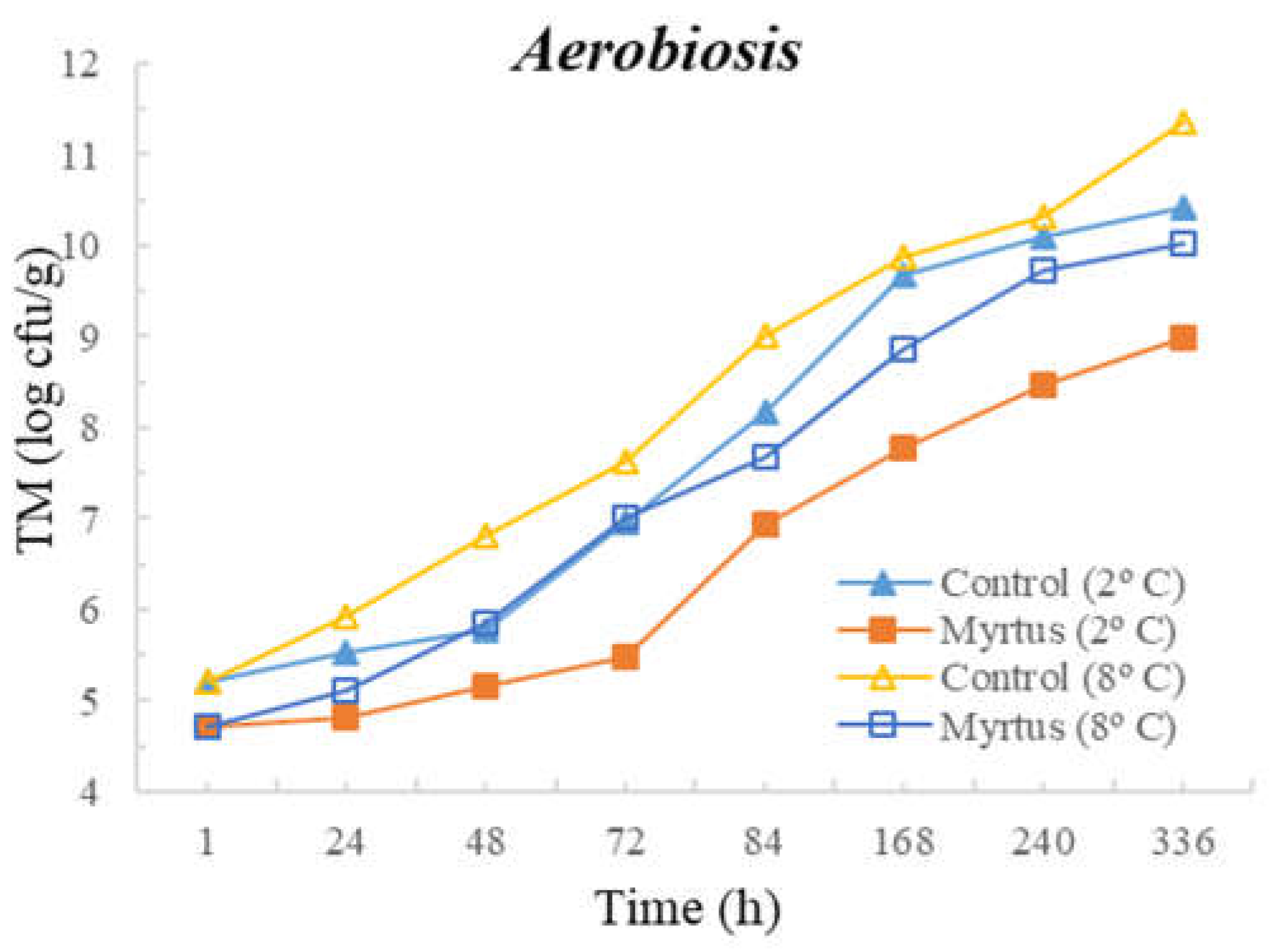
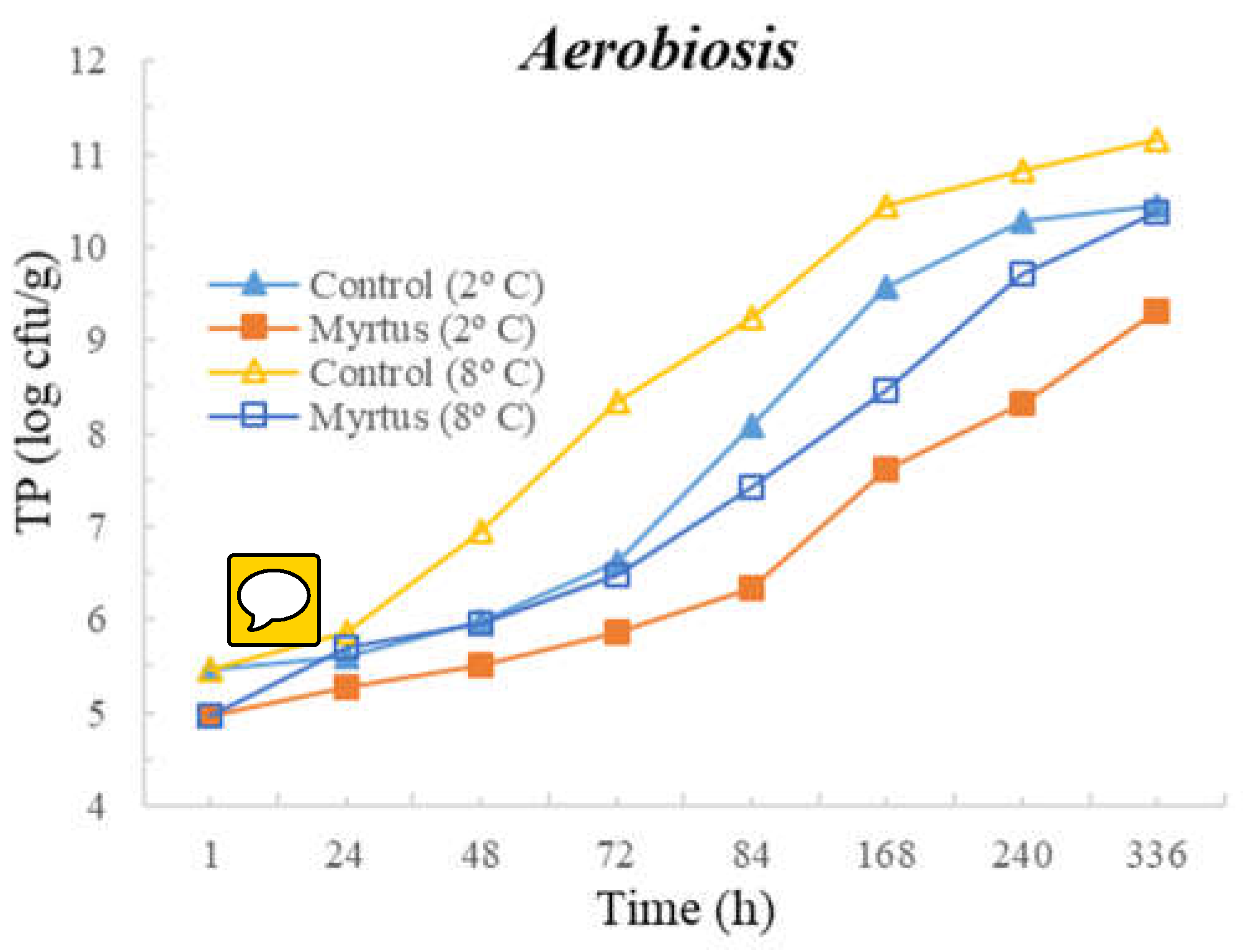
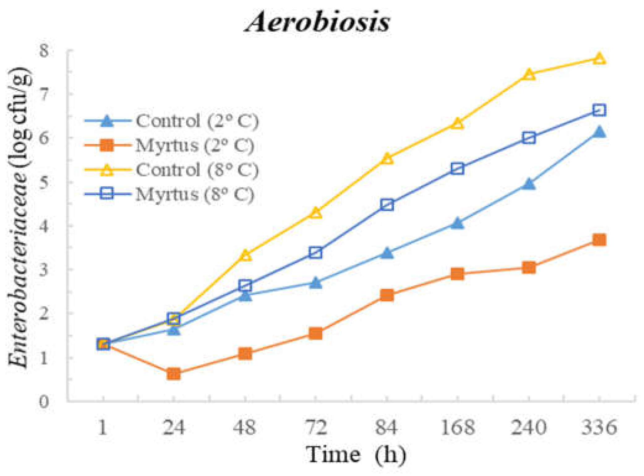
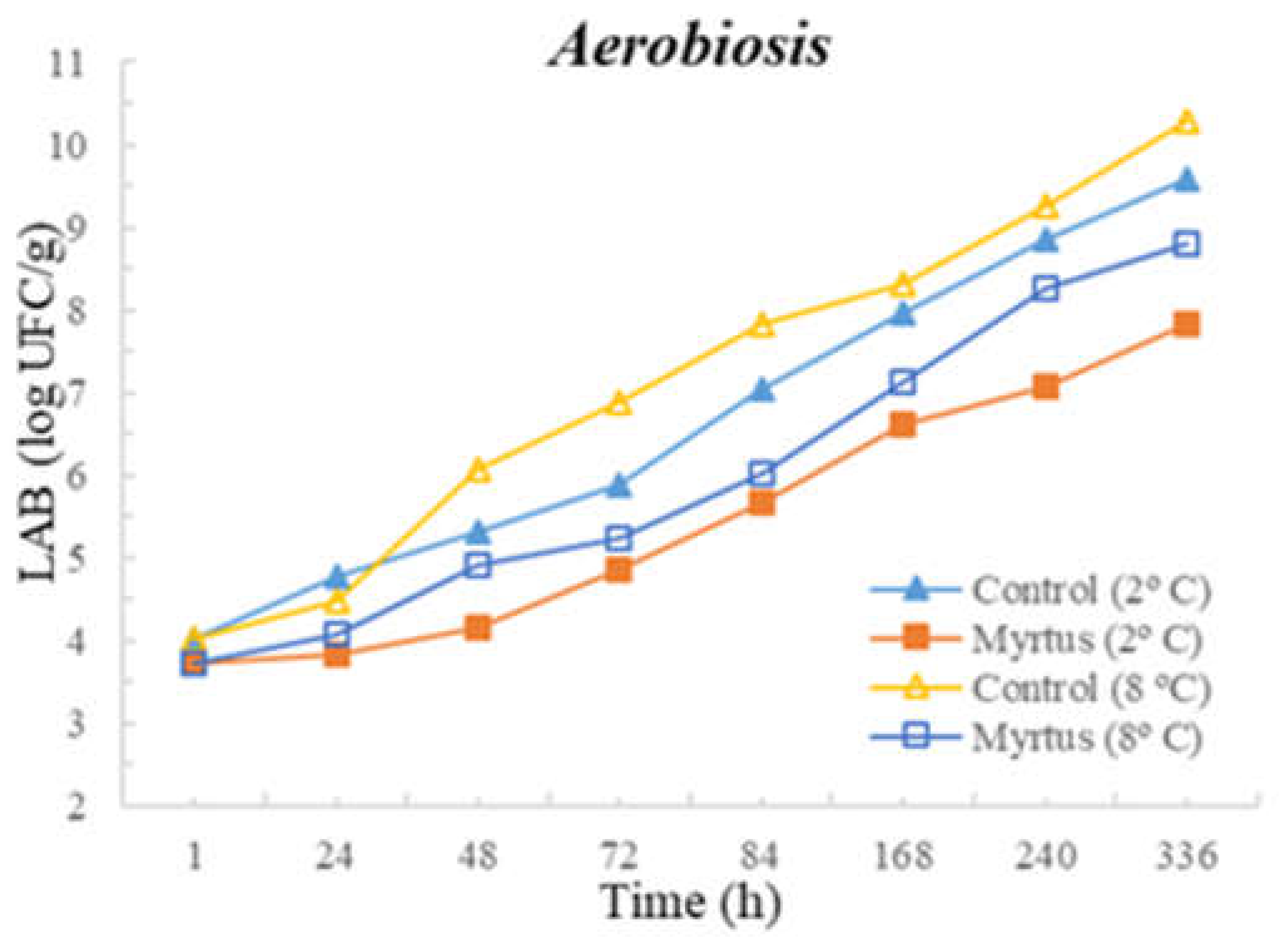
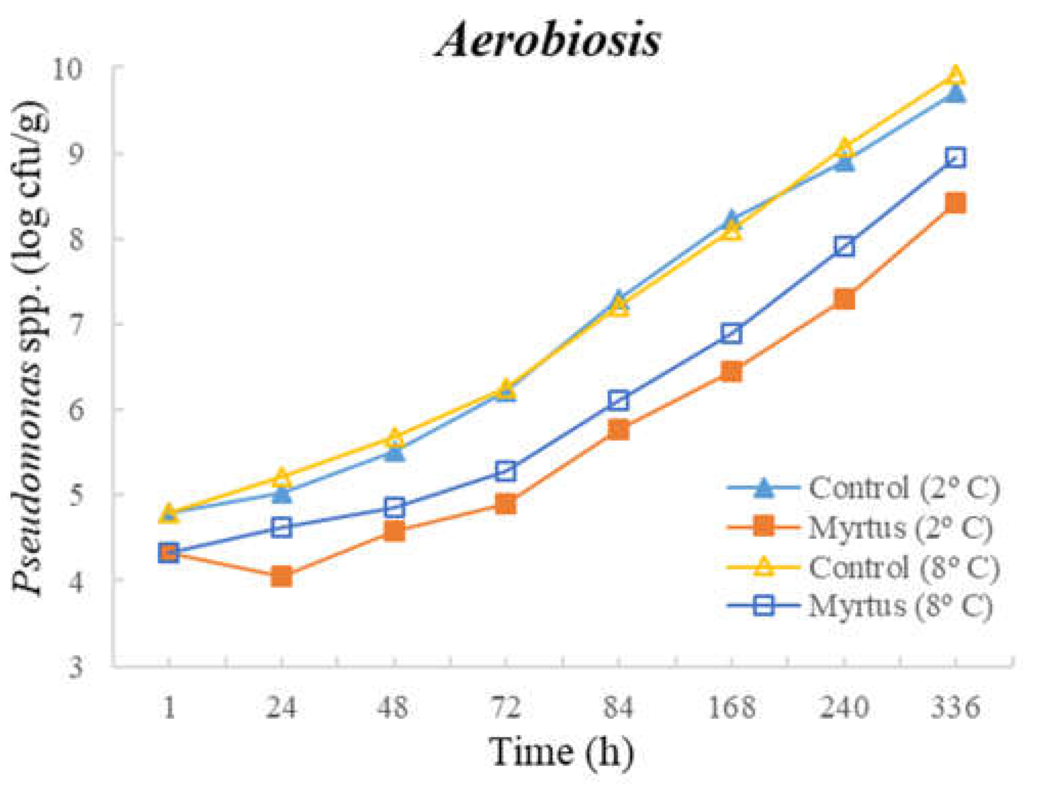
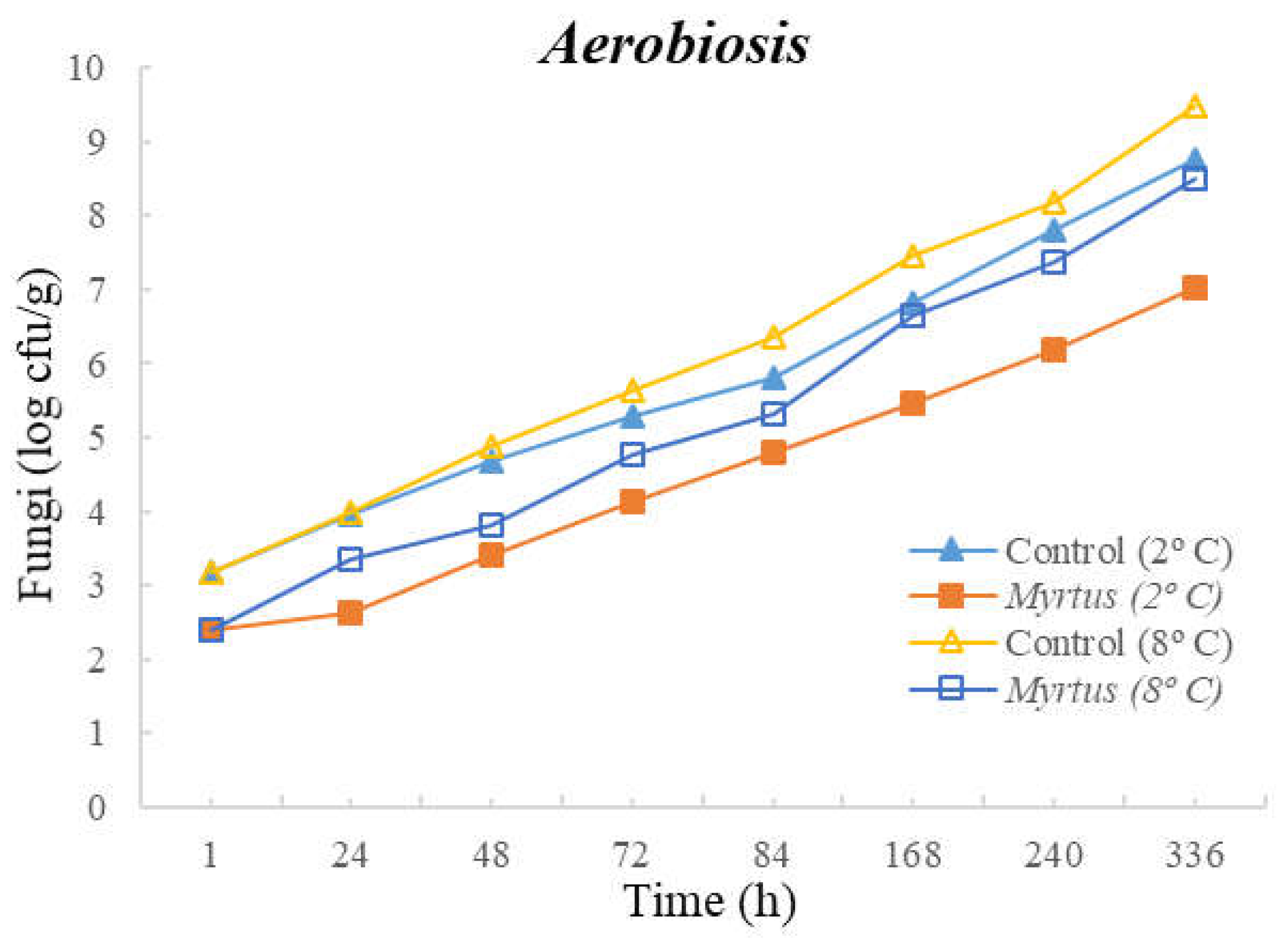
| Aerobiosis | Time (h) | TM | TP | Enterobacteriaceae | LAB | Pseudomonas spp. | Fungi | ||||||||||||
|---|---|---|---|---|---|---|---|---|---|---|---|---|---|---|---|---|---|---|---|
| Control | EO | Sig | Control | EO | Sig | Control | EO | Sig | Control | EO | Sig | Control | EO | Sig | Control | EO | Sig | ||
| T = 2 °C | 1 | 5.2 ± 0.8 a | 4.7 ± 0.8 a | NS | 5.5 ± 0.5 a | 5.0 ± 0.3 a | NS | 1.3 ± 1.2 a | 1.3 ± 1.1 ab | NS | 4.0 ± 2.0 a | 3.7 ± 1.4 a | NS | 4.8 ± 0.9 a | 4.3 ± 0.7 ab | NS | 3.2 ± 2.9 a | 2.4 ± 2.1 a | NS |
| 24 | 5.5 ± 0.5 ab | 4.8 ± 0.6 a | NS | 5.6 ± 0.4 a | 5.3 ± 0.1 ab | NS | 1.6 ± 1.5 a | 0.6 ± 1.1 a | NS | 4.8 ± 1.5 ab | 3.8 ± 1.8 ab | NS | 5.0 ± 0.6 a | 4.0 ± 0.4 a | NS | 4.0 ± 1.7 ab | 2.6 ± 0.8 a | NS | |
| 48 | 5.5 ± 0.3 ab | 5.0 ± 0.2 a | NS | 5.7 ± 0.0 a | 5.5 ± 0.0 ab | NS | 2.3 ± 0.1 ab | 1.2 ± 1.2 ab | NS | 5.3 ± 0.9 abA | 4.1 ± 0.0 ab | * | 5.5 ± 0.1 a | 4.6 ± 0.2 ab | ** | 4.7 ± 0.5 ab | 3.4 ± 0.3 ab | * | |
| 72 | 7.0 ± 0.8 bc | 5.5 ± 0.5 ab | * | 6.6 ± 0.5 ab | 5.9 ± 0.1 bc | NS | 2.7 ± 0.4 abc | 1.5 ± 1.4 ab | NS | 5.9 ± 1.4 abc | 4.9 ± 1.0 abc | NS | 6.2 ± 0.8 ab | 4.9 ± 0.4 ab | ** | 5.3 ± 0.6 abc | 4.1 ± 0.2 ab | * | |
| 84 | 8.2 ± 0.3 cd | 6.9 ± 0.5 bc | * | 8.1 ± 0.9 b | 6.3 ± 0.2 c | * | 3.4 ± 0.2 abc | 2.4 ± 0.6 ab | NS | 7.0 ± 1.0 abcd | 5.7 ± 0.6 abc | NS | 7.3 ± 0.4 bc | 5.8 ± 0.4 bc | * | 5.8 ± 0.5 abc | 4.8 ± 0.9 ab | NS | |
| 168 | 9.7 ± 0.6 de | 7.8 ± 0.2 cd | ** | 9.6 ± 0.8 c | 7.6 ± 0.5 d | * | 4.1 ± 0.2 cde | 2.9 ± 0.8 ab | NS | 8.0 ± 0.8 bcd | 6.6 ± 0.5 abc | NS | 8.2 ± 0.7 c | 6.4 ± 0.4 c | * | 6.8 ± 0.5 abc | 5.5 ± 1.1 ab | NS | |
| 240 | 10.1 ± 0.3 e | 8.5 ± 0.4 d | ** | 10.3 ± 0.2 c | 8.3 ± 0.2 d | *** | 5.0 ± 0.3 deA | 3.1 ± 1.0 abB | * | 8.9 ± 0.2 cd | 7.1 ± 0.5bc | NS | 8.9 ± 0.5 cd | 7.3 ± 0.6 cd | ** | 7.8 ± 0.5 bc | 6.2 ± 1.3 ab | NS | |
| 336 | 10.4 ± 0.5 e | 9.0 ± 0.0 d | ** | 10.5 ± 0.3 c | 9.3 ± 0.1 e | ** | 6.2 ± 0.8 eA | 3.7 ± 0.6 bB | * | 9.6 ± 0.3 d | 7.8 ± 1.0 c | NS | 9.7 ± 0.4 d | 8.4 ± 0.4 d | * | 8.8 ± 0.2 c | 7.0 ± 1.5 b | NS | |
| Sig | *** | *** | *** | *** | *** | * | *** | *** | *** | *** | ** | ** | |||||||
| Aerobiosis | Time (h) | TM | TP | Enterobacteriaceae | LAB | Pseudomonas spp. | Fungi | ||||||||||||
|---|---|---|---|---|---|---|---|---|---|---|---|---|---|---|---|---|---|---|---|
| Control | EO | Sig | Control | EO | Sig | Control | EO | Sig | Control | EO | Sig | Control | EO | Sig | Control | EO | Sig | ||
| 1 | 5.2 ± 0.8 a | 4.7 ± 0.8 a | NS | 5.5 ± 0.5 a | 5.0 ± 0.3 a | NS | 1.3 ± 1.2 ab | 1.0 ± 0.9 a | NS | 4.0 ± 2.0 ab | 3.7 ± 1.4 a | NS | 4.8 ± 0.9 ab | 4.3 ± 0.6 a | NS | 3.2 ± 2.9 ab | 2.4 ± 2.1 a | NS | |
| T = 8 °C | 24 | 5.9 ± 0.5 ab | 5.1 ± 0.4 a | NS | 5.9 ± 0.7 ab | 5.7 ± 0.2 abc | NS | 1.9 ± 1.6 abc | 1.9 ± 0.3 ab | NS | 4.5 ± 2.1 cb | 4.1 ± 1.4 ab | NS | 5.2 ± 0.7 ab | 4.6 ± 0.7 a | NS | 4.0 ± 1.7 abc | 3.4 ± 0.9 ab | NS |
| 48 | 6.8 ± 0.1 b | 5.7 ± 0.3 a | NS | 6.6 ± 0.2 b | 5.9 ± 0.0 bc | * | 3.4 ± 0.1 cde | 2.7 ± 0.4 ab | NS | 6.1 ± 0.2 abc | 4.9 ± 0.0 ab | * | 5.6 ± 0.2 ab | 4.9 ± 0.7 a | ** | 4.4 ± 0.2 abcd | 3.5 ± 0.6 ab | * | |
| 72 | 7.6 ± 1.2 bc | 7.0 ± 0.4 b | NS | 8.3 ± 0.3 c | 6.5 ± 0.1 c | * | 4.3 ± 0.5 def | 3.4 ± 0.6 bc | NS | 6.9 ± 0.8 bcd | 5.2 ± 0.9 abc | NS | 6.3 ± 0.2 bc | 5.3 ± 0.8 ab | * | 5.6 ± 0.6 abcd | 4.7 ± 0.8 ab | * | |
| 84 | 9.0 ± 0.7 cd | 7.7 ± 0.3 bc | * | 9.3 ± 0.3 c | 7.4 ± 0.3 d | * | 5.5 ± 0.9 efg | 4.5 ± 0.4 cd | NS | 7.8 ± 0.3 cde | 6.0 ± 0.6 abcd | NS | 7.2 ± 0.7 cd | 6.1 ± 0.5 abc | ** | 6.4 ± 0.4 bcde | 5.3 ± 0.7 abcd | NS | |
| 168 | 9.9 ± 0.4 de | 8.9 ± 0.2 cd | ** | 10.5 ± 0.1 d | 8.5 ± 0.4 e | * | 6.4 ± 0.5 fg | 5.3 ± 0.3 de | NS | 8.3 ± 0.6 cde | 7.1 ± 0.7 bcd | NS | 8.1 ± 0.7 de | 6.9 ± 0.6 bc | * | 7.5 ± 0.2 cde | 6.7 ± 0.2 bcd | NS | |
| 240 | 10.3 ± 0.3 de | 9.7 ± 0.2 d | ** | 10.8 ± 0.2 d | 9.7 ± 0.2 f | *** | 7.5 ± 0.8 g | 6.0 ± 0.4 de | * | 9.3 ± 0.6 de | 8.3 ± 0.3 cd | ** | 9.1 ± 0.6 cf | 7.9 ± 0.7 cd | * | 8.2 ± 0.6 de | 7.4 ± 0.2 cd | NS | |
| 336 | 11.4 ± 0.9 e | 10.0 ± 0.0 d | ** | 11.2 ± 0.3 d | 10.4 ± 0.3 f | ** | 7.8 ± 0.7 g | 6.6 ± 0.1 e | * | 10.3 ± 0.3 e | 8.8 ± 0.5 d | * | 9.9 ± 0.3 f | 8.9 ± 0.4 d | * | 9.5 ± 0.6 e | 8.5 ± 0.3 d | NS | |
| Sig | *** | *** | *** | *** | *** | *** | *** | *** | *** | *** | *** | ** | |||||||
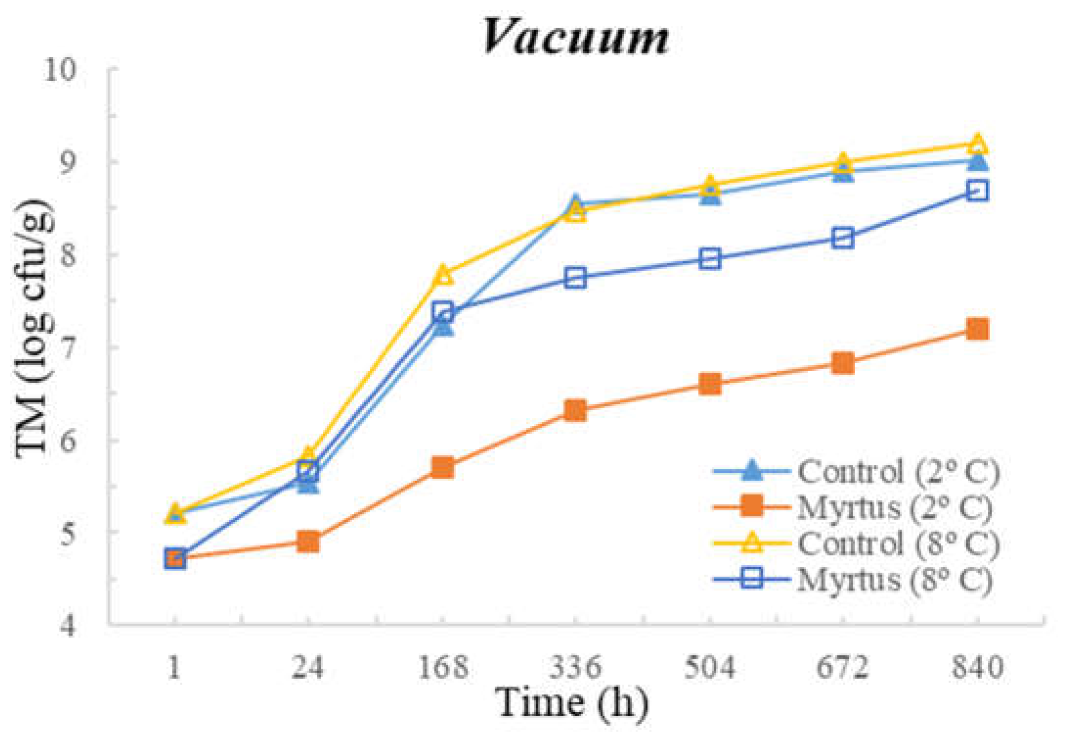
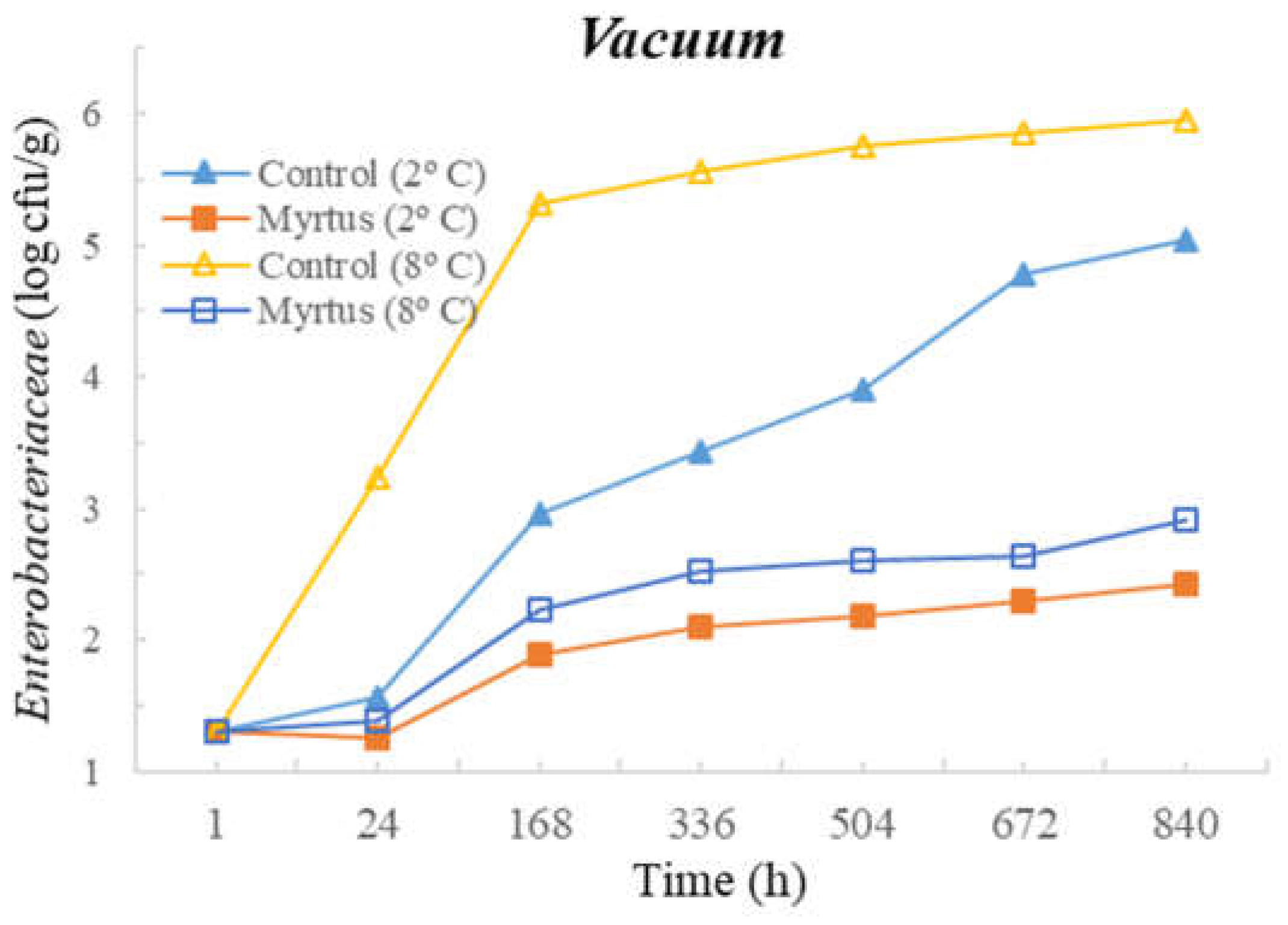
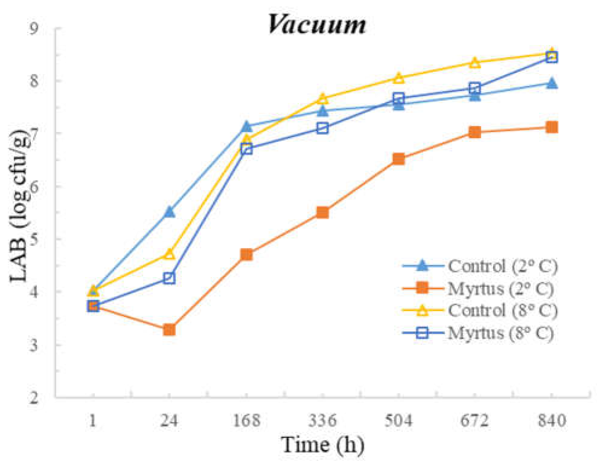
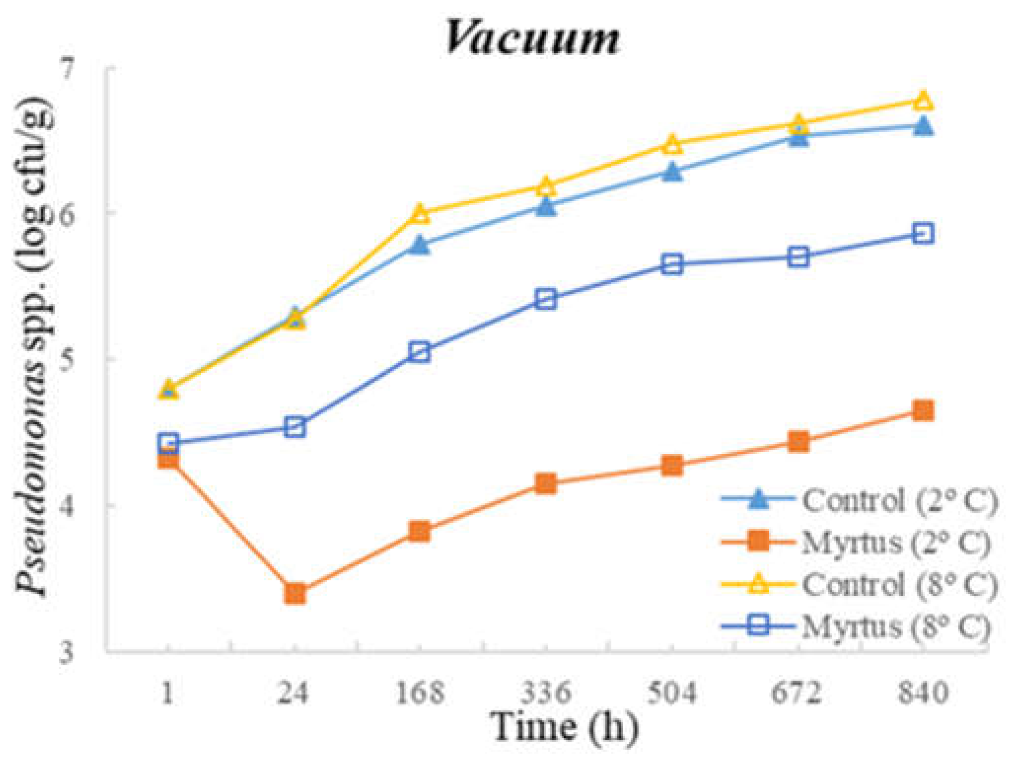
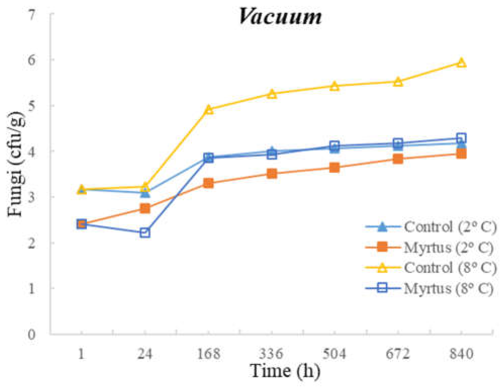
| Vacuum | Time (h) | TM | TP | Enterobacteriaceae | LAB | Pseudomonas spp. | Fungi | ||||||||||||
|---|---|---|---|---|---|---|---|---|---|---|---|---|---|---|---|---|---|---|---|
| Control | EO | Sig | Control | EO | Sig | Control | EO | Sig | Control | EO | Sig | Control | EO | Sig | Control | EO | Sig | ||
| T = 2 °C | 1 | 5.2 ± 0.8 a | 4.7 ± 0.8 a | NS | 5.5 ± 0.5 a | 45.0 ± 0.3 a | NS | 1.3 ± 1.2 a | 1.3 ± 1.2 a | NS | 4.0 ± 2.0 a | 3.7 ± 1.4 ab | NS | 4.8 ± 0.9 a | 4.3 ± 0.7 | NS | 3.2 ± 2.9 a | 2.4 ± 2.1 a | NS |
| 24 | 5.5 ± 1.0 ab | 4.9 ± 0.8 ab | * | 5.7 ± 0.5 b | 5.2 ± 1.3 ab | NS | 1.6 ± 1.4 a | 0.7 ± 1.2 a | NS | 5.5 ± 1.0 ab | 3.3 ± 1.6 a | NS | 5.3 ± 0.4 ab | 3.4 ± 0.3 | NS | 3.1 ± 0.9 a | 2.8 ± 0.5 a | NS | |
| 168 | 7.2 ± 1.0 bc | 5.7 ± 0.2 abcd | *** | 7.3 ± 0.3 bc | 5.8 ± 1.1 abc | NS | 3.0 ± 0.0 ab | 1.9 ± 1.7 a | NS | 7.2 ± 0.0 ab | 4.7 ± 0.7 abcd | ** | 5.8 ± 0.4 ab | 3.8 ± 0.5 | ** | 3.9 ± 0.3 a | 3.3 ± 0.2 a | NS | |
| 336 | 8.6 ± 0.1 c | 6.3 ± 0.2 bcde | *** | 8.1 ± 0.3 bc | 6.7 ± 0.8 abc | * | 3.4 ± 0.2 ab | 2.1 ± 1.9 a | NS | 7.4 ± 0.1 ab | 5.5 ± 0.4 abcd | *** | 6.1 ± 0.2 ab | 4.2 ± 0.5 | ** | 4.0 ± 0.3 a | 3.5 ± 0.2 a | NS | |
| 504 | 8,7 ± 0.0 c | 6.6 ± 0.1 cde | *** | 8.4 ± 0.2 bc | 6.8 ± 0.8 abc | ** | 3.9 ± 0.1 b | 1.2 ± 2.1 a | NS | 7.6 ± 0.1 ab | 6.5 ± 0.1 bcd | ** | 6.3 ± 0.2 b | 4.3 ± 0.5 | ** | 4.1 ± 0.4 a | 3.7 ± 0.2 a | NS | |
| 672 | 8.9 ± 0.0 c | 6.8 ± 0.2 de | *** | 8.5 ± 0.0 bc | 7.4 ± 0.4 bc | ** | 4.8 ± 0.2 b | 1.3 ± 2.3 a | NS | 7.7 ± 0.1 ab | 7.0 ± 0.1 cd | ** | 6.5 ± 0.3 b | 4.4 ± 0.5 | ** | 4.1 ± 0.3 a | 3.8 ± 0.2 a | NS | |
| 840 | 9.0 ± 0.0 c | 7.2 ± 0.2 e | *** | 8.7 ± 0.0 c | 7.6 ± 0.2 c | ** | 5.1 ± 0.1 b | 1.6 ± 2.8 a | NS | 8.0 ± 0.1 b | 7.1 ± 0.0 d | *** | 6.6 ± 0.2 b | 4.7 ± 0.3 | *** | 4.2 ± 0.3 a | 4.0 ± 0.1 a | NS | |
| Sig | *** | *** | *** | ** | *** | NS | ** | * | * | NS | NS | NS | |||||||
| Vacuum | Time (h) | TM | TP | Enterobacteriaceae | LAB | Pseudomonas spp. | Fungi | ||||||||||||
|---|---|---|---|---|---|---|---|---|---|---|---|---|---|---|---|---|---|---|---|
| Control | EO | Sig | Control | EO | Sig | Control | EO | Sig | Control | EO | Sig | Control | EO | Sig | Control | EO | Sig | ||
| T = 8 °C | 1 | 5.2 ± 0.8 a | 4.7 ± 0.8 a | NS | 5.5 ± 0.5 a | 5.0 ± 0.3 a | NS | 1.3 ± 1.2 a | 1.3 ± 1.1 a | NS | 4.0 ± 2.0 a | 3.7 ± 1.4 a | NS | 4.8 ± 0.9 a | 4.4 ± 0.6 a | NS | 3.2 ± 2.9 a | 2.4 ± 2.1 a | NS |
| 24 | 5.8 ± 0.6 a | 5.7 ± 1.1 ab | NS | 5.8 ± 0.2 a | 5.7 ± 1.1 a | NS | 3.2 ± 0.7 b | 1.7 ± 1.9 a | NS | 4.7 ± 1.4 ab | 4.3 ± 1.9 ab | NS | 5.3 ± 0.4 ab | 4.5 ± 0.6 ab | ** | 3.2 ± 2.9 a | 2.2 ± 2.1 a | NS | |
| 168 | 7.8 ± 0.6 b | 7.4 ± 0.8 bc | NS | 8.2 ± 0.5 b | 7.6 ± 0.3 b | NS | 5.3 ± 0.0 c | 2.6 ± 2.3 a | NS | 6.9 ± 0.5 abc | 6.7 ± 0.7 abc | NS | 6.0 ± 0.1 abc | 5.1 ± 0.3 abc | ** | 4.9 ± 0.7 a | 3.9 ± 0.3 a | * | |
| 336 | 8.5 ± 0.5 b | 7.8 ± 0.2 c | NS | 8.7 ± 0.2 bcA | 7.9 ± 0.2 b | ** | 5.6 ± 0.1 c | 2.9 ± 2.6 a | NS | 7.7 ± 0.2 bc | 7.1 ± 0.5 bc | NS | 6.2 ± 0.0 bc | 5.4 ± 0.2 abc | ** | 5.3 ± 0.5 a | 3.9 ± 0.2 a | * | |
| 504 | 8.8 ± 0.3b | 8.0 ± 0.5 c | NS | 8.9 ± 0.0 bcA | 8.1 ± 0.4 b | * | 5.8 ± 0.1 c | 1.7 ± 2.9 a | NS | 8.1 ± 0.2 c | 7.7 ± 0.5 c | NS | 6.5 ± 0.0 bc | 5.7 ± 0.1 abc | *** | 5.4 ± 0.4 a | 4.1 ± 0.4 a | * | |
| 672 | 9.0 ± 0.3 b | 8.2 ± 0.6 c | NS | 9.0 ± 0.1 bc | 8.2 ± 0.4 b | NS | 5.9 ± 0.0 c | 1.7 ± 3.0 a | NS | 8.4 ± 0.1 c | 7.9 ± 0.7 c | NS | 6.6 ± 0.1 bc | 5.7 ± 0.2 bc | *** | 5.5 ± 0.4 a | 4.2 ± 0.4 a | * | |
| 840 | 9.2 ± 0.3 b | 8.7 ± 0.7 c | NS | 9.2 ± 0.2 c | 8.8 ± 0.4 b | NS | 6.0 ± 0.0 c | 2.6 ± 2.7 a | NS | 8.5 ± 0.1 c | 8.5 ± 0.6 c | NS | 6.8 ± 0.0 cA | 5.9 ± 0.1 c | *** | 5.9 ± 0.0 a | 4.3 ± 0.4 a | ** | |
| Sig | *** | *** | *** | *** | *** | NS | *** | *** | *** | ** | NS | NS | |||||||
Disclaimer/Publisher’s Note: The statements, opinions and data contained in all publications are solely those of the individual author(s) and contributor(s) and not of MDPI and/or the editor(s). MDPI and/or the editor(s) disclaim responsibility for any injury to people or property resulting from any ideas, methods, instructions or products referred to in the content. |
© 2023 by the authors. Licensee MDPI, Basel, Switzerland. This article is an open access article distributed under the terms and conditions of the Creative Commons Attribution (CC BY) license (https://creativecommons.org/licenses/by/4.0/).
Share and Cite
Moura, D.; Vilela, J.; Saraiva, S.; Monteiro-Silva, F.; De Almeida, J.M.M.M.; Saraiva, C. Antimicrobial Effects and Antioxidant Activity of Myrtus communis L. Essential Oil in Beef Stored under Different Packaging Conditions. Foods 2023, 12, 3390. https://doi.org/10.3390/foods12183390
Moura D, Vilela J, Saraiva S, Monteiro-Silva F, De Almeida JMMM, Saraiva C. Antimicrobial Effects and Antioxidant Activity of Myrtus communis L. Essential Oil in Beef Stored under Different Packaging Conditions. Foods. 2023; 12(18):3390. https://doi.org/10.3390/foods12183390
Chicago/Turabian StyleMoura, Dirce, Joana Vilela, Sónia Saraiva, Filipe Monteiro-Silva, José M. M. M. De Almeida, and Cristina Saraiva. 2023. "Antimicrobial Effects and Antioxidant Activity of Myrtus communis L. Essential Oil in Beef Stored under Different Packaging Conditions" Foods 12, no. 18: 3390. https://doi.org/10.3390/foods12183390