
Recent Advances in Electrochemical Enzyme-Based Biosensors for Food and Beverage Analysis

Abstract
:1. Introduction
2. The Electrochemical Biosensor
3. Enzyme Immobilisation

4. Enzyme-Based Biosensors for Food Analysis
4.1. Saccharides
4.1.1. Glucose
4.1.2. Other Saccharides
4.2. Alcohol Beverages—Ethanol and Antioxidants
4.3. Organic Acids

| Analyte | Electrode | Enzyme | Transducer | Sensitivity | Detection Range | LOD | Food Matrices | Ref. |
|---|---|---|---|---|---|---|---|---|
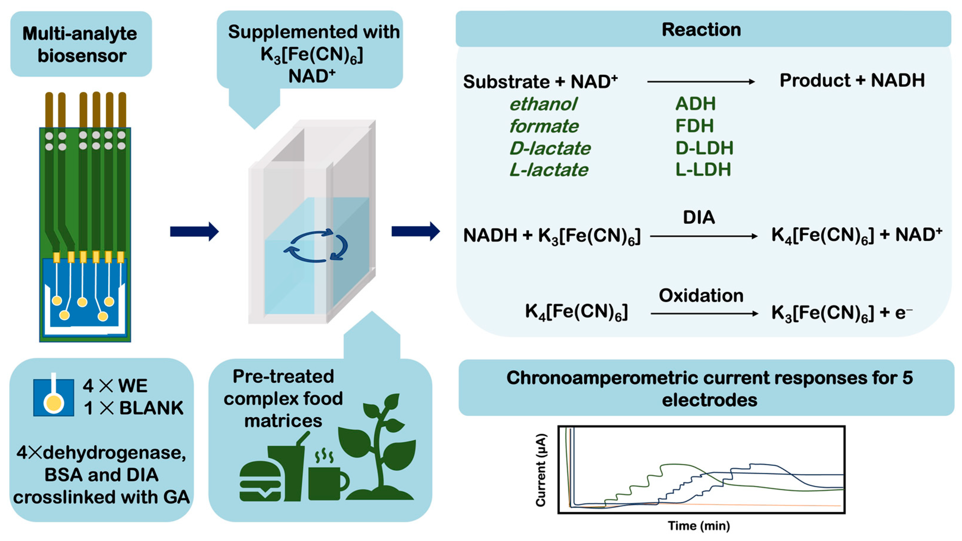
| L-Lactate | Pt/Ti/GA/BSA/ Glycerol | L-LDH, DIA | Amp | 37.2 μA mM−1 cm−2 | n.d | 0.7 μM | Maize and sugarcane silage | [84] |
| D-lactate | D-LDH, DIA | 28.4 μA mM−1 cm−2 | 0.7 μM | |||||
| Formate | FDH, DIA | 20.5 μA mM−1 cm−2 | 1.3 μM | |||||
| Acetate | Pt/GA/BSA/ Glycerol | AK, PK, PyOx | Amp | 0.27 µA mM−1 | 0–1.4 mM | n.d | Food waste | [86] |
| Propionate | PCT, SCAOx | 2.11 µA mM−1 | 0–1.5 mM | |||||
| L-Lactate | Cu-MOF/CS/Pt/SPCE | LOx | Amp | 14.65 µA mM−1; under inhibition 0.207 µA mM−1 | 0.00075–1.0 mM; under inhibition 4.0–50 mM | 0.75 μM | Red and white wines | [88] |
| L-Lactate | Pt/rGO/CNT/Au | LOx | Amp | 35.3 μA mM−1 cm−2 | 0.05–100 mM | 2.3 μM | Cow milk | [89] |
| L-Lactate | Pt/OPD/resorcinol/GA/BSA | LOx | Amp | n.d | 0.05–4.5 mM | 0.03 mM | Red and white wines | [87] |
| Malic, tartaric acids | SOx, FUM | |||||||
| L-Lactate | Pt/Pd/BSA/GA/ Dextran/Lactitol/Glycerol | LOx | Amp | 3.03 μA mM−1 cm−2 | 0.05–0.8 mM | 0.1 µM | Red and white wines | [90] |
| L-Lactate | GA/AuNPs-ERGO-PAH/SPE | L-LDH | Amp | I range: 1.08 μA mM−1cm−2; II range: 0.28 μA mM−1cm−2 | I range: 0.5–3 mM; II range: 4–16 mM | 1 µM | Yoghurt and wine | [79] |
| L-Lactate | Aluminium coated cellulose | LOx | Amp | 10.04 μA mM−1 cm−2 | 0.125–2 M | 0.23 M | Cow milk | [91] |
| L-Lactate | CF-H/PtMPs | LOx | Amp | 5233 A M−1m−2 | 0.005 mM–0.14 mM | 2 μM | Red wine | [80] |
| L-Lactate | PtZn/GE + PMS | Fcb2 | Amp | 1436 A M−1m−2 | 0.01–0.12 mM | 0.01 mM | Yoghurt | [81] |
| L-Lactate | Pt/Nafion | LOx | Amp | 0.4 µA mM−1 cm² | 50–350 µM | 31 µM | Lactic acid bacteria metabolites | [82] |
| Pyruvate | Cu-NF/tG/Au | HRP | Amp | 67.6 μA mM−1 cm−2 | 0.1–8.2 mM | 0.06 mM | Yeast metabolites | [20] |
| Pyruvate | GQD/PB/SPCE | POx | Amp | 40.8 μA mM−1 cm−2 | 10–100 μM | 0.91 μM | Fish serum samples | [83] |
4.4. Amino Acids, Biogenic Amines, and Purine Derivatives
4.5. Chemical Contaminants
4.5.1. Pesticides
4.5.2. Bisphenol A (BPA)
4.5.3. Formaldehyde
5. Improvement Strategies
5.1. Enzyme Engineering
5.2. Nanomaterials
5.3. Polymers
6. Conclusions—Challenges and Outlooks
Author Contributions
Funding
Data Availability Statement
Conflicts of Interest
Abbreviations
References
- Cifuentes, A. Food Analysis: Present, Future, and Foodomics. ISRN Anal. Chem. 2012, 2012, 801607. [Google Scholar] [CrossRef]
- Heineman, W.R.; Jensen, W.B. Leland C. Clark Jr. (1918–2005). Biosens. Bioelectron. 2006, 21, 1403–1404. [Google Scholar] [CrossRef]
- Naresh, V.; Lee, N. A Review on Biosensors and Recent Development of Nanostructured Materials-Enabled Biosensors. Sensors 2021, 21, 1109. [Google Scholar] [CrossRef] [PubMed]
- Alhamoud, Y.; Yang, D.; Fiati Kenston, S.S.; Liu, G.; Liu, L.; Zhou, H.; Ahmed, F.; Zhao, J. Advances in Biosensors for the Detection of Ochratoxin A: Bio-Receptors, Nanomaterials, and Their Applications. Biosens. Bioelectron. 2019, 141, 111418. [Google Scholar] [CrossRef] [PubMed]
- Castillo, J.; Gáspár, S.; Leth, S.; Niculescu, M.; Mortari, A.; Bontidean, I.; Soukharev, V.; Dorneanu, S.A.; Ryabov, A.D.; Csöregi, E. Biosensors for Life Quality—Design, Development and Applications. Sens. Actuators B Chem. 2004, 102, 179–194. [Google Scholar] [CrossRef]
- Polat, E.O.; Cetin, M.M.; Tabak, A.F.; Güven, E.B.; Uysal, B.Ö.; Arsan, T.; Kabbani, A.; Hamed, H.; Gül, S.B. Transducer Technologies for Biosensors and Their Wearable Applications. Biosensors 2022, 12, 385. [Google Scholar] [CrossRef]
- Thévenot, D.R.; Toth, K.; Durst, R.A.; Wilson, G.S. Electrochemical Biosensors: Recommended Definitions and Classification. Anal. Lett. 2001, 34, 635–659. [Google Scholar] [CrossRef]
- Chaubey, A.; Malhotra, B.D. Mediated Biosensors. Biosens. Bioelectron. 2002, 17, 441–456. [Google Scholar] [CrossRef]
- Jaffrezic-Renault, N.; Dzyadevych, S.V. Conductometric Microbiosensors for Environmental Monitoring. Sensors 2008, 8, 2569–2588. [Google Scholar] [CrossRef]
- Justino, C.I.L.; Freitas, A.C.; Pereira, R.; Duarte, A.C.; Rocha Santos, T.A.P. Recent Developments in Recognition Elements for Chemical Sensors and Biosensors. Trends Anal. Chem. 2015, 68, 2–17. [Google Scholar] [CrossRef]
- Sassolas, A.; Blum, L.J.; Leca-Bouvier, B.D. Immobilization Strategies to Develop Enzymatic Biosensors. Biotechnol. Adv. 2012, 30, 489–511. [Google Scholar] [CrossRef] [PubMed]
- Jesionowski, T.; Zdarta, J.; Krajewska, B. Enzyme Immobilization by Adsorption: A Review. Adsorption 2014, 20, 801–821. [Google Scholar] [CrossRef]
- Feizabadi, M.; Soleymanpour, A.; Faridnouri, H.; Ajloo, D. Improving Stability of Biosensor Based on Covalent Immobilization of Horseradish Peroxidase by γ-Aminobutyric Acid and Application in Detection of H2O2. Int. J. Biol. Macromol. 2019, 136, 597–606. [Google Scholar] [CrossRef] [PubMed]
- Yan, X.; Tang, J.; Tanner, D.; Ulstrup, J.; Xiao, X. Direct Electrochemical Enzyme Electron Transfer on Electrodes Modified by Self-Assembled Molecular Monolayers. Catalysts 2020, 10, 1458. [Google Scholar] [CrossRef]
- Suni, I.I. Substrate Materials for Biomolecular Immobilization within Electrochemical Biosensors. Biosensors 2021, 11, 239. [Google Scholar] [CrossRef]
- Felisardo, R.J.A.; Luque, A.M.; Silva, Q.S.; Soares, C.M.F.; Fricks, A.T.; Lima, Á.S.; Cavalcanti, E.B. Biosensor of Horseradish Peroxidase Immobilized onto Self-Assembled Monolayers: Optimization of the Deposition Enzyme Concentration. J. Electroanal. Chem. 2020, 879, 114784. [Google Scholar] [CrossRef]
- Nguyen, H.H.; Kim, M. An Overview of Techniques in Enzyme Immobilization. Appl. Sci. Converg. Technol. 2017, 26, 157–163. [Google Scholar] [CrossRef]
- Smith, S.; Goodge, K.; Delaney, M.; Struzyk, A.; Tansey, N.; Frey, M. A Comprehensive Review of the Covalent Immobilization of Biomolecules onto Electrospun Nanofibers. Nanomaterials 2020, 10, 2142. [Google Scholar] [CrossRef]
- Hassan, M.E.; Yang, Q.; Xiao, Z. Covalent Immobilization of Glucoamylase Enzyme onto Chemically Activated Surface of κ-Carrageenan. Bull. Natl. Res. Cent. 2019, 43, 102. [Google Scholar] [CrossRef]
- Yang, L.; Wu, N.; Bai, R.; Chen, M.; Dong, W.; Zhou, J.; Jiang, M. A Novel Strategy for the Detection of Pyruvate in Fermentation Processes Based on Well-Distributed Enzyme-Inorganic Hybrid Nanoflowers on Thiol Graphene Modified Gold Electrodes. Electrochim. Acta 2022, 427, 140855. [Google Scholar] [CrossRef]
- German, N.; Popov, A.; Ramanavicius, A.; Ramanaviciene, A. Development and Practical Application of Glucose Biosensor Based on Dendritic Gold Nanostructures Modified by Conducting Polymers. Biosensors 2022, 12, 641. [Google Scholar] [CrossRef]
- FAO/WHO. Carbohydrates in Human Nutrition. Report of a Joint FAO/WHO Expert Consultation. FAO Food Nutr. Pap. 1998, 66, 1–140. [Google Scholar]
- Brouns, F. Saccharide Characteristics and Their Potential Health Effects in Perspective. Front. Nutr. 2020, 7, 75. [Google Scholar] [CrossRef] [PubMed]
- Weibel, M.K.; Brights, H.J. The Glucose Oxidase Mechanism Interpretation of The PH Dependence. J. Biol. Chem. 1971, 246, 2734–2744. [Google Scholar] [CrossRef] [PubMed]
- Murugan, P.; Annamalai, J.; Atchudan, R.; Govindasamy, M.; Nallaswamy, D.; Ganapathy, D.; Reshetilov, A.; Sundramoorthy, A.K. Electrochemical Sensing of Glucose Using Glucose Oxidase/PEDOT:4-Sulfocalix [4]Arene/MXene Composite Modified Electrode. Micromachines 2022, 13, 304. [Google Scholar] [CrossRef]
- Krzyczmonik, P.; Socha, E.; Skrzypek, S. Electrochemical Detection of Glucose in Beverage Samples Using Poly(3,4-Ethylenedioxythiophene)-Modified Electrodes with Immobilized Glucose Oxidase. Electrocatalysis 2018, 9, 380–387. [Google Scholar] [CrossRef]
- Tan, B.; Baycan, F. An Enzymatic Glucose Biosensor Based on a Pencil Graphite Electrode Modified with Naphthalenedimide/3,4-Ethylenedioxythiophene Conjugated Polymer and Enriched with Au Nanoparticles. ChemistrySelect 2022, 7, e202103437. [Google Scholar] [CrossRef]
- Vukojević, V.; Djurdjić, S.; Ognjanović, M.; Fabián, M.; Samphao, A.; Kalcher, K.; Stanković, D.M. Enzymatic Glucose Biosensor Based on Manganese Dioxide Nanoparticles Decorated on Graphene Nanoribbons. J. Electroanal. Chem. 2018, 823, 610–616. [Google Scholar] [CrossRef]
- Soldatkina, O.V.; Kucherenko, I.S.; Soldatkin, O.O.; Pyeshkova, V.M.; Dudchenko, O.Y.; Akata Kurç, B.; Dzyadevych, S.V. Development of Electrochemical Biosensors with Various Types of Zeolites. Appl. Nanosci. 2019, 9, 737–747. [Google Scholar] [CrossRef]
- Smutok, O.; Kavetskyy, T.; Prokopiv, T.; Serkiz, R.; Wojnarowska-Nowak, R.; Šauša, O.; Novák, I.; Berek, D.; Melman, A.; Gonchar, M. New Micro/Nanocomposite with Peroxidase-like Activity in Construction of Oxidases-Based Amperometric Biosensors for Ethanol and Glucose Analysis. Anal. Chim. Acta 2021, 1143, 201–209. [Google Scholar] [CrossRef]
- Cohen, R.; Bitton, R.E.; Herzallh, N.S.; Cohen, Y.; Yehezkeli, O. Utilization of FAD-Glucose Dehydrogenase from T. Emersonii for Amperometric Biosensing and Biofuel Cell Devices. Anal. Chem. 2021, 93, 11585–11591. [Google Scholar] [CrossRef]
- Jeon, W.-Y.; Kim, H.-H.; Choi, Y.-B. Development of a Glucose Sensor Based on Glucose Dehydrogenase Using Polydopamine-Functionalized Nanotubes. Membranes 2021, 11, 384. [Google Scholar] [CrossRef]
- Yoo, E.H.; Lee, S.Y. Glucose Biosensors: An Overview of Use in Clinical Practice. Sensors 2010, 10, 4558–4576. [Google Scholar] [CrossRef]
- Kurbanoglu, S.; Zafar, M.N.; Tasca, F.; Aslam, I.; Spadiut, O.; Leech, D.; Haltrich, D.; Gorton, L. Amperometric Flow Injection Analysis of Glucose and Galactose Based on Engineered Pyranose 2-Oxidases and Osmium Polymers for Biosensor Applications. Electroanalysis 2018, 30, 1496–1504. [Google Scholar] [CrossRef]
- Jayakumar, K.; Reichhart, T.M.B.; Schulz, C.; Ludwig, R.; Felice, A.K.G.; Leech, D. An Oxygen Insensitive Amperometric Glucose Biosensor Based on An Engineered Cellobiose Dehydrogenase: Direct versus Mediated Electron Transfer Responses. ChemElectroChem 2022, 9, e202200418. [Google Scholar] [CrossRef]
- Pilo, M.; Farre, R.; Lachowicz, J.I.; Masolo, E.; Panzanelli, A.; Sanna, G.; Senes, N.; Sobral, A.; Spano, N. Design of Amperometric Biosensors for the Detection of Glucose Prepared by Immobilization of Glucose Oxidase on Conducting (Poly)Thiophene Films. J. Anal. Methods Chem. 2018, 2018, 1849439. [Google Scholar] [CrossRef] [PubMed]
- Jiménez-Fiérrez, F.; González-Sánchez, M.I.; Jiménez-Pérez, R.; Iniesta, J.; Valero, E. Glucose Biosensor Based on Disposable Activated Carbon Electrodes Modified with Platinum Nanoparticles Electrodeposited on Poly(Azure a). Sensors 2020, 20, 4489. [Google Scholar] [CrossRef]
- Bin, Z.; Feng, L.; Pengyun, W.; Jiaojiao, X. Tailoring Glucose Oxidase As Versatile Biocatalyst for High-Efficiency Electrochemical Sensing of Glucose in Honey. ACS Food Sci. Technol. 2021, 1, 1805–1813. [Google Scholar] [CrossRef]
- Wu, B.; Xu, H.; Shi, Y.; Yao, Z.; Yu, J.; Zhou, H.; Li, Y.; Chen, Q.; Long, Y. Microelectrode Glucose Biosensor Based on Nanoporous Platinum/Graphene Oxide Nanostructure for Rapid Glucose Detection of Tomato and Cucumber Fruits. Food Qual. Saf. 2022, 6, fyab030. [Google Scholar] [CrossRef]
- Caballero, B.; Trugo, L.C.; Finglas, P.M. Encyclopedia of Food Sciences and Nutrition, 2nd ed.; Academic Press: Amsterdam, The Netherlands, 2003. [Google Scholar]
- Yan, X.; Ma, S.; Tang, J.; Tanner, D.; Ulstrup, J.; Xiao, X.; Zhang, J. Direct Electron Transfer of Fructose Dehydrogenase Immobilized on Thiol-Gold Electrodes. Electrochim. Acta 2021, 392, 138946. [Google Scholar] [CrossRef]
- Bollella, P.; Gorton, L.; Antiochia, R. Direct Electron Transfer of Dehydrogenases for Development of 3rd Generation Biosensors and Enzymatic Fuel Cells. Sensors 2018, 18, 1319. [Google Scholar] [CrossRef]
- Bollella, P.; Hibino, Y.; Kano, K.; Gorton, L.; Antiochia, R. Enhanced Direct Electron Transfer of Fructose Dehydrogenase Rationally Immobilized on a 2-Aminoanthracene Diazonium Cation Grafted Single-Walled Carbon Nanotube Based Electrode. ACS Catal. 2018, 8, 10279–10289. [Google Scholar] [CrossRef]
- Bagal-Kestwal, D.R.; Chiang, B.-H. Electrochemical Invertase Probes with Nanocomposite of Microfibrillated Cellulose-Tragacanth Gum-Metal Nanoparticles for Direct Sucrose Analysis in Sweetened Beverages. J. Electrochem. Soc. 2019, 166, B720–B727. [Google Scholar] [CrossRef]
- Stredansky, M.; Redivo, L.; Magdolen, P.; Stredansky, A.; Navarini, L. Rapid Sucrose Monitoring in Green Coffee Samples Using Multienzymatic Biosensor. Food Chem. 2018, 254, 8–12. [Google Scholar] [CrossRef] [PubMed]
- Pyeshkova, V.M.; Saiapina, O.Y.; Soldatkin, O.O.; Dzyadevych, S.V. Enzyme Conductometric Biosensor for Maltose Determination Biopolym. Cell 2009, 25, 272–278. [Google Scholar] [CrossRef]
- Marconi, E.; Messia, M.C.; Palleschi, G.; Cubadda, R. A Maltose Biosensor for Determining Gelatinized Starch in Processed Cereal Foods. Cereal Chem. 2004, 81, 6–9. [Google Scholar] [CrossRef]
- Zajoncová, L.; Jílek, M.; Beranová, V.; Peč, P. A Biosensor for the Determination of Amylase Activity. Biosens. Bioelectron. 2004, 20, 240–245. [Google Scholar] [CrossRef] [PubMed]
- Shafiqul Islam, A.K.M.; Shin, T.C.; Ahmad, M.N. A Sol-Gel Biosensor for the Detection of Maltose in Starch Hydrolysis. Malays. J. Chem. 2020, 22, 69–77. [Google Scholar]
- Wijayanti, S.D.; Schachinger, F.; Ludwig, R.; Haltrich, D. Electrochemical and Biosensing Properties of an FAD-Dependent Glucose Dehydrogenase from Trichoderma Virens. Bioelectrochemistry 2023, 153, 108480. [Google Scholar] [CrossRef] [PubMed]
- Gursoy, O.; Sen Gursoy, S.; Cogal, S.; Celik Cogal, G. Development of a New Two-Enzyme Biosensor Based on Poly(Pyrrole-Co-3,4-Ethylenedioxythiophene) for Lactose Determination in Milk. Polym. Eng. Sci. 2018, 58, 839–848. [Google Scholar] [CrossRef]
- Gursoy, S.S.; Yildiz, A.; Cogal, G.C.; Gursoy, O. A Novel Lactose Biosensor Based on Electrochemically Synthesized 3,4-Ethylenedioxythiophene/Thiophene (EDOT/Th) Copolymer. Open Chem. 2020, 18, 974–985. [Google Scholar] [CrossRef]
- de Brito, A.R.; de Jesus, R.S.; de Tavares, I.M.; Silva, F.N.; Santana, N.B.; Barbosa Ferrão, S.P.; Bilal, M.; de Santana Santos, A.; Salay, L.C.; de Oliveira, J.R.; et al. Application of the Electrochemical Biosensor in the Detection of Lactose in Skimmed Milk. Surf. Interfaces 2021, 22, 100839. [Google Scholar] [CrossRef]
- Hu, X.; Robin, S.; O’Connell, S.; Walsh, G.; Wall, J.G. Engineering of a Fungal β-Galactosidase to Remove Product Inhibition by Galactose. Appl. Microbiol. Biotechnol. 2010, 87, 1773–1782. [Google Scholar] [CrossRef]
- Çakıroğlu, B.; Demirci, Y.C.; Gökgöz, E.; Özacar, M. A Photoelectrochemical Glucose and Lactose Biosensor Consisting of Gold Nanoparticles, MnO2 and g-C3N4 Decorated TiO2. Sens. Actuators B Chem. 2019, 282, 282–289. [Google Scholar] [CrossRef]
- Ahlawat, J.; Aggarwal, V.; Jaiwal, R.; Pundir, C.S. An Improved Amperometric Lactose Biosensor Based on Enzyme Nanoparticles. Int. J. Appl. Sci. Biotechnol. 2022, 10, 21–30. [Google Scholar] [CrossRef]
- Stoica, L.; Ludwig, R.; Haltrich, D.; Gorton, L. Third-Generation Biosensor for Lactose Based on Newly Discovered Cellobiose Dehydrogenase. Anal. Chem. 2006, 78, 393–398. [Google Scholar] [CrossRef] [PubMed]
- Tanne, J.; Kracher, D.; Dietzel, B.; Schulz, B.; Ludwig, R.; Lisdat, F.; Scheller, F.W.; Bier, F.F. Carboxylated or Aminated Polyaniline-Multiwalled Carbon Nanotubes Nanohybrids for Immobilization of Cellobiose Dehydrogenase on Gold Electrodes. Biosensors 2014, 4, 370–386. [Google Scholar] [CrossRef] [PubMed]
- Safina, G.; Ludwig, R.; Gorton, L. A Simple and Sensitive Method for Lactose Detection Based on Direct Electron Transfer between Immobilised Cellobiose Dehydrogenase and Screen-Printed Carbon Electrodes. Electrochim. Acta 2010, 55, 7690–7695. [Google Scholar] [CrossRef]
- Bollella, P.; Hibino, Y.; Kano, K.; Gorton, L.; Antiochia, R. Highly Sensitive Membraneless Fructose Biosensor Based on Fructose Dehydrogenase Immobilized onto Aryl Thiol Modified Highly Porous Gold Electrode: Characterization and Application in Food Samples. Anal. Chem. 2018, 90, 12131–12136. [Google Scholar] [CrossRef] [PubMed]
- Suzuki, Y.; Kano, K.; Shirai, O.; Kitazumi, Y. Diffusion-Limited Electrochemical D-Fructose Sensor Based on Direct Electron Transfer-Type Bioelectrocatalysis by a Variant of D-Fructose Dehydrogenase at a Porous Gold Microelectrode. J. Electroanal. Chem. 2020, 877, 114651. [Google Scholar] [CrossRef]
- Halpin, G.; ’McEntee, S.; Dwyer, C.; Lawless, F.; Dempsey, E. Lactose Biosensor Development and Deployment in Dairy Product Analysis. J. Electrochem. Soc. 2022, 169, 037528. [Google Scholar] [CrossRef]
- Pyeshkova, V.M.; Dudchenko, O.Y.; Soldatkin, O.O.; Alekseev, S.A.; Seker, T.; Kurc, B.A.; Dzyadevych, S.V. Development of Three-Enzyme Lactose Amperometric Biosensor Modified by Nanosized Poly (Meta-Phenylenediamine) Film. Appl. Nanosci. 2022, 12, 1267–1274. [Google Scholar] [CrossRef]
- Wollan, D.; Pham, D.T.; Wilkinson, K.L. Changes in Wine Ethanol Content Due to Evaporation from Wine Glasses and Implications for Sensory Analysis. J. Agric. Food Chem. 2016, 64, 7569–7575. [Google Scholar] [CrossRef]
- Alzeer, J.; Abou Hadeed, K. Ethanol and Its Halal Status in Food Industries. Trends Food Sci. Technol. 2016, 58, 14–20. [Google Scholar] [CrossRef]
- Goswami, P.; Chinnadayyala, S.S.R.; Chakraborty, M.; Kumar, A.K.; Kakoti, A. An Overview on Alcohol Oxidases and Their Potential Applications. Appl. Microbiol. Biotechnol. 2013, 97, 4259–4275. [Google Scholar] [CrossRef] [PubMed]
- Istrate, O.M.; Rotariu, L.; Bala, C. A Novel Amperometric Biosensor Based on Poly(Allylamine Hydrochloride) for Determination of Ethanol in Beverages. Sensors 2021, 21, 6510. [Google Scholar] [CrossRef]
- Madaci, A.; Raffin, G.; Hangouet, M.; Pages, C.; Jose, C.; Martin, M.; Ferkous, H.; Bouzid, A.; Bausells, J.; Alcacer, A.; et al. A Microconductometric Ethanol Sensor Prepared through Encapsulation of Alcohol Dehydrogenase in Chitosan: Application to the Determination of Alcoholic Content in Headspace above Beverages. J. Mater. Sci. Mater. Electron. 2021, 32, 17752–17763. [Google Scholar] [CrossRef]
- Arranz, S.; Chiva-Blanch, G.; Valderas-Martínez, P.; Medina-Remón, A.; Lamuela-Raventós, R.M.; Estruch, R. Wine, Beer, Alcohol and Polyphenols on Cardiovascular Disease and Cancer. Nutrients 2012, 4, 759–781. [Google Scholar] [CrossRef]
- García-Guzmán, J.J.; López-Iglesias, D.; Cubillana-Aguilera, L.; Lete, C.; Lupu, S.; Palacios-Santander, J.M.; Bellido-Milla, D. Assessment of the Polyphenol Indices and Antioxidant Capacity for Beers and Wines Using a Tyrosinase-Based Biosensor Prepared by Sinusoidal Current Method. Sensors 2019, 19, 66. [Google Scholar] [CrossRef] [PubMed]
- Mohtar, L.G.; Aranda, P.; Messina, G.A.; Nazareno, M.A.; Pereira, S.V.; Raba, J.; Bertolino, F.A. Amperometric Biosensor Based on Laccase Immobilized onto a Nanostructured Screen-Printed Electrode for Determination of Polyphenols in Propolis. Microchem. J. 2019, 144, 13–18. [Google Scholar] [CrossRef]
- Ye, Y.; Ji, J.; Sun, Z.; Shen, P.; Sun, X. Recent Advances in Electrochemical Biosensors for Antioxidant Analysis in Foodstuff. Trends Anal. Chem. 2020, 122, 115718. [Google Scholar] [CrossRef]
- Mossé Alhadeff, E.; Jackson Telles Bosco, A.; Fragale Pastusiak, C.; Anjos Correia, T.; Isabel Bojorge Ramirez, N. Development of an Ethanol Biosensor Based on Silver Nanoparticles/Polyaniline/Graphite/Epoxy Composite for Friendly Analytical Application. In Biosensors for Environmental Monitoring; IntechOpen: London, UK, 2019. [Google Scholar]
- Wang, S.; Yao, Z.; Yang, T.; Zhang, Q.; Gao, F. Editors’ Choice—An Enzymatic Electrode Integrated with Alcohol Dehydrogenase and Chloranil in Liquid-Crystalline Cubic Phases on Carbon Nanotubes for Sensitive Amperometric Detection of NADH and Ethanol. J. Electrochem. Soc. 2019, 166, G116–G121. [Google Scholar] [CrossRef]
- Prasanna Kumar, S.; Parashuram, L.; Suhas, D.P.; Krishnaiah, P. Carboxylated Graphene-Alcohol Oxidase Thin Films Modified Graphite Electrode as an Electrochemical Sensor for Electro-Catalytic Detection of Ethanol. Mater. Sci. Energy Technol. 2020, 3, 159–166. [Google Scholar] [CrossRef]
- Đurđić, S.; Stanković, V.; Vlahović, F.; Ognjanović, M.; Kalcher, K.; Veličković, T.Ć.; Mutić, J.; Stanković, D.M. Laccase Polyphenolic Biosensor Supported on MnO2 @GNP Decorated SPCE: Preparation, Characterization, and Analytical Application. J. Electrochem. Soc. 2021, 168, 037510. [Google Scholar] [CrossRef]
- Zrinski, I.; Pungjunun, K.; Martinez, S.; Zavašnik, J.; Stanković, D.; Kalcher, K.; Mehmeti, E. Evaluation of Phenolic Antioxidant Capacity in Beverages Based on Laccase Immobilized on Screen-Printed Carbon Electrode Modified with Graphene Nanoplatelets and Gold Nanoparticles. Microchem. J. 2020, 152, 104282. [Google Scholar] [CrossRef]
- Shi, Y.; Pu, D.; Zhou, X.; Zhang, Y. Recent Progress in the Study of Taste Characteristics and the Nutrition and Health Properties of Organic Acids in Foods. Foods 2022, 11, 3408. [Google Scholar] [CrossRef]
- Istrate, O.M.; Rotariu, L.; Bala, C. Amperometric L-Lactate Biosensor Based upon a Gold Nanoparticles/Reduced Graphene Oxide/Polyallylamine Hydrochloride Modified Screen-Printed Graphite Electrode. Chemosensors 2021, 9, 74. [Google Scholar] [CrossRef]
- Smutok, O.; Kavetskyy, T.; Prokopiv, T.; Serkiz, R.; Šauša, O.; Novák, I.; Švajdlenková, H.; Maťko, I.; Gonchar, M.; Katz, E. Biosensor Based on Peroxidase-Mimetic Nanozyme and Lactate Oxidase for Accurate L-Lactate Analysis in Beverages. Biosensors 2022, 12, 1042. [Google Scholar] [CrossRef]
- Demkiv, O.; Gayda, G.; Stasyuk, N.; Moroz, A.; Serkiz, R.; Kausaite-Minkstimiene, A.; Gonchar, M.; Nisnevitch, M. Flavocytochrome B2-Mediated Electroactive Nanoparticles for Developing Amperometric L-Lactate Biosensors. Biosensors 2023, 13, 587. [Google Scholar] [CrossRef]
- Ozoglu, O.; Uzunoglu, A.; Unal, M.A.; Gumustas, M.; Ozkan, S.A.; Korukluoglu, M.; Gunes Altuntas, E. Electrochemical Detection of Lactate Produced by Foodborne Presumptive Lactic Acid Bacteria. J. Biosci. Bioeng. 2023, 135, 313–320. [Google Scholar] [CrossRef]
- Thirumalai, D.; Kim, S.; Kim, S.; Chang, S.C. Reagentless Amperometric Pyruvate Biosensor Based on a Prussian Blue- and Enzyme Nanoparticle-Modified Screen-Printed Carbon Electrode. ACS Omega 2020, 5, 30123–30129. [Google Scholar] [CrossRef]
- Pilas, J.; Yazici, Y.; Selmer, T.; Keusgen, M.; Schöning, M.J. Application of a Portable Multi-Analyte Biosensor for Organic Acid Determination in Silage. Sensors 2018, 18, 1470. [Google Scholar] [CrossRef] [PubMed]
- Pilas, J.; Yazici, Y.; Selmer, T.; Keusgen, M.; Schöning, M.J. Optimization of an Amperometric Biosensor Array for Simultaneous Measurement of Ethanol, Formate, D- and L-Lactate. Electrochim. Acta 2017, 251, 256–262. [Google Scholar] [CrossRef]
- Röhlen, D.L.; Pilas, J.; Dahmen, M.; Keusgen, M.; Selmer, T.; Schöning, M.J. Toward a Hybrid Biosensor System for Analysis of Organic and Volatile Fatty Acids in Fermentation Processes. Front. Chem. 2018, 6, 284. [Google Scholar] [CrossRef]
- Milovanovic, M.; Žeravík, J.; Obořil, M.; Pelcová, M.; Lacina, K.; Cakar, U.; Petrovic, A.; Glatz, Z.; Skládal, P. A Novel Method for Classification of Wine Based on Organic Acids. Food Chem. 2019, 284, 296–302. [Google Scholar] [CrossRef]
- Cunha-Silva, H.; Arcos-Martinez, M.J. Dual Range Lactate Oxidase-Based Screen Printed Amperometric Biosensor for Analysis of Lactate in Diversified Samples. Talanta 2018, 188, 779–787. [Google Scholar] [CrossRef]
- Hashemzadeh, S.; Omidi, Y.; Rafii-Tabar, H. Amperometric Lactate Nanobiosensor Based on Reduced Graphene Oxide, Carbon Nanotube and Gold Nanoparticle Nanocomposite. Microchim. Acta 2019, 186, 680. [Google Scholar] [CrossRef]
- Shkotova, L.; Bohush, A.; Voloshina, I.; Smutok, O.; Dzyadevych, S. Amperometric Biosensor Modified with Platinum and Palladium Nanoparticles for Detection of Lactate Concentrations in Wine. SN Appl. Sci. 2019, 1, 306. [Google Scholar] [CrossRef]
- Phamonpon, W.; Promphet, N.; Ummartyotin, S.; Rodthongkum, N. A Dual-Lactate Sensor for Milk Spoilage Based on Modified Recycled UHT Milk Carton Cellulose Surface. J. Clean. Prod. 2022, 363, 132519. [Google Scholar] [CrossRef]
- Jadán Piedra, F.; Rojas, C.; Latorre Castro, G.B.; Maldonado Alvarado, P. Selective Determination of Lysine in Mozzarella Cheese Using a Novel Potentiometric Biosensor. Food Biotechnol. 2023, 37, 41–53. [Google Scholar] [CrossRef]
- Nohwal, B.; Chaudhary, R.; Kumar, P.; Pundir, C.S. Fabrication and Application of an Amperometric Lysine Biosensor Based on Covalently Immobilized Lysine Oxidase Nanoparticles onto Au Electrode. Int. J. Biol. Macromol. 2020, 146, 907–915. [Google Scholar] [CrossRef] [PubMed]
- Tang, S.; Wang, C.; Liu, K.; Luo, B.; Dong, H.; Wang, X.; Hou, P.; Li, A. In Vivo Detection of Glutamate in Tomatoes by an Enzyme-Based Electrochemical Biosensor. ACS Omega 2022, 7, 30535–30542. [Google Scholar] [CrossRef] [PubMed]
- Mentana, A.; Nardiello, D.; Palermo, C.; Centonze, D. Accurate Glutamate Monitoring in Foodstuffs by a Sensitive and Interference-Free Glutamate Oxidase Based Disposable Amperometric Biosensor. Anal. Chim. Acta 2020, 1115, 16–22. [Google Scholar] [CrossRef] [PubMed]
- Bachrach, U. Naturally Occurring Polyamines: Interaction with Macromolecules. Curr. Protein Pept. Sci. 2005, 6, 559–566. [Google Scholar] [CrossRef] [PubMed]
- Koçoǧlu, İ.O.; Erden, P.E.; Kiliç, E. Disposable Biogenic Amine Biosensors for Histamine Determination in Fish. Anal. Methods 2020, 12, 3802–3812. [Google Scholar] [CrossRef] [PubMed]
- Kaçar, C.; Erden, P.E.; Dalkiran, B.; İnal, E.K.; Kiliç, E. Amperometric Biogenic Amine Biosensors Based on Prussian Blue, Indium Tin Oxide Nanoparticles and Diamine Oxidase– or Monoamine Oxidase–Modified Electrodes. Anal. Bioanal. Chem. 2020, 412, 1933–1946. [Google Scholar] [CrossRef] [PubMed]
- Erden, P.E.; Erdoğan, Z.Ö.; Öztürk, F.; Koçoğlu, İ.O.; Kılıç, E. Amperometric Biosensors for Tyramine Determination Based on Graphene Oxide and Polyvinylferrocene Modified Screen-Printed Electrodes. Electroanalysis 2019, 31, 2368–2378. [Google Scholar] [CrossRef]
- Trevisani, M.; Cecchini, M.; Fedrizzi, G.; Corradini, A.; Mancusi, R.; Tothill, I.E. Biosensing the Histamine Producing Potential of Bacteria in Tuna. Front. Microbiol. 2019, 10, 1844. [Google Scholar] [CrossRef] [PubMed]
- Torre, R.; Costa-Rama, E.; Lopes, P.; Nouws, H.P.A.; Delerue-Matos, C. Amperometric Enzyme Sensor for the Rapid Determination of Histamine. Anal. Methods 2019, 11, 1155–1158. [Google Scholar] [CrossRef]
- Torre, R.; Costa-Rama, E.; Nouws, H.P.A.; Delerue-Matos, C. Diamine Oxidase-Modified Screen-Printed Electrode for the Redox-Mediated Determination of Histamine. J. Anal. Sci. Technol. 2020, 11, 5. [Google Scholar] [CrossRef]
- Hidouri, S.; Errachid, A.H.; Baussels, J.; Korpan, Y.I.; Ruiz-Sanchez, O.; Baccar, Z.M. Potentiometric Sensing of Histamine Using Immobilized Enzymes on Layered Double Hydroxides. J. Food Sci. Technol. 2021, 58, 2936–2942. [Google Scholar] [CrossRef] [PubMed]
- Nontipichet, N.; Khumngern, S.; Choosang, J.; Thavarungkul, P.; Kanatharana, P.; Numnuam, A. An Enzymatic Histamine Biosensor Based on a Screen-Printed Carbon Electrode Modified with a Chitosan–Gold Nanoparticles Composite Cryogel on Prussian Blue-Coated Multi-Walled Carbon Nanotubes. Food Chem. 2021, 364, 130396. [Google Scholar] [CrossRef] [PubMed]
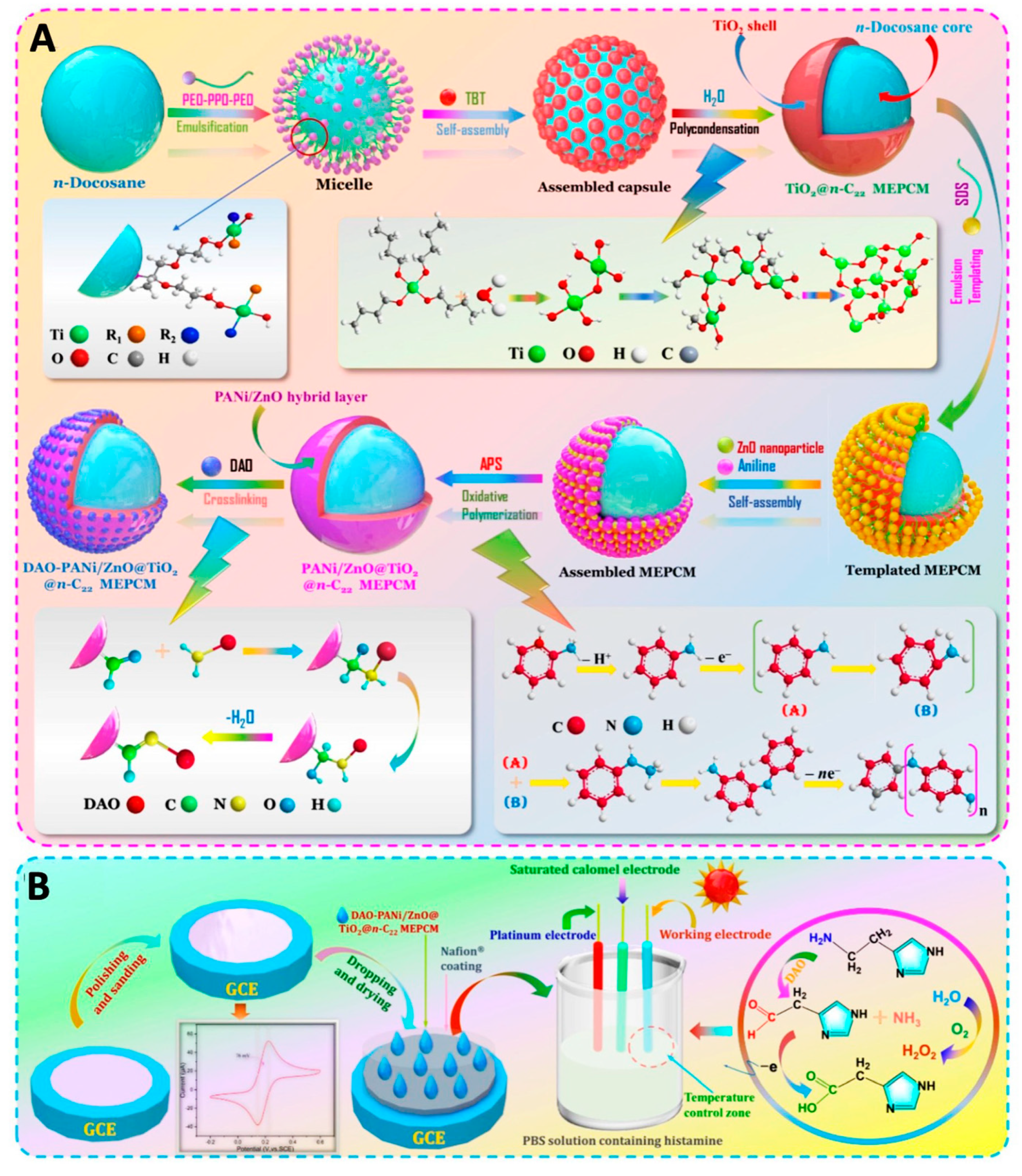
- Tian, X.; Liu, H.; Liu, H.; Wang, X. Immobilizing Diamine Oxidase on Electroactive Phase-Change Microcapsules to Construct Thermoregulatory Smart Biosensor for Enhancing Detection of Histamine in Foods. Food Chem. 2022, 397, 133759. [Google Scholar] [CrossRef] [PubMed]
- Erol, E.; Yildirim, E.; Cete, S. Construction of Biosensor for Hypoxanthine Determination by Immobilization of Xanthine Oxidase and Uricase in Polypyrrole-Paratoluenesulfonate Film. J. Solid State Electrochem. 2020, 24, 1695–1707. [Google Scholar] [CrossRef]
- Boluda, A.; Casado, C.M.; Alonso, B.; García Armada, M.P. Efficient Oxidase Biosensors Based on Bioelectrocatalytic Surfaces of Electrodeposited Ferrocenyl Polyclosiloxanes—Pt Nanoparticles. Chemosensors 2021, 9, 81. [Google Scholar] [CrossRef]
- Sharma, N.K.; Monika; Kaushal, A.; Thakur, S.; Thakur, N.; Sheetal; Kumar, D.; Bhalla, T.C. Nanohybrid Electrochemical Enzyme Sensor for Xanthine Determination in Fish Samples. 3 Biotech 2021, 11, 212. [Google Scholar] [CrossRef]
- Erden, P.E.; Kaçar Selvi, C.; Kılıç, E. A Novel Tyramine Biosensor Based on Carbon Nanofibers, 1-Butyl-3-Methylimidazolium Tetrafluoroborate and Gold Nanoparticles. Microchem. J. 2021, 170, 106729. [Google Scholar] [CrossRef]
- Lionetto, M.G.; Caricato, R.; Calisi, A.; Giordano, M.E.; Schettino, T. Acetylcholinesterase as a Biomarker in Environmental and Occupational Medicine: New Insights and Future Perspectives. BioMed Res. Int. 2013, 2013, 321213. [Google Scholar] [CrossRef] [PubMed]
- Jiang, B.; Lu, M.; Xu, M. Amperometric Sensing of Organophosphorus Pesticides Based on Covalently Attached Multilayer Assemblies of Diazo-Resin, Prussian Blue Single-Walled Carbon Nanotubes, and Acetylcholinesterase. Rev. Roum. Chim. 2019, 64, 763–774. [Google Scholar] [CrossRef]
- Palanivelu, J.; Chidambaram, R. Acetylcholinesterase with Mesoporous Silica: Covalent Immobilization, Physiochemical Characterization, and Its Application in Food for Pesticide Detection. J. Cell. Biochem. 2019, 120, 10777–10786. [Google Scholar] [CrossRef]
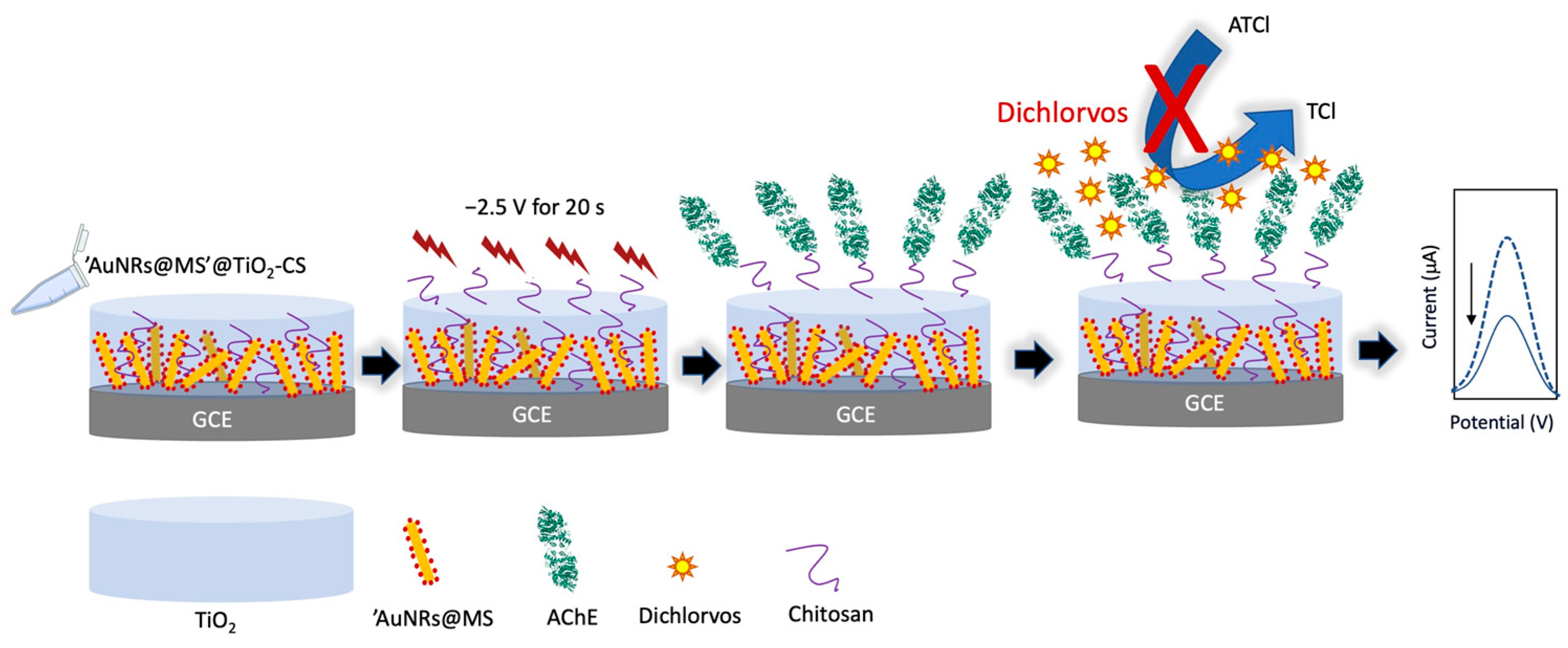
- Cui, H.F.; Zhang, T.T.; Lv, Q.Y.; Song, X.; Zhai, X.J.; Wang, G.G. An Acetylcholinesterase Biosensor Based on Doping Au Nanorod@SiO2 Nanoparticles into TiO2-Chitosan Hydrogel for Detection of Organophosphate Pesticides. Biosens. Bioelectron. 2019, 141, 111452. [Google Scholar] [CrossRef] [PubMed]
- Cui, H.F.; Wu, W.W.; Li, M.M.; Song, X.; Lv, Y.; Zhang, T.T. A Highly Stable Acetylcholinesterase Biosensor Based on Chitosan-TiO2-Graphene Nanocomposites for Detection of Organophosphate Pesticides. Biosens. Bioelectron. 2018, 99, 223–229. [Google Scholar] [CrossRef] [PubMed]
- Hu, H.; Wang, B.; Li, Y.; Wang, P.; Yang, L. Acetylcholinesterase Sensor with Patterned Structure for Detecting Organophosphorus Pesticides Based on Titanium Dioxide Sol-Gel Carrier. Electroanalysis 2020, 32, 1834–1842. [Google Scholar] [CrossRef]
- Mahmoudi, E.; Fakhri, H.; Hajian, A.; Afkhami, A.; Bagheri, H. High-Performance Electrochemical Enzyme Sensor for Organophosphate Pesticide Detection Using Modified Metal-Organic Framework Sensing Platforms. Bioelectrochemistry 2019, 130, 107348. [Google Scholar] [CrossRef]
- Bilal, S.; Mudassir Hassan, M.; Fayyaz ur Rehman, M.; Nasir, M.; Jamil Sami, A.; Hayat, A. An Insect Acetylcholinesterase Biosensor Utilizing WO3/g-C3N4 Nanocomposite Modified Pencil Graphite Electrode for Phosmet Detection in Stored Grains. Food Chem. 2021, 346, 128894. [Google Scholar] [CrossRef]
- Zhao, G.; Zhou, B.; Wang, X.; Shen, J.; Zhao, B. Detection of Organophosphorus Pesticides by Nanogold/Mercaptomethamidophos Multi-Residue Electrochemical Biosensor. Food Chem. 2021, 354, 129511. [Google Scholar] [CrossRef] [PubMed]
- Li, X.; Gao, X.; Gai, P.; Liu, X.; Li, F. Degradable Metal-Organic Framework/Methylene Blue Composites-Based Homogeneous Electrochemical Strategy for Pesticide Assay. Sens. Actuators B Chem. 2020, 323, 128701. [Google Scholar] [CrossRef]
- Ma, J.; Yuan, J.; Xu, Y.; Jiang, Y.; Bai, W.; Zheng, J. Ultrasensitive Electrochemical Determination of Bisphenol A in Food Samples Based on a Strategy for Activity Enhancement of Enzyme: Layer-by-Layer Self-Assembly of Tyrosinase between Two-Dimensional Porphyrin Metal–Organic Framework Nanofilms. Chem. Eng. J. 2022, 446, 137001. [Google Scholar] [CrossRef]
- Zhao, S.; Zhou, T.; Khan, A.; Chen, Z.; Liu, P.; Li, X. A Novel Electrochemical Biosensor for Bisphenol A Detection Based on Engineered Escherichia Coli Cells with a Surface-Display of Tyrosinase. Sens. Actuators B Chem. 2022, 353, 131063. [Google Scholar] [CrossRef]
- Ben Messaoud, N.; Ghica, M.E.; Dridi, C.; Ben Ali, M.; Brett, C.M.A. A Novel Amperometric Enzyme Inhibition Biosensor Based on Xanthine Oxidase Immobilised onto Glassy Carbon Electrodes for Bisphenol A Determination. Talanta 2018, 184, 388–393. [Google Scholar] [CrossRef] [PubMed]
- Jinadasa, B.K.K.K.; Elliott, C.; Jayasinghe, G.D.T.M. A Review of the Presence of Formaldehyde in Fish and Seafood. Food Control. 2022, 136, 108882. [Google Scholar] [CrossRef]
- Kundu, M.; Rajesh, D.; Krishnan, P.; Gajjala, S. Comparative Studies of Screen-Printed Electrode Based Electrochemical Biosensor with the Optical Biosensor for Formaldehyde Detection in Corn. Food Bioprocess Technol. 2021, 14, 726–738. [Google Scholar] [CrossRef]
- Nurlely; Ahmad, M.; Heng, L.Y.; Tan, L.L. Potentiometric Enzyme Biosensor for Rapid Determination of Formaldehyde Based on Succinimide-Functionalized Polyacrylate Ion-Selective Membrane. Measurement 2021, 175, 109112. [Google Scholar] [CrossRef]
- Suzuki, N.; Lee, J.; Loew, N.; Takahashi-Inose, Y.; Okuda-Shimazaki, J.; Kojima, K.; Mori, K.; Tsugawa, W.; Sode, K. Engineered Glucose Oxidase Capable of Quasi-Direct Electron Transfer after a Quick-and-Easy Modification with a Mediator. Int. J. Mol. Sci. 2020, 21, 1137. [Google Scholar] [CrossRef]
- Hibino, Y.; Kawai, S.; Kitazumi, Y.; Shirai, O.; Kano, K. Protein-Engineering Improvement of Direct Electron Transfer-Type Bioelectrocatalytic Properties of D-Fructose Dehydrogenase. Electrochemistry 2019, 87, 47–51. [Google Scholar] [CrossRef]
- Wheeler, L.C.; Lim, S.A.; Marqusee, S.; Harms, M.J. The Thermostability and Specificity of Ancient Proteins. Curr. Opin. Struct. Biol. 2016, 38, 37–43. [Google Scholar] [CrossRef] [PubMed]
- Ito, K.; Okuda-Shimazaki, J.; Kojima, K.; Mori, K.; Tsugawa, W.; Asano, R.; Ikebukuro, K.; Sode, K. Strategic Design and Improvement of the Internal Electron Transfer of Heme b Domain-Fused Glucose Dehydrogenase for Use in Direct Electron Transfer-Type Glucose Sensors. Biosens. Bioelectron. 2021, 176, 112911. [Google Scholar] [CrossRef]
- Viehauser, M.C.; Breslmayr, E.; Scheiblbrandner, S.; Schachinger, F.; Ma, S.; Ludwig, R. A Cytochrome B-Glucose Dehydrogenase Chimeric Enzyme Capable of Direct Electron Transfer. Biosens. Bioelectron. 2022, 196, 113704. [Google Scholar] [CrossRef]
- Liu, Y.; Zhang, J.; Cheng, Y.; Jiang, S.P. Effect of Carbon Nanotubes on Direct Electron Transfer and Electrocatalytic Activity of Immobilized Glucose Oxidase. ACS Omega 2018, 3, 667–676. [Google Scholar] [CrossRef]
- Kizling, M.; Dzwonek, M.; Wieckowska, A.; Bilewicz, R. Gold Nanoparticles in Bioelectrocatalysis—The Role of Nanoparticle Size. Curr. Opin. Electrochem. 2018, 12, 113–120. [Google Scholar] [CrossRef]
- Bollella, P.; Hibino, Y.; Conejo-Valverde, P.; Soto-Cruz, J.; Bergueiro, J.; Calderón, M.; Rojas-Carrillo, O.; Kano, K.; Gorton, L. The Influence of the Shape of Au Nanoparticles on the Catalytic Current of Fructose Dehydrogenase. Anal. Bioanal. Chem. 2019, 411, 7645–7657. [Google Scholar] [CrossRef] [PubMed]
- Tîlmaciu, C.M.; Morris, M.C. Carbon Nanotube Biosensors. Front. Chem. 2015, 3, 59. [Google Scholar] [CrossRef]
- Mallick, S.; Singh, K.R.; Nayak, V.; Singh, J.; Singh, R.P. Potentialities of Core@shell Nanomaterials for Biosensor Technologies. Mater. Lett. 2022, 306, 130912. [Google Scholar] [CrossRef]
- Yang, Z.; Zhang, C.; Zhang, J.; Bai, W. Potentiometric Glucose Biosensor Based on Core-Shell Fe3O4-Enzyme-Polypyrrole Nanoparticles. Biosens. Bioelectron. 2014, 51, 268–273. [Google Scholar] [CrossRef] [PubMed]
- Song, M.; Lin, X.; Peng, Z.; Xu, S.; Jin, L.; Zheng, X.; Luo, H. Materials and Methods of Biosensor Interfaces with Stability. Front. Mater. 2021, 7, 583739. [Google Scholar] [CrossRef]
- Le, T.H.; Kim, Y.; Yoon, H. Electrical and Electrochemical Properties of Conducting Polymers. Polymers 2017, 9, 150. [Google Scholar] [CrossRef]
- Cichosz, S.; Masek, A.; Zaborski, M. Polymer-Based Sensors: A Review. Polym. Test. 2018, 67, 342–348. [Google Scholar] [CrossRef]
- Yuqing, M.; Jianrong, C.; Xiaohua, W. Using Electropolymerized Non-Conducting Polymers to Develop Enzyme Amperometric Biosensors. Trends Biotechnol. 2004, 22, 227–231. [Google Scholar] [CrossRef]
- He, J.; Su, J.; Wang, J.; Zhang, L. Synthesis of Water-Free PEDOT with Polyvinylpyrrolidone Stabilizer in Organic Dispersant System. Org. Electron. 2018, 53, 117–126. [Google Scholar] [CrossRef]
- Yuan, M.; Minteer, S.D. Redox Polymers in Electrochemical Systems: From Methods of Mediation to Energy Storage. Curr. Opin. Electrochem. 2019, 15, 1–6. [Google Scholar] [CrossRef]
- Alsaoub, S.; Barwe, S.; Andronescu, C.; Pöller, S.; Ruff, A.; Schuhmann, W. Poly(Benzoxazine)s Modified with Osmium Complexes as a Class of Redox Polymers for Wiring of Enzymes to Electrode Surfaces. Chempluschem 2015, 80, 1178–1185. [Google Scholar] [CrossRef] [PubMed]
- Zafar, M.N.; Wang, X.; Sygmund, C.; Ludwig, R.; Leech, D.; Gorton, L. Electron-Transfer Studies with a New Flavin Adenine Dinucleotide Dependent Glucose Dehydrogenase and Osmium Polymers of Different Redox Potentials. Anal. Chem. 2012, 84, 334–341. [Google Scholar] [CrossRef] [PubMed]
- Jayakumar, K.; Bennett, R.; Leech, D. Electrochemical Glucose Biosensor Based on an Osmium Redox Polymer and Glucose Oxidase Grafted to Carbon Nanotubes: A Design-of-Experiments Optimisation of Current Density and Stability. Electrochim. Acta 2021, 371, 137845. [Google Scholar] [CrossRef]
- Vidal, J.C.; Bonel, L.; Ezquerra, A.; Hernández, S.; Bertolín, J.R.; Cubel, C.; Castillo, J.R. Electrochemical Affinity Biosensors for Detection of Mycotoxins: A Review. Biosens. Bioelectron. 2013, 49, 146–158. [Google Scholar] [PubMed]


| Electrochemical | Optical | Mass-Based | |
|---|---|---|---|
| Working principle | Detection of the potential/gradient of oxidation and reduction reactions from enzymes and metabolites. | Chemical or biological reactions produce light signals (visible, ultraviolet and infrared) that are measured by a transducer and converted into data for analysis | Production of electrical signals based on applied mechanical force |
| Advantages | User-friendly Miniaturization Fast detection Low detection limit | High sensitivity and selectivity No electrical interference | Simplicity No optical interference Stable output |
| Drawbacks | Unstable current and voltage Less selectivity Limited shelf life | Bulky instruments Requirement of sample pre-treatment | Low sensitivity Interference induces by nonspecific binding |
| Analytes | Electrode | Enzymes | Transducer | Sensitivity | Detection Range | LOD | Food Matrices | Ref. |
|---|---|---|---|---|---|---|---|---|
| Glucose | Nafion/MnO2-GNR/SPCE | GOx | Amp | 56.32 μA mM−1 cm−2 | 0.1–1.4 mmol L−1 | 0.05 mM | Honey | [28] |
| Glucose | PEDOT/PAA/GOx PEDOT/AA/GOx | GOx | Amp | 2.74 × 10⁻⁴ A M−1 2.57 × 10⁻⁴ A M−1 | 0.96–30 mM 1.86–30 mM | 0.29 mM 0.56 mM | Grape juice, honey | [26] |
| Glucose | Poly(2,2-bithiophene)/Pt disk | GOx | Amp | 1.5 × 10⁻³ A mM−1 | 0.09–5.20 mM | 30 μM | Pear, apricot, and peach fruit juices | [36] |
| Poly(4,4′-bithiophene derivative/Pt disk | 3.4 × 10⁻⁴ A mM−1 | 0.15–5.20 mM | 50 μM | |||||
| Glucose Galactose | Os polymer/graphite rod | POx | Amp | n.d | 0.1–15 mM 0.1–10 mM | 8.5 μM 3.2 μM | n.d | [34] |
| Glucose | PtNPs-poly(Azure-A)-aSPCE | GOx | Amp | 42.7 μA mM−1 cm−2 | 20 μM–2.3 mM | 7.6 μM | Commercial orange, pineapple, and peach juices | [37] |
| Glucose | MWCNTs/Nafion/GCE | GOx | Amp | 23.3 μA mM−1 cm−2 | 50 μM–1 mM | 0.58 μM | Honey | [38] |
| 32.4 μA mM−1 cm−2 | 1–3 mM | 4.94 μM | ||||||
| Glucose Alcohol | CF(Hemin-AuNPs)/graphite rod | GOx | Amp | 909.5 A M−1 m−2 | 0.1–0.9 mM | 0.05 mM | Grape must and wine | [30] |
| AOx | 4089 A M−1 m−2 | 0.01–0.15 mM | 0.005 mM | |||||
| Glucose | NPPt/GO/Nafion | GOx | Amp | 11.64 μA.L.mmol−1 cm−2 | 0.1–4 mmol L−1 4–20.0 mmol L−1 | 13 μM | Tomato, cucumber | [39] |
| Glucose | PEDOT:SCX/MXene/GOx/GCE | GOx | Amp | n.d | 0.5–8 mM | 0.0225 mM | Fruit juice | [25] |
| Glucose | Ppy/GOx/DGNs/ Graphite rod | GOx | Amp | 59.4 μA mM−1 cm−2 | 0.1–19.9 mmol L−1 | 0.070 mM | Wine, coconut milk, almond milk, apple juice, mandarin juice | [21] |
| PANI/GOx/DGNs/ Graphite rod | 43.9 μA mM−1 cm−2 | 0.3–19.9 mmol L−1 | 0.18 mM | |||||
| Glucose | AuNPs/PENDI/PGE | GOx | Amp | 0.172 μA mM−1 cm−2 | 0.0009–0.33 mM | 0.0407 mM | Dextrose solution, orange juice | [27] |
| Analytes | Electrode | Enzymes | Transducer | Sensitivity | Detection Range | LOD | Food Matrices | Ref. |
|---|---|---|---|---|---|---|---|---|
| Fructose | 4-MPh/h-PG/polycrystalline Au electrodes | FDH | Amp | 175 μA mM−1 cm−2 | 0.05–5 mM | 0.3 μM | Honey, tomato juice, apple juice, pineapple juice, energy drinks | [60] |
| Fructose | Au microdisk electrode | FDH | Amp | 200 μA mM−1 cm−2 | up to 2 mM | n.d | Fruit juice, carbonated drinks, honey | [61] |
| Sucrose | Chitosan/planar Au electrode | Invertase mutarotase GDH | Amp | 0.65 nA μM−1 | 10–1200 μM | 8.4 μM | Green coffee beans | [45] |
| Sucrose | CuNPs-MFC-IGT/AuSPE | Invertase GOx | Amp | 3.7 μA M−1 | 0.01 nM–100 μM | 0.01 nM | Sweetened tea beverages | [44] |
| Sucrose | PEI/GA/ silicalite-modified stainless steel electrodes | Invertase mutarotase GOx | Cond | n.d | 0.0035–4 mM | 3.5 μM | Orange nectar, orange juice, apple juice | [29] |
| Maltose | Sol-gel-MWCNTs/PVC tube | α-1,4-glucosidase GOx | Amp | 29.15 μA mM−1 cm−2 | 0.5–5 mM | 2.4 × 10⁻² mM | n.d | [49] |
| Maltose | GDH/Os polymer/Graphite rod | GDH | Amp | 1.7 μA mM−1 cm−2 | 0.5–15 mM | 0.45 mM | n.d | [50] |
| Lactose | Poly(Pyrrole-co-EDOT)/Pt disc electrode | β-gal GalOx | CV | 1.08 A M−1 cm−2 | 0.198–2.301 mM | 1.4 × 10⁻⁵ M | Whole, low-fat, skimmed milk | [51] |
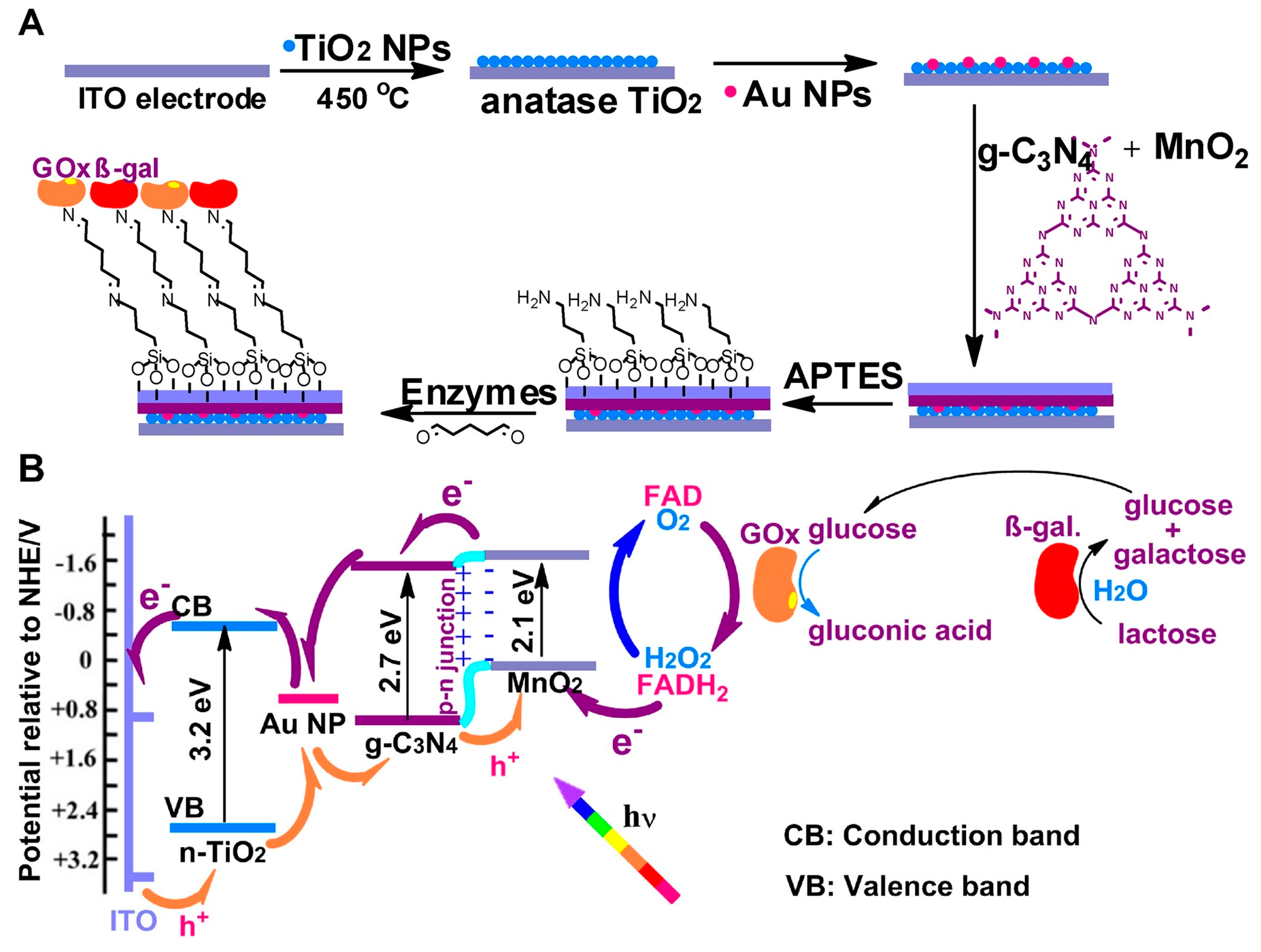
| Lactose Glucose | GOx-β-Gal/Au NPs-graphitic C3N4-MnO2-TiO2/ITO | β-gal GOx | Photo-electrochemical | 1.66 μA mM−1 cm−2 (lactose) | 0.008–2.50 mM (lactose) | 0.23 μM (lactose) | n.d | [55] |
| 1.54 μA mM−1 cm−2 (glucose) | 0.004–1.75 mM (glucose) | 0.12 μM (glucose) | ||||||
| Lactose | β-gal/MWCNTs/ carbon paste electrode | β-gal | Amp | 1.06 μA mmol−1L cm−2 | up to 0.025 mM | 0.15 mM | Skimmed milk | [53] |
| Lactose | Enzyme nanoparticles/Au-wire electrode | β-gal GOx | CV | n.d | 1–10 mg mL−1 | 1 mg mL−1 | Processed milk | [56] |
| Lactose | Chitosan/enzyme/GCE | β-gal GOx | Pot | 9.41 × 10⁻⁴ C cm−2 mM−1 | 5.83 × 10⁻³ to 1.65 × 10⁻² M | 1.38 mM | Whey permeates, milk protein isolates | [62] |
| Lactose | Poly (meta-phenylenediamine)/Pt disk electrode | β-gal mutarotase GOx | Amp | n.d | 0.01–1.25 mM | 0.005 mM | Milk | [63] |
| Analytes | Electrode | Enzymes | Transducer | Sensitivity | Detection Range | LOD | Food Matrices | Ref. |
|---|---|---|---|---|---|---|---|---|
| Ethanol | AgNPs/PANI/Graphite epoxy composites | AOx HRP | SWV | 6.899 μA L g−1 | Up to 0.35 g L−1 | 3.48 × 10⁻³ g L−1 | n.d | [73] |
| Ethanol | TCBQ-LCPs/SWCNTs | ADH | Amp | 0.5188 μA mM−1 | 0.2–13 mM | 0.05 mM | Beer, red wine, Chinese liquor | [74] |
| Ethanol | Graphite/(PDDA-CG/electrode | AOx | Amp | n.d | 250–1500 μM | 50 μM | White and red wine, whisky, vodka | [75] |
| Ethanol | PAH/SPE | ADH | Amp | 13.45 μA mM−1 cm−2 | 0.05–2 mM | 20 μM | Commercial beer | [67] |
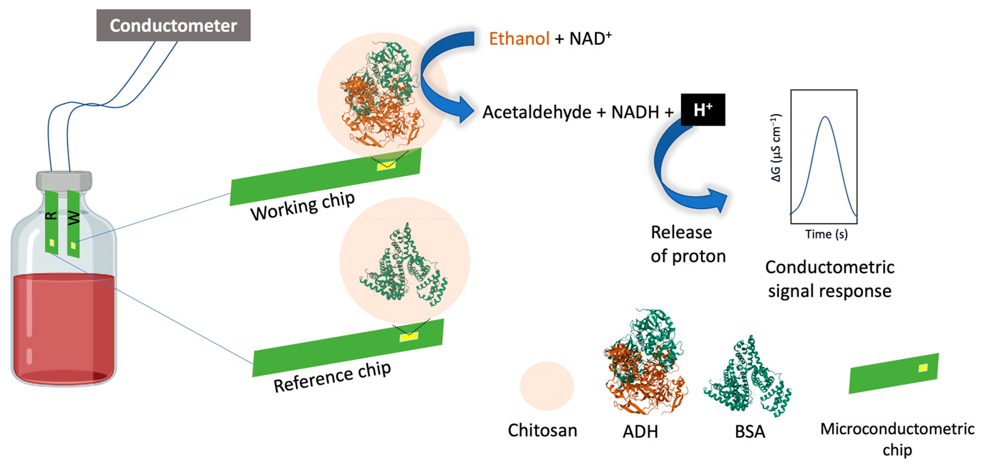
| Ethanol | chitosan/interdigitated Au electrodes | ADH | Cond | 36.8 μS cm−1 (v/v)−1 | n.d | 1200 ppm (220 mM) | Red wine | [68] |
| Polyphenols | Ppy/AuNPs/SPCE | Lacc | Amp | n.d | 1–250 μM | 0.83 μM | Propolis | [71] |
| Polyphenols | PEDOT/SNGC | Tyr | Amp | 2.4 × 10⁻⁴ μA μM−1 | 10–300 μM | 4.33 μM | Beers and wines | [70] |
| Polyphenols | GNP-MnO2/SPCE | Lacc | Amp | 455 nA µM−1 | 5–320 μM | 1.9 μM | Commercial white & red Wine | [76] |
| Hydroquinone | AuNPs/GNP/SPCE | Lacc | Amp | 0.0029 μA μM−1 | 2–120 μM | 1.5 μM | Wine & Blueberry syrup | [77] |
| Analytes | Electrode | Enzymes | Transducer | Sensitivity | Detection Range | LOD | Food Matrices | Ref. |
|---|---|---|---|---|---|---|---|---|
| L-Lysine | Au electrode | LyOx | Amp | n.d | 10–800 μM | 10 μM | Milk | [93] |
| L-Lysine | Pt electrode | LyOx | Pot | n.d | 30–1300 μM | 0.03 mM | Mozzarella | [92] |
| L-Glutamate | SPPtE/oxidised Ppy/GA-BSA | GluOx | Amp | 18.3 mA M−1 cm−2 | 0.005–1 mM | 1.8 μM | Stock cube, ketchup, Parmigiano Reggiano cheese | [95] |
| L-Glutamate | Nafion/carboxylated MWNTs)/ Au-Pt NPs/SPE | GluOx | Amp | n.d | 2 μM–16 mM | 0.14 μM | Tomatoes | [94] |
| Tyramine | PVF/GO/SPCE | DAO MAO | Amp | 7.99 μA mM−1 11.98 μA mM−1 | 0.012–0.99 μM 0.010–0.99 μM | 0.61 μM | Cheese | [99] |
| Tyramine | AuNPs/CNFs-IL-chitosan/GCE | Tyr | DPV | n.d | 10–60 μM | 3.16 μM | Wine | [109] |
| Histamine | BSA/GA/SPCE | DAO HRP | Amp | 1.31–1.59 μA mM−1 | 2–20 μg mL−1 | 0.11 μM | Yellowfin tuna fillets | [100] |
| Histamine | BSA/GA/SPCE | DAO | Amp | 3.8 nA L mg−1 | 1–75 mg L−1 | 0.5 mg L−1 | Mackerel and hake fish | [101] |
| Histamine | TiO2-carboxylated MWCNTs-RU-chitosan/SPCE | DAO MAO | Amp | 3.39 μA mM−1 2.20 μA mM−1 | 9.9–1100 µM 56–1100 µM | 6.9 µM 36 µM | Fish | [97] |
| Histamine | PB/ITONPs/SPCE | DAO MAO | Amp | 1.84 μA mM−1 0.06 μA mM−1 | 6–690 μM 2–32,000 μM | 1.9 μM 2.0 μM | Cheese | [98] |
| Histamine | GA/[Fe(CN)6]3−/SPCE | DAO | Amp | 8.9 nA L mg−1cm−2 | 5–75 mg L−1 | 0.97 mg L−1 | Tuna and mackerel | [102] |
| Histamine | LDH/µ-ISE microelectrode | DAO HRP | Pot | n.d | 10−8–10−3 M | <10 nM | n.d | [103] |
| Histamine | Chitosan-AuNPs/PB/MWCNTs/SPCE | DAO | Amp | 1.319 ± 0.055 nA μmol−1 L at pH 7.50 | 2.5–125 μM 125–400 μM | 1.81 μM (0.2 ppm) | Fish and shrimp | [104] |
| Histamine | DAO-PANI/ZnO@TiO2@n-C22 MEPCM | DAO | DPV | 28.57 μA mM−1 cm−2 | n.d | 0.473 μM | Milk, Beer, Orange juice | [105] |
| Xanthine | AuNPs/carboxylated/MWCNTs/SPCE | XOx | CV | 2.388 μA cm−2 μM−1 | n.d | 1.14 nM | Fish | [108] |
| Xanthine | PtNPs/FPP/Pt disk electrode | XOx | Amp | 1.10 A M−1 cm−2 | 0.01–0.1 mM 0.1–1.4 mM | 48 nM | Fish | [107] |
| Hypoxanthine | Ppy-paratoluenesulfonate-enzymes/Pt electrode | XOx Uricase | Amp | n.d | 5–5000 µM | 5 µM | Fish | [106] |
| Analytes | Electrode | Enzymes | Transducer | Sensitivity | Detection Range | LOD | Food Matrices | Ref. |
|---|---|---|---|---|---|---|---|---|
| Malathion Methyl parathion | DAR/PB-SWCNTs/GCE | AChE | CV | n.d | 10−6–10−12 g L−1 | 3.11 × 10⁻⁴ ng L−1 1.88 × 10⁻⁴ ng L−1 | Tap water, purified water, Chinese cabbage | [111] |
| Monocrotophos Dimethoate | mesoporous SiNPs/GCE | AChE | CV | n.d | 0.001–0.003 mg L−1 | 2.51 × 10³ ng L−1 1.5 × 10³ ng L−1 | Soft drinks | [112] |
| Dichlovos Fenthion | Chitosan/‘AuNRs@ mesoporous SiO2′@TiO2-chitosan/GCE | AChE | CV and EIS | n.d | 0.018–13.6 μM | 5.3 nM 1.3 nM | Cabbage | [113] |
| Dichlorvos | Chitosan/TiO2 /GCE | AChE | DPV | n.d | 1.13–22,600 nM | 0.23 nM | Cabbage | [115] |
| Phosmet | WO3/graphitic-C3N4/Pencil graphite electrode | AChE | Amp | 15 μA nM−1 cm−2 | 5–125 nM | 3.6 nM | Wheat flour | [117] |
| Eleven organo-phosphorus pesticides Methomyl | AuNPs/mercaptomethamidophos/mercaptohexanol/GCE | AChE | DPV and EIS | n.d | 0.1–1500 ng mL−1 | 19 to 77 ng L−1 81 ng L−1 | Apple and cabbage | [118] |
| Paraoxon | Ce/Zr-based MOF/MWCNTs/GCE | AChE | Amp and DPV | n.d | 0.01–150 nM | 0.004 nM | Spinach, cabbage | [116] |
| Paraoxon | zeolitic imidazolate framework-8/Methylene blue/ITO | AChE | DPV | n.d | 20–4000 ng mL−1 | 1.7 × 10³ ng L−1 | Apple, eggplant | [119] |
| Bisphenol A (BPA) | XOx/GCE | XOx | Amp | n.d | up to 41 nM | 1.0 nM | Mineral water | [122] |
| Bisphenol A (BPA) | Cu–TCPP | Tyr | DPV | n.d | 3.5 nM–18.9 μM | 1.2 nM | Milk | [120] |
| Bisphenol A (BPA) | GCE | Tyr | Amp | n.d | 0.00001–0.1 μM | 0.01 nM | Commercial canned teas and juices | [121] |
| Formaldehyde | SPCE | FdDH | CV | 352 μA mg−1 L cm−2 | 0.01–0.5 mg L−1 | 0.03 mg L−1 | Corn | [124] |
| Formaldehyde | pnBA-NAS/pHEMA/ Ag/AgCl screen printed electrode | AOx | Pot | 59.23 mV/decade | 0.5–220 mM | 0.1 mM | Fish | [125] |
Disclaimer/Publisher’s Note: The statements, opinions and data contained in all publications are solely those of the individual author(s) and contributor(s) and not of MDPI and/or the editor(s). MDPI and/or the editor(s) disclaim responsibility for any injury to people or property resulting from any ideas, methods, instructions or products referred to in the content. |
© 2023 by the authors. Licensee MDPI, Basel, Switzerland. This article is an open access article distributed under the terms and conditions of the Creative Commons Attribution (CC BY) license (https://creativecommons.org/licenses/by/4.0/).
Share and Cite
Wijayanti, S.D.; Tsvik, L.; Haltrich, D. Recent Advances in Electrochemical Enzyme-Based Biosensors for Food and Beverage Analysis. Foods 2023, 12, 3355. https://doi.org/10.3390/foods12183355
Wijayanti SD, Tsvik L, Haltrich D. Recent Advances in Electrochemical Enzyme-Based Biosensors for Food and Beverage Analysis. Foods. 2023; 12(18):3355. https://doi.org/10.3390/foods12183355
Chicago/Turabian StyleWijayanti, Sudarma Dita, Lidiia Tsvik, and Dietmar Haltrich. 2023. "Recent Advances in Electrochemical Enzyme-Based Biosensors for Food and Beverage Analysis" Foods 12, no. 18: 3355. https://doi.org/10.3390/foods12183355