Article

Antimicrobial and Antibiofilm Effect of ε-Polylysine against Salmonella Enteritidis, Listeria monocytogenes, and Escherichia coli in Tryptic Soy Broth and Chicken Juice

1
Department of Food Science and Biotechnology of Animal Resource, Konkuk University, Seoul 05029, Korea
2
Sempio Fermentation Research Center, Sempio Foods Company, Cheongju 28156, Korea
*
Author to whom correspondence should be addressed.
Foods 2021, 10(9), 2211; https://doi.org/10.3390/foods10092211
Submission received: 30 July 2021 / Revised: 13 September 2021 / Accepted: 15 September 2021 / Published: 17 September 2021
(This article belongs to the Section Food Microbiology)

Abstract

:
ε-Polylysine (ε-PL) is a safe food additive that is used in the food industry globally. This study evaluated the antimicrobial and antibiofilm activity of antibacterial peptides (ε-PL) against food poisoning pathogens detected in chicken (Salmonella Enteritidis, Listeria monocytogenes, and Escherichia coli). The results showed that minimum inhibitory concentrations (MICs) ranged between 0.031–1.0 mg/mL, although most bacterial groups (75%) showed MICs of 1.0 mg/mL. The reduction in the cell viability of pathogens due to ε-PL depended on the time and concentration, and 1/2 × MIC of ε-PL killed 99.99% of pathogens after 10 h of incubation. To confirm biofilm inhibition and degradation effects, crystal violet assay and confocal laser scanning microscopy (CLSM) were used. The biofilm formation rates of four bacterial groups (Salmonella, Listeria, E. coli, and multi-species bacteria) were 10.36%, 9.10%, 17.44%, and 21.37% at 1/2 × MIC of ε-PL, respectively. Additionally, when observed under a CLSM, ε-PL was found to induce biofilm destruction and bacterial cytotoxicity. These results demonstrated that ε-PL has the potential to be used as an antibiotic and antibiofilm material for chicken meat processing.

1. Introduction

Foodborne diseases commonly occur due to food being contaminated by bacteria, bacterial toxins, viruses, parasites, chemicals, and other agents. Researchers have identified more than 250 foodborne diseases [1]. Common symptoms of foodborne diseases include vomiting, nausea, diarrhea, and abdominal cramps. In some cases, severe symptoms can even lead to death. Nearly one in ten people in the world, (approximately 600 million) get food poisoning through contaminated food, and 420,000 people die as a result each year, resulting in 33 million years of disability-adjusted life years. The combined productive and medical costs due to contaminated food in low- and middle-income countries is USD110 billion annually. Food contamination can occur at every stage of the food processing chain, including food production, packaging, and storage [2]. Salmonella, Campylobacter, enterohemorrhagic Escherichia coli, Listeria, and Vibrio cholerae are among the most common foodborne pathogens [3].
Globally, people enjoy eating poultry meat and its products. However, it is particularly important to pay attention to sanitation as chicken meat and its products can transfer foodborne pathogens [4]. Raw chicken meat is commonly contaminated by Campylobacter and occasionally by Clostridium perfringens and Salmonella [4]. Listeria monocytogenes and E. coli have also been detected in cooked chicken [5,6]. The global broiler meat production amounted to approximately 100.6 Mt in 2020, and is predicted to grow to approximately 102 Mt in 2021, which is 20 Mt more than what it was in 2012 [7]. In addition, the global consumption of poultry meat is expected to reach 144.87 kt by 2029 [8].
Microorganisms normally live in complicated biological communities, known as biofilms, which provide higher resistance against external physical and chemical attacks such as high temperature, pH, humidity, bactericides, and other antibiotics [9]. Biofilm formation occurs in three steps: bacterial adhesion, maturation, and bacterial dispersion [10]. Adhesion is initiated by the movement of planktonic cells using bacterial nanofibers such as pili and flagella. Subsequently, physicochemical interactions such as electrostatic interaction, hydrophobicity, van der Waals force, and acid-based interactions form bonds between the bacterial cell and the surface [11]. When the attachment is stable, polysaccharides, as the main element of exopolysaccharide (EPS), act as important mediators of attachment. This makes the bacterial adhesion irreversible, creating conditions suitable for maturation [10]. In the maturation stage, bacteria attached to the surface mature and organize a microcolony. Bacteria then perform quorum sensing (QS), an intercellular communication, through various autoinducer secretions [12]. Finally, in the dispersion step, bacteria in the microcolonies disperse when a polysaccharide enzyme disbands the EPS of the biofilm.
Multi-species biofilms are biofilms composed of multiple microbial species. Each microbial species has its own characteristics and reveals some developed and distinct functions on multi-species biofilms that do not exist in their mono-species biofilms [13]. Multi-species biofilms are commonly found in nature, the food industry, and medical devices. They are related to numerous amounts of human bacterial infections, have severe human health impacts, and increase the economic burden on food processing and health-care systems. The intricacy of multi-species biofilms has been associated with their metabolism, interactions, and communication. Additionally, pathogens in multi-species biofilms are more resistant to antimicrobials than those in mono-species biofilms [14].
Antimicrobial peptides (AMPs) are oligopeptides that consist of five or more amino acids [15], and are endowed with a wide range of antibacterial effects against both Gram-positive and Gram-negative bacteria, viruses, and parasites [16]. AMPs occur in all living organisms, ranging from bacteria to plants and animals. AMPs derived from bacteria are known as bacteriocins, and account for 7.36% of the total antibacterial peptide database [16]. Bacteriocins are manufactured by several species to kill other bacterial species for accessing nutrients and space in a limited environment. ε-Polylysine (ε-PL) is a cationic peptide, l-lysine, consisting of 25–35 residues [17]. It is produced by Streptomyces albulus and Lysinopolymerus, and can inhibit most bacteria, yeast, and viruses as AMPs [18]. In addition, ε-PL has demonstrated a biofilm removal effect [15]. However, research on its antibacterial effects on multiple bacterial pathogens in chicken meat is scarce. In this study, we investigated the antimicrobial and antibiofilm activities of ε-PL against multiple microbial species. In addition, we used chicken juices, the extract of chicken breast, instead of a growth medium, to better understand the relationship between the environment of chickens and the pathogenic bacteria.

2. Materials and Methods

2.1. Strains, Growth Conditions, and Preparation of Experimental Group

Four biofilm-forming pathogens, Salmonella Enteritidis KCCM 12021, Listeria monocytogenes H7962 serotype 4, Escherichia coli O157:H4 FRIK 125, and E. coli ATCC 25922 were used in this study. The stock cultures of the pathogen species were frozen at −80 °C in broth media supplemented with glycerol (20% w/v). Stocks were transferred to tryptic soy agar (TSA) twice to activate the strains. S. Enteritidis KCCM 12021, E. coli O157:H4 FRIK 125, and E. coli ATCC 25922 were cultured in tryptic soy broth (TSB; Difco Laboratories, Franklin Lakes, NJ, USA) at 37 °C for 18 h. L. monocytogenes H7962 serotype 4 was cultured in TSB supplemented with yeast extract (0.6% w/v) (TSB-YE) at 37 °C for 18 h.
Four experimental groups were prepared after an 18 h incubation, depending on the bacterial species inoculated. Of these, three were mono-species cultures and the other was a multi-species culture. Each mono-species culture was adjusted to a final concentration of 1 × 10⁵ CFU/mL. The cultures of E. coli O157:H4 FRIK 125 and E. coli ATCC 25922 were combined in a 1:1 ratio. The multi-species culture was adjusted such that the final concentration of each species was 1 × 10⁵ CFU/mL.

2.2. Preparation of Chicken Juice

Raw chicken breast meat was purchased from retail outlets (Seoul, Korea). Chicken juice (CJ) was prepared using the method described by Wang et al. [19] with some modifications. Meats were cleaned of fats and fasciae and then minced using a sterilized knife. Minced chicken meat (100 g) was mixed with 200 mL of sterile water in a filter bag (InterScience, Weymouth, MA, USA). After sealing the mouth of the bag, the filter bag was homogenized using a stomacher (IUL Instruments, Barcelona, Spain) for 90 s. The homogenized mixture was centrifuged at 8000× g for 15 min. The supernatant was filtered using a 0.45 μm pore size syringe filter (Advantec, Tokyo, Japan), and the filtered fluid was used as the CJ.

2.3. Minimum Inhibitory Concentration (MIC) of ε-PL

The MICs of ε-PL (Shin Seung Hichem Co., Ltd., Seoul, Korea, 50% purity, food grade) were detected using a two-fold dilution method and TSB in 96-well polystyrene microtiter plates (SPL, Gyeonggi-do, Korea) [20]. ε-PL was diluted at double concentrations from 1.95 μg/mL to 8 mg/mL, and 50 μL of each concentration was mixed with 50 μL of bacterial culture adjusted to 1 × 10⁵ CFU/mL in the 96-well plates. The 96-well plates were incubated at 37 °C for 24 h, and the MIC was determined as the lowest concentration of ε-PL that inhibited bacterial growth.

2.4. Time-Kill Assay

Time-kill assays were conducted by culturing bacterial cultures in TSB, with the same volume of ε-PL at concentrations of 1/2 × MIC, 1 × MIC, and 2 × MIC at 37 °C, and a non-treated medium was used as a control [21]. Bacterial cultures were prepared in four groups at a final concentration of 1 × 10⁵ CFU/mL each, using the same protocol as described in Section 2.1. Aliquots were collected at 0, 1, 2, 4, 6, 8, and 10 h of incubation and then cultured in appropriate agar media at 37 °C for 24 h, followed by counting viable cell colonies. Single-species bacterial cultures were plated on TSA, and the multi-species bacterial culture was plated on xylose lysine deoxycholate agar (XLD; BD Difco) for Salmonella, Oxford agar (MB cell, Seoul, Korea) supplemented with Oxford supplement (MB cell) for Listeria, Eosin methylene blue agar (EMB; BD Difco) for E. coli, and TSA for total cell counts (Salmonella + Listeria + E. coli).

2.5. Biofilm Assay

The biofilm assay was conducted using the method described by Yu et al. [22], with some modifications. To investigate the inhibition effect on the formation of biofilms, bacterial cultures were diluted to a final concentration of 1.0 × 10⁵ CFU/mL, and 50 μL of each was mixed in a 96-well plate with volume equivalent to that of ε-PL, with concentrations ranging from 1/2 × MIC to 2 × MIC, using a non-treated TSB as a control. Bacterial cultures were divided into four groups, as described in Section 2.1. Following incubation at 37 °C for 24 h, the bacterial culture was eliminated, and the wells were rinsed twice with sterilized water to remove planktonic cells. Each 96-well plate was dried at 37 °C for 15 min, and 100 μL of 0.1% crystal violet was transferred to each well to stain the biofilms. After 30 min of reaction time, the wells were carefully rinsed with distilled water. Each well was filled with 100 μL of dissolving solution (10% acetic acid and 30% methanol) to dissolve the crystal violet. Optical density (OD) was measured at 570 nm using a microplate reader (Emax, Molecular Devices, Radnor, PA, USA).
To investigate the degradation effect of ε-PL on mature biofilms, bacterial cultures were adjusted to 1.0 × 10⁵ CFU/mL, and 100 μL of each was incubated in 96-well plates at 37 °C for 24 h. The bacterial suspension was placed in 100 μL of ε-PL at concentrations of 1/2 × MIC to 2 × MIC, followed by incubation at 37 °C for 24 h. The subsequent crystal violet assay was conducted as described above. The biofilm formation rate (%) was determined using the following formula:
Biofilm formation rate (%) = ODtreatment/ODcontrol × 100
where ODtreatment and ODcontrol are defined as the absorbance at 570 nm in a dissolving solution.
To investigate the biofilm inhibition and degradation of ε-PL on CJ, the strains were cultured in broth medium at 37 °C for 18 h. The bacterial culture was centrifuged at 12000× g at 4 °C for 5 min, and the cell supernatant was discarded. The pellet was washed twice with phosphate-buffered saline (PBS; pH 7.4; Hyclone, Logan, UT, USA) and dissolved in CJ and diluted to a concentration of 1.0 × 10⁵ CFU/mL. ε-PL was diluted in the CJ ranging from 1/2 × MIC to 2 × MIC. The same method was conducted as described above, except that TSB was substituted for CJ.

2.6. Hydrophobicity and Auto-Aggregation

The bacterial surface hydrophobicity of food poisoning pathogens was investigated in the presence of ε-PL, following Yang et al. [23] with some modifications. The bacterial culture was adjusted to a concentration of 1.0 × 10⁵ CFU/mL and prepared in four groups, as described in Section 2.1. The bacteria were treated with ε-PL (1/2 × MIC) in TSB at 37 °C for 4 h. Non-treated cells were used as the control.
Cultured cells were centrifuged at 12,000× g at 4 °C for 5 min, and the precipitates were rinsed twice. Collected cells were re-dissolved in PBS to adjust the OD600 to 0.5 ± 0.05 (ODinitial). Two milliliters of each suspension was mixed with chloroform (0.5 mL) and vortexed for 2 min. After being static at room temperature for 15 min, the upper aqueous layer was collected and measured at 600 nm (ODtreatment). The hydrophobicity (%) was calculated using the following equation:
Hydrophobicity (%) = (1 − ODtreatment / ODinitial) × 100
Auto-aggregation was conducted using a modification of the method presented by Kim et al. [24]. Cell culture was adjusted to a final concentration of 1.0 × 10⁵ CFU/mL and ε-PL solution at 1/2 × MIC was blended in a ratio of 1:1 and incubated at 37 °C for 24 h. Non-treated cells were used as control. Five milliliters of the mixture was collected and statically incubated at 4 °C for 24 h. After incubation, the upper aqueous layer was measured at 600 nm (ODtreatment). The sample was then vortexed and measured at 600 nm (ODinitial). Auto-aggregation (%) was calculated using the following equation:
Auto-aggregation (%) = (1 − ODtreatment/ODinitial) × 100

2.7. Total EPS Production Rate

The total EPS production rate of food-poisoning pathogens was determined according to Song et al. [25], with some modifications. Cell cultures were treated with 1/2 × MIC of ε-PL in TSB at 37 °C for 24 h. Non-treated cells were used as the control. Incubated cells were centrifuged at 8000× g at 25 °C for 10 min and the precipitates were dispersed in 1.0 mL of 1.5 M sodium chloride. The cell emulsion was centrifuged at 5000× g at 25 °C for 10 min, and 60 μL of the supernatant was mixed with 60 μL of 5% phenol and 4 mL of sulfuric acid by vortexing. Then, the mixtures were incubated at 30 °C for 10 min, and the absorbance was measured at 490 nm. The total EPS production rate (%) was calculated using the following equation:
Total EPS production rate (%) = ODtreatment/ODcontrol × 100
where ODtreatment and ODcontrol were defined as the absorbance (OD490) of the phenol-sulfuric acid solution, treated with ε-PL and the control, respectively.

2.8. Confocal Laser Scanning Microscopy (CLSM)

CLSM was used to investigate the anti-biofilm effects of ε-PL against biofilm-forming pathogens, as previously described, with some modifications [26]. Before the experiment, to sterilize the glass coupons, glass coupons (2.0 cm × 2.0 cm × 0.2 cm) were sonicated with distilled water 2–3 times and autoclaved at 121 °C for 15 min, followed by air-drying at 60 °C over 2 days.
Strains were cultured in appropriate broth media at 37 °C for 18 h, and their density was diluted using TSB to adjust the concentration to 1 × 10⁵ CFU/mL for cell suspension.
To investigate the inhibition effects on biofilm formation, two milliliters of cell suspension was transferred to each well of a 6-well plate (SPL, Gyeonggi-do, Korea) containing glass coupons. Two milliliters of ε-PL solution was adjusted to concentrations of 1 × MIC using TSB and added to each well of the plates. The control experiment used the non-treated medium. The 6-well plates were incubated at 37 °C for 24 h to form bacterial biofilms on the glass coupons. Then, the glass coupons were rinsed twice with PBS to remove planktonic cells.
To investigate the eradication effects on mature biofilms, four milliliters of cell suspension was added to each well of a 6-well plate with glass coupons. After that, the 6-well plates were incubated at 37 °C for 24 h. Cell cultures were substituted to four milliliters of ε-PL solution at 1 × MIC, and then re-incubated for 37 °C for 24. The subsequent process was the same as that described above.
For CLSM observation, each specimen was dyed as follows: 1 μM/mL each of SYTO9 (Invitrogen™, ThermoFisher Scientific, Carlsbad, CA, USA) and propidium iodide (PI; Invitrogen™, ThermoFisher Scientific) were used to dye the living cells on glass coupons at room temperature for 20 min without light. Following staining, the glass coupons were rinsed twice with PBS and dried at room temperature for 30 min. The specimens were then investigated using a Zeiss LSM 800 microscope (Carl Zeiss, Oberkochen, Germany).

2.9. Statistical Analysis

The results are presented as the mean ± standard error. All experiments were conducted in triplicates. SPSS version 18.0 (SPSS Inc., Chicago, IL, USA) was used for statistical analysis. One-way analysis of variance (ANOVA) was used to determine the significance of the differences.

3. Results and Discussion

3.1. Antimicrobial Activity

3.1.1. Minimum Inhibitory Concentration

The antimicrobial effect of ε-PL was estimated by investigating the MIC against the chosen foodborne pathogens (Table 1). S. Enteritidis KCCM 12021, L. monocytogenes H7962 serotype 4, E. coli O157:H4 FRIK 125, and E. coli ATCC 25922 were selected for this study. These species are representative of food-poisoning pathogens detected in poultry meat. In addition, they can form bacterial biofilms, which are a source of antimicrobial resistance [27,28,29,30]. When S. Enteritidis KCCM 12021, L. monocytogenes H7962 serotype 4, and E. coli were independently cultured, the MICs of ε-PL were 1.0, 0.031, and 1.0 mg/mL, respectively. The MIC of ε-PL in the co-cultured strain was 1.0 mg/mL. ε-PL has been shown to have antimicrobial effects against Gram-positive and Gram-negative bacteria, yeast, and fungi [31,32,33]. Some reports suggest that the optimal MIC levels are 0.1 and 0.05 mg/mL for S. Enteritidis and S. typhimurium, respectively [32]. In addition, L. monocytogenes and E. coli showed MIC values at 125 and 19.53 μg/mL, respectively [31,33]. These differences can be attributed to the use of the food grade (low purity) and the tested strain.

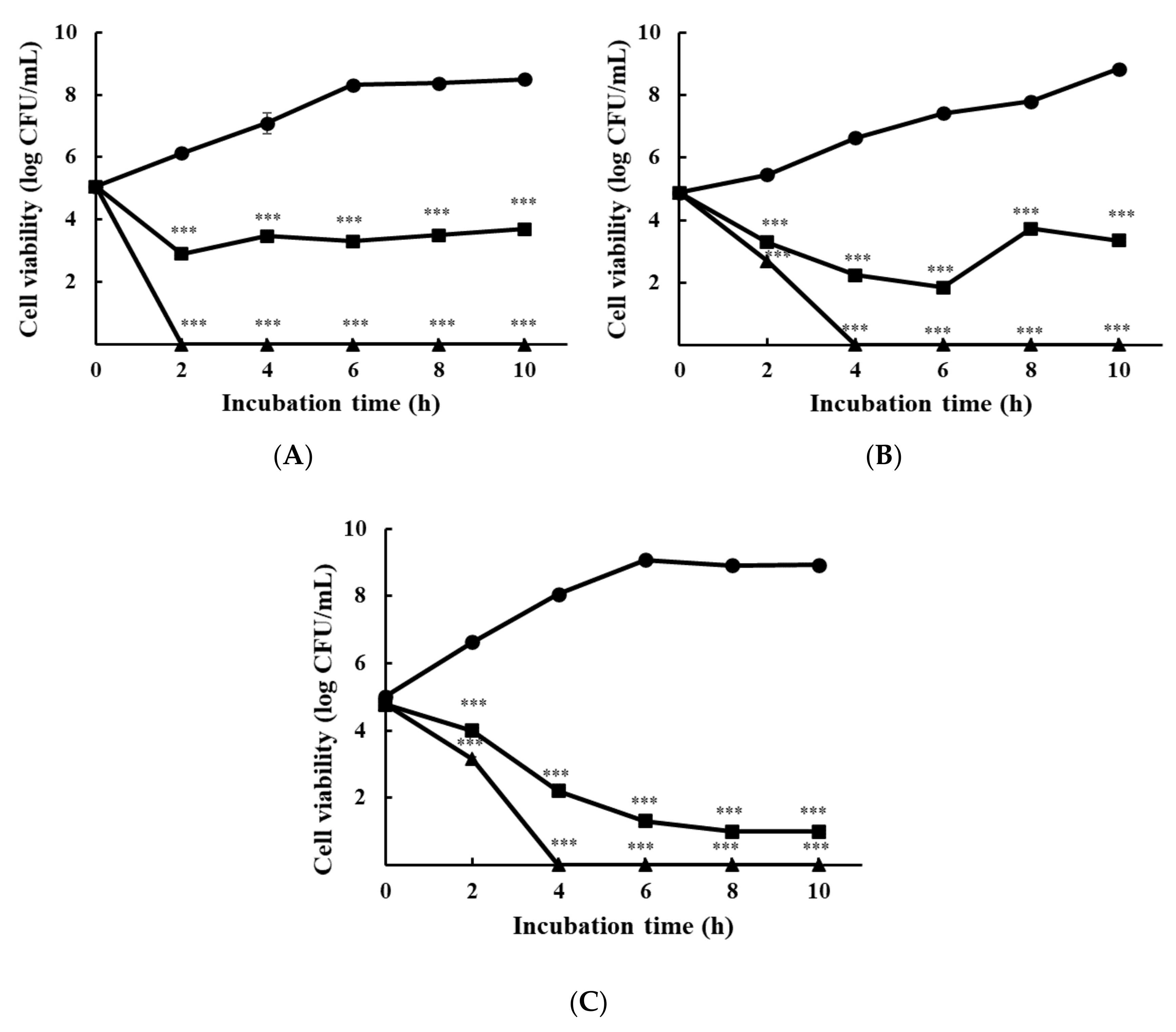
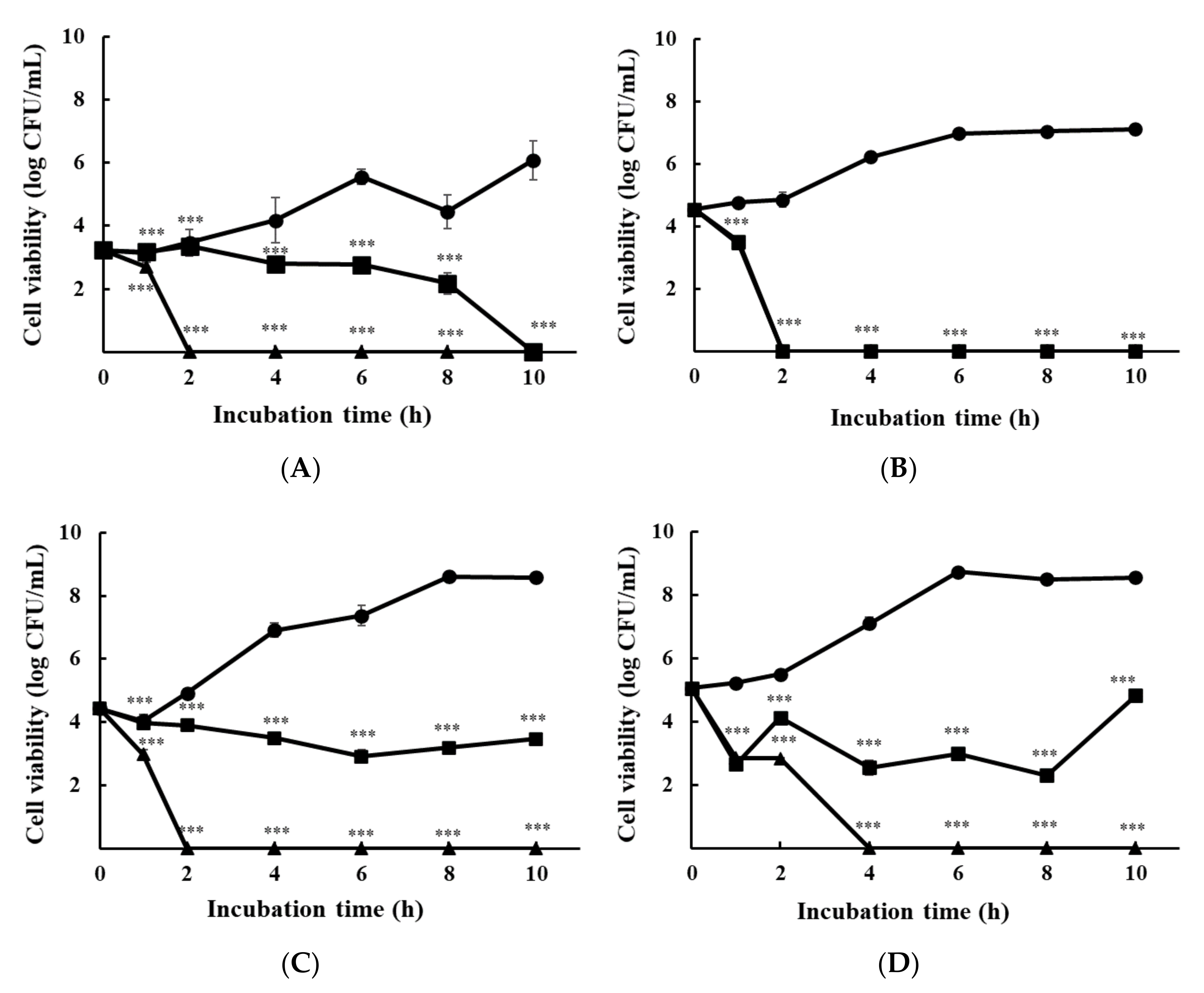
3.1.2. Time-Kill Analysis

Figure 1 shows the growth inhibition of foodborne pathogens due to ε-PL. Each strain was cultured independently in TSB and spread on TSA plates. All strains exhibited a concentration-dependent activity. In the control, cell viabilities for S. Enteritidis KCCM 12021, L. monocytogenes H7962 serotype 4, and the E. coli group, cultured at 10 h incubation, reached 8.49 ± 0.09 log CFU/mL, 8.85 ± 0.01 log CFU/mL, and 8.94 ± 0.08 log CFU/mL, respectively. ε-PL at 1 × MIC showed bactericidal activity against all bacteria within 4 h, while at 1/2 × MIC showed bacteriostatic activity against all bacteria within 4 h except to L. monocytogenes. The E. coli group exhibited a gradual decline on the cell viability depending on incubation time.
The influence of ε-PL on multi-species cultured bacteria is shown in Figure 2. After co-culturing multiple species in TSB, each species was differentiated by spreading the multi-species culture on a selective agar medium. Multi-species cultures showed a slightly different tendency from mono-species cultures, particularly at 1/2 × MIC. S. Enteritidis KCCM 12021 was diminished after 6 h of incubation and was extinguished at 10 h (Figure 2A). The cell viability of L. monocytogenes H7962 serotype 4 at 1/2 × MIC reached zero after 2 h (Figure 2B). The cell viability of the E. coli group with 1/2 × MIC of ε-PL remained stable over time, and did not significantly increase or decrease (Figure 2C). When all strains were cultured together, 1/2 × MIC of ε-PL killed 99.99% of the pathogens (approximately 1.4 × 10⁷ cells) after 4 h of treatment (Figure 2D). All cell viabilities showed an extinction at 1 × MIC of ε-PL after 2 or 4 h.
ε-PL has a cationic surface in water because of its positively charged amino acid residues, and thus inhibits the growth of pathogens sensitive to it [12]. Generally, cationic ε-PL is able to induce transitions in the structure of the peptidoglycan layer in the cell wall [34] and later interoperate with the cell membrane using electrostatic attraction, which increases the cell permeability [35], subsequently leading to the disruption of the cell membrane. Consequently, water-soluble proteins and ions are released from the cytoplasm. In addition, ε-PL enhances reactive oxygen species production, resulting in the inhibition of metabolism in bacteria [36,37].

3.2. Antibiofilm Effect

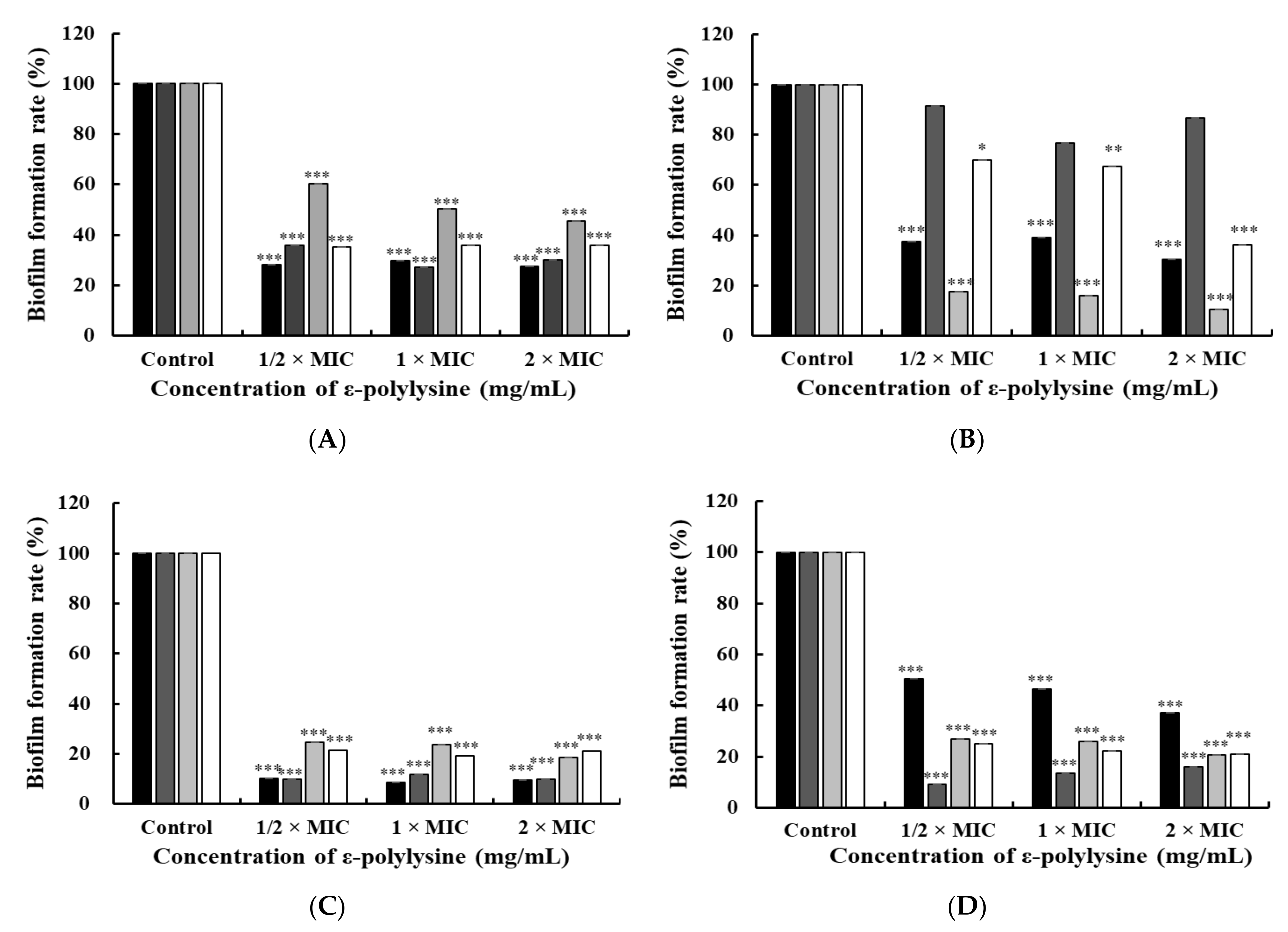
3.2.1. Crystal Violet Staining Assay

Biofilm inhibition effects have been investigated in the initial stages of biofilm formation [38]. The inhibitory effect of ε-PL on biofilms formed by foodborne pathogens was investigated using the TSB (Figure 3A) and CJ media (Figure 3C). Compared to the control, there was a significant difference in concentrations above 1/2 × MIC, regardless of the culture medium or bacterial species (p < 0.05). Figure 3A shows that biofilm formation rates of S. Enteritidis KCCM 12021, L. monocytogenes H7962 serotype 4, E. coli, and multi-species strains at 1/2 × MIC were 28.12%, 35.87%, 60.38%, and 35.28%, respectively. Likewise, in the CJ medium, biofilm formation rates for S. Enteritidis KCCM 12021, L. monocytogenes H7962 serotype 4, the E. coli group, and multi-species strains at 1/2 × MIC were 10.36%, 9.79%, 24.73%, and 21.37%, respectively (Figure 3C).
Biofilm degradation affects the removal activity of mature biofilms of foodborne pathogens [39]. The experiment was conducted in TSB (Figure 3B) and CJ media (Figure 3D). The biofilm formation rate at 1/2 × MIC in TSB were 37.49%, 91.64%, 17.44%, and 69.83% for S. Enteritidis KCCM 12021, L. monocytogenes H7962 serotype 4, E. coli group, and multi-species strains, respectively (Figure 3B). ε-PL had a significant degradation effect on mature S. Enteritidis, the E. coli group, and multi-species cell biofilms at concentrations above 1/2 × MIC (p < 0.05). However, the L. monocytogenes H7962 serotype 4 did not show a significant effect. Biofilm formation levels in the CJ medium were 50.41%, 9.1%, 26.73%, and 24.92% for S. Enteritidis KCCM 12021, L. monocytogenes H7962 serotype 4, the E. coli group, and multi-species strains at 1/2 × MIC (Figure 3D). ε-PL significantly diminished the biofilm formation rate in all groups at concentrations above 1/2 × MIC (p < 0.05).
Interestingly, ε-PL had inhibitory and degradative effects against bacterial biofilms at concentrations below the MIC, suggesting that ε-PL is an economical antibiofilm agent. Compared to the inhibitory effects of initial biofilm formation (Figure 3A,C) with the degradation of mature biofilm (Figure 3B,D), the biofilm formation rate in the initial stage was generally lower than that of the mature biofilm, except for the E. coli group. In addition, the biofilm formation rate of ε-PL in the CJ medium was generally lower than that in the TSB, for both the inhibitory and degradative effects of the biofilm. As biofilm formation and the bacterial cells remaining were both influenced by carbohydrates [40], there were differences between TSB and CJ.
Some researchers have demonstrated the anti-biofilm effects of ε-PL. ε-PL is a mediator of the inhibition of biofilm formation generated by Salmonella sp., which is associated with the potential molecular mechanisms [12]. ε-PL downregulated the expression of genes related to cellulose formation, curli amyloid fiber production, flagella-related motility, and QS, and upregulated genes controlling the synthesis of colonic acid against Salmonella sp. Other studies have reported the reduction in biofilms formed by Salmonella sp., P. aeruginosa, S. aureus, and L. monocytogenes when treated with ε-PL [41].

3.2.2. Bacterial Surface Properties and EPS Production

Hydrophobicity and auto-aggregation are the principal properties that form biofilms during the initial stage [42]. Cell surface hydrophobicity is known to regulate bacterial adhesion on diverse surfaces [38], while their auto-aggregation contributes to the topography, morphology, and maturation of the zenithal biofilm community. EPS is the main component of biofilms, provides structural stability as well as defense against antibiotics, and promotes antimicrobial resistance [43]. L. monocytogenes H7962 serotype 4 treated with 1/2 × MIC of ε-PL showed significantly lower levels of hydrophobicity, auto-aggregation, and EPS production than the control group (without ε-PL), with decreased levels of 6.4%, 21.65%, and 25%, respectively. In addition, the EPS production (%) of multi-species culture at 1/2 × MIC of ε-PL was reduced by 43.71% as compared to that of the control group (p < 0.05). These results suggest that ε-PL inhibits the formation of biofilms of L. monocytogenes at the initial stage by weakening bacterial surface properties, and this characteristic is identically manifested in a co-culture of L. monocytogenes H7962 serotype 4 with Gram-negative strains in a limited area. However, S. Enteritidis, and E. coli strains did not show significant effects on bacterial surface properties or EPS production (data not shown).

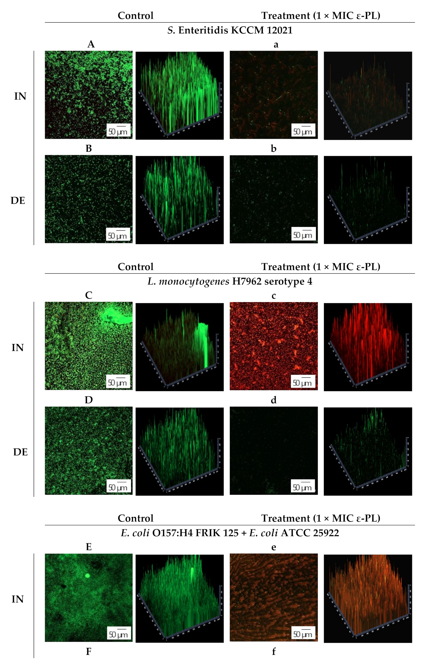
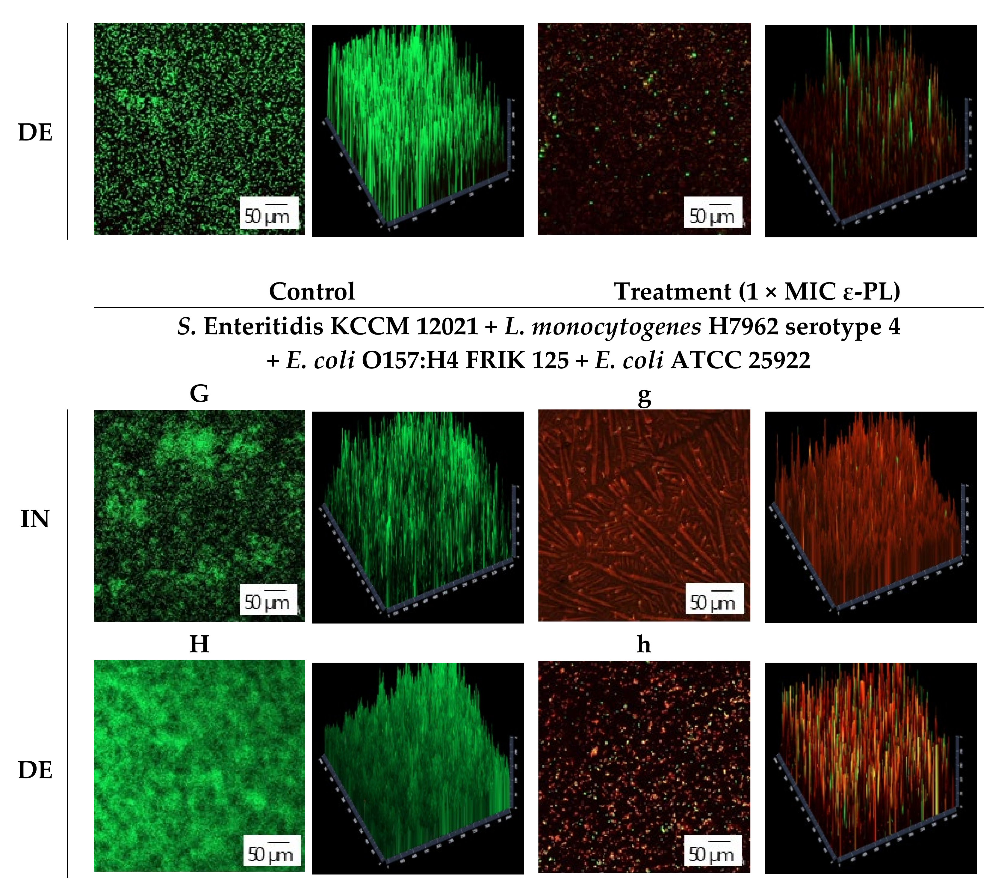
3.3. CLSM Observation

The inhibitory and degradation effects of ε-PL on the selective pathogen biofilms were optically analyzed using CLSM (Figure 4). Syto9 and PI differ in terms of the penetration capability of cell membranes [44]. Syto9 could penetrate both dead and live cells because of its high permeability, whereas PI has high molecular mass that it can only penetrate cells with damaged membranes. As shown in the CLSM images, ε-PL caused damage to live biofilm cells of S. Enteritidis KCCM 12021, L. monocytogenes H7962 serotype 4, and the E. coli group. The biofilm of the control showed abundant live colonies (green fluorescence), whereas biofilms treated with ε-PL showed few cells or increasing dead cells (red fluorescence). Some photographs of samples treated with 1 × MIC of ε-PL showed a rare-shaped structure due to the adsorption of ε-PL (Figure 4e,g), whereas few other samples showed death of most cells with no reduction in the total cell count (Figure 4c), which demonstrated the powerful penetrability of ε-PL to deeper biofilms, although the rate of eradication of the biofilm slightly decreased. Nevertheless, these results are worthwhile as green fluorescence (live cells) is rarely observed in both types. Visual observation by CLSM proved that ε-PL could exhibit anti-biofilm activity against Salmonella, Listeria, and E. coli, as also evidenced by the crystal violet assay results of the ε-PL antibiofilm activity (Section 3.2.1).

4. Conclusions

This study demonstrated that ε-PL has antibacterial and antibiofilm effects against food poisoning pathogens detected in chicken, such as S. Enteritidis, L. monocytogenes, and E. coli. ε-PL decreased cell viability depending on the time and concentration and induced the collapse of the cell membrane of pathogens, which proved the antibacterial effect of ε-PL. The antibiofilm effect of ε-PL was also evidenced by the reduction in biofilm formation and the destruction of the morphological form, as observed using CLSM. Antibiofilm effects were also observed in CJ. This study will help promote the safety of chicken meat processing, as it imitates the composition and microbiome of chicken meat to obtain more predictable results in contaminated environments. Therefore, ε-PL has the potential to be used as an antibiotic and antibiofilm material in the food industry, while accounting for the sanitary concerns of the consumers.

Author Contributions

Conceptualization, D.-U.L., H.H.Y., S.-C.J., J.-H.P., D.-H.L., N.-K.L. and H.-D.P.; methodology, D.-U.L. and Y.J.P.; validation, D.-U.L., N.-K.L. and H.-D.P.; formal analysis and investigation, D.-U.L. and Y.J.P.; data curation, D.-U.L., H.H.Y., N.-K.L. and H.-D.P.; writing—original draft preparation, D.-U.L. and N.-K.L.; writing—review and editing, D.-U.L., S.-C.J., D.-H.L., N.-K.L. and H.-D.P.; supervision, H.-D.P.; project administration, D.-H.L., N.-K.L. and H.-D.P. All authors have read and agreed to the published version of the manuscript.

Funding

This research received no external funding.

Data Availability Statement

Not applicable.

Conflicts of Interest

All authors and Sempio Foods Company has no conflict of interest with this research.

References

  1. Foodborne Germs and Illnesses. Available online: https://www.cdc.gov/foodsafety/foodborne-germs.html (accessed on 18 March 2020).
  2. Nerín, C.; Aznar, M.; Carrizo, D. Food contamination during food process. Trends Food Sci. Technol. 2016, 48, 63–68. [Google Scholar] [CrossRef]
  3. Food Safety. Available online: https://www.who.int/news-room/fact-sheets/detail/food-safety (accessed on 30 April 2020).
  4. Chicken and Food Poisoning. Available online: https://www.cdc.gov/foodsafety/chicken.html/ (accessed on 25 May 2020).
  5. McLauchlin, J.; Aird, H.; Amar, C.; Barker, C.; Dallman, T.; Elviss, N.; Jørgensen, F.; Willis, C. Listeria monocytogenes in cooked chicken: Detection of an outbreak in the United Kingdom (2016 to 2017) and analysis of L. monocytogenes from unrelated monitoring of foods (2013 to 2017). J. Food Protect. 2020, 83, 2041–2052. [Google Scholar] [CrossRef] [PubMed]
  6. Díaz-Jiménez, D.; García-Meniño, I.; Fernández, J.; García, V.; Mora, A. Chicken and turkey meat: Consumer exposure to multidrug-resistant Enterobacteriaceae including mcr-carriers, uropathogenic E. coli and high-risk lineages such as ST131. Int. J. Food Microbiol. 2020, 331, 108750. [Google Scholar] [CrossRef] [PubMed]
  7. Chicken Meat Production Worldwide from 2012 to 2021 (in 1000 Metric Tons). Available online: https://www.statista.com/statistics/237637/production-of-poultry-meat-worldwide-since-1990/ (accessed on 20 April 2021).
  8. Projected Poultry Meat Consumption Worldwide from 2021 to 2030. Available online: https://www.statista.com/statistics/739951/poultry-meat-consumption-worldwide/ (accessed on 20 July 2021).
  9. Hu, W.S.; Nam, D.M.; Choi, J.Y.; Kim, J.S.; Koo, O.K. Anti-attachment, anti-biofilm, and antioxidant properties of Brassicaceae extracts on Escherichia coli O157:H7. Food Sci. Biotechnol. 2019, 28, 1881–1890. [Google Scholar] [CrossRef] [PubMed]
  10. Kim, U.; Kim, J.H.; Oh, S.W. Review of multi-species biofilm formation from foodborne pathogens multi-species biofilms and removal methodology. Crit. Rev. Food Sci. 2021, 1–11. [Google Scholar] [CrossRef]
  11. Bos, R.; Van der Mei, H.C.; Busscher, H.J. Physico-chemistry of initial microbial adhesive interactions—its mechanisms and methods for study. FEMS Microbiol. Rev. 1999, 23, 179–230. [Google Scholar] [CrossRef]
  12. Shen, C.; Islam, M.T.; Masuda, Y.; Honjoh, K.I.; Miyamoto, T. Transcriptional changes involved in inhibition of biofilm formation by ε-polylysine in Salmonella Typhimurium. Appl. Microbiol. Biotechnol. 2020, 104, 5427–5436. [Google Scholar] [CrossRef]
  13. Joshi, R.V.; Gunawan, C.; Mann, R. We are one: Multispecies metabolism of a biofilm consortium and their treatment strategies. Front. Microbiol. 2021, 12, 635432. [Google Scholar] [CrossRef]
  14. Burmølle, M.; Ren, D.; Bjarnsholt, T.; Sørensen, S.J. Interactions in multispecies biofilms: Do they actually matter? Trends Microbiol. 2014, 22, 84–91. [Google Scholar] [CrossRef]
  15. Bahar, A.A.; Ren, D. Antimicrobial peptides. Pharmaceuticals 2013, 6, 1543–1575. [Google Scholar] [CrossRef] [Green Version]
  16. Kumar, P.; Kizhakkedathu, J.N.; Straus, S.K. Antimicrobial peptides: Diversity, mechanism of action and strategies to improve the activity and biocompatibility in vivo. Biomolecules 2018, 8, 4. [Google Scholar] [CrossRef] [Green Version]
  17. Dodd, A.; Swanevelder, D.; Zhou, N.; Brady, D.; Hallsworth, J.E.; Rumbold, K. Streptomyces albulus yields ε-poly-l-lysine and other products from salt contaminated glycerol waste. J. Ind. Microbiol. Biotechnol. 2018, 45, 1083–1090. [Google Scholar] [CrossRef] [PubMed]
  18. Shukla, S.C.; Singh, A.; Pandey, A.K.; Mishra, A. Review on production and medical applications of ε-polylysine. Biochem. Eng. J. 2012, 65, 70–81. [Google Scholar] [CrossRef]
  19. Wang, Y.; Hong, X.; Liu, J.; Zhu, J.; Chen, J. Interactions between fish isolates Pseudomonas fluorescens and Staphylococcus aureus in dual-species biofilms and sensitivity to carvacrol. Food Microbiol. 2020, 91, 103506. [Google Scholar] [CrossRef] [PubMed]
  20. Pontes, E.K.U.; Melo, H.M.; Nogueira, J.W.A.; Firmino, N.C.S.; Carvalho, M.G.; Catunda, F.E.A., Jr.; Cavalcante, T.T.A. Antibiofilm activity of the essential oil of citronella (Cymbopogon nardus) and its major component, geraniol, on the bacterial biofilms of Staphylococcus aureus. Food Sci. Biotechnol. 2019, 28, 633–639. [Google Scholar] [CrossRef]
  21. Bae, W.Y.; Kim, H.Y.; Kim, K.T.; Paik, H.D. Inhibitory effects of Inula britannica extract fermented by Lactobacillus plantarum KCCM 11613P on coagulase activity and growth of Staphylococcus aureus including methicillin-resistant strains. J. Food Biochem. 2019, 43, e12785. [Google Scholar] [CrossRef] [PubMed]
  22. Yu, H.H.; Song, Y.J.; Yu, H.S.; Lee, N.K.; Paik, H.D. Investigating the antimicrobial and antibiofilm effects of cinnamaldehyde against Campylobacter spp. using cell surface characteristics. J. Food Sci. 2020, 85, 157–164. [Google Scholar] [CrossRef]
  23. Yang, S.J.; Lee, J.E.; Lim, S.M.; Kim, Y.J.; Lee, N.K.; Paik, H.D. Antioxidant and immune-enhancing effects of probiotic Lactobacillus plantarum 200655 isolated from kimchi. Food Sci. Biotechnol. 2019, 28, 491–499. [Google Scholar] [CrossRef] [PubMed]
  24. Kim, Y.J.; Yu, H.H.; Song, Y.J.; Park, Y.J.; Lee, N.K.; Paik, H.D. Anti-biofilm effect of the cell-free supernatant of probiotic Saccharomyces cerevisiae against Listeria monocytogenes. Food Control 2021, 121, 107667. [Google Scholar] [CrossRef]
  25. Song, Y.J.; Yu, H.H.; Kim, Y.J.; Lee, N.-K.; Paik, H.D. Anti-Biofilm Activity of Grapefruit Seed Extract against Staphylococcus aureus and Escherichia coli. J. Microbiol. Biotechnol. 2019, 29, 1177–1183. [Google Scholar] [CrossRef]
  26. Liu, G.; Ren, G.; Zhao, L.; Cheng, L.; Wang, C.; Sun, B. Antibacterial activity and mechanism of bifidocin A against Listeria monocytogenes. Food Control 2017, 73, 854–861. [Google Scholar] [CrossRef]
  27. Guard-Petter, J. The chicken, the egg and Salmonella enteritidis. Environ. Microbiol. 2001, 3, 421–430. [Google Scholar] [CrossRef] [PubMed]
  28. Čabarkapa, I.; Čolović, R.; Đuragić, O.; Popović, S.; Kokić, B.; Milanov, D.; Pezo, L. Anti-biofilm activities of essential oils rich in carvacrol and thymol against Salmonella Enteritidis. Biofouling 2019, 35, 361–375. [Google Scholar] [CrossRef]
  29. Rodríguez-Campos, D.; Rodríguez-Melcón, C.; Alonso-Calleja, C.; Capita, R. Persistent Listeria monocytogenes isolates from a poultry-processing facility form more biofilm but do not have a greater resistance to disinfectants than sporadic strains. Pathogens 2019, 8, 250. [Google Scholar] [CrossRef] [PubMed] [Green Version]
  30. Crecencio, R.B.; Brisola, M.C.; Bitner, D.; Frigo, A.; Rampazzo, L.; Borges, K.A.; Furian, T.Q.; Salle, C.T.P.; Moraes, H.L.S.; Fari, G.A.; et al. Antimicrobial susceptibility, biofilm formation and genetic profiles of Escherichia coli isolated from retail chicken meat. Infect. Genet. Evol. 2020, 84, 104355. [Google Scholar] [CrossRef]
  31. Shao, Z.; Yang, Y.; Fang, S.; Li, Y.; Chen, J.; Meng, Y. Mechanism of the antimicrobial activity of whey protein-ε-polylysine complexes against Escherichia coli and its application in sauced duck products. Int. J. Food Microbiol. 2020, 328, 108663. [Google Scholar] [CrossRef] [PubMed]
  32. Lin, L.; Liao, X.; Surendhiran, D.; Cui, H. Preparation of ε-polylysine/chitosan nanofibers for food packaging against Salmonella on chicken. Food Packag. Shelf Life 2018, 17, 131–141. [Google Scholar] [CrossRef]
  33. Martínez-Ramos, A.R.; Ibarra-Sánchez, L.A.; Amaya-Llano, S.L.; Miller, M.J. Evaluation of combinations of nisin, lauric arginate, and ε-polylysine to control Listeria monocytogenes in queso fresco. J. Dairy Sci. 2020, 103, 11152–11162. [Google Scholar] [CrossRef]
  34. Tan, Z.; Shi, Y.; Xing, B.; Hou, Y.; Cui, J.; Jia, S. The antimicrobial effects and mechanism of ε-poly-lysine against Staphylococcus aureus. Bioresour. Bioprocess. 2019, 6, 1–10. [Google Scholar] [CrossRef]
  35. Hyldgaard, M.; Mygind, T.; Vad, B.S.; Stenvang, M.; Otzen, D.E.; Meyer, R.L. The antimicrobial mechanism of action of epsilon-poly-l-lysine. Appl. Environ. Microbiol. 2014, 80, 7758–7770. [Google Scholar] [CrossRef] [Green Version]
  36. Ye, R.; Xu, H.; Wan, C.; Peng, S.; Wang, L.; Xu, H.; Aguilar, Z.P.; Xiong, Y.; Zeng, Z.; Wei, H. Antibacterial activity and mechanism of action of ε-poly-l-lysine. Biochem. Biophys. Res. Commun. 2013, 439, 148–153. [Google Scholar] [CrossRef]
  37. Lin, L.; Gu, Y.; Li, C.; Vittayapadung, S.; Cui, H. Antibacterial mechanism of ε-poly-lysine against Listeria monocytogenes and its application on cheese. Food Control 2018, 91, 76–84. [Google Scholar] [CrossRef]
  38. Wang, Y.; Samaranayake, L.P.; Dykes, G.A. Tea extracts modulate oral biofilm development by altering bacterial hydrophobicity and aggregation. Arch. Oral Biol. 2021, 122, 105032. [Google Scholar] [CrossRef] [PubMed]
  39. Duarte, A.; Luís, Â.; Oleastro, M.; Domingues, F.C. Antioxidant properties of coriander essential oil and linalool and their potential to control Campylobacter spp. Food Control 2016, 61, 115–122. [Google Scholar] [CrossRef]
  40. Santana, I.L.; Goncalves, L.M.; Vasconcellos, A.A.; Silva, W.J.; Cury, J.A.; Cury, A.A.D.B. Dietary carbohydrates modulate Candida albicans biofilm development on the denture surface. PLoS ONE 2013, 8, e64645. [Google Scholar]
  41. Islam, M.T.; Oishi, A.; Machida, C.; Ogura, A.; Kin, S.; Honjoh, K.I.; Miyamoto, T. Combined effects of selected food additives on adhesion of various foodborne pathogens onto microtiter plate and cabbage leaves. Food Control 2014, 46, 233–241. [Google Scholar] [CrossRef]
  42. Choi, N.Y.; Bae, Y.M.; Lee, S.Y. Cell surface properties and biofilm formation of pathogenic bacteria. Food Sci. Biotechnol. 2015, 24, 2257–2264. [Google Scholar] [CrossRef]
  43. Misba, L.; Zaidi, S.; Khan, A.U. A comparison of antibacterial and antibiofilm efficacy of phenothiazinium dyes between Gram positive and Gram negative bacterial biofilm. Photodiagn. Photodyn. 2017, 18, 24–33. [Google Scholar] [CrossRef]
  44. Olszewska, M.A.; Gędas, A.; Simões, M. The effects of eugenol, trans-cinnamaldehyde, citronellol, and terpineol on Escherichia coli biofilm control as assessed by culture-dependent and -independent methods. Molecules 2020, 25, 2641. [Google Scholar] [CrossRef] [PubMed]
Figure 1. Time-kill analysis of ε-PL at 1/2 × MIC, 1 × MIC, and 2 × MIC against three mono-species cultured bacteria plated on TSA: (A) S. Enteritidis KCCM 12021; (B) L. monocytogenes H7962 serotype 4; and (C) E. coli O157:H4 FRIK 125 + E. coli ATCC 25922. All data are expressed as the mean ± standard error. Circle, Control; square, 1/2 × MIC; triangle, 1 × MIC. *** p < 0.001, compared to the control group.
Foods 10 02211 g001
Figure 2. Time-kill analysis of ε-PL at 1/2 × MIC, 1 × MIC, and 2 × MIC against three multi-species cultured bacteria plated on a selective agar medium. Cell viability using (A) XLD agar for S. Enteritidis KCCM 12021; (B) Oxford agar for L. monocytogenes H7962 serotype 4; (C) EMB agar for E. coli O157:H4 FRIK 125 + E. coli ATCC 25922; and (D) TSA agar for S. Enteritidis KCCM 12021 + L. monocytogenes H7962 serotype 4 + E. coli O157:H4 FRIK 125 + E. coli ATCC 25922. All data are expressed as the mean ± standard error. Circle, Control; square, 1/2 × MIC; triangle, 1 × MIC. *** p < 0.001, compared to the control group.
Foods 10 02211 g002
Figure 3. Antibiofilm assay of ε-PL at 1/2 × MIC, 1 × MIC, and 2 × MIC against the bacterial species (S. Enteritidis KCCM 12021, L. monocytogenes H7962 serotype 4, E. coli O157:H4 FRIK 125, and E. coli ATCC 25922). (A) inhibition of biofilm formation in TSB medium for 24 h; (B) degradation of mature biofilm in TSB medium for 24 h; (C) inhibition of biofilm formation in the CJ medium for 24 h; and (D) degradation of mature biofilm in the CJ medium for 24 h. ε-PL concentrations were relative to the MIC for each organism. All data are expressed as the mean ± standard error and the experiments were conducted in triplicate. Black square, S. Enteritidis KCCM 12021; dark gray square, L. monocytogenes H7962 serotype 4; light gray square, E. coli ATCC 25922 + E. coli O157:H4 FRIK 125; white square, S. Enteritidis KCCM 12021 + L. monocytogenes H7962 serotype 4 + E. coli ATCC 25922 + E. coli O157:H4 FRIK 125. All data are expressed as the mean ± standard error and the experiments were conducted in triplicate. * p < 0.05, ** p < 0.01, *** p < 0.001, compared to the control group.
Foods 10 02211 g003
Figure 4. Confocal laser scanning microscopy (CLSM) images of bacterial biofilms cells stained with SYTO 9 (live/green) and PI (dead/red) on glass coupons (100 x magnification). Control and treatment (1 × MIC of ε-PL) are marked in capital letters and small letters, respectively. IN, inhibition of biofilms; DE, degradation of mature biofilms. (A,a,B,b) S. Enteritidis KCCM 12021; (C,c,D,d) L. monocytogenes H7962 serotype 4; (E,e,F,f) E. coli O157:H4 FRIK 125 + E. coli ATCC 25922; (G,g,H,h); S. Enteritidis KCCM 12021 + L. monocytogenes H7962 serotype 4 + E. coli O157:H4 FRIK 125 + E. coli ATCC 25922.
Foods 10 02211 g004aFoods 10 02211 g004b
Table 1. Minimum inhibitory concentration (MIC) values obtained by ε-PL against mono-species cultured strains and multi-species cultured strains.
Serial NumberStrains 1MIC Value (mg/mL)
Salmonella Enteritidis KCCM 12021Listeria monocytogenes H7962 Serotype 4Escherichia coli O157:H4 FRIK 125Escherichia coli
ATCC 25922
1+1.0
2+0.031
3++1.0
4++++1.0
1 +, a strain inoculated; −, a stain not inoculated.
Publisher’s Note: MDPI stays neutral with regard to jurisdictional claims in published maps and institutional affiliations.

