Proteomic Characterization of Bacteriophage Peptides from the Mastitis Producer Staphylococcus aureus by LC-ESI-MS/MS and the Bacteriophage Phylogenomic Analysis

 , , , and
Abstract
:
1. Introduction
2. Materials and Methods
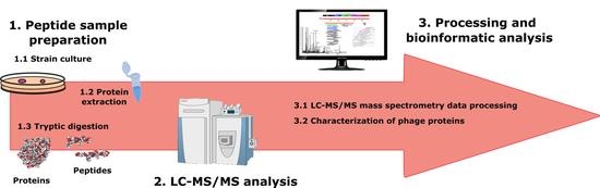
2.1. Bacteria
2.2. Protein Extraction and Peptide Sample Preparation
2.3. Shotgun LC-MS/MS Analysis
2.4. LC-MS/MS Mass Spectrometry Data Processing
2.5. Selection of Potential Peptide Biomarkers
2.6. Phage Genome Comparison and Relatedness
3. Results
3.1. S. aureus Proteome Repository
3.2. Phage Peptides Determined from the Analyzed S. aureus Strains
3.3. Staphylococcus spp. Phage Genome Comparisons and Their Relatedness
3.4. Identification of Peptides of Virulence Factors
4. Discussion
Supplementary Materials
Author Contributions
Funding
Institutional Review Board Statement
Informed Consent Statement
Data Availability Statement
Acknowledgments
Conflicts of Interest
References
- Böhme, K.; Morandi, S.; Cremonesi, P.; Fernández No, I.C.; Barros-Velázquez, J.; Castiglioni, B.; Brasca, M.; Cañas, B.; Calo-Mata, P. Characterization of Staphylococcus aureus strains isolated from Italian dairy products by MALDI-TOF mass fingerprinting. Electrophoresis 2012, 33, 2355–2364. [Google Scholar] [CrossRef]
- Forsman, P.; Tilsala-Timisjärvi, A.; Alatossava, T. Identification of staphylococcal and streptococcal causes of bovine mastitis using 16S-23S rRNA spacer regions. Microbiology 1997, 143, 3491–3500. [Google Scholar] [CrossRef] [Green Version]
- Carrera, M.; Böhme, K.; Gallardo, J.M.; Barros-Velázquez, J.; Cañas, B.; Calo-Mata, P. Characterization of foodborne strains of Staphylococcus aureus by shotgun proteomics: Functional networks, virulence factors and species-specific peptide biomarkers. Front. Microbiol. 2017, 8, 2458. [Google Scholar] [CrossRef] [PubMed] [Green Version]
- Rainard, P.; Foucras, G.; Fitzgerald, J.R.; Watts, J.L.; Koop, G.; Middleton, J.R. Knowledge gaps and research priorities in Staphylococcus aureus mastitis control. Transbound. Emerg. Dis. 2018, 65, 149–165. [Google Scholar] [CrossRef] [Green Version]
- Abril, A.G.; Villa, T.G.; Barros-Velázquez, J.; Cañas, B.; Sánchez-Pérez, A.; Calo-Mata, P.; Carrera, M. Staphylococcus aureus exotoxins and their detection in the dairy industry and mastitis. Toxins 2020, 12, 537. [Google Scholar] [CrossRef]
- Xia, G.; Wolz, C. Phages of Staphylococcus aureus and their impact on host evolution. Infect. Genet. Evol. 2014, 21, 593–601. [Google Scholar] [CrossRef] [PubMed]
- Fortier, L.C.; Sekulovic, O. Importance of prophages to evolution and virulence of bacterial pathogens. Virulence 2013, 4, 354–365. [Google Scholar] [CrossRef] [PubMed]
- Menouni, R.; Hutinet, G.; Petit, M.A.; Ansaldi, M. Bacterial genome remodeling through bacteriophage recombination. FEMS Microbiol. Lett. 2015, 362, 1–10. [Google Scholar] [CrossRef] [Green Version]
- Deghorain, M.; Van Melderen, L. The staphylococci phages family: An overview. Viruses 2012, 4, 3316–3335. [Google Scholar] [CrossRef] [Green Version]
- Feiner, R.; Argov, T.; Rabinovich, L.; Sigal, N.; Borovok, I.; Herskovits, A.A. A new perspective on lysogeny: Prophages as active regulatory switches of bacteria. Nat. Rev. Microbiol. 2015, 13, 641–650. [Google Scholar] [CrossRef]
- Penadés, J.R.; Chen, J.; Quiles-Puchalt, N.; Carpena, N.; Novick, R.P. Bacteriophage-mediated spread of bacterial virulence genes. Curr. Opin. Microbiol. 2015, 23, 171–178. [Google Scholar] [CrossRef]
- Brüssow, H.; Desiere, F. Comparative phage genomics and the evolution of Siphoviridae: Insights from dairy phages. Mol. Microbiol. 2001, 39, 213–222. [Google Scholar] [CrossRef]
- Canchaya, C.; Fournous, G.; Brüssow, H. The impact of prophages on bacterial chromosomes. Mol. Microbiol. 2004, 53, 9–18. [Google Scholar] [CrossRef]
- Uchiyama, J.; Taniguchi, M.; Kurokawa, K.; Takemura-Uchiyama, I.; Ujihara, T.; Shimakura, H.; Sakaguchi, Y.; Murakami, H.; Sakaguchi, M.; Matsuzaki, S. Adsorption of Staphylococcus viruses S13’ and S24-1 on Staphylococcus aureus strains with different glycosidic linkage patterns of wall teichoic acids. J. Gen. Virol. 2017, 98, 2171–2180. [Google Scholar] [CrossRef]
- Moon, B.Y.; Park, J.Y.; Hwang, S.Y.; Robinson, D.A.; Thomas, J.C.; Fitzgerald, J.R.; Park, Y.H.; Seo, K.S. Phage-mediated horizontal transfer of a Staphylococcus aureus virulence-associated genomic island. Sci. Rep. 2015, 5, 9784. [Google Scholar] [CrossRef] [Green Version]
- Koskella, B.; Brockhurst, M.A. Bacteria-phage coevolution as a driver of ecological and evolutionary processes in microbial communities. FEMS Microbiol. Rev. 2014, 38, 916–931. [Google Scholar] [CrossRef] [Green Version]
- Chakravorty, S.; Helb, D.; Burday, M.; Connell, N.; Alland, D. A detailed analysis of 16S ribosomal RNA gene segments for the diagnosis of pathogenic bacteria. J. Microbiol. Methods 2007, 69, 330–339. [Google Scholar] [CrossRef] [Green Version]
- Ivnitski, D.; Abdel-hamid, I.; Atanasov, P.; Wilkins, E. Biosensors for detection of pathogenic bacteria. Biosens. Bioelectron. 1999, 14, 599–624. [Google Scholar] [CrossRef]
- Abril, A.G.; Carrera, M.; Böhme, K.; Barros, J.; CANAS, B.; Rama, J.L.R.; Villa, T.G.; Calo-Mata, P. Characterization of bacteriophage peptides of pathogenic Streptococcus by LC-ESI-MS/MS: Bacteriophage phylogenomics and their relationship to their host. Front. Microbiol. 2020, 11, 1241. [Google Scholar] [CrossRef]
- Gantzer, C.; Maul, A.; Audic, J.M.; Pharmacie, D. Detection of infectious enteroviruses, enterovirus genomes, somatic coliphages, and bacteroides fragilis phages in treated wastewater. Appl. Environ. Microbiol. 1998, 64, 4307–4312. [Google Scholar] [CrossRef] [Green Version]
- Böhme, K.; Fernández-No, I.C.; Barros-Velázquez, J.; Gallardo, J.M.; Cañas, B.; Calo-Mata, P. Rapid species identification of seafood spoilage and pathogenic Gram-positive bacteria by MALDI-TOF mass fingerprinting. Electrophoresis 2011, 32, 2951–2965. [Google Scholar] [CrossRef] [PubMed]
- Branquinho, R.; Sousa, C.; Lopes, J.; Pintado, M.E.; Peixe, L.V.; Osorio, H. Differentiation of Bacillus pumilus and Bacillus safensis using MALDI-TOF-MS. PLoS ONE 2014, 9, e110127. [Google Scholar] [CrossRef] [PubMed]
- Lasch, P.; Beyer, W.; Nattermann, H.; Stämmler, M.; Siegbrecht, E.; Grunow, R.; Naumann, D. Identification of Bacillus anthracis by using matrix-assisted laser desorption ionization-time of flight mass spectrometry and artificial neural networks. Appl. Environ. Microbiol. 2009, 75, 7229–7242. [Google Scholar] [CrossRef] [PubMed] [Green Version]
- Quintela-Baluja, M.; Böhme, K.; Fernández-No, I.C.; Alnakip, M.E.; Caamano, S.; Barros-Velázques, J.; Calo-mata, P. MALDI-TOF Mass Spectrometry, a rapid and reliable method for the identification of bacterial species in food-microbiology Laboratories. Nov. Food Preserv. Microb. Assess. Tech. 2014, 353–385. [Google Scholar]
- Craigie, J.; Yen, C.H. The Demonstration of Types of B. typhosus by means of preparations of type ii vi phage: I. Principles and Technique on JSTOR. Can. J. Public Health 1938, 29, 484–496. [Google Scholar]
- Chanishvili, N. Nanotechnology to Aid Chemical and Biological Defense; Springer: Berlin/Heidelberg, Germany, 2015; pp. 17–33. [Google Scholar]
- Lavigne, R.; Ceyssens, P.; Robben, J. Phage proteomics: Applications of mass spectrometry. In Bacteriophages; Humana Press: Totowa, NJ, USA, 2009; Volume 502, pp. 239–251. [Google Scholar]
- Rees, J.C.; Voorhees, K.J. Simultaneous detection of two bacterial pathogens using bacteriophage amplification coupled with matrix-assisted laser desorption/ionization time-of-flight mass spectrometry. Rapid Commun. Mass Spectrom. 2005, 19, 2757–2761. [Google Scholar] [CrossRef]
- Richter, Ł.; Janczuk-richter, M.; Niedzió, J.; Paczesny, J.; Ho, R. Recent advances in bacteriophage-based methods for bacteria detection. Drug Discov. Today 2018, 23, 448–455. [Google Scholar] [CrossRef]
- Singh, A.; Poshtiban, S.; Evoy, S. Recent advances in bacteriophage based biosensors for food-borne pathogen detection. Sensors 2013, 13, 1763–1786. [Google Scholar] [CrossRef] [PubMed] [Green Version]
- Calo-Mata, P.; Carrera, M.; Böhme, K.; Caamaño-Antelo, S.; Gallardo, J.M.; Barros-Velázquez, J.; Cañas, B. Novel Peptide Biomarker discovery for detection and identification of bacterial pathogens by LC-ESI-MS/MS. J. Anal. Bioanal. Tech. 2016, 7, 296. [Google Scholar]
- Pfrunder, S.; Grossmann, J.; Hunziker, P.; Brunisholz, R.; Gekenidis, M.-T.; Drissner, D. Bacillus cereus group-type strain-specific diagnostic peptides. J. Proteome Res. 2016, 15, 3098–3107. [Google Scholar]
- Serafim, V.; Ring, C.; Pantoja, L.; Shah, H.S.A. Rapid identification of E. coli bacteriophages using Mass Spectrometry. J. Proteom. Enzymol. 2017, 6, 1000130. [Google Scholar]
- Morandi, S.; Brasca, M.; Lodi, R.; Cremonesi, P.; Castiglioni, B. Detection of classical enterotoxins and identification of enterotoxin genes in Staphylococcus aureus from milk and dairy products. Vet. Microbiol. 2007, 124, 66–72. [Google Scholar] [CrossRef]
- Giebel, R.; Worden, C.; Rust, S.M.; Kleinheinz, G.T.; Robbins, M.; Sandrin, T.R. Microbial fingerprinting using matrix-assisted laser desorption ionization time-of-flight mass spectrometry (MALDI-TOF MS) applications and challenges. Adv. Appl. Microbiol. 2010, 71, 149–184. [Google Scholar]
- Böhme, K.; Fernández-No, I.C.; Barros-Velázquez, J.; Gallardo, J.M.; Calo-Mata, P.; Cañas, B. Species differentiation of seafood spoilage and pathogenic gram-negative bacteria by MALDI-TOF mass fingerprinting. J. Proteome Res. 2010, 9, 3169–3183. [Google Scholar] [CrossRef]
- Böhme, K.; Fernández-No, I.C.; Barros-Velázquez, J.; Gallardo, J.M.; Cañas, B.; Calo-Mata, P. Comparative analysis of protein extraction methods for the identification of seafood-borne pathogenic and spoilage bacteria by MALDI-TOF mass spectrometry. Anal. Methods 2010, 2, 1941. [Google Scholar] [CrossRef] [Green Version]
- Carrera, M.; Cañas, B.; Gallardo, J.M. The sarcoplasmic fish proteome: Pathways, metabolic networks and potential bioactive peptides for nutritional inferences. J. Proteomics 2013, 78, 211–220. [Google Scholar] [CrossRef]
- Käll, L.; Canterbury, J.D.; Weston, J.; Noble, W.S.; MacCoss, M.J. Semi-supervised learning for peptide identification from shotgun proteomics datasets. Nat. Methods 2007, 4, 923–925. [Google Scholar] [CrossRef]
- Altschul, S.F.; Gish, W.; Miller, W.; Myers, E.W.; Lipman, D.J. Basic local alignment search tool. J. Mol. Biol. 1990, 215, 403–410. [Google Scholar] [CrossRef]
- Meier-Kolthoff, J.P.; Göker, M. VICTOR: Genome-based phylogeny and classification of prokaryotic viruses. Bioinformatics 2017, 33, 3396–3404. [Google Scholar] [CrossRef] [Green Version]
- Lucchini, S.; Desiere, F.; Brüssow, H. Similarly organized lysogeny modules in temperate Siphoviridae from low GC content gram-positive bacteria. Virology 1999, 263, 427–435. [Google Scholar] [CrossRef] [Green Version]
- Bateman, A.; Coggill, P.; Finn, R.D. DUFs: Families in search of function. Acta Crystallogr. Sect. F Struct. Biol. Cryst. Commun. 2010, 66, 1148–1152. [Google Scholar] [CrossRef] [PubMed] [Green Version]
- Goodacre, N.F.; Gerloff, D.L.; Uetz, P. protein domains of unknown function are essential in Bacteria. MBio 2014, 5, e00744-13. [Google Scholar] [CrossRef] [PubMed] [Green Version]
- Durante-Rodríguez, G.; Mancheño, J.M.; Díaz, E.; Carmona, M. Refactoring the λ phage lytic/lysogenic decision with a synthetic regulator. Microbiologyopen 2016, 5, 575–581. [Google Scholar] [CrossRef] [Green Version]
- Van Wamel, W.J.; Rooijakkers, S.H.; Ruyken, M.; van Kessel, K.P.; van Strijp, J.A. The Innate Immune Modulators staphylococcal complement inhibitor and chemotaxis inhibitory protein of Staphylococcus aureus are located on β the innate immune modulators staphylococcal complement inhibitor and chemotaxis inhibitory protein of Staphylococcus. J. Bacteriol. 2006, 188, 1310–1315. [Google Scholar] [PubMed] [Green Version]
- Gual, A.; Camacho, A.G.; Alonso, J.C. Functional analysis of terminase large subunit, G2P, of Bacillus subtilis bacteriophage SPP1. J. Biol. Chem. 2000, 275, 35311–35319. [Google Scholar] [CrossRef] [PubMed] [Green Version]
- Simmonds, P. Methods for virus classification and the challenge of incorporating metagenomic sequence data. J. Gen. Virol. 2015, 96, 1193–1206. [Google Scholar] [CrossRef] [PubMed]
- Argov, T.; Azulay, G.; Pasechnek, A.; Stadnyuk, O.; Ran-Sapir, S.; Borovok, I.; Sigal, N.; Herskovits, A.A. Temperate bacteriophages as regulators of host behavior. Curr. Opin. Microbiol. 2017, 38, 81–87. [Google Scholar] [CrossRef] [PubMed]
- Angelopoulou, A.; Warda, A.K.; Hill, C.; Ross, R.P. Non-antibiotic microbial solutions for bovine mastitis–live biotherapeutics, bacteriophage, and phage lysins. Crit. Rev. Microbiol. 2019, 45, 564–580. [Google Scholar] [CrossRef]
- Lin, D.M.; Koskella, B.; Lin, H.C. Phage therapy: An alternative to antibiotics in the age of multi-drug resistance. World J. Gastrointest. Pharmacol. Ther. 2017, 8, 162. [Google Scholar] [CrossRef]
- Dams, D.; Briers, Y. Enzybiotics: Enzyme-based antibacterials as therapeutics. In Advances in Experimental Medicine and Biology; Springer: New York, NY, USA, 2019; Volume 1148, pp. 233–253. [Google Scholar]
- Ganaie, M.Y.; Qureshi, S.; Kashoo, Z.; Wani, S.A.; Hussain, M.I.; Kumar, R.; Maqbool, R.; Sikander, P.; Banday, M.S.; Malla, W.A.; et al. Isolation and characterization of two lytic bacteriophages against Staphylococcus aureus from India: Newer therapeutic agents against Bovine mastitis. Vet. Res. Commun. 2018, 42, 289–295. [Google Scholar] [CrossRef]
- Geng, H.; Zou, W.; Zhang, M.; Xu, L.; Liu, F.; Li, X.; Wang, L.; Xu, Y. Evaluation of phage therapy in the treatment of Staphylococcus aureus-induced mastitis in mice. Folia Microbiol. 2019, 65, 339–351. [Google Scholar] [CrossRef]
- Kwan, T.; Liu, J.; DuBow, M.; Gros, P.; Pelletier, J. The complete genomes and proteomes of 27 Staphylococcus aureus bacteriophages. Proc. Natl. Acad. Sci. USA 2005, 102, 5174–5179. [Google Scholar] [CrossRef] [Green Version]
- Bae, T.; Baba, T.; Hiramatsu, K.; Schneewind, O. Prophages of Staphylococcus aureus Newman and their contribution to virulence. Mol. Microbiol. 2006, 62, 1035–1047. [Google Scholar] [CrossRef] [PubMed]
- Kuroda, M.; Ohta, T.; Uchiyama, I.; Baba, T.; Yuzawa, H.; Kobayashi, I.; Cui, L.; Oguchi, A.; Aoki, K.; Nagai, Y.; et al. Whole genome sequencing of meticillin-resistant Staphylococcus aureus. Lancet 2001, 357, 1225–1240. [Google Scholar] [CrossRef]
- Keary, R.; McAuliffe, O.; Ross, R.P.; Hill, C.; O’Mahony, J.; Coffey, A. Genome analysis of the staphylococcal temperate phage DW2 and functional studies on the endolysin and tail hydrolase. Bacteriophage 2014, 4, e28451. [Google Scholar] [CrossRef]
- Van der Mee-Marquet, N.; Corvaglia, A.R.; Valentin, A.S.; Hernandez, D.; Bertrand, X.; Girard, M.; Kluytmans, J.; Donnio, P.Y.; Quentin, R.; François, P. Analysis of prophages harbored by the human-adapted subpopulation of Staphylococcus aureus CC398. Infect. Genet. Evol. 2013, 18, 299–308. [Google Scholar] [CrossRef] [Green Version]
- García, P.; Martínez, B.; Obeso, J.M.; Lavigne, R.; Lurz, R.; Rodríguez, A. Functional genomic analysis of two Staphylococcus aureus phages isolated from the dairy environment. Appl. Environ. Microbiol. 2009, 75, 7663–7673. [Google Scholar] [CrossRef] [Green Version]
- Yoon, H.; Yun, J.; Lim, J.A.; Roh, E.; Jung, K.S.; Chang, Y.; Ryu, S.; Heu, S. Characterization and genomic analysis of two Staphylococcus aureus bacteriophages isolated from poultry/livestock farms. J. Gen. Virol. 2013, 94, 2569–2576. [Google Scholar] [CrossRef]
- Iandolo, J.J.; Worrell, V.; Groicher, K.H.; Qian, Y.; Tian, R.; Kenton, S.; Dorman, A.; Ji, H.; Lin, S.; Loh, P.; et al. Comparative analysis of the genomes of the temperate bacteriophages φ11, φ12 and φ13 of Staphylococcus aureus 8325. Gene 2002, 289, 109–118. [Google Scholar] [CrossRef]
- Zhang, M.; Ito, T.; Li, S.; Jin, J.; Takeuchi, F.; Lauderdale, T.-L.Y.; Higashide, M.; Hiramatsu, K. Identification of the third type of PVL phage in ST59 methicillin-resistant Staphylococcus aureus (MRSA) strains. FEMS Microbiol. Lett. 2011, 323, 20–28. [Google Scholar] [CrossRef] [PubMed] [Green Version]
- El Haddad, L.; Moineau, S. Characterization of a novel panton-valentine leukocidin (PVL)-encoding staphylococcal phage and its naturally PVL-lacking variant. Appl. Environ. Microbiol. 2013, 79, 2828–2832. [Google Scholar] [CrossRef] [PubMed] [Green Version]
- Liu, J.; Dehbi, M.; Moeck, G.; Arhin, F.; Banda, P.; Bergeron, D.; Callejo, M.; Ferretti, V.; Ha, N.; Kwan, T.; et al. Antimicrobial drug discovery through bacteriophage genomics. Nat. Biotechnol. 2004, 22, 185–191. [Google Scholar] [CrossRef] [PubMed]
- Kraushaar, B.; Hammerl, J.A.; Kienöl, M.; Heinig, M.L.; Sperling, N.; Thanh, M.D.; Reetz, J.; Jäckel, C.; Fetsch, A.; Hertwig, S. Acquisition of virulence factors in livestock-associated MRSA: Lysogenic conversion of CC398 strains by virulence gene-containing phages. Sci. Rep. 2017, 7, 1–13. [Google Scholar] [CrossRef] [Green Version]
- Narita, S.; Kaneko, J.; Chiba, J.I.; Piémont, Y.; Jarraud, S.; Etienne, J.; Kamio, Y. Phage conversion of Panton-Valentine leukocidin in Staphylococcus aureus: Molecular analysis of a PVL-converting phage, φSLT. Gene 2001, 268, 195–206. [Google Scholar] [CrossRef]
- Chang, Y.; Lee, J.H.; Shin, H.; Heu, S.; Ryu, S. Characterization and complete genome sequence analysis of Staphylococcus aureus bacteriophage SA12. Virus Genes 2013, 47, 389–393. [Google Scholar] [CrossRef]
- Xiao, X.M.; Ito, T.; Kondo, Y.; Cho, M.; Yoshizawa, Y.; Kaneko, J.; Katai, A.; Higashiide, M.; Li, S.; Hiramatsu, K. Two different Panton-Valentine leukocidin phage lineages predominate in Japan. J. Clin. Microbiol. 2008, 46, 3246–3258. [Google Scholar]
- Kaneko, J.; Kimura, T.; Narita, S.; Tomita, T.; Kamio, Y. Complete nucleotide sequence and molecular characterization of the temperate staphylococcal bacteriophage φPVL carrying Panton-Valentine leukocidin genes. Gene 1998, 215, 57–67. [Google Scholar] [CrossRef]
- Mariem, B.J.J.; Ito, T.; Zhang, M.; Jin, J.; Li, S.; Ilhem, B.B.B.; Adnan, H.; Han, X.; Hiramatsu, K. Molecular characterization of methicillin-resistant Panton-valentine leukocidin positive Staphylococcus aureus clones disseminating in Tunisian hospitals and in the community. BMC Microbiol. 2013, 13, 2. [Google Scholar] [CrossRef] [PubMed] [Green Version]
- Christie, G.E.; Matthews, A.M.; King, D.G.; Lane, K.D.; Olivarez, N.P.; Tallent, S.M.; Gill, S.R.; Novick, R.P. The complete genomes of Staphylococcus aureus bacteriophages 80 and 80α-Implications for the specificity of SaPI mobilization. Virology 2010, 407, 381–390. [Google Scholar] [CrossRef] [Green Version]
- Frígols, B.; Quiles-Puchalt, N.; Mir-Sanchis, I.; Donderis, J.; Elena, S.F.; Buckling, A.; Novick, R.P.; Marina, A.; Penadés, J.R. Virus Satellites Drive Viral Evolution and Ecology. PLoS Genet. 2015, 11, e1005609. [Google Scholar] [CrossRef]
- Botka, T.; Růžičková, V.; Konečná, H.; Pantůček, R.; Rychlík, I.; Zdráhal, Z.; Petráš, P.; Doškař, J. Complete genome analysis of two new bacteriophages isolated from impetigo strains of Staphylococcus aureus. Virus Genes 2015, 51, 122–131. [Google Scholar] [CrossRef]
- Yamaguchi, T.; Hayashi, T.; Takami, H.; Nakasone, K.; Ohnishi, M.; Nakayama, K.; Yamada, S.; Komatsuzawa, H.; Sugai, M. Phage conversion of exfoliative toxin A production in Staphylococcus aureus. Mol. Microbiol. 2000, 38, 694–705. [Google Scholar] [CrossRef] [PubMed]
- Santiago-Rodriguez, T.M.; Naidu, M.; Jones, M.B.; Ly, M.; Pride, D.T. Identification of staphylococcal phage with reduced transcription in human blood through transcriptome sequencing. Front. Microbiol. 2015, 6, 216. [Google Scholar] [CrossRef] [PubMed]
- Matsuzaki, S.; Yasuda, M.; Nishikawa, H.; Kuroda, M.; Ujihara, T.; Shuin, T.; Shen, Y.; Jin, Z.; Fujimoto, S.; Nasimuzzaman, M.D.; et al. Experimental protection of mice against lethal Staphylococcus aureus infection by novel bacteriophage φMR11. J. Infect. Dis. 2003, 187, 613–624. [Google Scholar] [CrossRef] [PubMed] [Green Version]
- Carroll, D.; Kehoe, M.A.; Cavanagh, D.; Coleman, D.C. Novel organization of the site-specific integration and excision recombination functions of the Staphylococcus aureus serotype F virulence-converting phages φ13 and φ42. Mol. Microbiol. 1995, 16, 877–893. [Google Scholar] [CrossRef] [PubMed]
- Hoshiba, H.; Uchiyama, J.; Kato, S.I.; Ujihara, T.; Muraoka, A.; Daibata, M.; Wakiguchi, H.; Matsuzaki, S. Isolation and characterization of a novel Staphylococcus aureus bacteriophage, φMR25, and its therapeutic potential. Arch. Virol. 2010, 155, 545–552. [Google Scholar] [CrossRef] [PubMed]
- Varga, M.; Pantůček, R.; Růžičková, V.; Doškař, J. Molecular characterization of a new efficiently transducing bacteriophage identified in meticillin-resistant Staphylococcus aureus. J. Gen. Virol. 2016, 97, 258–268. [Google Scholar] [CrossRef]
- Pantůček, R.; Doškař, J.; Růžičková, V.; Kašpárek, P.; Oráčová, E.; Kvardová, V.; Rosypal, S. Identification of bacteriophage types and their carriage in Staphylococcus aureus. Arch. Virol. 2004, 149, 1689–1703. [Google Scholar] [CrossRef]
- Chang, Y.; Shin, H.; Lee, J.-H.; Park, C.; Paik, S.-Y.; Ryu, S. Isolation and genome characterization of the virulent Staphylococcus aureus bacteriophage SA97. Viruses 2015, 7, 5225–5242. [Google Scholar] [CrossRef] [Green Version]
- Zou, D.; Kaneko, J.; Narita, S.; Kamio, Y. Prophage, φpv83-pro, carrying panton-valentine leukocidin genes, on the Staphylococcus aureus p83 chromosome: Comparative analysis of the genome structures of φpv83-pro, φpvl, φ11, and other phages. Biosci. Biotechnol. Biochem. 2000, 64, 2631–2643. [Google Scholar] [CrossRef] [Green Version]
- Utter, B.; Deutsch, D.R.; Schuch, R.; Winer, B.Y.; Verratti, K.; Bishop-Lilly, K.; Sozhamannan, S.; Fischetti, V.A. Beyond the Chromosome: The prevalence of unique extra-chromosomal bacteriophages with integrated virulence genes in pathogenic Staphylococcus aureus. PLoS ONE 2014, 9, e100502. [Google Scholar] [CrossRef]
- Sanchini, A.; Del Grosso, M.; Villa, L.; Ammendolia, M.G.; Superti, F.; Monaco, M.; Pantosti, A. Typing of Panton-Valentine leukocidin-encoding phages carried by methicillin-susceptible and methicillin-resistant Staphylococcus aureus from Italy. Clin. Microbiol. Infect. 2014, 20, O840–O846. [Google Scholar] [CrossRef] [PubMed] [Green Version]
- Jia, H.; Bai, Q.; Yang, Y.; Yao, H. Complete genome sequence of Staphylococcus aureus siphovirus phage JS01. Genome Announc. 2013, 1, 797–810. [Google Scholar] [CrossRef] [PubMed] [Green Version]
- Jeon, J.; D’Souza, R.; Hong, S.K.; Lee, Y.; Yong, D.; Choi, J.; Lee, K.; Chong, Y. Complete genome sequence of the bacteriophage YMC/09/04/R1988 MRSA BP: A lytic phage from a methicillin-resistant Staphylococcus aureus isolate. FEMS Microbiol. Lett. 2014, 359, 144–146. [Google Scholar] [CrossRef] [PubMed]
- Zeman, M.; Mašlaňová, I.; Indráková, A.; Šiborová, M.; Mikulášek, K.; Bendíčková, K.; Plevka, P.; Vrbovská, V.; Zdráhal, Z.; Doškař, J.; et al. Staphylococcus sciuri bacteriophages double-convert for staphylokinase and phospholipase, mediate interspecies plasmid transduction, and package mecA gene. Sci. Rep. 2017, 7, 46319. [Google Scholar] [CrossRef] [PubMed]
- Perez-Riverol, Y.; Csordas, A.; Bai, J.; Bernal-Llinares, M.; Hewapathirana, S.; Kundu, D.J.; Inuganti, A.; Griss, J.; Mayer, G.; Eisenacher, M.; et al. The PRIDE database and related tools and resources in 2019: Improving support for quantification data. Nucleic Acids Res. 2019, 47, 442–450. [Google Scholar] [CrossRef]

 , yellow-filled diamond IQQLADYFNVPK (cluster A-specific),
  , brown-filled diamond HAGYVRC*KLF (cluster A-specific),
  , black-outlined diamond IYDRNSDTLDGLPVVNLK (cluster A.1-specific),
  , red=outlined diamond AVAELLKEINR (cluster A.2-specific),
  , pink-filled diamond KSNVEAFSNAVK (cluster A.1),
  , gray-filled diamond QKNVLNYANEQLDEQNKV (cluster A.1),
  , brown-outlined diamond MPVYKDGNTGKWYFSI (cluster A-specific),
  , dark gray-filled diamond KLYIIEEYVKQGM (cluster A.1-specific),
  , purple-outlined diamond EVPNEPDYIVIDVC*EDYSASK (cluster A.1-specific),
  , orange-filled diamond AYINITGLGFAK (cluster B.1-specific),
  , yellow-outlined diamond TSIELITGFTK (cluster B.2-specific),
  , red-filled diamond VSYTLDDDDFITDVETAK (cluster D-specific),
  , green-filled diamond LLHALPTGNDSGGDKLLPK (cluster D-specific),
  , black-filled diamond RVSYTLDDDDFITDVETAKELKL (cluster D-specific),
  , purple-filled diamond LYVGVFNPEATK (cluster D-specific,
  , blue-filled diamond ELAEAIGVSQPTVSNWIQQTK (cluster D-specific);
  , light green-filled diamond VLEMIFLGEDPK (cluster D-specific),
  , orange-outlined diamond KAMIKASPK (cluster D-specific) and
  , gray-outlined diamond GMPTGTNVYAVKGGIADK (cluster D-specific).
, yellow-filled diamond IQQLADYFNVPK (cluster A-specific), , brown-filled diamond HAGYVRC*KLF (cluster A-specific), , black-outlined diamond IYDRNSDTLDGLPVVNLK (cluster A.1-specific), , red=outlined diamond AVAELLKEINR (cluster A.2-specific), , pink-filled diamond KSNVEAFSNAVK (cluster A.1), , gray-filled diamond QKNVLNYANEQLDEQNKV (cluster A.1), , brown-outlined diamond MPVYKDGNTGKWYFSI (cluster A-specific), , dark gray-filled diamond KLYIIEEYVKQGM (cluster A.1-specific), , purple-outlined diamond EVPNEPDYIVIDVC*EDYSASK (cluster A.1-specific), , orange-filled diamond AYINITGLGFAK (cluster B.1-specific), , yellow-outlined diamond TSIELITGFTK (cluster B.2-specific), , red-filled diamond VSYTLDDDDFITDVETAK (cluster D-specific), , green-filled diamond LLHALPTGNDSGGDKLLPK (cluster D-specific), , black-filled diamond RVSYTLDDDDFITDVETAKELKL (cluster D-specific), , purple-filled diamond LYVGVFNPEATK (cluster D-specific, , blue-filled diamond ELAEAIGVSQPTVSNWIQQTK (cluster D-specific); , light green-filled diamond VLEMIFLGEDPK (cluster D-specific), , orange-outlined diamond KAMIKASPK (cluster D-specific) and , gray-outlined diamond GMPTGTNVYAVKGGIADK (cluster D-specific).
, gray-outlined diamond GMPTGTNVYAVKGGIADK (cluster D-specific).
, yellow-filled diamond IQQLADYFNVPK (cluster A-specific), , brown-filled diamond HAGYVRC*KLF (cluster A-specific), , black-outlined diamond IYDRNSDTLDGLPVVNLK (cluster A.1-specific), , red=outlined diamond AVAELLKEINR (cluster A.2-specific), , pink-filled diamond KSNVEAFSNAVK (cluster A.1), , gray-filled diamond QKNVLNYANEQLDEQNKV (cluster A.1), , brown-outlined diamond MPVYKDGNTGKWYFSI (cluster A-specific), , dark gray-filled diamond KLYIIEEYVKQGM (cluster A.1-specific), , purple-outlined diamond EVPNEPDYIVIDVC*EDYSASK (cluster A.1-specific), , orange-filled diamond AYINITGLGFAK (cluster B.1-specific), , yellow-outlined diamond TSIELITGFTK (cluster B.2-specific), , red-filled diamond VSYTLDDDDFITDVETAK (cluster D-specific), , green-filled diamond LLHALPTGNDSGGDKLLPK (cluster D-specific), , black-filled diamond RVSYTLDDDDFITDVETAKELKL (cluster D-specific), , purple-filled diamond LYVGVFNPEATK (cluster D-specific, , blue-filled diamond ELAEAIGVSQPTVSNWIQQTK (cluster D-specific); , light green-filled diamond VLEMIFLGEDPK (cluster D-specific), , orange-outlined diamond KAMIKASPK (cluster D-specific) and , gray-outlined diamond GMPTGTNVYAVKGGIADK (cluster D-specific).

| Strain | Protein | Peptide | Bacteria with 100% Homology Based on the NCBI Protein Database | Phages with 100% Homology Based on the NCBI Protein Database |
|---|---|---|---|---|
| S4 | Uncharacterized phage protein | IRLPYYDVK | Staphylococcus aureus | Staphylococcus phage StauST398-2 |
| S4 | Uncharacterized phage protein | AVAELLKEINR | Staphylococcus argenteus Staphylococcus simiae Staphylococcus aureus | Staphylococcus virus 71 Staphylococcus virus 55 Staphylococcus virus 88 |
| S4 | Major capsid protein | LLHALPTGNDSGGDKLLPK | Staphylococcus aureus Staphylococcus xylosus Staphylococcus muscae Staphylococcus haemolyticus Staphylococcus argenteus Streptococcus pneumoniae | Staphylococcus phage phiSa2wa_st72 Staphylococcus phage phiSa2wa_st121mssa Staphylococcus phage vB_SauS_phi2 Staphylococcus phage StauST398-2 Staphylococcus phage LH1 Staphylococcus phage phiSa2wa_st30 Staphylococcus virus phi12 Staphylococcus virus 3a Staphylococcus virus phiSLT Staphylococcus phage tp310-2 Staphylococcus phage vB_SauS_JS02 Staphylococcus phage R4 Staphylococcus phage vB_SauS_fPfSau02 Staphylococcus phage SA137ruMSSAST121PVL |
| S4 | Major capsid protein | RVSYTLDDDDFITDVETAKELKL | Staphylococcus aureus 12S01399 Staphylococcus aureus Staphylococcus aureus A9299 Staphylococcus aureus A9765 Staphylococcus argenteus Staphylococcus aureus A6300 Staphylococcus sp. Terrabacteria group Escherichia coli | Staphylococcus phage LH1 Staphylococcus phage StauST398-2 Staphylococcus phage vB_SauS_phi2 Staphylococcus phage R4 |
| S7 | Major tail protein | LYVGVFNPEATK | Staphylococcus aureus | Staphylococcus phage vB_SauS_ phi2 Staphylococcus virus phi12 Staphylococcus virus phiSLT Staphylococcus phage R4 Staphylococcus phage vB_SauS_JS02 Staphylococcus phage SH-St 15644 Staphylococcus virus 3a Staphylococcus phage P240 |
| S8 | Uncharacterized phage protein | M*NDSNQGLQANPQYTIHYLSQEITR | Staphylococcus aureus | Staphylococcus phage phiN315 |
| S8 | Major tail protein | AYINITGLGFAK | Staphylococcus aureus Staphylococcus argenteus Pararheinheimera mesophila | Staphylococcus phage phiNM3 Staphylococcus phage P282 Staphylococcus phage StauST398-4 Staphylococcus phage phiN315 Staphylococcus phage phi7247PVL Staphylococcus phage phiSa2wa_st22 Staphylococcus virus 77 Staphylococcus phage P954 |
| S9 | Major capsid protein | IYDRNSDTLDGLPVVNLK | Staphylococcus aureus Staphylococcus argenteus | Staphylococcus virus 85 Staphylococcus phage SP5 Staphylococcus virus phiETA2 Staphylococcus phage phiNM2 Staphylococcus virus SAP26 Staphylococcus phage SA12 Staphylococcus virus Baq Sau1 |
| S11 and S20 | Phage repressor, Cro/CI family | ELAEAIGVSQPTVSNWIQQTK | Staphylococcus aureus Staphylococcus argenteus Staphylococcus sciuri | Staphylococcus virus IPLA35 Staphylococcus phage SMSAP5 Staphylococcus phage vB_SauS_phi2 |
| S11 and S20 | Phage repressor, Cro/CI family | IQQLADYFNVPK | Staphylococcus aureus Staphylococcus sciuri Staphylococcus pseudintermedius Staphylococcus devriesei Staphylococcus warneri Staphylococcus capitis Staphylococcus argenteus | Staphylococcus phage SMSAP5 Staphylococcus phage vB_SauS_phi2 Staphylococcus virus IPLA35 |
| S12 S10 and S14 | Complement inhibitor | IYNEIDEALKSK | Staphylococcus aureus, Enterobacter sp. IF2SW-B1 Klebsiella pneumoniae | Staphylococcus phage 13 Staphylococcus phage phiNM3 Staphylococcus phage StauST398-1 |
| S20 | Major capsid protein | VSYTLDDDDFITDVETAK | Staphylococcus aureus Staphylococcus haemolyticus Staphylococcus saprophyticus Staphylococcus warneri Staphylococcus argenteus Streptococcus pneumoniae Staphylococcus sciuri | Staphylococcus phage phiSa2wa_st72 Staphylococcus phage tp310-2 Staphylococcus phage phiSa2wa_st121mssa Staphylococcus phage vB_SauS_phi2 Staphylococcus phage StauST398-2 Staphylococcus virus 3a Staphylococcus phage LH1 Staphylococcus phage phiSa2wa_st30 Staphylococcus virus phi12 Staphylococcus virus phiSLT Staphylococcus phage vB_SauS_JS02 Staphylococcus phage R4 Staphylococcus phage vB_SauS_fPfSau02 Staphylococcus phage SA137ruMSSAST121PVL |
| S20 | Phage protein (DUF2479 domain) | SIINGKLDSQWTVPNEHK | Staphylococcus aureus | Staphylococcus phage DW2 Staphylococcus virus IPLA88 |
| S18 | N-acetylmuramoyl-L-alanine amidase | KEAGNYTVANVK | Bacilli, Staphylococcus argenteus Staphylococcus aureus Staphylococcus sp. HMSC34H10 | Staphylococcus phage tp310-1 Staphylococcus phage tp310-2 Staphylococcus phage phi2958PVL Staphylococcus phage PVL Staphylococcus phage SA137ruMSSAST121PVL Staphylococcus virus IPLA35 |
| S4 | Phage protein NrdI | VETFLENETNQNNLIAVM*SSGNRNWGTNFAIAGDTISK | Staphylococcus haemolyticus Staphylococcus hominis Staphyloccus aureus Staphylococcus aureus subsp. aureus Z172 | |
| S12 | Complement inhibitor | IYNEIDEALK | Staphylococcus. Aureus Klebsiella pneumoniae Enterobacter sp. IF2SW-B1 | Staphylococcus phage StauST398-1 Staphylococcus virus 13 |
| S10 | Complement inhibitor | IYNEIDEALKSKY | Staphylococcus. aureus Klebsiella pneumoniae Enterobacter sp. IF2SW-B2 | Staphylococcus phage StauST398-1 Staphylococcus virus 13 |
| S10 | DDE-type integrase/transposase/recombinase | PC*PALM*NKRNSIATIHR | Staphylococcus aureus | |
| S9 | DNA primase phage-associated | LLHHFYNPENTTALSFNDLNDKFKPANLQGKLVNIAD | Staphylococcus aureus, Staphylococcus haemolyticus Staphylococcus capiti, Staphylococcus epidermidis Staphylococcus warneri Staphylococcus sp. HMSC077D08 Corynebacterium propinquum, Staphylococcus sp. U Staphylococcus lugdunensis Staphylococcus sp. HMSC077B09 | Uncultured Caudovirales Phage |
| S2 | Phage repressor, Cro/CI family | AAHLEGELTDDEWQR | Staphylococcus haemolyticus Staphylococcus warneri Staphylococcus agnetis, Staphylococcus chromogenes Staphylococcus haemolyticus Staphylococcus sp. 58-22 Staphylococcus capitis Staphylococcus pasteuri Bacillales Staphylococcus chromogenes Staphylococcus agnetis Escherichia coli, Staphylococcus aureus 08-02906 Staphylococcus aureus VET0383R, Staphylococcus aureus VET0098R Staphylococcus aureus M1487 Staphylococcus aureus, Staphylococcus aureus A6300 Staphylococcus aureus subsp. aureus str. Newman Staphylococcus aureus subsp. aureus WBG10049, Staphylococcus aureus A9635, Staphylococcus aureus subsp. aureus MN8 | Staphylococcus virus 71 Staphylococcus phage phiSa2wa_st1 Staphylococcus phage phiSa2wa_st5 Staphylococcus phage Henu2 Staphylococcus phage ROSA Staphylococcus phage phi7401PVL |
| S2 | Phage repressor, Cro/CI family | VLDYADYIR | Staphylococcus aureus Staphylococcus epidermidis Staphylococcus warneri Staphylococcus agnetis Staphylococcus warneri Staphylococcus chromogenes, staphylococcus spp. Staphylococcus schleiferi Staphylococcus simulans Staphylococcus haemolyticus, Staphylococcus pettenkoferi Staphylococcus lugdunensis Escherichia coli | Staphylococcus virus 71 Staphylococcus phage phiSa2wa_st1 Staphylococcus phage phiSa2wa_st5 Staphylococcus phage Henu2 Staphylococcus phage ROSA Staphylococcus phage phi7401PVL |
| S9 | DNA-binding protein | SLDNM*SLK | Striga asiática Staphylococcus aureus subsp. aureus 112808A Staphylococcus aureus A8819 Staphylococcus argenteus Staphylococcus spp. Pseudomonas aeruginosa Flectobacillus sp. BAB-3569 Eoetvoesia caeni Arabidopsis thaliana, Coxiellaceae bacterium, Clostridia bacterium | Staphylococcus phage vB_SauS_phi2 |
| S19 | DUF2479, Phage tail fiber, BppU family phage baseplate upper protein | HAGYVRC*KLF | Staphylococcus aureus, Staphylococcus sp. HMSC055H07 Staphylococcus argenteus, Staphylococcus sp. KY49P Staphylococcus sp. HMSC035F11 Pseudomonas aeruginosa Escherichia coli | Staphylococcus phage SA97 Staphylococcus virus 55 uncultured Caudovirales phage Staphylococcus virus 85 Staphylococcus virus 80 Staphylococcus virus phiETA3 Staphylococcus virus phiETA2 Staphylococcus phage 55-2 Staphylococcus phage B166 Staphylococcus phage B236 Staphylococcus virus SAP26 Staphylococcus virus 88 Staphylococcus virus phiETA Staphylococcus virus 11 Staphylococcus phage SP5 Staphylococcus virus 69 Staphylococcus phage ROSA Staphylococcus phage TEM123 Staphylococcus virus 92 Staphylococcus phage StauST398-1 Staphylococcus virus phiNM2 Staphylococcus virus phiNM1 Staphylococcus virus 29 Staphylococcus phage vB_SauS-SAP27 Staphylococcus virus 80alpha Staphylococcus phage HSA84 Staphylococcus virus phiMR11 Staphylococcus phage SAP33 Staphylococcus phage 3MRA |
| S12 | Phage protein (DUF4393 domain) | NSPIDLNSTEISLNNLER | Staphylococcus aureus Staphylococcus spp. Staphylococcus argenteus | Staphylococcus phage StauST398-1 |
| S12 | Phage protein (DUF669 domain) | MNFNLNLQGAQELGN | Staphylococcus capitis Staphylococcus epidermidis Staphylococcus caprae Staphylococcus devriesei Staphylococcus warneri | Staphylococcus virus phiMR11 |
| S10 | GNAT family N-acetyltransferase | IINYARQNNYESLLTSIVSNNIGAK | Staphylococcus aureus Staphylococcus aureus subsp. anaerobius Staphylococcus aureus subsp. aureus Mu50 Staphylococcus hominis Escherichia coli | |
| S5 | Holin, phage phi LC3 family | SQDSNLTPELSTKAPK | Staphylococcus aureus | Staphylococcus phage HSA84 Staphylococcus phage SP5 |
| S6 | ImmA/IrrE family metallo-endopeptidase | EKAKIFGDFDMNDSGVYDEENSTIIYNPLDSITR | Staphylococcus aureus subsp. aureus H19 Staphylococcus aureus Staphylococcus aureus subsp. aureus Staphylococcus aureus subsp. aureus 21204 | |
| S16 | Involved in the expression of fibrinogen-binding protein phage-associated | ESINANTYINQNLEK | Staphylococcus aureus | |
| S16 | Involved in the expression of fibrinogen-binding protein phage-associated | VAVLSTPLVTSFESK | Staphylococcus aureus | |
| S17 | N-6 DNA methylase; N6_Mtase domain-containing protein | KDGEILFDAIDIYLRNK | Staphylococcus aureus | Staphylococcus phage phi-42 |
| S4 | Phage DNA-binding protein | GDM*FVVITIM*MQQIK | Staphylococcus aureus Staphylococcus warneri | |
| S9 | Phage terminase | KLYIIEEYVKQGM | Staphylococcus aureus Staphylococcus argenteus Staphylococcus sp. HMSC58E11 Allobacillus sp. SKP4-8 | Staphylococcus virus Baq_Sau1 Staphylococcus virus phiETA2 Staphylococcus virus 69 Staphylococcus virus 11 Staphylococcus virus 80alpha |
| S14 | Integrase | M*PVYKDGNTGKWYFSI | Staphylococcus aureus | Staphylococcus phage B166 Staphylococcus virus phiMR25 Staphylococcus virus 88 |
| S4 | Phage repressor | ISKVQQLADYFNVPK | Staphylococcus aureus, Staphylococcus chromogenes Staphylococcus hyicus | Staphylococcus virus 80 |
| S13 | Toxin Phage protein; Pathogenicity island protein | NLDGVWLGDLILIKRGLSDR | Staphylococcus aureus, Staphylococcus sp. HMSC58E11, Staphylococcus argenteus, Escherichia coli | Staphylococcus phage phiSa2wa_st80 Staphylococcus phage 3MRA Staphylococcus phage phiSa2wa_st5 |
| S16 | Toxin Phage protein; Pathogenicity island protein | SDREKAGILFEELAHNK | Staphylococcus aureus Escherichia coli Staphylococcus argenteus Staphylococcus sp. HMSC58E11 | Staphylococcus phage 3MRA Staphylococcus phage phiSa2wa_st5 Staphylococcus phage phiSa2wa_st80 Staphylococcus phage phiJB Staphylococcus phage phi7401PVL |
| S6 | PBSX family phage terminase | QADNTYVHHSTYLNNPFISKQFIQEAESAKQR | Staphylococccus spp. | |
| S11 | PBSX family phage terminase | QGVSHLFKVTKSPM*R | Staphylococcus aureus Staphylococcus lentus Staphylococcus sciuri | |
| S20 | Phage-related cell wall hydrolase; Peptidase C51; CHAP domain- | EVPNEPDYIVIDVC*EDYSASK | Staphylococcus argenteus Staphylococcus sp. HMSC36F05 | Staphylococcus virus IPLA88 Staphylococcus virus phiNM2 Staphylococcus phage SAP40 Staphylococcus phage phi 53 Staphylococcus virus phiNM4 Staphylococcus phage SA12 Staphylococcus virus 69 Staphylococcus phage SA97 Staphylococcus phage TEM123 Staphylococcus virus 11 Staphylococcus virus phiMR25 Staphylococcus virus 53 Staphylococcus phage SAP33 |
| S5 | Phage antirepressor Ant | QDWLAM*EVLPAIR | Staphylococcus aureus, Staphylococcus simulans Staphylococcus argenteus Staphylococcus pseudintermedius | Staphylococcus phage SA75 Staphylococcus phage SA13 |
| S11 | Phage capsid protein | M*AEETNSNVTEETEVNE | Staphylococcus, aureus Staphylococcus spp. | |
| S4 | Phage encoded lipoprotein | IHDKELDDPSEEESKLTQEEENSI | Staphylococcus aureus, Staphylococcus capitis, Staphylococcus epidermidis, Staphylococcus cohnii, Staphylococcus haemolyticus | Staphylococcus phage SPbeta-like |
| S2 | Phage head morphogenesis protein | KDVQRIVSHVT | Staphylococcus aureus Staphylococcus argenteus | |
| S9 | YhgE/Pip, Phage infection protein | LNEYM*PNIEKLLNVASNDIPAQFPK | Staphylococcusaureus, Staphylococcus haemolyticus Staphylococcus sp. HMSC34C02 | |
| S14 | Minor structural protein | KTTSEALKEVLSDT | Staphylococcus aureus | |
| S4 | Phage portal protein | EPKPVDATGADDPLKPDDRM*ITNFHANLVDQKVSY | Staphylococcus aureus | |
| S5 | Phage protein | VHISEFKYPLYM*DFLGTKGELE | Staphylococcusaureus Staphylococcus haemolyticus | |
| S15 | Phage protein | MSHNALTTGIGIGAGAG | Staphylococcus aureus | |
| S2 | Phage protein | EITDGEISSVLTM*M* | Staphylococcus aureus, Staphylococcus hominis Staphylococcus epidermidis | |
| S20 | Phage recombination protein Bet | KSSTTYEVNGETVK | Staphylococcus aureus, Staphylococcus sciuri | |
| S2 | Phage resistance protein | ESVDTGEITANTTRTVK | Staphylococcus aureus Staphylococcus fleurettii Staphylococcus pasteuri Staphylococcus epidermidis Staphylococcus warneri Staphylococcus schleiferi Escherichia coli | |
| S13 | Tail tape measure protein | GM*PTGTNVYAVKGGIADK | Staphylococcus aureus, Staphylococcus saprophyticus, Staphylococcus pseudoxylosus | Staphylococcus phage phiSa2wa_st5 Staphylococcus phage phi3A Staphylococcus phage SH-St 15,644 Staphylococcus virus 3a |
| S3 | Tail tape measure protein | VQHPGKLVNKVM*SGLNINFGGGANATAK | Staphylococcus aureus | |
| S4 | Tail tape measure protein | QM*MEGLSGVMDLAAVSGEDLGAVSDIVTDGLTAFGLKAKDSG | Staphylococcus aureus | |
| S2 | Tail tape measure protein | AEEAGVTVKQL | Staphylococcus aureus Staphylococcus cohnii Staphylococcus sp. HMSC061H04 Staphylococcus hominis Staphylococcus capitis Staphylococcus cohnii Staphylococcus sp. HMSC061H04 Staphylococcus sp. HMSC067G10 Staphylococcus Staphylococcus haemolyticus Enterococcus faecium Staphylococcus epidermidis Staphylococcus sp. HMSC067G10 Staphylococcus haemolyticus Enterococcus faecium Staphylococcus epidermidis | Staphylococcus phage SPbeta-like |
| S10 | Phage repressor, Cro/CI family | QKNVLNYANEQLDEQNKV | Staphylococcus aureus, Bacilli, Staphylococcus hyicus Staphylococcus epidermidis | Staphylococcus virus phiNM2 Staphylococcus virus 53 Staphylococcus virus 80alpha |
| S13 | Phage protein | KSNVEAFSNAVK | Staphylococcus aureus | Staphylococcus virus 80alpha Staphylococcus virus phiNM1 Staphylococcus virus phiNM2 |
| S11 | Phage protein | PYHDLSDERIM*EELKK | Staphylococcus aureus Staphylococcus argenteus taphylococcus schweitzeri | Staphylococcus virus phiETA2 Staphylococcus phage P630 Staphylococcus virus SAP26 Staphylococcus phage B236 Staphylococcus virus 88 Staphylococcus prophage phiPV83 |
| S4 | Minor structural protein | LNDNISNINTIV | Pseudomonas aeruginosa E. coli Pararheinheimera mesophila Staphylococcus pseudintermedius Staphylococcus epidermidis, Staphylococcus sp. KY49P Staphylococcus argenteus Staphylococcus schleiferi Staphylococcus hyicus Staphylococcus sp. HMSC063H12 Staphylococcus aureus | Staphylococcus virus 77 Staphylococcus phage P630 Staphylococcus phage SA780ruMSSAST101 Staphylococcus phage phiSa119 Staphylococcus phage phiN315 Staphylococcus phage SA7 Staphylococcus phage JS01 Staphylococcus phage StauST398-4 Staphylococcus virus 13 Staphylococcus phage 23MRA Staphylococcus virus 108PVL Staphylococcus phage phiBU01 Staphylococcus phage PVL Staphylococcus phage tp310-1 Staphylococcus phage P954 Staphylococcus phage SA345ruMSSAST8 Staphylococcus phage phiNM3 Staphylococcus virus 77 Staphylococcus phage phiSa2wa_st22 Staphylococcus phage SA1014ruMSSAST7 Staphylococcus phage P282 Staphylococcus prophage phiPV83 Staphylococcus phage 3 AJ-2017 Staphylococcus phage SAP090B Staphylococcus phage IME1346_01 Staphylococcus phage phi5967PVL Staphylococcus phage P1105 Staphylococcus phage IME1361_01 |
| S9 | PhiETA ORF58-like protein | GMVASMQMQVVQVNVLTM*ELAQQNAMLTQQLTELK | Staphylococcus aureus | |
| S4 | Phage portal protein | TEQLPRLEML | Staphylococcus aureus, Staphylococcus sp. HMSC063A07, Staphylococcus lugdunensis, Staphylococcus sp. HMSC068D08, Staphylococcus sp. HMSC069E09 | |
| S4 | Prophage, terminase | KDRYSSVSY | Staphylococcus aureus, Staphylococcus delphini, Staphylococcus pseudintermedius, Staphylococcus agnetis, Staphylococcus epidermidis, Staphylococcus hominis, Staphylococcus haemolyticus, Paenibacillus sophorae | Staphylococcus phage SPbeta-like |
| S4 | Prophage tail domain; Peptidase | VLEM*IFLGEDPK | Staphylococcus aureus E. coli Bacilli | Staphylococcus phage phi7401PVL Staphylococcus phage phiSa2wa_st121mssa Staphylococcus virus 3a Staphylococcus virus phiSLT Staphylococcus phage tp310-2 Staphylococcus phage SA137ruMSSAST121PVL Staphylococcus phage phiSa2wa_st5 Staphylococcus phage phiSa2wa_st1 Staphylococcus phage SH-St 15644 Staphylococcus phage phi2958PVL Staphylococcus virus IPLA35 Staphylococcus phage P240 Staphylococcus phage vB_SauS_JS02 Staphylococcus virus 42e Staphylococcus virus phi12 Staphylococcus phage phiSa2wa_st72 Staphylococcus phage vB_SauS_fPfSau02 Staphylococcus phage phiSa2wa_st30 Staphylococcus phage vB_SauS_phi2 Staphylococcus phage StauST398-2 |
| S15 | Site-specific integrase | VEELEDSEIHKK | Staphylococcus aureus, Staphylococcus epidermidis Staphylococcus haemolyticus Staphylococcus condimenti Staphylococcus sp. HMSC035D11 Staphylococcus warneri | uncultured Caudovirales phage Sequence ID: ASN72447.1 |
| S13 | Site-specific integrase | KEAGSIINLHTINNALKSAC*R | Staphylococcus aureus Staphylococcus sp. | |
| S6 | Site-specific integrase | YLNRNFVFTNHK | Staphylococcus aureus, Staphylococcus argenteus Staphylococcus cohini Staphylococcus lugdunensis Staphylococcus caeli Staphylococcus sp. 47.1 | |
| S9 | Terminase large subunit | KAMIKASPK | Staphylococcusaureus Escherichia coli Staphylococcus sp. HMSC74F04 Staphylococcus sp. HMSC055H07 Cutibacterium acnes Staphylococcus warneri Brevibacillus laterosporus Bacillus cihuensis Paenibacillus larvae | Staphylococcus phage vB_SauS_JS02 Staphylococcus phage Staphylococcus phage phiSa2wa_st5 Staphylococcus phage LH1 Staphylococcus phage phiSa2wa_st1 Staphylococcus phage phiSa2wa_st121mssa Staphylococcus virus IPLA35 Staphylococcus phage tp310-2 Staphylococcus virus phiSLT Staphylococcus phage StauST398-2 Staphylococcus phage vB_SauS_phi2 Staphylococcus virus phi12 Staphylococcus phage SMSAP5 Staphylococcus phage phi2958PVL Staphylococcus virus 3a Staphylococcus phage YMC/09/04/R1988 |
| S20 | Phage repressor, Cro/CI family | RIQQLADYFNVPK | Staphylococcus aureus Staphylococcus pettenkoferi Staphylococcus pettenkoferi Staphylococcus capitis Staphylococcus devriesei | Staphylococcus phage vB_SauS_phi2 Staphylococcus virus IPLA35 |
| S4 | Transposase B from transposon Tn554 O | WDRRNLPLPDDK | Staphylococcus aureus, Staphylococcuspettenkoferi Staphylococcushominis, Quasibacillus thermotolerans Staphylococcaceae Staphylococcusvitulinus Streptococcus suis Staphylococcusfelis Salinicoccus roseus Staphylococcus epidermidis Staphylococcuslentus Staphylococcuswarneri Staphylococcusepidermidis Staphylococcuschromogenes Staphylococcus sp. HMSC058E01 Enterococcus faecium Staphylococcus epidermidis VCU065 Staphylococcus cohnii Negativicoccus succinicivorans Eubacteriaceae bacterium Staphylococcus Enterococcus faecium Enterococcus Staphylococcus fleurettii Staphylococcus sp. 47.1 Bacilli Staphylococcus sp. SKL71207 Lactobacillales | |
| S13 | Uncharacterized phage protein | C*VSGIAGGAVTGGTTLGLAGAG | Staphylococcus aureus Staphylococcus argenteus Staphylococcus schweitzeri Staphylococcus schweitzeri Staphylococcus hyicus Staphylococcus agnetis | |
| S13 | Uncharacterized phage protein | DIITVYC*PENGTATDEY | Staphylococcus aureus | |
| S20 | Uncharacterized phage protein | QTDVPSWVPM*VLR | Staphylococcusaureus Staphylococcus sp. HMSC74F04 Bacilli Staphylococcus Staphylococcus argenteus Staphylococcus sp. HMSC063H12 | |
| S12 | Uncharacterized phage protein | IIINHDEIDLL | Staphylococcus aureus Staphylococcus epidermidis Staphylococcus hominis Staphylococcus haemolyticus Staphylococcus sp. HMSC067G10 Staphylococcus haemolyticus Staphylococcus epidermidis Staphylococcus petrasii Staphylococcus capitis | Staphylococcus phage SPbeta-like |
| S14 | Uncharacterized phage protein | TSIELITGFTK | Staphylococcus aureus, Staphylococcus sciuri, Staphylococcus schweitzeri, Staphylococcus spp. | Staphylococcus phage phi879, Staphylococcus phage phi575, Staphylococcus phage PVL, Staphylococcus prophage phiPV83, Staphylococcus phage SA45ruMSSAST97 |
| S3 | Uncharacterized phage protein | EFRNKLNELGADK | Staphylococcusaureus, Streptococcus pneumoniae, Terrabacteria group | Staphylococcus phage phi7401PVL, Staphylococcus phage tp310-2, Staphylococcus phage vB_SauS_phi2, Staphylococcus virus IPLA35, Staphylococcus phage phiSa2wa_st30, Staphylococcus virus 47, Staphylococcus virus 3a |
| S3 | Phage repressor, Cro/CI family | HLEEVDIR | Staphylococcusaureus, Paxillus involutus ATCC 200175, Brassica cretica, Staphylococcus epidermidis, Staphylococcus spp., Enterobacter hormaechei | |
| S4 | YhgE/Pip; Phage infection protein | APQSTSVKK | Staphylococcusaureus, Staphylococcusschweitzeri, Staphylococcus sp. | |
| S4 | YhgE/Pip Phage infection protein | ALNFAADDVPAQFPK | S. aureus, Staphylococcus sp. HMSC36A10, Staphylococcus sp. HMSC34H10, Pseudomonas aeruginosa, E. coli |
| Protein | Peptide | Phages | Cluster Located |
|---|---|---|---|
| Major capsid protein | VSYTLDDDDFITDVETAK | Staphylococcus phage phiSa2wa_st72 Staphylococcus phage tp310-2 Staphylococcus phage phiSa2wa_st121mssa Staphylococcus phage vB_SauS_phi2 Staphylococcus phage StauST398-2 Staphylococcus virus 3a Staphylococcus phage LH1 Staphylococcus phage phiSa2wa_st30 Staphylococcus virus phi12 Staphylococcus virus phiSLT Staphylococcus phage vB_SauS_JS02 Staphylococcus phage R4 Staphylococcus phage vB_SauS_fPfSau02 Staphylococcus phage SA137ruMSSAST121PVL | Cluster D |
| Major capsid protein | LLHALPTGNDSGGDKLLPK | Staphylococcus phage phiSa2wa_st72 Staphylococcus phage phiSa2wa_st121mssa Staphylococcus phage vB_SauS_phi2 Staphylococcus phage StauST398-2 Staphylococcus phage LH1 Staphylococcus phage phiSa2wa_st30 Staphylococcus virus phi12 Staphylococcus virus 3ª Staphylococcus virus phiSLT Staphylococcus phage tp310-2 Staphylococcus phage vB_SauS_JS02 Staphylococcus phage R4 Staphylococcus phage vB_SauS_fPfSau02 Staphylococcus phage SA137ruMSSAST121PVL | Cluster D |
| Major capsid protein | RVSYTLDDDDFITDVETAKELKL | Staphylococcus phage LH1 Staphylococcus phage StauST398-2 Staphylococcus phage vB_SauS_phi2 Staphylococcus phage R4 | Cluster D |
| Major tail protein | LYVGVFNPEATK | Staphylococcus phage vB_SauS_ phi2 Staphylococcus virus phi12 Staphylococcus virus phiSLT Staphylococcus phage R4 Staphylococcus phage vB_SauS_JS02 Staphylococcus phage SH-St 15644 Staphylococcus virus 3a Staphylococcus phage P240 | Cluster D |
| Phage repressor, Cro/CI family | ELAEAIGVSQPTVSNWIQQTK | Staphylococcus virus IPLA35 Staphylococcus phage SMSAP5 Staphylococcus phage vB_SauS_phi2 | Cluster D |
| Phage repressor, Cro/CI family | IQQLADYFNVPK | Staphylococcus virus IPLA35 Staphylococcus phage SMSAP5 Staphylococcus phage vB_SauS_phi2 | Cluster D |
| Major tail protein | AYINITGLGFAK | Staphylococcus phage phiNM3 Staphylococcus phage StauST398-4 Staphylococcus phage P282 Staphylococcus phage phiN315 Staphylococcus phage phi7247PVL Staphylococcus phage phiSa2wa_st22 Staphylococcus virus 77 Staphylococcus phage P954 | Cluster B.1 |
| Major capsid protein | IYDRNSDTLDGLPVVNLK | Staphylococcus virus 85 Staphylococcus phage SP5 Staphylococcus virus phiETA2 Staphylococcus phage phiNM Staphylococcus virus SAP26 Staphylococcus phage SA12 Staphylococcus virus Baq Sau1 | Cluster A.1 |
| Uncharacterized phage protein | AVAELLKEINR | Staphylococcus virus 71 Staphylococcus virus 55 Staphylococcus virus 88 | Cluster A.2 |
| DUF2479, Phage tail fiber, BppU family phage baseplate upper protein | HAGYVRCKLF | Staphylococcus phage SA97 Staphylococcus virus 55 uncultured Caudovirales phage Staphylococcus virus 85 Staphylococcus virus 80 Staphylococcus virus phiETA3 Staphylococcus virus phiETA2 Staphylococcus phage 55-2 Staphylococcus phage B166 Staphylococcus phage B236 Staphylococcus virus SAP26 Staphylococcus virus 88 Staphylococcus virus phiETA Staphylococcus virus 11 Staphylococcus phage SP5 Staphylococcus virus 69 Staphylococcus phage ROSA Staphylococcus phage TEM123 Staphylococcus virus 92 Staphylococcus phage StauST398-1 Staphylococcus virus phiNM2 Staphylococcus virus phiNM1 Staphylococcus virus 29 Staphylococcus phage vB_SauS-SAP27 Staphylococcus virus 80alpha Staphylococcus phage HSA84 Staphylococcus virus phiMR11 Staphylococcus phage SAP33 Staphylococcus phage 3MRA | Cluster A |
| Phage terminase | KLYIIEEYVKQGM | Staphylococcus virus Baq_Sau1 Staphylococcus virus phiETA2 Staphylococcus virus 69 Staphylococcus virus 11 Staphylococcus virus 80alpha | Cluster A.1 |
| Phage-related cell wall hydrolase; Peptidase C51; CHAP domain- | EVPNEPDYIVIDVC*EDYSASK | Staphylococcus virus IPLA88 Staphylococcus virus phiNM2 Staphylococcus phage SAP40 Staphylococcus phage phi 53 Staphylococcus virus phiNM4 Staphylococcus phage SA12 Staphylococcus virus 69 Staphylococcus phage SA97 Staphylococcus phage TEM123 Staphylococcus virus 11 Staphylococcus virus phiMR25 Staphylococcus virus 53 Staphylococcus phage SAP33 | Cluster A.1 |
| Prophage_tail domain-; Peptidase | VLEM*IFLGEDPK | Staphylococcus phage phi7401PVL Staphylococcus phage phiSa2wa_st121mssa Staphylococcus virus 3a Staphylococcus virus phiSLT Staphylococcus phage tp310-2 Staphylococcus phage SA137ruMSSAST121PVL Staphylococcus phage phiSa2wa_st5 Staphylococcus phage phiSa2wa_st1 Staphylococcus phage SH-St 15644 Staphylococcus phage phi2958PVL Staphylococcus virus IPLA35 Staphylococcus phage P240 Staphylococcus phage vB_SauS_JS02 Staphylococcus virus 42e Staphylococcus virus phi12 Staphylococcus phage phiSa2wa_st72 Staphylococcus phage vB_SauS_fPfSau02 Staphylococcus phage phiSa2wa_st30 Staphylococcus phage vB_SauS_phi2 Staphylococcus phage StauST398-2 | Cluster D |
| Terminase large subunit | KAM*IKASPK | Staphylococcus phage vB_SauS_JS02 Staphylococcus phage Staphylococcus phage phiSa2wa_st5 Staphylococcus phage LH1 Staphylococcus phage phiSa2wa_st1 Staphylococcus phage phiSa2wa_st121mssa Staphylococcus virus IPLA35 Staphylococcus phage tp310-2 Staphylococcus virus phiSLT Staphylococcus phage StauST398-2 Staphylococcus phage vB_SauS_phi2 Staphylococcus virus phi12 Staphylococcus phage SMSAP5 Staphylococcus phage phi2958PVL Staphylococcus virus 3a Staphylococcus phage YMC/09/04/R1988 | Cluster D |
| Uncharacterized phage protein | TSIELITGFTK | Staphylococcus phage phi879, Staphylococcus phage phi575, Staphylococcus phage PVL, Staphylococcus prophage phiPV83, Staphylococcus phage SA45ruMSSAST97 | Cluster B2 |
| Uncharacterized phage protein | EFRNKLNELGADK | Staphylococcus phage phi7401PVL, Staphylococcus phage tp310-2, Staphylococcus phage vB_SauS_phi2, Staphylococcus virus IPLA35, Staphylococcus phage phiSa2wa_st30, Staphylococcus virus 47, Staphylococcus virus 3a | Cluster D |
| Phage protein | KSNVEAFSNAVK | Staphylococcus virus 80alpha Staphylococcus virus phiNM1 Staphylococcus virus phiNM2 | Cluster A.1 |
| Phage repressor, Cro/CI family | QKNVLNYANEQLDEQNKV | Staphylococcus virus phiNM2 Staphylococcus virus 53 Staphylococcus virus 80alpha | Cluster A.1 |
| Tail tape measure protein | GM*PTGTNVYAVKGGIADK | Staphylococcus phage phiSa2wa_st5 Staphylococcus phage phi3A Staphylococcus phage SH-St 15,644 Staphylococcus virus 3a | Cluster D |
| integrase | M*PVYKDGNTGKWYFSI | Staphylococcus phage B166 Staphylococcus virus phiMR25 Staphylococcus virus 88 | Cluster A |
Publisher’s Note: MDPI stays neutral with regard to jurisdictional claims in published maps and institutional affiliations. |
© 2021 by the authors. Licensee MDPI, Basel, Switzerland. This article is an open access article distributed under the terms and conditions of the Creative Commons Attribution (CC BY) license (https://creativecommons.org/licenses/by/4.0/).
Share and Cite
Abril, A.G.; Carrera, M.; Böhme, K.; Barros-Velázquez, J.; Cañas, B.; Rama, J.-L.R.; Villa, T.G.; Calo-Mata, P. Proteomic Characterization of Bacteriophage Peptides from the Mastitis Producer Staphylococcus aureus by LC-ESI-MS/MS and the Bacteriophage Phylogenomic Analysis. Foods 2021, 10, 799. https://doi.org/10.3390/foods10040799
Abril AG, Carrera M, Böhme K, Barros-Velázquez J, Cañas B, Rama J-LR, Villa TG, Calo-Mata P. Proteomic Characterization of Bacteriophage Peptides from the Mastitis Producer Staphylococcus aureus by LC-ESI-MS/MS and the Bacteriophage Phylogenomic Analysis. Foods. 2021; 10(4):799. https://doi.org/10.3390/foods10040799
Chicago/Turabian StyleAbril, Ana G., Mónica Carrera, Karola Böhme, Jorge Barros-Velázquez, Benito Cañas, José-Luis R. Rama, Tomás G. Villa, and Pilar Calo-Mata. 2021. "Proteomic Characterization of Bacteriophage Peptides from the Mastitis Producer Staphylococcus aureus by LC-ESI-MS/MS and the Bacteriophage Phylogenomic Analysis" Foods 10, no. 4: 799. https://doi.org/10.3390/foods10040799