
Luminescence and Scintillation in the Niobium Doped Oxyfluoride Rb4Ge5O9F6:Nb

 , , , , , and
Abstract
:
1. Introduction
2. Experimental Section
2.1. Reagents
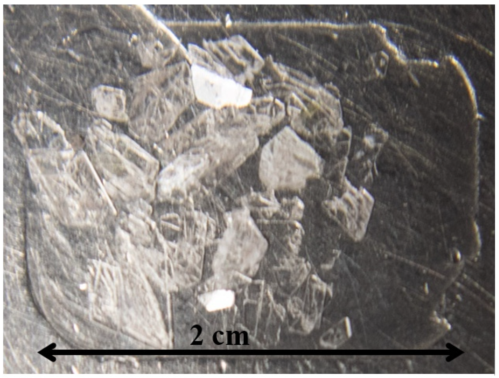
2.2. Crystal Growth
2.3. Single-Crystal X-ray Diffraction
2.4. Powder X-ray Diffraction (PXRD)
2.5. Inductively Coupled Plasma Optical Emission Spectrometry (ICP-OES)
2.6. Optical Properties
2.7. Scintillation
2.8. Extended X-ray Absorption Fine Structure (EXAFS)
3. Results and Discussion
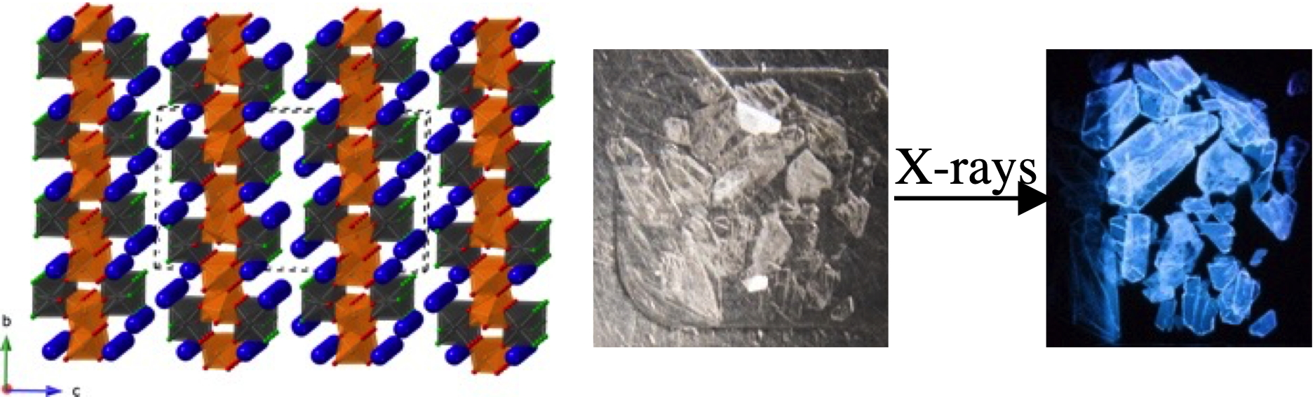
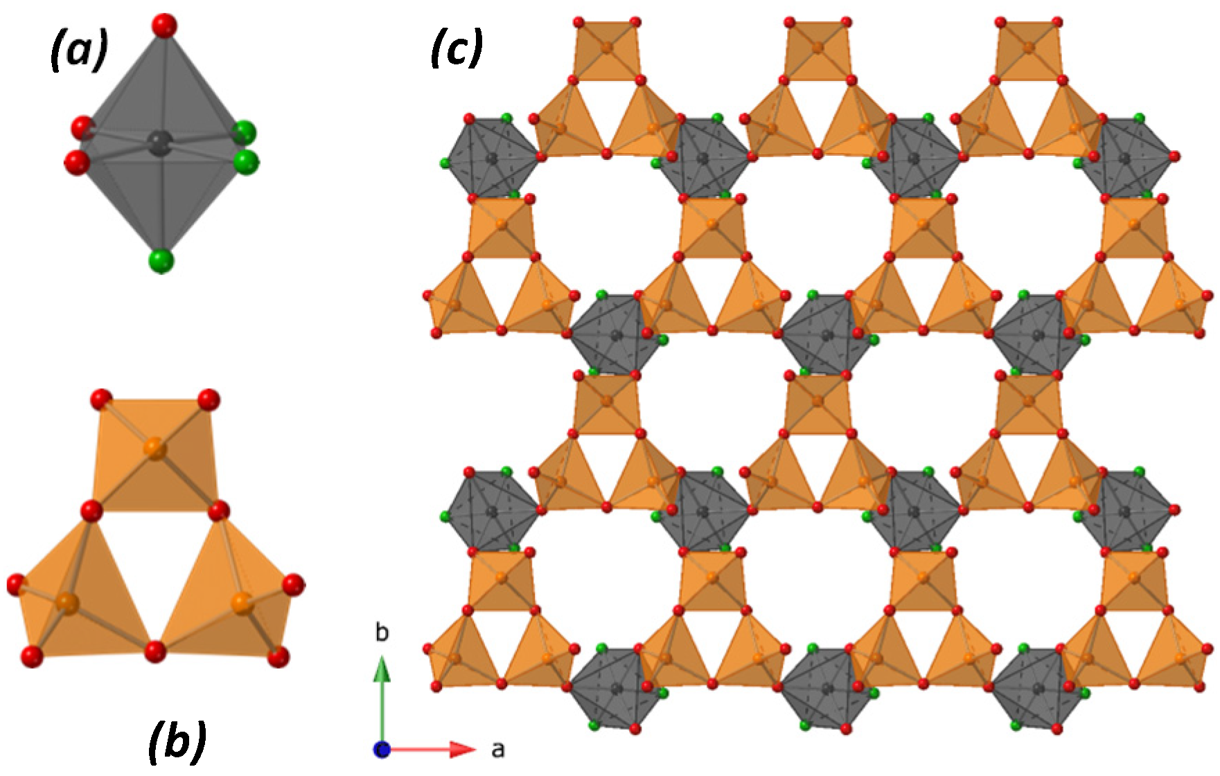
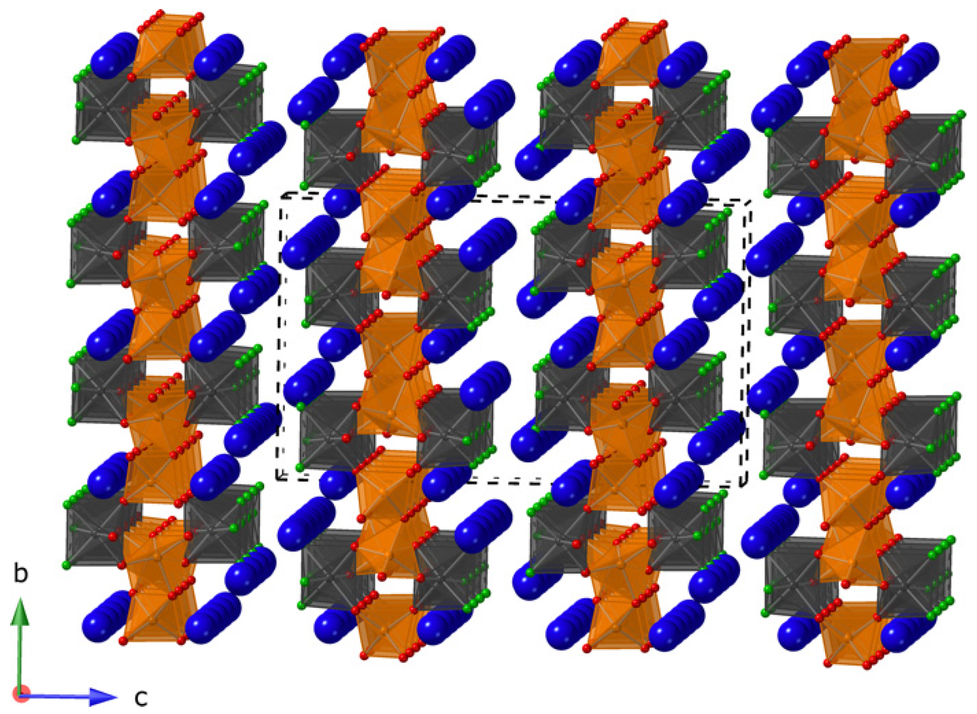
3.1. Crystal Structure
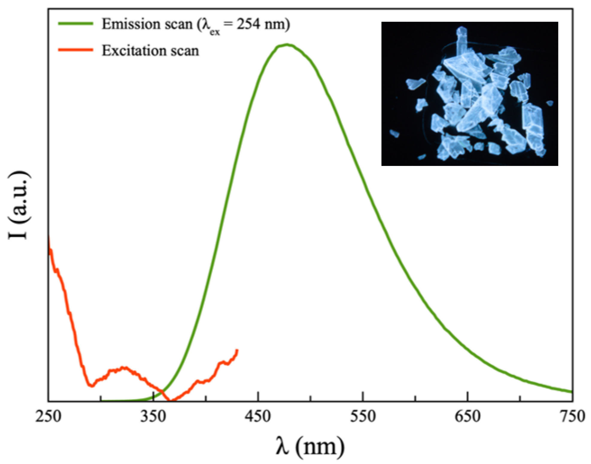
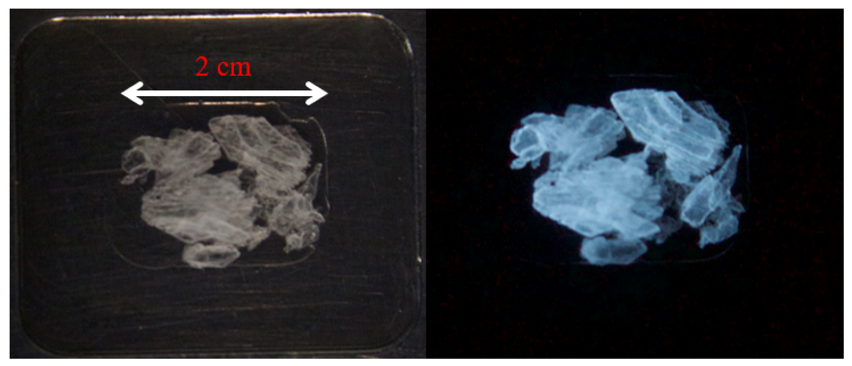
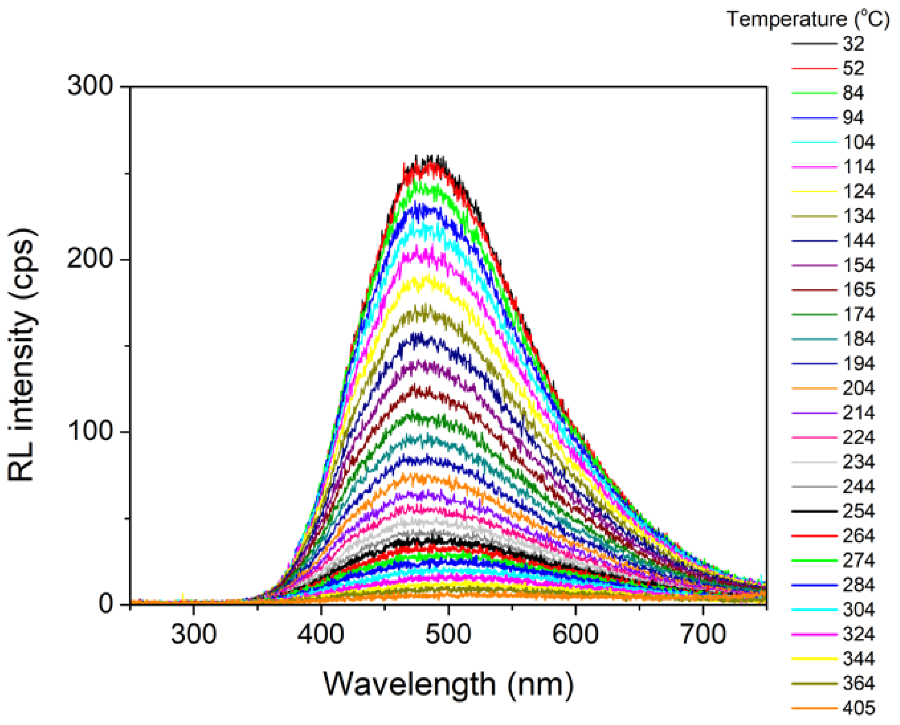
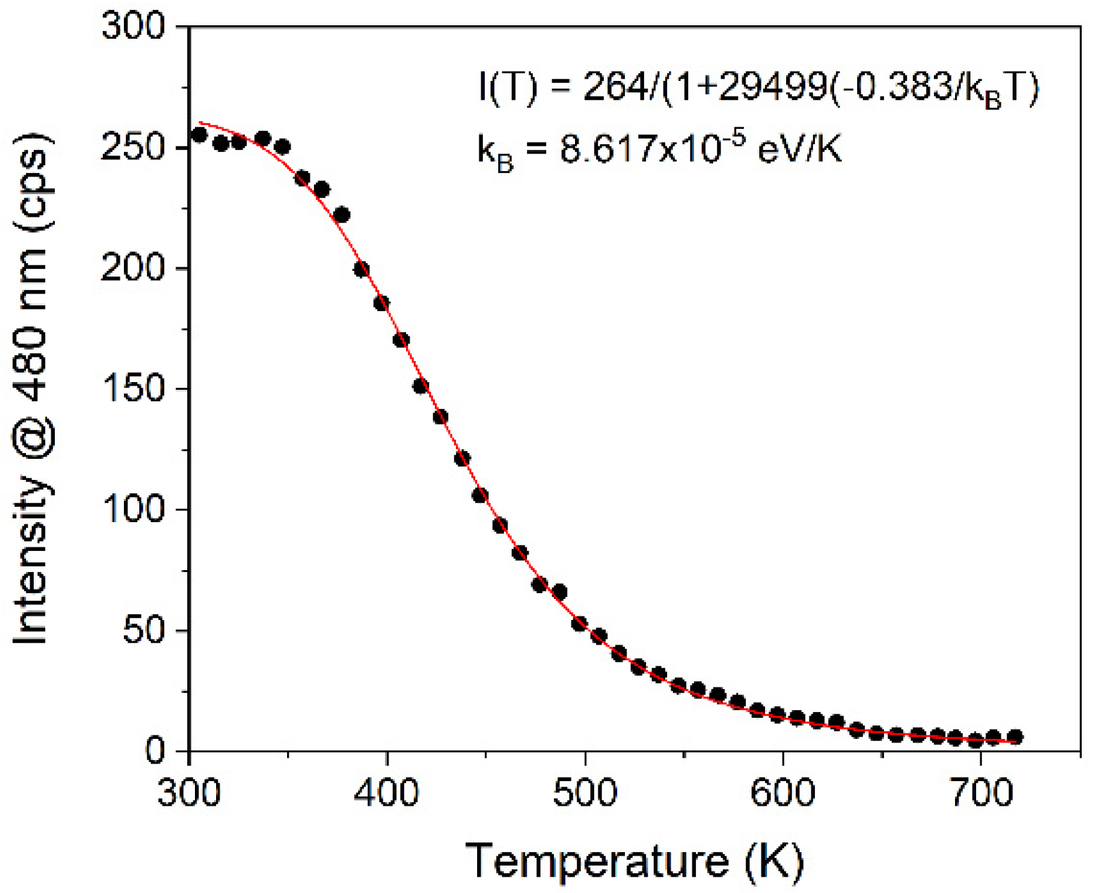
3.2. Optical Properties
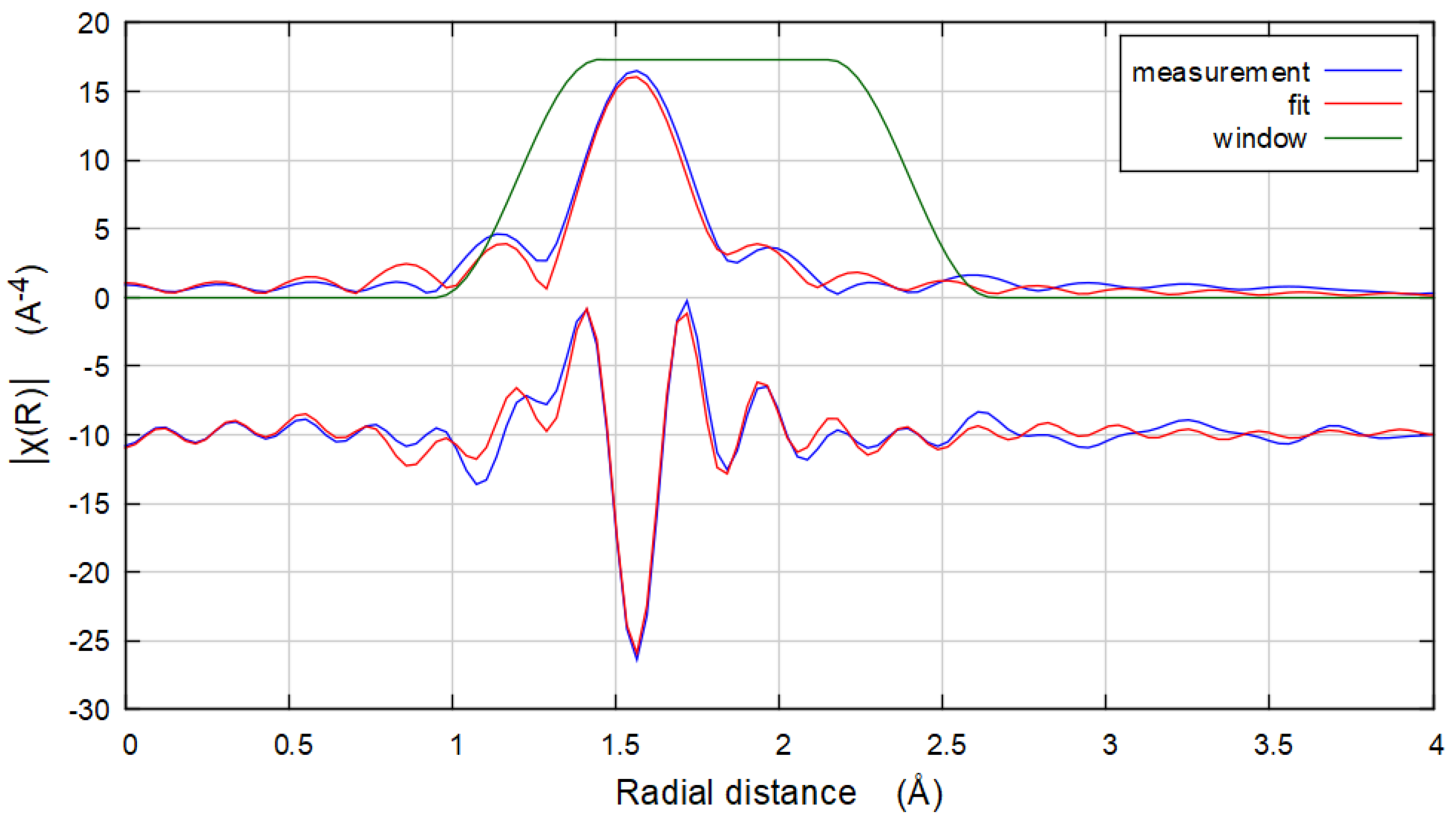
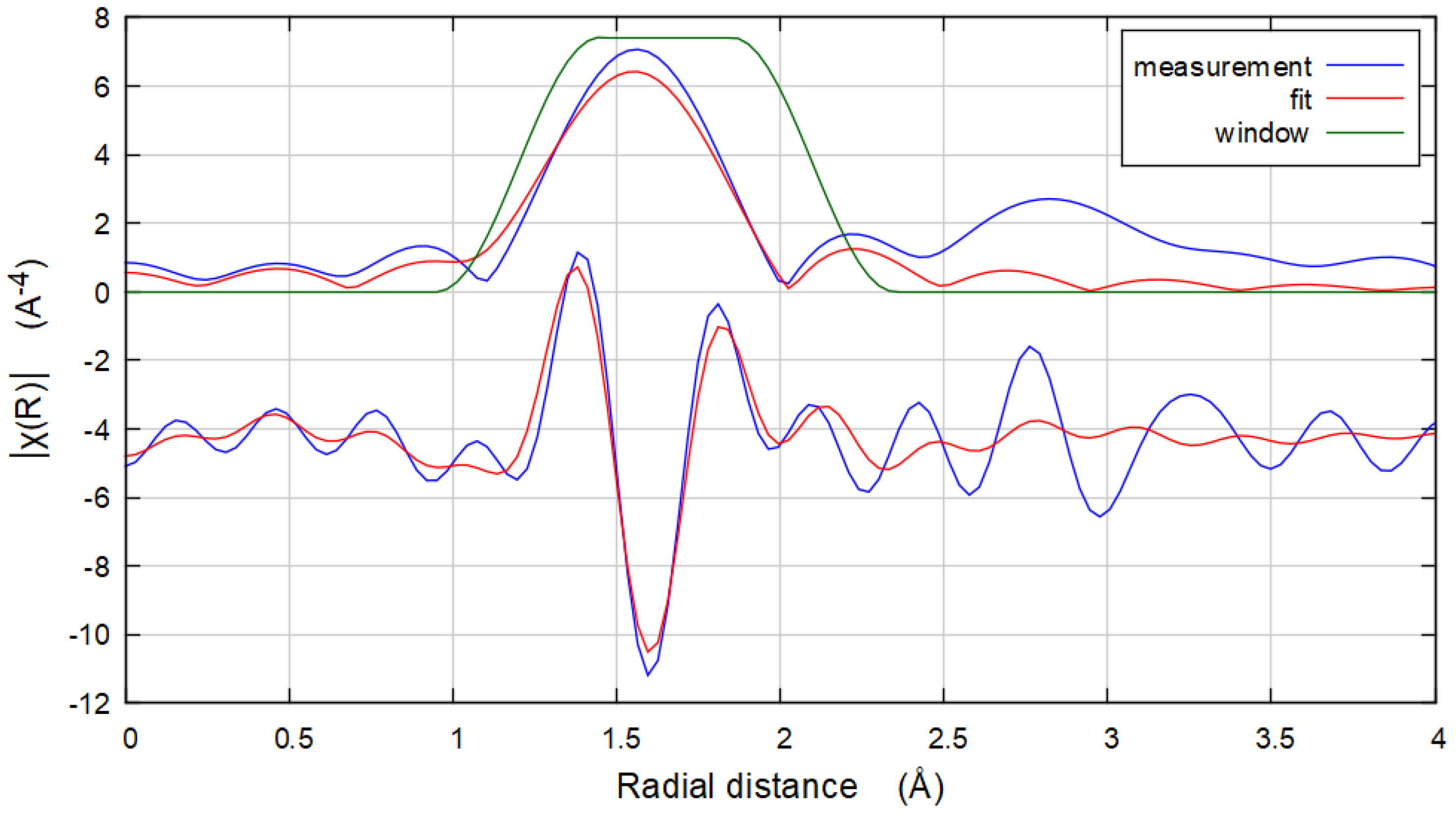
3.3. EXAFS
4. Conclusions
Supplementary Materials
Author Contributions
Funding
Data Availability Statement
Conflicts of Interest
References
- Yoshida, E.; Hirano, Y.; Tashima, H.; Inadama, N.; Nishikido, F.; Moriya, T.; Omura, T.; Watanabe, M.; Murayama, H.; Yamaya, T. The X’tal cube PET detector with a monolithic crystal processed by the 3D sub-surface laser engraving technique: Performance comparison with glued crystal elements. Nucl. Instrum. Methods Phys. Res. Sect. A 2013, 723, 83–88. [Google Scholar] [CrossRef]
- Tous, J.; Blazek, K.; Pína, L.; Sopko, B. High-resolution imaging of biological and other objects with an X-ray digital camera. Appl. Radiat. Isot. 2010, 68, 651–653. [Google Scholar] [CrossRef] [PubMed]
- Danevich, F.A.; Chernyak, D.M.; Dubovik, A.M.; Grinyov, B.V.; Henry, S.; Kraus, H.; Kudovbenko, V.M.; Mikhailik, V.B.; Nagornaya, L.L.; Podviyanuk, R.B.; et al. MgWO4-A new crystal scintillator. Nucl. Instrum. Methods Phys. Res. Sect. A 2009, 608, 107–115. [Google Scholar] [CrossRef]
- Carone, D.; Jacobsohn, L.G.; Breton, L.S.; zur Loye, H.-C. Synthesis, structure, and scintillation of Rb4Ta2Si8O23. Solid State Sci. 2022, 127, 106861. [Google Scholar] [CrossRef]
- Juillerat, C.A.; Kocevski, V.; Morrison, G.; Karakalos, S.G.; Patil, D.; Misture, S.T.; Besmann, T.M.; zur Loye, H.-C. Flux crystal growth of uranium(v) containing oxyfluoride perovskites. Inorg. Chem. Front. 2019, 6, 3203–3214. [Google Scholar] [CrossRef]
- Morrison, G.; zur Loye, H.-C. Flux Growth of [NaK6F][(UO2)3(Si2O7)2] and [KK6Cl][(UO2)3(Si2O7)2]: The Effect of Surface Area to Volume Ratios on Reaction Products. Cryst. Growth Des. 2016, 16, 1294–1299. [Google Scholar] [CrossRef]
- Morrison, G.; Tran, T.T.; Halasyamani, P.S.; zur Loye, H.-C. K8(K5F)U6Si8O40: An Intergrowth Uranyl Silicate. Inorg. Chem. 2016, 55, 3215–3217. [Google Scholar] [CrossRef]
- Morrison, G.; Smith, M.D.; zur Loye, H.-C. Understanding the Formation of Salt-Inclusion Phases: An Enhanced Flux Growth Method for the Targeted Synthesis of Salt-Inclusion Cesium Halide Uranyl Silicates. J. Am. Chem. Soc. 2016, 138, 7121–7129. [Google Scholar] [CrossRef]
- Spagnuolo, N.R.; Morrison, G.; zur Loye, H.-C. Synthesis and crystal structure of [(Cs6F)(Cs3AgF)][Ge14O32] through alkali halide flux growth. Solid State Sci. 2019, 97, 105973. [Google Scholar] [CrossRef]
- Latshaw, A.M.; Wilkins, B.O.; Hughey, K.D.; Yeon, J.; Williams, D.E.; Tran, T.T.; Halasyamani, P.S.; zur Loye, H.-C. A5RE4X[TO4]4 crystal growth and photoluminescence. Fluoride flux synthesis of sodium and potassium rare earth silicate oxyfluorides. CrystEngComm 2015, 17, 4654–4661. [Google Scholar] [CrossRef]
- Latshaw, A.M.; Morrison, G.; zur Loye, K.D.; Myers, A.R.; Smith, M.D.; zur Loye, H.-C. Intrinsic blue-white luminescence, luminescence color tunability, synthesis, structure, and polymorphism of K3YSi2O7. CrystEngComm 2016, 18, 2294–2302. [Google Scholar] [CrossRef]
- Morrison, G.; Latshaw, A.M.; Spagnuolo, N.R.; zur Loye, H.-C. Observation of Intense X-ray Scintillation in a Family of Mixed Anion Silicates, Cs3RESi4O10F2(RE = Y, Eu–Lu), Obtained via an Enhanced Flux Crystal Growth Technique. J. Am. Chem. Soc. 2017, 139, 14743–14748. [Google Scholar] [CrossRef]
- Ayer, G.B.; Klepov, V.V.; Smith, M.D.; Hu, M.; Yang, Z.; Martin, C.R.; Morrison, G.; zur Loye, H.-C. BaWO2F4: A mixed anion X-ray scintillator with excellent photoluminescence quantum efficiency. Dalton Trans. 2020, 49, 10734–10739. [Google Scholar] [CrossRef] [PubMed]
- Hizhnyi, Y.; Nedilko, S.G.; Chornii, V.P.; Nikolaenko, T.M.; Slobodyanik, N.S.; Sheludko, V.I. Structure of the fluorine states in cadmium molybdate host studied by the electronic band structure calculations of CdMoO4, CdMoO4:F and CdMoO3F2 crystals. Comput. Mater. Sci. 2014, 87, 12–18. [Google Scholar] [CrossRef]
- Kozlov, A.V.; Pustovarov, V.A.; Sarychev, M.N.; Isaenko, L.I. Luminescence Spectroscopy of Rb2KTiOF5 Oxyfluoride Single Crystals. AIP Conf. Proc. 2017, 1886, 020012. [Google Scholar]
- Srivastava, A.M.; Ackerman, J.F. Synthesis and luminescence properties of Cs2NbOF5 and Cs2NbOCl5 with isolated [NbOX5]2- (X = F-, Cl-) octahedra. Mater. Res. Bull. 1991, 26, 443–448. [Google Scholar] [CrossRef]
- Krause, L.; Herbst-Irmer, R.; Sheldrick, G.M.; Stalke, D. Comparison of Silver and Molybdenum Microfocus X-ray sources for Single-Crystal Structure Determination. J. Appl. Crystallogr. 2015, 48, 3–10. [Google Scholar] [CrossRef] [Green Version]
- Bruker. APEX3, SAINT+, TWINABS, and SADABS; Bruker AXS Inc.: Madison, WI, USA, 2015. [Google Scholar]
- Dolomanov, O.V.; Bourhis, L.J.; Gildea, R.J.; Howard, J.A.; Puschmann, H. OLEX2: A Complete Structure Solution, Refinement and Analysis Program. J. Appl. Crystallogr. 2009, 42, 334–341. [Google Scholar] [CrossRef]
- Sheldrick, G.M. Crystal Structure Refinement with SHELXL. Acta Crystallogr. Sect. C Struct. Chem. 2015, 71, 3–8. [Google Scholar] [CrossRef]
- Spek, A.L. Structure Validation in Chemical Crystallography. Acta Crystallogr. Sect. D Biol. Crystallogr. 2009, 65, 148–155. [Google Scholar] [CrossRef]
- Ravel, B.; Newville, M. ATHENA, ARTEMIS, HEPHAESTUS: Data analysis for X-ray absorption spectroscopy using IFEFFIT. J. Synchrotron. Radiat. 2005, 12, 537–541. [Google Scholar] [CrossRef] [PubMed] [Green Version]
- Conradsson, T.; Zou, X.; Dadachov, M.S. Synthesis and Crystal Structure of a Novel Germanate: (NH4)4[(GeO2)3(GeO1.5F3)2]·0.67H2O. Inorg. Chem. 2000, 39, 1716–1720. [Google Scholar] [CrossRef] [PubMed]
- Aluker, E.D. Temperature dependence of scintillation response in NaI(Tl), KI(Tl), CsI(Tl). Phys Lett. 1965, 14, 17–18. [Google Scholar] [CrossRef]
- Aluker, E.D.; Mezina, I.P. The temperature dependence of radioluminescence yield in alkali halide crystal phosphors. Phys. Stat. Sol. 1967, 19, 35–40. [Google Scholar] [CrossRef]
- Wu, Y.; Meng, F.; Li, Q.; Koschan, M.; Melcher, C.L. Role of Ce4+ in the scintillation mechanism of codoped Gd3Ga3Al2O12:Ce. Phys. Rev. Appl. 2014, 2, 044009. [Google Scholar] [CrossRef] [Green Version]
- Trofimov, A.A.; Jacobsohn, L.G. Radioluminescence of Lu3Al5O12:Ce single crystal and transparent polycrystalline ceramic at high temperatures. Ceram. Int. 2020, 46, 26335–26338. [Google Scholar] [CrossRef]
- Botter-Jensen, L.; McKeever, S.W.S.; Wintle, A.G. Optically Stimulated Luminescence Dosimetry; Elsevier: Amsterdam, The Netherlands, 2003. [Google Scholar]
- Smith, R.W.; Hu, C.; Liu, J.; Mei, W.-N.; Lin, K.-J. Structure and antiferroelectric properties of cesium niobate, Cs2Nb4O11. J. Solid State Chem. 2007, 180, 1193–1197. [Google Scholar] [CrossRef]


| Empirical Formula | Rb4Ge5O9F6 | Rb4Ge5O9F6:Nb |
|---|---|---|
| Temperature (K) | 302(2) | 303(2) |
| Wavelength (Å) | 0.71073 | 0.71073 |
| Space group, Z | Pbcn, 4 | Pbcn, 4 |
| Unit cell dimensions (Å) | a = 6.98430(10) b = 11.7265(2) c = 19.2732(3) | a = 6.9960(3) b = 11.7464(6) c = 19.3341(9) |
| Volume (Å3) | 1578.50(4) | 1588.83(13) |
| Density (calculated) (g/cm3) | 4.051 | 4.025 |
| Absorption coefficient (mm−1) | 21.768 | 21.626 |
| F(000) | 1736 | 1736 |
| Crystal size (mm × mm × mm) | 0.06 × 0.03 × 0.02 | 0.04 × 0.03 × 0.005 |
| Theta range for data collection (°) | 3.40 to 36.31 | 3.39 to 31.61 |
| Index ranges | −9 ≤ h ≤ 9, −16 ≤ k ≤16, −27 ≤ l ≤ 24 | −9 ≤ h ≤ 9, −16 ≤ k ≤ 16, −27 ≤ l ≤ 27 |
| Reflections collected | 36,504 | 106,148 |
| Independent reflections | 2297 [R(int) = 0.0410] | 2317 [R(int) = 0.0890] |
| Data/restraints/parameters | 2297/0/120 | 2317/0/119 |
| Goodness-of-fit on F2 | 1.144 | 1.089 |
| Final R indices [I > 2sigma(I)] | R1 = 0.0152, wR2 = 0.0335 | R1 = 0.0245, wR2 = 0.0513 |
| R indices (all data) | R1 = 0.0164, wR2 = 0.0338 | R1 = 0.0335, wR2 = 0.0563 |
| Largest diff. peak and hole | 0.728 and −0.464 e−/Å3 | 1.464 and −0.873 e−/Å3 |
| Interaction | Rb4Ge5O9F6 | Rb4Ge5O9F6:Nb |
|---|---|---|
| Ge1-F1 | 1.8290(12) | 1.828(2) |
| Ge1-F2 | 1.8252(12) | 1.831(2) |
| Ge1-F3 | 1.8374(12) | 1.841(2) |
| Ge1-O1 | 1.8237(14) | 1.833(3) |
| Ge1-O2 | 1.8569(14) | 1.863(3) |
| Ge1-O5 | 1.8527(14) | 1.861(3) |
| Ge2-O1 | 1.7168(14) | 1.714(3) |
| Ge2-O2 | 1.7607(10) | 1.727(3) |
| Ge2-O3 | 1.7232(14) | 1.7614(18) |
| Ge2-O4 | 1.7777(13) | 1.779(3) |
| Ge3-O4 | 1.7727(13) | 1.772(3) |
| Ge3-O4 | 1.7727(13) | 1.772(3) |
| Ge3-O5 | 1.7269(14) | 1.728(3) |
| Ge3-O5 | 1.7270(14) | 1.728(3) |
Publisher’s Note: MDPI stays neutral with regard to jurisdictional claims in published maps and institutional affiliations. |
© 2022 by the authors. Licensee MDPI, Basel, Switzerland. This article is an open access article distributed under the terms and conditions of the Creative Commons Attribution (CC BY) license (https://creativecommons.org/licenses/by/4.0/).
Share and Cite
Carone, D.; Klepov, V.V.; Misture, S.T.; Schaeperkoetter, J.C.; Jacobsohn, L.G.; Aziziha, M.; Schorne-Pinto, J.; Thomson, S.A.J.; Hines, A.T.; Besmann, T.M.; et al. Luminescence and Scintillation in the Niobium Doped Oxyfluoride Rb4Ge5O9F6:Nb. Inorganics 2022, 10, 83. https://doi.org/10.3390/inorganics10060083
Carone D, Klepov VV, Misture ST, Schaeperkoetter JC, Jacobsohn LG, Aziziha M, Schorne-Pinto J, Thomson SAJ, Hines AT, Besmann TM, et al. Luminescence and Scintillation in the Niobium Doped Oxyfluoride Rb4Ge5O9F6:Nb. Inorganics. 2022; 10(6):83. https://doi.org/10.3390/inorganics10060083
Chicago/Turabian StyleCarone, Darren, Vladislav V. Klepov, Scott T. Misture, Joseph C. Schaeperkoetter, Luiz G. Jacobsohn, Mina Aziziha, Juliano Schorne-Pinto, Stuart A. J. Thomson, Adrian T. Hines, Theodore M. Besmann, and et al. 2022. "Luminescence and Scintillation in the Niobium Doped Oxyfluoride Rb4Ge5O9F6:Nb" Inorganics 10, no. 6: 83. https://doi.org/10.3390/inorganics10060083