Article

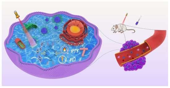
A Near-Infrared BODIPY-Based Rhomboidal Metallacycle for Imaging-Guided Photothermal Therapy

1
College of Material, Chemistry and Chemical Engineering, Key Laboratory of Organosilicon Chemistry and Material Technology, Ministry of Education, Key Laboratory of Organosilicon Material Technology, Hangzhou Normal University, Hangzhou 310036, China
2
Department of Chemistry, University of Utah, 315 South 1400 East, Room 2020, Salt Lake City, UT 84112, USA
*
Authors to whom correspondence should be addressed.
These authors contributed equally to this work.
Inorganics 2022, 10(6), 80; https://doi.org/10.3390/inorganics10060080
Submission received: 23 May 2022 / Revised: 9 June 2022 / Accepted: 10 June 2022 / Published: 13 June 2022
(This article belongs to the Special Issue Metallamacrocycles and Metallacages: Foundations and Applications)

Abstract

:
Although boron dipyrromethene (BODIPY)-based metallacycles are expected to be promising candidates for imaging probes and therapeutic agents, their biomedical applications are restricted by their short absorption/emission wavelengths. In this work, we report a rhombic metallacycle M with broad absorption in the near-infrared (NIR) range and emissions at wavelengths >800 nm, which exhibits an efficient photothermal conversion capacity. Metallacycle M was encapsulated via Pluronic F127 to fit the biotic environment, resulting in the generation of F127/M nanoparticles (NPs) with high hydrophilicity and biocompatibility. In vitro studies demonstrated that the F127/M NPs underwent efficient cellular uptake and exhibited satisfactory photothermal therapeutic activity. Furthermore, in vivo experiments revealed that tumor growth was effectively inhibited, and the degree of undesirable biological damage was minimal in treatment with F127/M NPs and laser irradiation. Finally, the F127/M NPs could be visualized through NIR fluorescence imaging in living mice, thereby allowing their distribution to be monitored in order to enhance treatment accuracy during photothermal therapy. We envision that such BODIPY-based metallacycles will provide emerging opportunities for the development of novel therapeutic agents for biomedical applications.

Graphical Abstract

1. Introduction

Coordination-driven self-assembly provides a feasible and effective strategy for obtaining metal-organic macrocycles (MOCs) with well-defined morphologies and sizes based on the metal–ligand bonds present between the organic donors and the metal acceptors [1,2,3,4,5]. On account of the diverse metal centers and ligands available for their construction, MOCs can be easily constructed from the bottom up [6,7,8,9], and they can serve as integrated function platforms for extensive applications (e.g., optical materials [10,11,12], chemical sensors [13,14,15], catalysis [16,17], bioimaging [18,19], and biotherapy [20,21]) via pre- and post-self-assembly functional modifications. For example, fluorescent metallacycles with anticancer potential have attracted widespread interest in medicinal chemistry, not only because of the adjustable bioactivity of the metal centers and organic ligands, but also due to the fact that they can be visually localized in vivo [22]. Thus, the judicious selection of organic donors and metal acceptors plays an essential role in achieving superior fluorescent metallacycles for theranostics [23,24].
As an extensively investigated organic chromophore, boron dipyrromethene (BODIPY) is of particular interest because of its excellent properties, such as its facile synthesis and structural diversification, its high absorption coefficients and fluorescence quantum yields, its high photostability, and its excellent chemical stability [25,26,27,28,29,30,31]. Owing to these advantages, BODIPYs have been widely applied in photovoltaic devices [32,33,34,35], fluorescent probes [36,37,38,39], bioimaging [40,41,42], photodynamic therapy (PDT) [43,44,45,46], and photothermal therapy (PTT) [47,48,49,50]. It has been demonstrated that integrating BODIPYs as building blocks into metallacycles may result in a range of advantages [22]. More specifically, when well-defined metallacycles are employed, BODIPY units can endow the metallacycles with fluorescent characteristics and bioactivity, and the photophysical properties of the formed BODIPY-based metallacycles can be modulated by directional transition dipole moments or efficient energy transfer. Based on these factors, a number of efforts were made to develop BODIPY-based metallacycles for bioapplications [23,51,52,53]. For example, Huang et al. [54] reported two high-emission platinum(II)-centered metallacycles containing BODIPY-derived ligands, which were suitable for fluorescence imaging-guided chemo-photodynamic synergistic therapy upon light irradiation at 400–700 nm. However, strong absorption in the near-infrared (NIR) region (650–1700 nm) is desirable to allow deeper penetration and produce minimal biological damage within healthy tissue. To date, very few BODIPY-based metallacycles have been reported that exhibit intense NIR absorption [51].
Herein, we describe an NIR-absorbing and emissive metallacycle M constructed via coordination-driven self-assembly using 120° dipyridyl BODIPY ligand 1 and 60° phenanthrene-based diplatinum(II) 2 as the building blocks (Scheme 1). Based on a Knoevenagel condensation, a N, N-diethyl-4-aminophenyl group was introduced into the 3,5-positions of the BODIPY cores to obtain BODIPY ligands with strong NIR absorption properties. Due to its NIR emission, the visualization and localization of metallacycle M were also investigated. Furthermore, metallacycle M was encapsulated in micelles of Pluronic F127 to produce F127/M nanoparticles (NPs), whose photothermal conversion behaviors and fluorescent imaging properties were evaluated both in vitro and in vivo. The tumor growth inhibition ability of the F127/M NPs was also examined to determine the potential for MOCs to act as versatile platforms for theranostics.

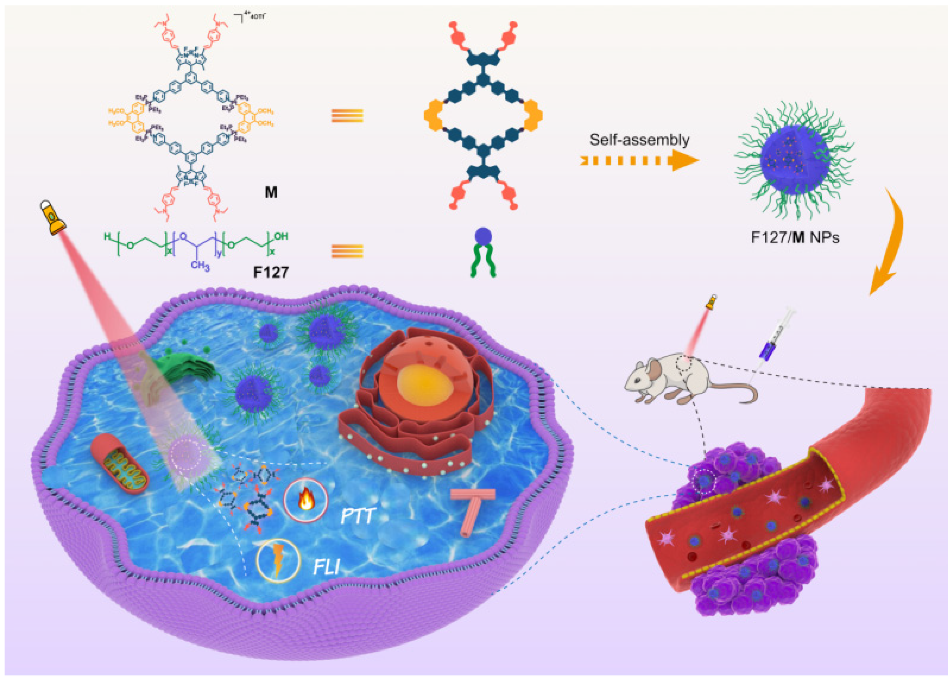
2. Results and Discussion

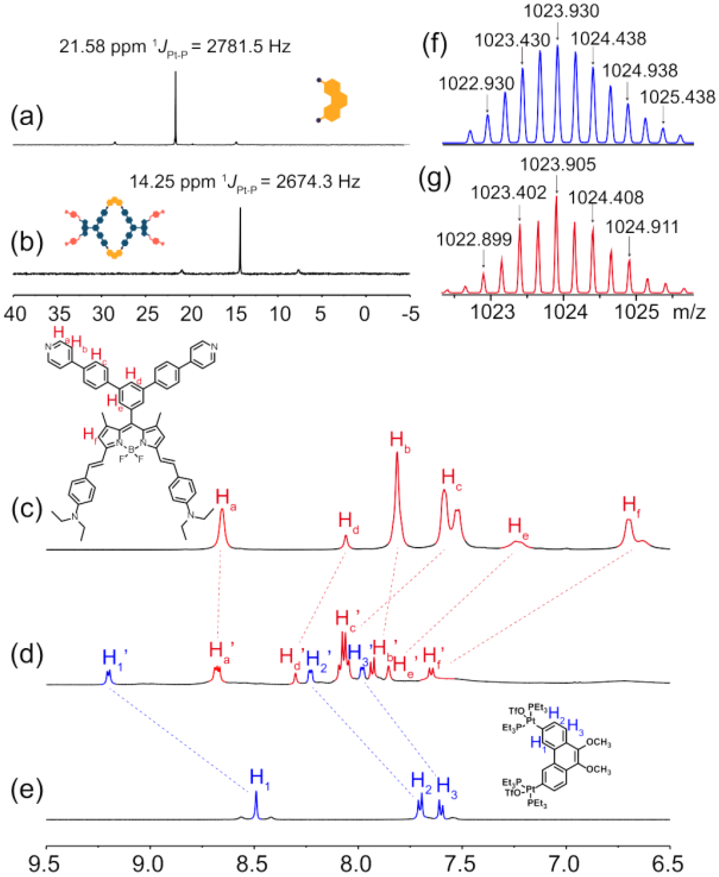
2.1. Synthesis of Metallacycle M

As outlined in Scheme S1, BODIPY ligand 1 was initially synthesized via a Knoevenagel condensation reaction and a following palladium-catalyzed coupling reaction. Subsequently, the coordination-driven self-assembly of BODIPY-based 120° dipyridyl donor 1 with phenanthrene-based 60° diplatinum(II) acceptor 2 produced rhomboidal Pt(II) metallacycle M in an excellent yield (>90%). The metallacycle was then characterized using multinuclear NMR spectroscopy (i.e., 1H and 31P{1H}) and electrospray ionization time-of-flight mass spectrometry (ESI-TOF-MS). In the 31P{1H} NMR spectrum, a sharp singlet peak was observed for M at 14.25 ppm, with concomitant 195Pt satellite peaks also being present (JPt-P = 2674.3 Hz), thereby indicating a single phosphorus environment. In addition, an upfield shift by 7.33 ppm in comparison with the corresponding signal of acceptor 2 confirmed the formation of metal–ligand bonds (Figure 1a,b). As shown in the 1H NMR spectrum of M (Figure 1c–e), downfield shifts were observed for the α-pyridyl Ha protons (i.e., from 8.65 to 8.68 ppm) and for the β-pyridyl Hb proton (i.e., from 7.81 to 7.93 ppm), compared to the corresponding shifts of the free BODIPY-based dipyridyl ligand 1; these shifts were attributed to the reduced electron densities caused by coordination with the metal. In addition, aromatic protons Hc, Hd, He, and Hf of ligand 1 and aromatic protons H1, H2, and H3 of Pt(II) acceptor 2 shifted upfield. ESI-TOF-MS provided further evidence of the formation of M, wherein isotopically resolved peaks were observed at m/z values of 1414.945 and 1023.905, which corresponded to [M−3OTf]3+ and [M−4OTf]4+, respectively (Figure 1g and Figure S17).

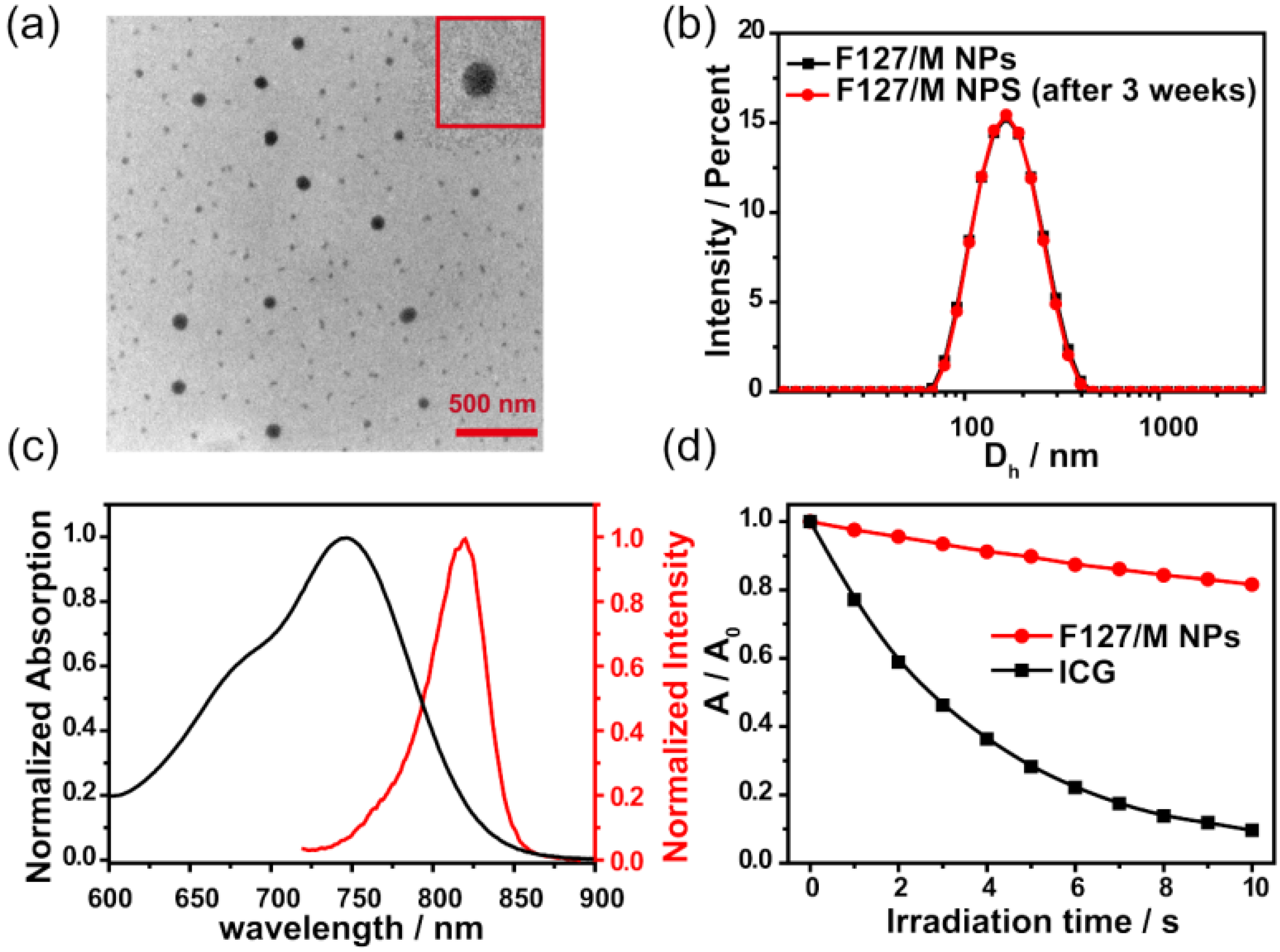
2.2. Fabrication and Characterization of F127/M NPs

Owing to the hydrophobicity of M, we introduced F127 for encapsulation via the precipitation method to yield F127/M NPs with enhanced solubility, biocompatibility, and tumor accumulation properties. As depicted in Figure 2a,b, transmission electron microscopy (TEM) and dynamic light scattering (DLS) were used to confirm the morphology and size of the NPs, and both techniques indicated a uniform size of ~150 nm, thereby indicating their potential for accumulating in tumor tissues owing to the enhanced permeability and retention (EPR) effect [55]. Furthermore, after three weeks in storage, it was found that the NP size remained relatively constant, thereby demonstrating their excellent colloidal stability (Figure 2b). The F127/M NPs displayed broad absorption in the NIR range from 600 to 900 nm, with a maximum absorption peak being observed at 747 nm in an aqueous solution; this represented a more pronounced red shift compared to that observed in acetone (i.e., 716 nm). Based on the UV–vis absorption spectrum of M, the M-loading efficiency of the F127/M NPs was calculated as 10% (Figure S20). In addition, the maximum emission peak of the F127/M NPs was 820 nm in the NIR-I window (Figure 2c) and the fluorescent quantum yield was 0.31%.
The colloidal stability of M is an important property of phototherapeutic agents for applications in vivo, which was measured by M treatment with irradiation. Insignificant changes in 31P{1H} NMR spectra of M were detected after irradiation for 10 min (Figure S20), indicating the structural stability of M. Furthermore, from comparison between the relative absorbance changes in F127/M NPs and ICG in aqueous media upon irradiation, F127/M NPs exhibited better photobleaching resistance, aiding in the maintenance of the phototherapy effect (Figure 2d and Figure S21).

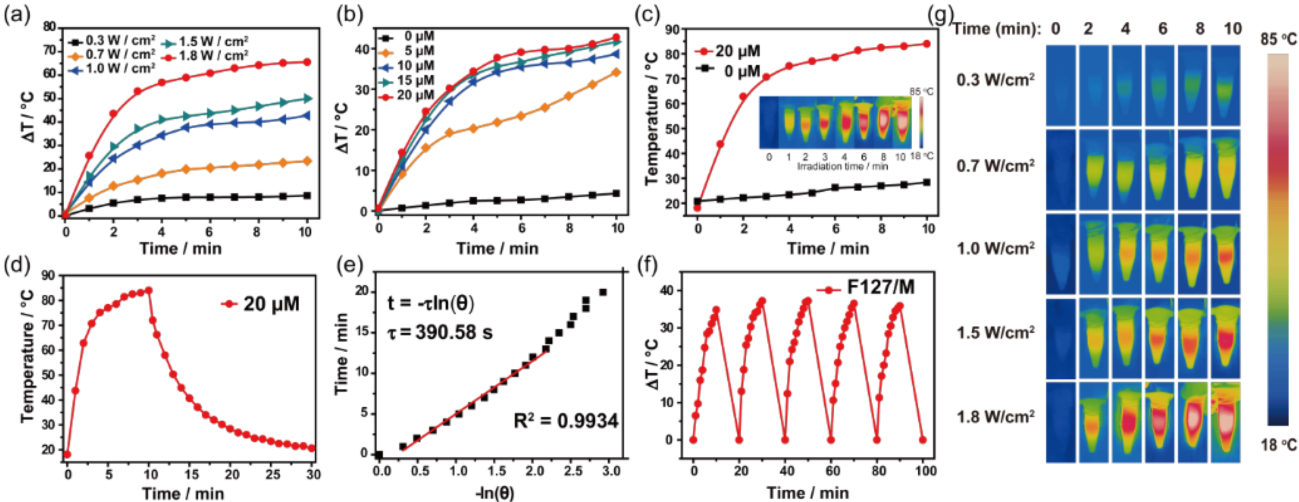
2.3. Photothermal Properties of F127/M NPs

Subsequently, we systematically evaluated the photothermal properties of the F127/M NPs under 660 nm laser exposure. As shown in Figure 3a–c, upon 660 nm laser irradiation for 10 min, the temperature of the F127/M NP solution elevated in a concentration- and power-density-dependent manner. With an increase in F127/M NP concentration (from 0 to 20 μM), the temperature of the F127/M NP solution eventually rose by 4.3 to 43 °C, and it was 8.4 to 63.9 °C warmer upon increasing power density (from 0.3 to 1.8 W/cm2), revealing efficient photothermal conversion with tunability. Based on the relevant literature [49], the photothermal conversion efficiency (PCE) of the F127/M NPs was calculated to be 36% based on the cooling process (Figure 3d). Photothermal stability is a necessary factor for photothermal agents during cancer treatment. Gratifyingly, the F127/M NPs were confirmed to possess high photothermal stability, as demonstrated by the results that the temperature elevation of the F127/M NP aqueous solution remained unaltered after five heating/cooling cycles (Figure 3e). Overall, these results indicate that the F127/M NPs possessed efficient photothermal conversion properties and high photostability, which are beneficial for their use as promising photothermal agents.

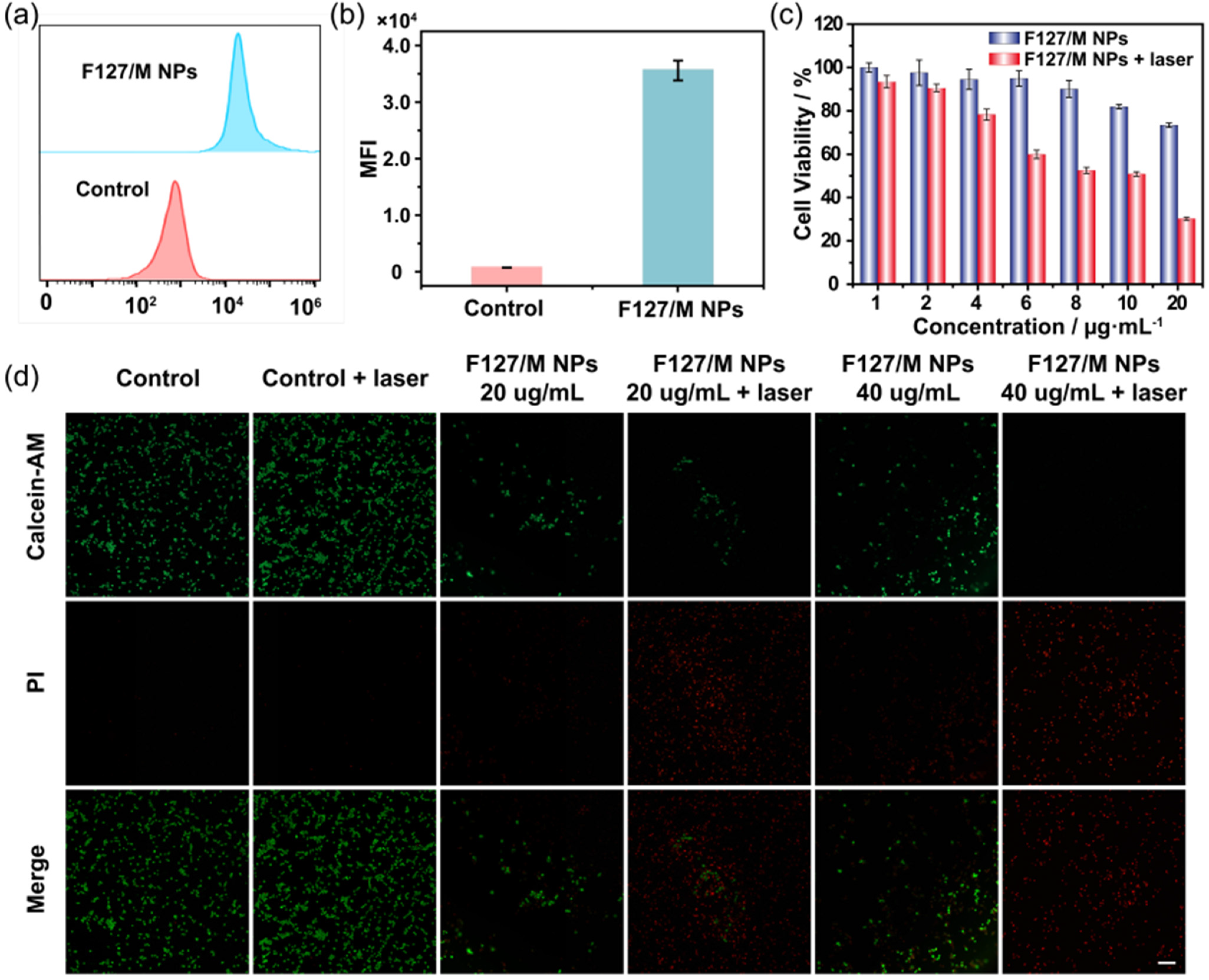
2.4. In Vitro Photonic Cytotoxicity and Antitumor Therapy

To determine the in vitro photonic cytotoxicity of the FM127/M NPs, we initially assessed their cellular uptake via flow cytometry (FCM). As shown in Figure 4a, the group treated with F127/M NPs exhibited an extremely higher fluorescence intensity than the control group, thereby indicating that the F127/M NPs could successfully enter the cells. In addition, the quantitative mean fluorescence intensity (MFI) also intuitively verified their cellular internalization (Figure 4b). Based on these observations and the satisfactory photothermal properties of the F127/M NPs, we were encouraged to investigate their efficacy at the cytological level. Thus, the phototoxicity and dark toxicity of the F127/M NPs in U87 cancer cells were examined using an MTT assay (Figure 4c). >80% cell viability was observed, even at high concentrations of F127/M NPs in darkness, thereby indicating the good biocompatibility of the F127/M NPs. In contrast, under 660 nm laser irradiation at 1.0 W/cm2, the cell viability gradually decreased with an increase in F127/M NP concentration, which suggested dose-dependent cytotoxicity. To further verify the phototherapeutic properties of the F127/M NPs, calcein-AM (green) and propidium iodide (PI, red) dyes were used to perform live–dead cell co-staining fluorescence experiments. As shown in Figure 4d, the control group (without the F127/M NPs) exhibited a uniform and bright green fluorescence even under laser irradiation, indicating that essentially no cell damage occurred under laser irradiation alone. In addition, the green fluorescence of the F127/M NP group demonstrated their stability in darkness. However, distinct red fluorescence was observed in the U87 cells treated with the F127/M NPs plus irradiation (660 nm, 1.0 W/cm2), and the area of red fluorescence increased upon increasing the NP concentration from 20 to 40 μg/mL. These results therefore confirm the strong photothermal potential of the F127/M NPs, which demonstrates their potential for use in PTT applications in vivo.

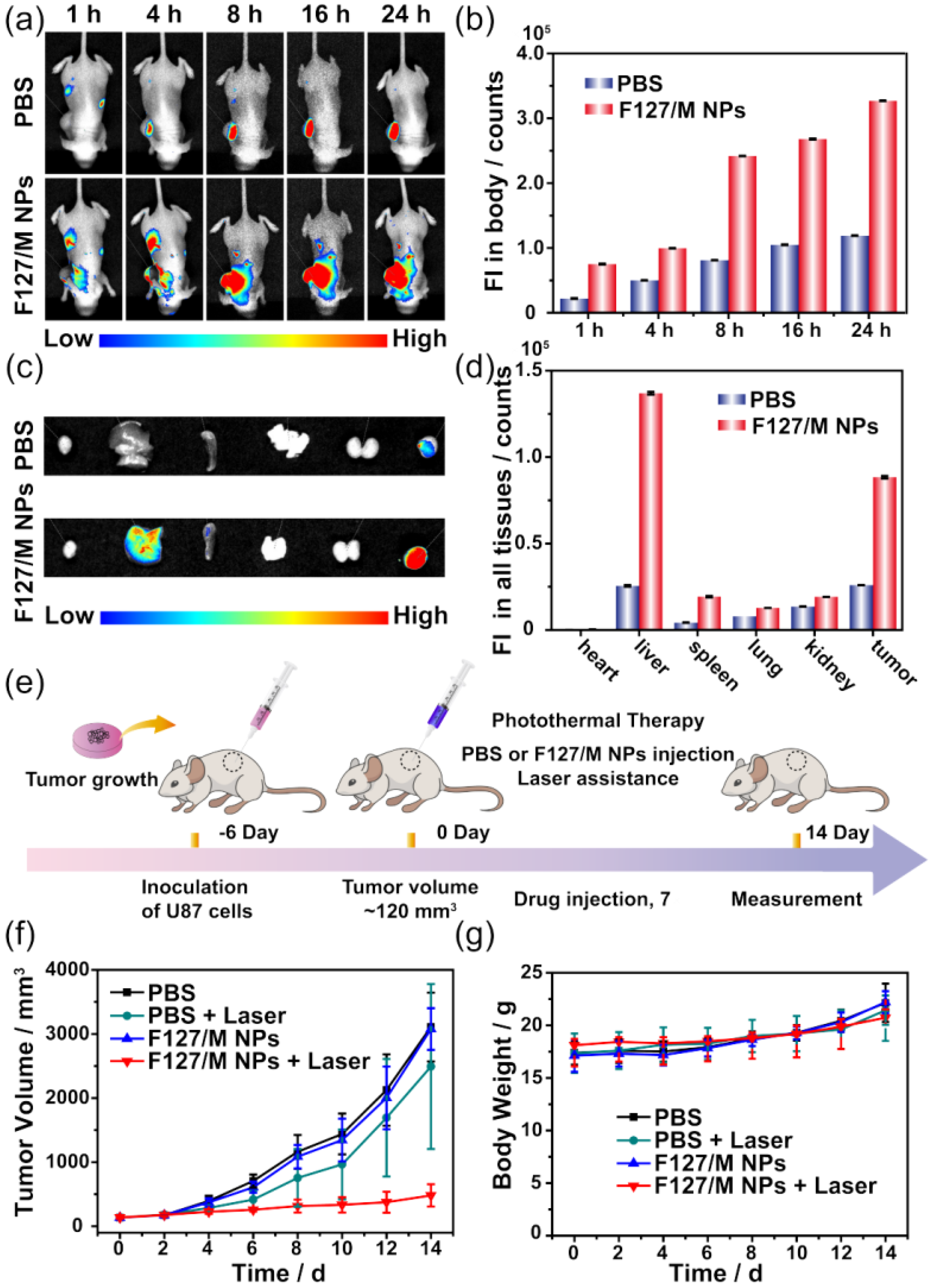
2.5. In Vivo Imaging and Antitumor Effect

In vivo imaging is essential for both drug localization and tumor phototherapy. The intense absorption and emission provided by F127/M NPs in the NIR region indicated their strong potential for use in fluorescence imaging in vivo. Thus, we injected U87 tumor-bearing mice with F127/M NPs or PBS and evaluated the fluorescence images recorded over time (i.e., 1, 4, 8, 16, and 24 h). As shown in Figure 5a,b, the fluorescence signal in the group treated with F127/M NPs gradually increased at the tumor site over time, reaching the brightest red fluorescence after 24 h. Subsequently, the effective accumulation of F127/M NPs in tumors was further verified by ex vivo fluorescence imaging of a tumor and of the major organs (i.e., heart, liver, spleen, lung, and kidney). As shown in Figure 5c,d, the F127/M NPs mainly accumulated in the liver and in the tumor, which was likely due to hypermetabolism in the mouse models, in addition to the EPR effect described above. These results indicate that F127/M NPs could effectively accumulate at the tumor site, and the fluorescent signal could be used to offer therapeutic guidance and enhance the in vivo antitumor effects of this system.
Encouraged by the remarkable in vitro anticancer activity and in vivo imaging results obtained for the F127/M NPs, we further evaluated their PTT efficiency using the U87 tumor-bearing mouse model in vivo (Figure 5e). PBS or F127/M NPs were injected intratumorally into U87 tumor-bearing mice, followed by laser irradiation treatment (660 nm, 1.0 W/cm2) at a post-administration time of 10 min. Subsequently, tumor volume and mouse body weight were recorded every 2 d for 14 d. As shown in Figure 5f, in the absence of irradiation, both PBS and F127/M NP groups exhibited the fastest tumor growth among the various groups, with the tumor volumes reaching ~3100 mm3. The PBS + Laser group also showed rapid tumor growth, but it was slower compared to the above two groups. This was likely due to the fact that continuous laser irradiation at 1.0 W/cm2 was capable of producing a slight temperature increase, thereby inhibiting tumor growth to a certain extent. In contrast, the F127/M NP + Laser group demonstrated significant suppression of tumor growth in mice within the 14 d period. The photographic images recorded for the four mouse tumor groups after 14 d directly confirmed that the F127/M NPs could effectively inhibit tumor growth under constant laser irradiation (Figure S22). Finally, to examine the biosafety of the F127/M NPs, we evaluated their systemic toxicity based on the mouse body weight from U87 tumor-bearing nude mice after the different treatments. During the 14 d experimental period, the body weights of all mice gradually increased (Figure 5g), which indicates that the F127/M NPs could be used as biocompatible and biosafe anticancer agents for tumor therapy.

3. Conclusions

In summary, a near-infrared (NIR) boron dipyrromethene (BODIPY)-based rhomboidal metallacycle M was successfully formed by the coordination-driven self-assembly of strongly NIR-absorbing BODIPY ligands and phenanthrene-based Pt(II) acceptors, which not only served as diagnostic agents for bioimaging, but also could be utilized as a class of efficient photothermal reagents for photothermal therapy. In addition, following encapsulation by Pluronic F127, the F127/M nanoparticles (NPs) demonstrated enhanced photothermal performance with a high photothermal conversion efficiency of 36% and remarkable photobleaching resistance. In vitro studies showed that the F127/M NPs, with good biocompatibility, were successfully taken up by tumor cells, and that under laser irradiation, they selectively and efficiently damaged these cells. Moreover, the F127/M NPs also exhibited excellent antitumor PTT effects in vivo. Ultimately, this work presents a strategy for the construction of diagnostic and therapeutic agents through a BODIPY-based metallacycle, wherein the properties can be modulated by varying the individual building blocks rather than the overall design. Therefore, this work inspires insight into the development of metallacycle-based imaging-guided therapeutic agents.

Supplementary Materials

The following supporting information can be downloaded at: https://www.mdpi.com/article/10.3390/inorganics10060080/s1, Scheme S1: Synthetic route to M and chemical structures of compounds; Figure S1–S19: NMR spectra, ESI-HRMS spectra, FT-IR spectra and fluorescence spectra; Figure S20: (a) Concentration-dependent absorbance and (b) the standard curve of M in acetone; Figure S21: UV–vis absorption spectra and 31P{1H} NMR spectra; Figure S22: Photographs of U87 tumor-bearing nude mice. [49,56,57].

Author Contributions

Conceptualization, S.Y. and P.J.S.; methodology: S.Y., P.J.S., Y.L. and Y.S.; formal analysis: J.Z., J.Y., W.L. and Y.F.; investigation, J.Z., J.Y., W.L. and Y.F.; data curation, J.Y., J.Z. and W.L.; writing—original draft preparation, J.Z. and J.Y.; writing—review and editing, S.Y., P.J.S., Y.S. and Y.L.; funding acquisition, S.Y. and Y.L. All authors have read and agreed to the published version of the manuscript.

Funding

This research was funded by the National Natural Science Foundation of China (grant numbers 21971049 and 51903070) and “Ten-thousand Talents Plan” of Zhejiang Province (grant number 2019R52040).

Institutional Review Board Statement

The mice were housed at the Laboratory Animal Center of Hangzhou Normal University with use license number SYXK (Zhejiang) 2020-0026, and cultivated in a pathogen-free environment with appropriate humidity and temperature. All animal procedures were performed in accordance with the animal care and use guidelines of the Organizational Animal Care and Use Committee.

Informed Consent Statement

Not applicable.

Data Availability Statement

Not applicable.

Conflicts of Interest

The authors declare no conflict of interest.

References

  1. Zhao, J.; Zhou, Z.X.; Li, G.F.; Stang, P.J.; Yan, X.Z. Light-emitting self-assembled metallacages. Natl. Sci. Rev. 2021, 8, nwab045. [Google Scholar] [CrossRef] [PubMed]
  2. Sun, Y.; Chen, C.; Stang, P.J. Soft Materials with Diverse Suprastructures via the Self-Assembly of Metal–Organic Complexes. Acc. Chem. Res. 2019, 52, 802–817. [Google Scholar] [CrossRef] [PubMed]
  3. Sepehrpour, H.; Fu, W.; Sun, Y.; Stang, P.J. Biomedically Relevant Self-Assembled Metallacycles and Metallacages. J. Am. Chem. Soc. 2019, 141, 14005–14020. [Google Scholar] [CrossRef] [PubMed]
  4. Li, Y.; Zhang, J.; Li, H.; Fan, Y.; He, T.; Qiu, H.; Yin, S. Metallacycle/Metallacage-Cored Fluorescent Supramolecular Assemblies with Aggregation-Induced Emission Properties. Adv. Optical Mater. 2020, 8, 1902190. [Google Scholar] [CrossRef]
  5. Li, B.; He, T.; Fan, Y.; Yuan, X.; Qiu, H.; Yin, S. Recent developments in the construction of metallacycle/metallacage-cored supramolecular polymers via hierarchical self-assembly. Chem. Commun. 2019, 55, 8036–8059. [Google Scholar] [CrossRef]
  6. Xu, L.; Shen, X.; Zhou, Z.; He, T.; Zhang, J.; Qiu, H.; Saha, M.L.; Yin, S.; Stang, P.J. Metallacycle-Cored Supramolecular Polymers: Fluorescence Tuning by Variation of Substituents. J. Am. Chem. Soc. 2018, 140, 16920–16924. [Google Scholar] [CrossRef]
  7. Chen, F.; Lin, X.; Li, Y.; Xu, D.; Qiu, H.; Yin, S. Metallacycle-crosslinked supramolecular polymers constructed by amino–YNE click reaction with enhanced mechanical properties. Supramol. Mater. 2022, 1, 100003. [Google Scholar] [CrossRef]
  8. Zhang, Y.-Y.; Chen, F.; Li, Y.; Qiu, H.-Y.; Zhang, J.-J.; Yin, S.-C. Supramolecular Polymer Networks with Enhanced Mechanical Properties: The Marriage of Covalent Polymer and Metallacycle. Chin. J. Chem. 2021, 39, 2731–2737. [Google Scholar] [CrossRef]
  9. Zhang, Q.; Tang, D.; Zhang, J.; Ni, R.; Xu, L.; He, T.; Lin, X.; Li, X.; Qiu, H.; Yin, S.; et al. Self-Healing Heterometallic Supramolecular Polymers Constructed by Hierarchical Assembly of Triply Orthogonal Interactions with Tunable Photophysical Properties. J. Am. Chem. Soc. 2019, 141, 17909–17917. [Google Scholar] [CrossRef]
  10. Hu, Y.X.; Hao, X.T.; Xu, L.; Xie, X.; Xiong, B.; Hu, Z.; Sun, H.; Yin, G.-Q.; Li, X.; Peng, H.; et al. Construction of supramolecular liquid-crystalline metallacycles for holographic storage of colored images. J. Am. Chem. Soc. 2020, 142, 6285–6294. [Google Scholar] [CrossRef]
  11. Chen, L.; Chen, C.L.; Sun, Y.; Lu, S.; Huo, H.; Tan, T.; Li, A.; Li, X.; Ungar, G.; Liu, F.; et al. Luminescent metallacycle-cored liquid crystals induced by metal coordination. Angew. Chem. Int. Ed. 2020, 59, 10143–10150. [Google Scholar] [CrossRef] [PubMed]
  12. Zhang, M.; Yin, S.; Zhang, J.; Zhou, Z.; Saha Manik, L.; Lu, C.; Stang Peter, J. Metallacycle-cored supramolecular assemblies with tunable fluorescence including white-light emission. Proc. Natl. Acad. Sci. USA 2017, 114, 3044–3049. [Google Scholar] [CrossRef] [PubMed] [Green Version]
  13. Zhang, M.; Saha, M.L.; Wang, M.; Zhou, Z.; Song, B.; Lu, C.; Yan, X.; Li, X.; Huang, F.; Yin, S.; et al. Multicomponent platinum(II) cages with tunable emission and amino acid sensing. J. Am. Chem. Soc. 2017, 139, 5067–5074. [Google Scholar] [CrossRef]
  14. Saha, M.L.; Yan, X.Z.; Stang, P.J. Photophysical properties of organoplatinum(II) compounds and derived self-assembled metallacycles and metallacages: Fluorescence and its applications. Acc. Chem. Res. 2016, 49, 2527–2539. [Google Scholar] [CrossRef]
  15. Xu, L.; Wang, Y.X.; Chen, L.J.; Yang, H.B. Construction of multiferrocenyl metallacycles and metallacages via coordination-driven self-assembly: From structure to functions. Chem. Soc. Rev. 2020, 49, 3889–3919. [Google Scholar]
  16. Hong, T.; Zhang, Z.B.; Sun, Y.; Tao, J.-J.; Tang, J.-D.; Xie, C.; Wang, M.; Chen, F.; Xie, S.-S.; Li, S.; et al. Chiral metallacycles as catalysts for asymmetric conjugate addition of styrylboronic acids to α,β-Enones. J. Am. Chem. Soc. 2020, 142, 10244–10249. [Google Scholar] [CrossRef]
  17. Kaphan David, M.; Levin Mark, D.; Bergman Robert, G.; Raymond Kenneth, N.; Toste, F.D. A supramolecular microenvironment strategy for transition metal catalysis. Science 2015, 350, 1235–1238. [Google Scholar] [CrossRef] [Green Version]
  18. Zhu, H.; Li, Q.; Shi, B.; Ge, F.; Liu, Y.; Mao, Z.; Zhu, H.; Wang, S.; Yu, G.; Huang, F.; et al. Dual-Emissive Platinum(II) Metallacage with a Sensitive Oxygen Response for Imaging of Hypoxia and Imaging-Guided Chemotherapy. Angew. Chem. Int. Ed. 2020, 59, 20208–20214. [Google Scholar] [CrossRef]
  19. Fan, Y.; Zhang, J.; Li, Y.; Chen, Q.; Ni, Z.; Zhou, H.; Yu, J.; Qiu, H.; Yin, S. Amphiphilic rhomboidal metallacycles with aggregation-induced emission and aggregation-caused quenching luminogens for white-light emission and bioimaging. Mater. Chem. Front. 2022, 6, 633–643. [Google Scholar] [CrossRef]
  20. Yu, G.C.; Zhang, M.M.; Saha, M.L.; Mao, Z.; Chen, J.; Yao, Y.; Zhou, Z.; Liu, Y.; Gao, C.; Huang, F.; et al. Antitumor activity of a unique polymer that incorporates a fluorescent self-assembled metallacycle. J. Am. Chem. Soc. 2017, 139, 15940–15949. [Google Scholar] [CrossRef] [Green Version]
  21. Zhou, Z.X.; Liu, J.P.; Rees, T.W.; Wang, H.; Li, X.; Chao, H.; Stang, P.J. Heterometallic Ru-Pt metallacycle for two-photon photodynamic therapy. Proc. Natl. Acad. Sci. USA 2018, 115, 5664. [Google Scholar] [CrossRef] [PubMed] [Green Version]
  22. Gupta, G.; Sun, Y.; Das, A.; Stang, P.J.; Yeon Lee, C. BODIPY based metal-organic macrocycles and frameworks: Recent therapeutic developments. Coord. Chem. Rev. 2022, 452, 214308. [Google Scholar] [CrossRef] [PubMed]
  23. Xu, Y.; Li, C.; Lu, S.; Wang, Z.; Liu, S.; Yu, X.; Li, X.; Sun, Y. Construction of emissive ruthenium(II) metallacycle over 1000 nm wavelength for in vivo biomedical applications. Nat. Commun. 2022, 13, 2009. [Google Scholar] [CrossRef] [PubMed]
  24. Ding, Y.; Tong, Z.; Jin, L.; Ye, B.; Zhou, J.; Sun, Z.; Yang, H.; Hong, L.; Huang, F.; Wang, W.; et al. An NIR Discrete Metallacycle Constructed from Perylene Bisimide and Tetraphenylethylene Fluorophores for Imaging-Guided Cancer Radio-Chemotherapy. Adv. Mater. 2022, 34, 2106388. [Google Scholar] [CrossRef] [PubMed]
  25. Lu, H.; Mack, J.; Yang, Y.C.; Shen, Z. Structural modification strategies for the rational design of red/NIR region BODIPYs. Chem. Soc. Rev. 2014, 43, 4778–4823. [Google Scholar] [CrossRef] [Green Version]
  26. Sun, W.; Zhao, X.; Fan, J.; Du, J.; Peng, X. Boron Dipyrromethene Nano-Photosensitizers for Anticancer Phototherapies. Small 2019, 15, 1804927. [Google Scholar] [CrossRef]
  27. Liu, M.; Ma, S.; She, M.; Chen, J.; Wang, Z.; Liu, P.; Zhang, S.; Li, J. Structural modification of BODIPY: Improve its applicability. Chin. Chem. Lett. 2019, 30, 1815–1824. [Google Scholar] [CrossRef]
  28. Bassan, E.; Gualandi, A.; Cozzi, P.G.; Ceroni, P. Design of BODIPY dyes as triplet photosensitizers: Electronic properties tailored for solar energy conversion, photoredox catalysis and photodynamic therapy. Chem. Sci. 2021, 12, 6607–6628. [Google Scholar] [CrossRef]
  29. Ito, F.; Nagai, T.; Ono, Y.; Yamaguchi, K.; Furuta, H.; Nagamura, T. Photophysical properties of 2-picolinoylpyrrole boron complex in solutions. Chem. Phys. Lett. 2007, 435, 283–288. [Google Scholar] [CrossRef]
  30. Loudet, A.; Burgess, K. BODIPY Dyes and Their Derivatives:  Syntheses and Spectroscopic Properties. Chem. Rev. 2007, 107, 4891–4932. [Google Scholar] [CrossRef]
  31. Squeo, B.M.; Gregoriou, V.G.; Avgeropoulos, A.; Baysec, S.; Allard, S.; Scherf, U.; Chochos, C.L. BODIPY-based polymeric dyes as emerging horizon materials for biological sensing and organic electronic applications. Prog. Polym. Sci. 2017, 71, 26–52. [Google Scholar] [CrossRef]
  32. Klfout, H.; Stewart, A.; Elkhalifa, M.; He, H. BODIPYs for dye-sensitized solar cells. ACS Appl. Mater. Interfaces 2017, 9, 39873–39889. [Google Scholar] [CrossRef]
  33. Ma, X.; Azeem, E.A.; Liu, X.L.; Cheng, Y.; Zhu, C. Synthesis and tunable chiroptical properties of chiral BODIPY-based D-π-A conjugated polymers. J. Mater. Chem. C 2014, 2, 1076–1084. [Google Scholar] [CrossRef]
  34. Bucher, L.; Desbois, N.; Harvey, P.D.; Sharma, G.D.; Gros, C.P. Porphyrins and BODIPY as Building Blocks for Efficient Donor Materials in Bulk Heterojunction Solar Cells. Solar RRL 2017, 1, 1700127. [Google Scholar] [CrossRef]
  35. Miao, J.; Wang, Y.; Liu, J.; Wang, L. Organoboron molecules and polymers for organic solar cell applications. Chem. Soc. Rev. 2022, 51, 153–187. [Google Scholar] [CrossRef] [PubMed]
  36. Li, S.; Liu, K.; Kuang, G.; Masuda, T.; Zhang, A. Thermoresponsive Helical Poly(phenylacetylene)s. Macromolecules 2014, 47, 3288–3296. [Google Scholar] [CrossRef]
  37. Boens, N.; Leen, V.; Dehaen, W. Fluorescent indicators based on BODIPY. Chem. Soc. Rev. 2012, 41, 1130–1172. [Google Scholar] [CrossRef]
  38. Nguyen, V.-N.; Ha, J.; Cho, M.; Li, H.; Swamy, K.M.K.; Yoon, J. Recent developments ofBODIPY-based colorimetric and fluorescent probes for the detection of reactive oxygen/nitrogen species and cancer diagnosis. Coord. Chem. Rev. 2021, 439, 213936. [Google Scholar] [CrossRef]
  39. Zhang, J.; Wang, N.; Ji, X.; Tao, Y.; Wang, J.; Zhao, W. BODIPY-Based Fluorescent Probes for Biothiols. Chem. Eur. J. 2020, 26, 4172–4192. [Google Scholar] [CrossRef]
  40. Zhang, Y.F.; Bo, S.W.; Feng, T.; Qin, X.; Wan, Y.; Jiang, S.; Li, C.; Lin, J.; Wang, T.; Zhou, X.; et al. A versatile theranostic nanoemulsion for architecture-dependent multimodal imaging and dually augmented photodynamic therapy. Adv. Mater. 2019, 31, 1806444. [Google Scholar] [CrossRef]
  41. Chen, X.; Liu, Y.C.; Cui, J.J.; Wu, F.Y.; Xiao, Q. A galactosidase-activatable fluorescent probe for detection of bacteria based on BODIPY. Molecules 2021, 26, 6072. [Google Scholar] [CrossRef] [PubMed]
  42. Ni, Y.; Kannadorai, R.K.; Yu, S.W.K.; Chang, Y.-T.; Wu, J. Push–pull type meso-ester substituted BODIPY near-infrared dyes as contrast agents for photoacoustic imaging. Org. Biomol. Chem. 2017, 15, 4531–4535. [Google Scholar] [CrossRef] [PubMed]
  43. Caruso, E.; Malacarne, M.C.; Marras, E.; Papa, E.; Bertato, L.; Banfi, S.; Gariboldi, M.B. New BODIPYs for photodynamic therapy (PDT): Synthesis and activity on human cancer cell lines. Biorg. Med. Chem. 2020, 28, 115737. [Google Scholar] [CrossRef] [PubMed]
  44. Zhang, T.; Ma, C.; Sun, T.T.; Xie, Z. Unadulterated BODIPY nanoparticles for biomedical applications. Coord. Chem. Rev. 2019, 390, 76–85. [Google Scholar] [CrossRef]
  45. Wang, Z.; Huang, L.; Yan, Y.; El-Zohry, A.M.; Toffoletti, A.; Zhao, J.; Barbon, A.; Dick, B.; Mohammed, O.F.; Han, G. Elucidation of the Intersystem Crossing Mechanism in a Helical BODIPY for Low-Dose Photodynamic Therapy. Angew. Chem. Int. Ed. 2020, 59, 16114–16121. [Google Scholar] [CrossRef]
  46. Yogo, T.; Urano, Y.; Ishitsuka, Y.; Maniwa, F.; Nagano, T. Highly Efficient and Photostable Photosensitizer Based on BODIPY Chromophore. J. Am. Chem. Soc. 2005, 127, 12162–12163. [Google Scholar] [CrossRef]
  47. Xi, D.M.; Xiao, M.; Cao, J.F.; Zhao, L.; Xu, N.; Long, S.; Fan, J.; Shao, K.; Sun, W.; Yan, X.; et al. NIR light-driving barrier-free group rotation in nanoparticles with an 88.3% photothermal conversion efficiency for photothermal therapy. Adv. Mater. 2020, 32, 1907855. [Google Scholar] [CrossRef]
  48. Lin, Y.; Yin, J.; Li, X.; Pan, C.; Kuang, G. Luminescent BODIPY-based Porous Organic Polymer for CO2 Adsorption. J. Wunan Univ. Technol. 2019, 34, 440–445. [Google Scholar] [CrossRef]
  49. Su, M.; Han, Q.; Yan, X.; Liu, Y.; Luo, P.; Zhai, W.; Zhang, Q.; Li, L.; Li, C. A Supramolecular Strategy to Engineering a Non-photobleaching and Near-Infrared Absorbing Nano-J-Aggregate for Efficient Photothermal Therapy. ACS Nano 2021, 15, 5032–5042. [Google Scholar] [CrossRef]
  50. Wang, X.; Lin, W.; Zhang, W.; Li, C.; Sun, T.; Chen, G.; Xie, Z. Amphiphilic redox-sensitive NIR BODIPY nanoparticles for dual-mode imaging and photothermal therapy. J. Colloid Interface Sci. 2019, 536, 208–214. [Google Scholar] [CrossRef]
  51. Li, G.; Zhang, X.; Zhao, W.; Zhao, W.; Li, F.; Xiao, K.; Yu, Q.; Liu, S.; Zhao, Q. Stable and Well-Organized Near-Infrared Platinum(II)–Acetylide-Based Metallacycles-Mediated Cancer Phototherapy. ACS Appl. Mater. Interfaces 2020, 12, 20180–20190. [Google Scholar] [CrossRef] [PubMed]
  52. Gupta, G.; Das, A.; Ghate, N.B.; Kim, T.; Ryu, J.Y.; Lee, J.; Mandal, N.; Lee, C.Y. Novel BODIPY-based Ru(ii) and Ir(iii) metalla-rectangles: Cellular localization of compounds and their antiproliferative activities. Chem. Commun. 2016, 52, 4274–4277. [Google Scholar] [CrossRef] [PubMed]
  53. Gupta, G.; You, Y.; Hadiputra, R.; Jung, J.; Kang, D.-K.; Lee, C.Y. Heterometallic BODIPY-Based Molecular Squares Obtained by Self-Assembly: Synthesis and Biological Activities. ACS Omega 2019, 4, 13200–13208. [Google Scholar] [CrossRef] [Green Version]
  54. Zhou, J.; Zhang, Y.Z.; Yu, G.C.; Crawley, M.R.; Fulong, C.R.P.; Friedman, A.E.; Sengupta, S.; Sun, J.; Li, Q.; Huang, F.; et al. Highly emissive self-assembled BODIPY-platinum supramolecular triangles. J. Am. Chem. Soc. 2018, 140, 7730–7736. [Google Scholar] [CrossRef]
  55. Fang, J.; Islam, W.; Maeda, H. Exploiting the dynamics of the EPR effect and strategies to improve the therapeutic effects of nanomedicines by using EPR effect enhancers. Adv. Drug Deliv. Rev. 2020, 157, 142–160. [Google Scholar] [CrossRef] [PubMed]
  56. Li, Y.; Yuan, X.C.; Yu, J.L.; Fan, Y.; He, T.; Lu, S.; Li, X.; Qiu, H.; Yin, S. Amphiphilic rhomboidal organoplatinum(II) metallacycles with encapsulated doxorubicin for synergistic cancer therapy. ACS Appl. Bio Mater. 2020, 3, 8061–8068. [Google Scholar] [CrossRef] [PubMed]
  57. Sun, W.; Chen, R.; Cheng, X.J.; Marin, L. Bodipy-based chemosensors for highly sensitive and selective detection of Hg2+ ions. New J. Chem. 2018, 42, 19224–19231. [Google Scholar] [CrossRef]
Scheme 1. Preparation of the F127/M NPs from metallacycle M and F127, and a schematic illustration of their application in imaging-guided photothermal therapy.
Inorganics 10 00080 sch001
Figure 1. 31P{1H} NMR spectra of (a) 2 and (b) M (202 MHz, CD3OD, 298 K). Partial 1H NMR spectra of (c) ligand 1, (d) metallacycle M, and (e) acceptor 2 (500 MHz, CD2Cl2, 298 K). (f) Calculated (blue) and (g) experimental (red) ESI-TOF-MS peaks of [M−4OTf]4+.
Inorganics 10 00080 g001
Figure 2. (a) TEM image and (b) DLS results for F127/M NPs immediately after preparation and after three weeks of storage. (c) Normalized UV–vis absorption and fluorescence spectra of the NPs in water (λex = 660 nm). (d) Relative absorption changes recorded for aqueous solutions of F127/M NPs or ICG at 745 or 779 nm, respectively, over different irradiation times (660 nm, 1.0 W/cm2).
Inorganics 10 00080 g002
Figure 3. (a) Photothermal conversion of F127/M NPs (20 μM) under laser irradiation at different power densities (660 nm, 0.3–1.8 W/cm2). (b) Photothermal conversion of F127/M NPs at different concentrations (5–20 μM) under laser irradiation (660 nm, 1.0 W/cm2). (c) Photothermal conversion of F127/M NPs at different concentrations (0 and 20 μM) under laser irradiation (660 nm, 1.0 W/cm2). Insert: IR thermal images of F127/M NPs. (d) Photothermal heating curves of F127/M NPs under irradiation (660 nm, 1.8 W/cm2) for 10 min and subsequent natural cooling. (e) Time constants for calculation of the PCE by applying the linear time data from the cooling period to the negative natural logarithm of temperature. (f) Photothermal stability of F127/M NPs under laser irradiation (660 nm, 1.0 W/cm2) over five heating/cooling cycles. (g) IR thermal images of F127/M NPs following irradiation at different laser power densities (660 nm, 0.3–1.8 W/cm2).
Inorganics 10 00080 g003
Figure 4. (a,b) Flow cytometry of U87 cells incubated with F127/M NPs. (c) Cell viability of U87 cells incubated with F127/M NPs at different concentrations in darkness and after laser irradiation (660 nm, 1.0 W/cm2, 30 s). (d) Calcein-AM-/PI-containing fluorescence imaging of U87 cells after different treatments. Laser irradiation: 660 nm, 1.0 W/cm2, 5 min. Scale bar: 50 µm.
Inorganics 10 00080 g004
Figure 5. (a) Fluorescence images and (b) fluorescence intensities of tumor in U87 tumor-bearing mice at different time intervals (1, 4, 8, 16, and 24 h) after treatment with PBS or F127/M NPs. (c) Ex vivo fluorescence imaging and (d) ex vivo fluorescence intensities of a tumor and of the major organs (i.e., heart, live, spleen, lung, and kidney) after treatment with PBS or F127/M NPs. (e) Schematic illustration of the in vivo treatment process. Changes in the (f) tumor volume and (g) body weight of the U87 tumor-bearing mice in the different treatment groups.
Inorganics 10 00080 g005
Publisher’s Note: MDPI stays neutral with regard to jurisdictional claims in published maps and institutional affiliations.

