Single-Shot Phase-Contrast Imaging with a Single Grating

Abstract
:1. Introduction
2. Methods
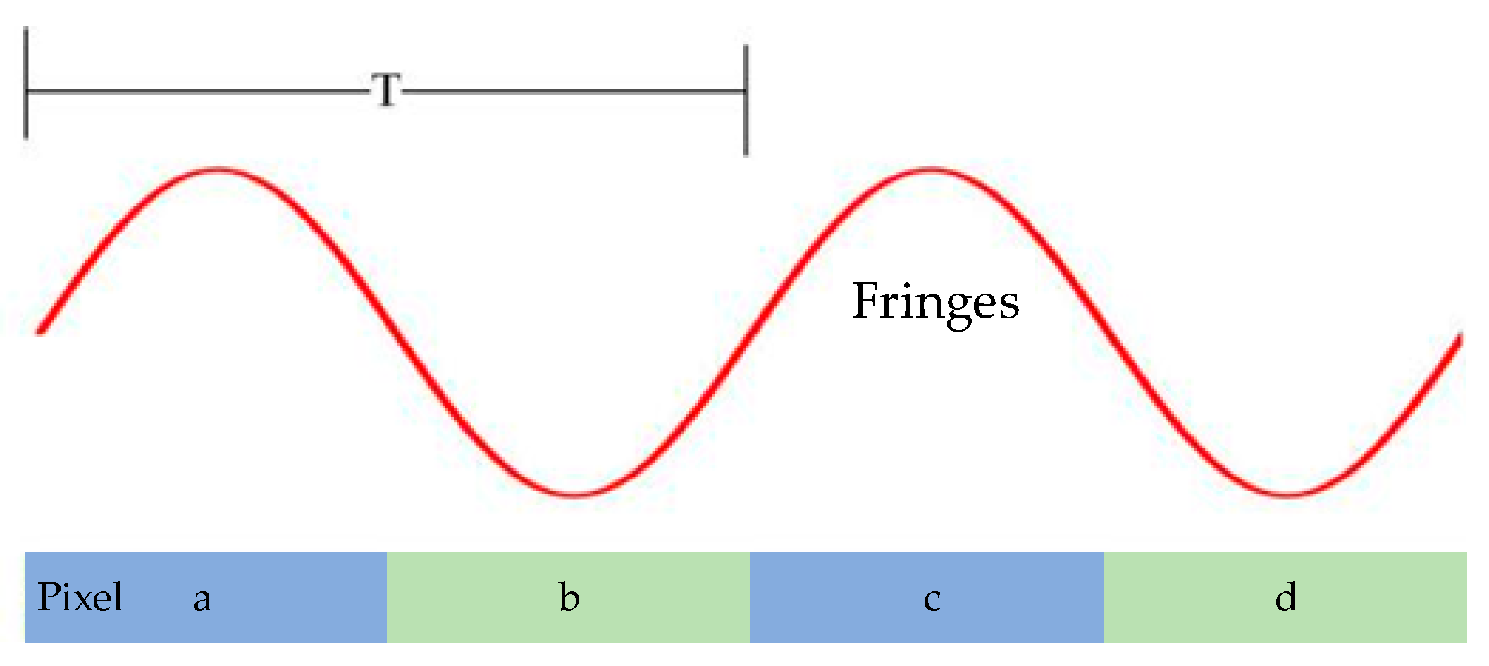
2.1. Data Model
2.2. Background Signals
2.3. Attenuation and Phase Information Reconstruction
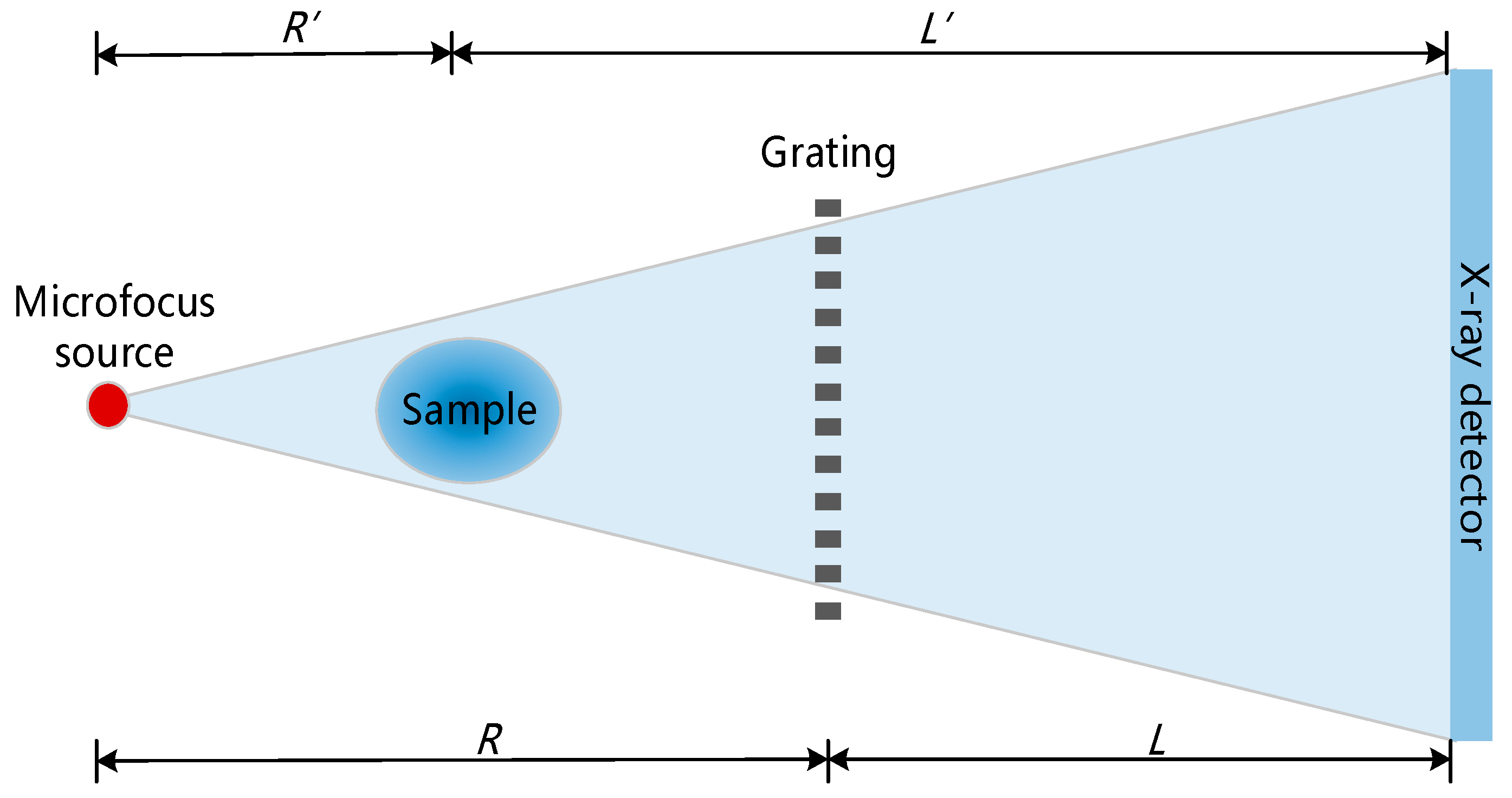
3. Experiments
3.1. Grating
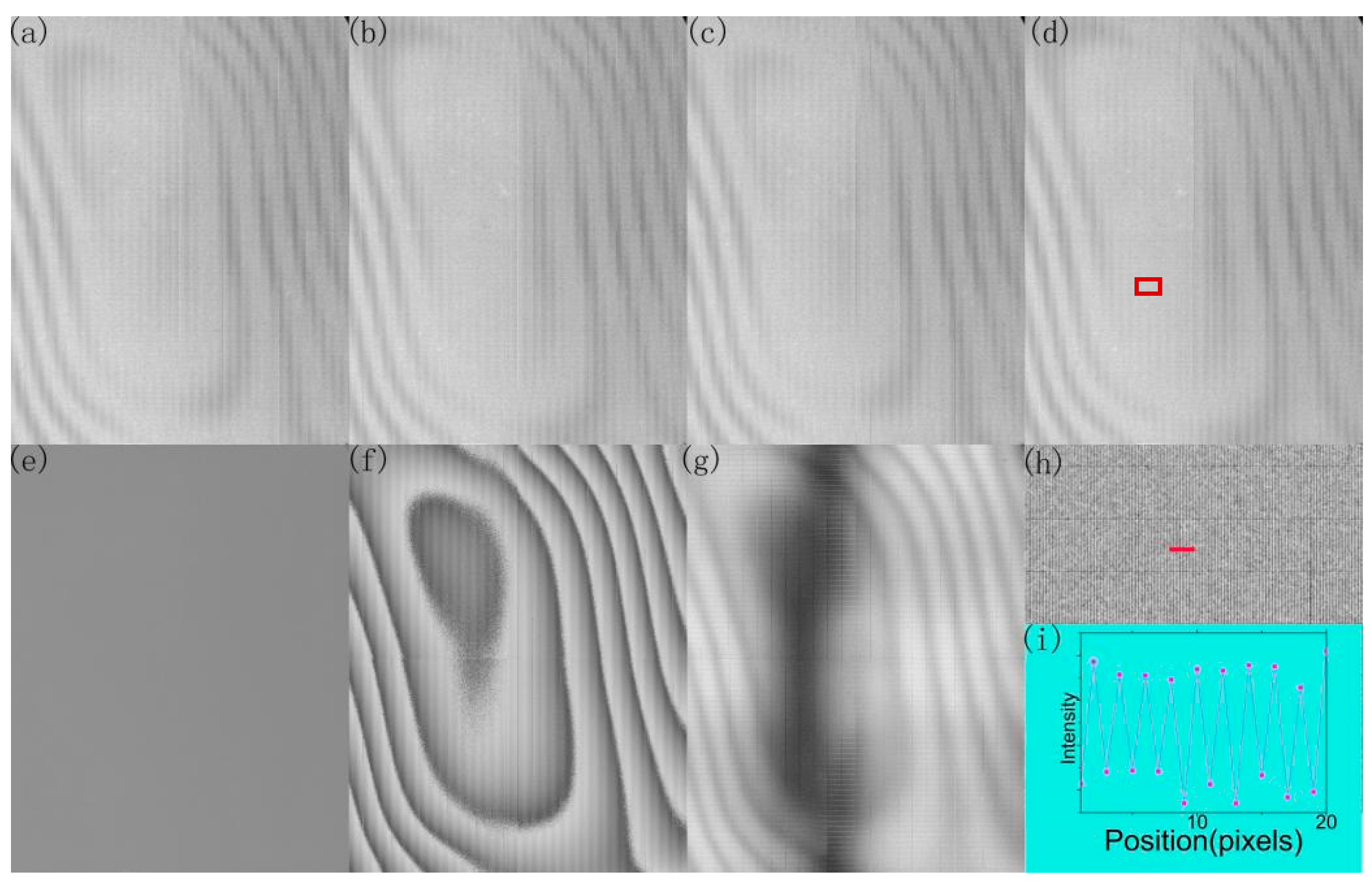
3.2. Background Signals
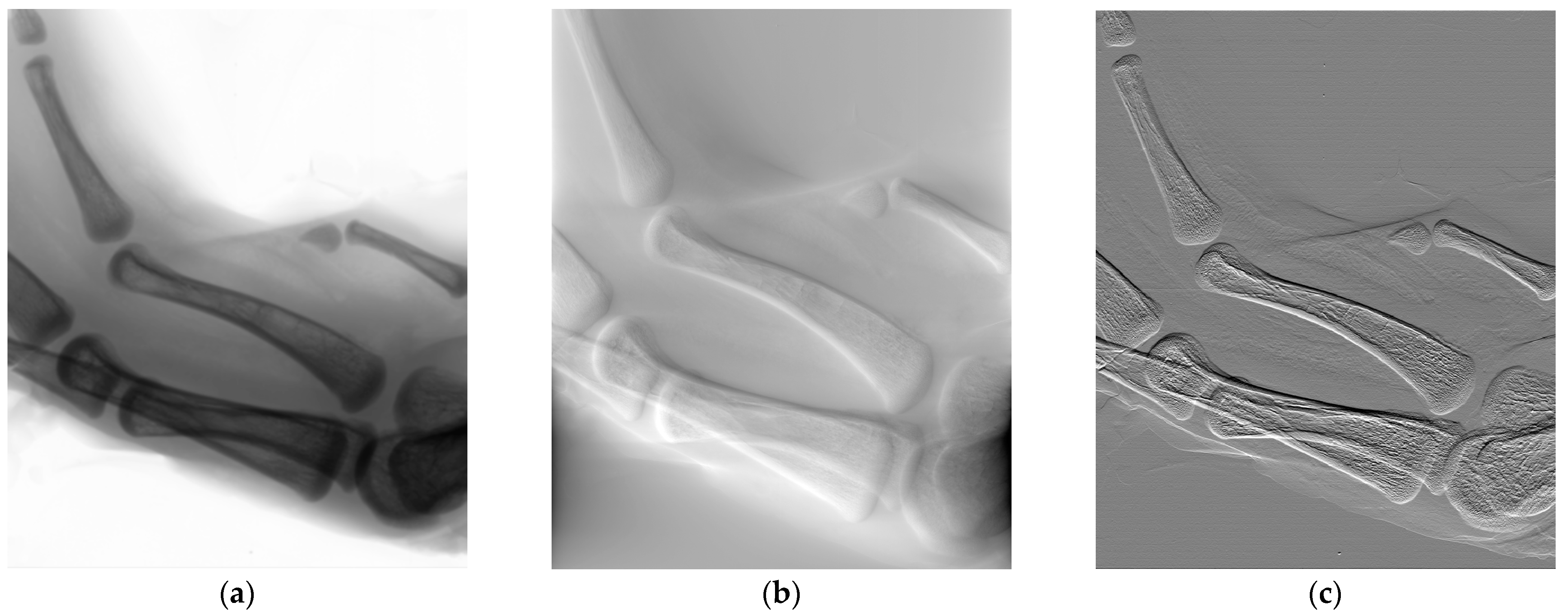
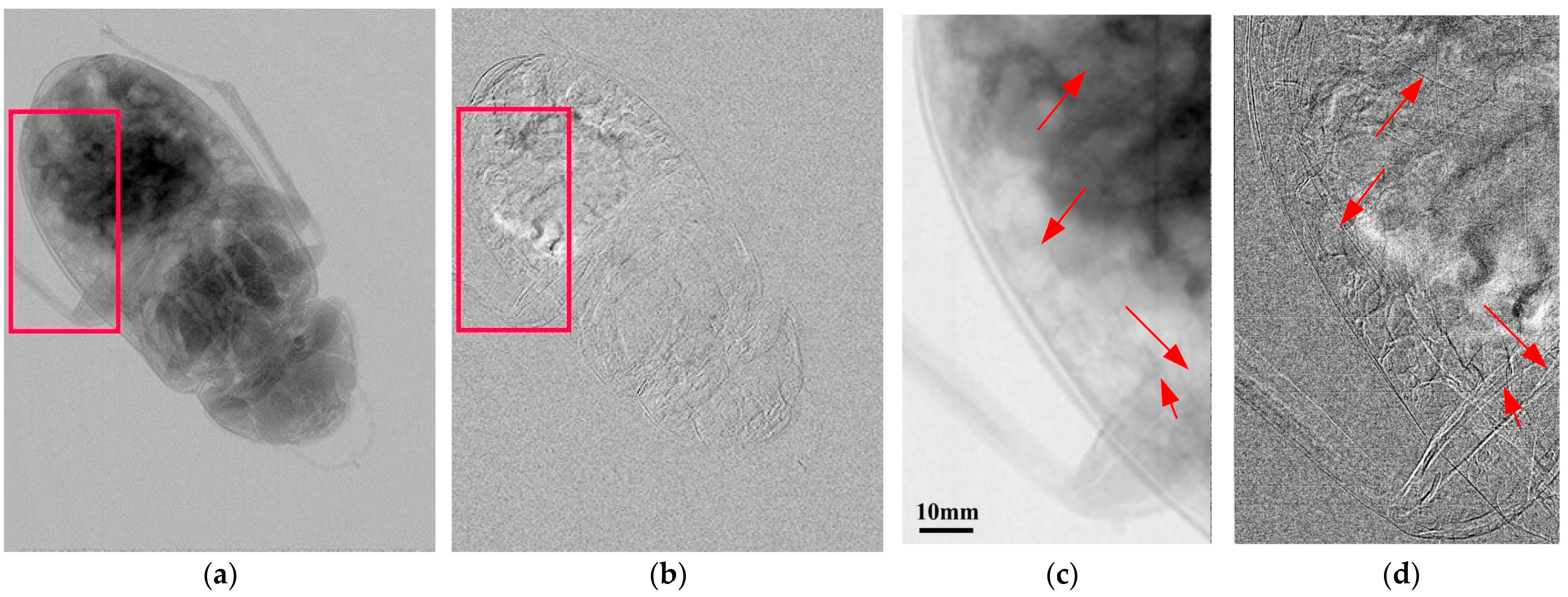
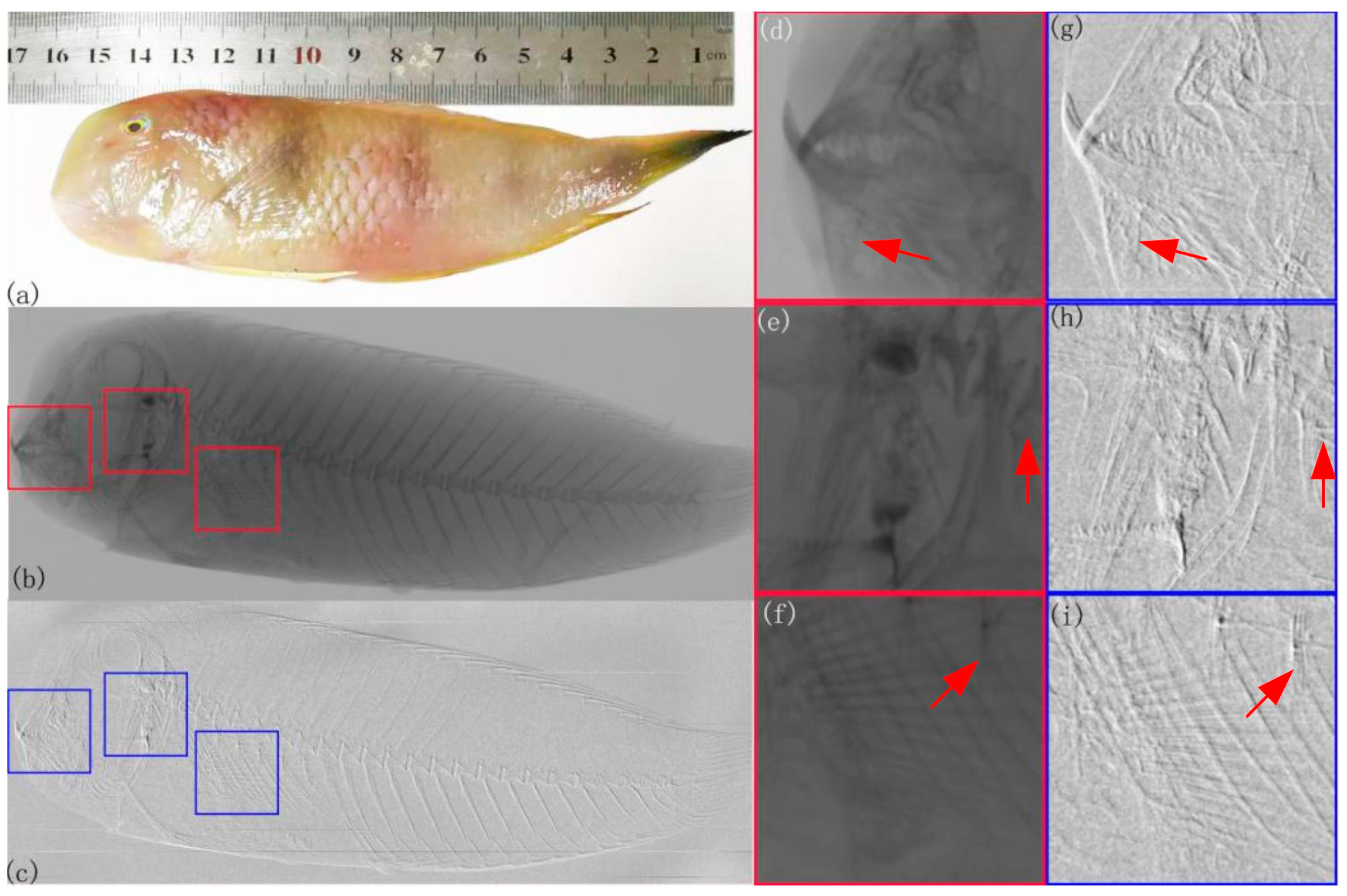
3.3. Micrography
3.4. Large Field of View
4. Discussion and Conclusions
Author Contributions
Funding
Institutional Review Board Statement
Informed Consent Statement
Data Availability Statement
Conflicts of Interest
References
- Pil-Ali, A.; Adnani, S.; Karim, K.S. Self-aligned multi-layer X-ray absorption grating using large-area fabrication methods for X-ray phase-contrast imaging. Sci. Rep. 2023, 13, 2508. [Google Scholar] [CrossRef] [PubMed]
- Andrejewski, J.; De Marco, F.; Willer, K.; Noichl, W.; Gustschin, A.; Koehler, T.; Meyer, P.; Kriner, F.; Fischer, F.; Braun, C.; et al. Whole-body X-ray dark-field radiography of a human cadaver. Eur. Radiol. Exp. 2021, 5, 6. [Google Scholar] [CrossRef]
- Momose, A.; Yashiro, W.; Kuwabara, H.; Kawabata, K. Grating-based X-ray phase imaging using multiline X-ray source. Jpn. J. Appl. Phys. 2009, 48, 076512. [Google Scholar] [CrossRef]
- Arboleda, C.; Wang, Z.; Stampanoni, M. Tilted-grating approach for scanning-mode X-ray phase contrast imaging. Opt. Express 2014, 22, 15447–15458. [Google Scholar] [CrossRef]
- Liu, H.; Liu, M.; Jiang, X.; Luo, J.; Song, Y.; Chu, X.; Zan, G. Multimodal Image Fusion for X-ray Grating Interferometry. Sensors 2023, 23, 3115. [Google Scholar] [CrossRef]
- Pfeiffer, F.; Bech, M.; Bunk, O.; Kraft, P.; Eikenberry, E.F.; Bronnimann, C.; Grunzweig, C.; David, C. Hard-X-ray dark-field imaging using a grating interferometer. Nature Mater. 2008, 7, 134–137. [Google Scholar] [CrossRef] [PubMed]
- Pinkert-Leetsch, D.; Frohn, J.; Ströbel, P.; Alves, F.; Salditt, T.; Missbach-Guentner, J. Three-dimensional analysis of human pancreatic cancer specimens by phase-contrast based X-ray tomography–the next dimension of diagnosis. Cancer Imaging 2023, 23, 43. [Google Scholar] [CrossRef]
- Momose, A.; Yashiro, W.; Harasse, S.; Kuwabara, H. Four-dimensional X-ray phase tomography with talbot interferometry and white synchrotron radiation: Dynamic observation of a living worm. Opt. Express 2011, 19, 8423–8432. [Google Scholar] [CrossRef] [PubMed]
- Miki, H.; Yoneyama, A.; Hirano, K. Visualizing morphological structures of rice grains in precooked products using synchrotron radiation X-ray phase-contrast computed tomography. Food Funct. 2023, 14, 87–93. [Google Scholar] [CrossRef]
- Takeda, Y.; Yashiro, W.; Suzuki, Y.; Aoki, S.; Hattori, T.; Momose, A. X-ray phase imaging with single phase grating. Jpn. J. Appl. Phys. 2007, 46, L89–L91. [Google Scholar] [CrossRef]
- Wang, Z.; Huang, Z.; Zhang, L.; Kang, K.; Zhu, P. Fast X-ray phase-contrast imaging using high resolution detector. IEEE Trans. Nucl. Sci. 2009, 56, 1383–1388. [Google Scholar] [CrossRef]
- Morimoto, N.; Fujino, S.; Ohshima, K.; Harada, J.; Hosoi, T.; Watanabe, H.; Shimura, T. Two dimensional X-ray phase imaging using single grating interferometer with embedded X-ray targets. Opt. Express 2015, 23, 16582–16588. [Google Scholar] [CrossRef] [PubMed]
- Morimoto, N.; Fujino, S.; Ito, Y.; Yamazaki, A.; Sano, I.; Hosoi, T.; Watanabe, H.; Shimura, T. Design and demonstration of phase gratings for 2d single grating interferometer. Opt. Express 2015, 23, 29399–29412. [Google Scholar] [CrossRef] [PubMed]
- Morimoto, N.; Fujino, S.; Ohshima, K.; Harad, J.; Hosoi, T.; Watanabe, H.; Shimura, T. X-ray phase contrast imaging by compact Talbot-Lau interferometer with a single transmission grating. Opt. Lett. 2014, 39, 4297–4300. [Google Scholar] [CrossRef] [PubMed]
- Takeda, M.; Ina, H.; Kobayashi, S. Fourier-transform method of fringe pattern analysis for computer-based topography and interferometry. J. Opt. Soc. Am. 1982, 72, 156–160. [Google Scholar] [CrossRef]
- Itoh, H.; Nagai, K.; Sato, G.; Yamaguchi, K.; Nakamura, T.; Kondoh, T.; Ouchi, C.; Teshima, T.; Setomoto, Y.; Den, T. Two-dimensional grating-based X-ray phase-contrast imaging using fourier transform phase retrieval. Opt. Express 2011, 19, 3339–3346. [Google Scholar] [CrossRef]
- Wen, H.H.; Bennett, E.E.; Kopace, R.; Stein, A.F.; Pai, V. Single-shot X-ray differential phase-contrast and diffraction imaging using two-dimensional transmission gratings. Opt. Lett. 2010, 35, 1932–1934. [Google Scholar] [CrossRef]
- Lim, H.; Lee, J.; Lee, S.; Cho, H.; Lee, H.; Jeon, D. Low-density foreign body detection in food products using single-shot grid-based dark-field X-ray imaging. J. Food Eng. 2022, 335, 111189. [Google Scholar] [CrossRef]
- Krejci, F.; Jakubek, J.; Kroupa, M. Hard X-ray phase contrast imaging using single absorption grating and hybrid semiconductor pixel detector. Rev. Sci. Instrum. 2010, 81, 113702. [Google Scholar] [CrossRef]
- Xu, Y.; Tao, S.; Bian, Y.; Bai, L.; Tian, Z.; Hao, X.; Kuang, C.; Liu, X. Single-shot grating-based X-ray phase contrast imaging via generative adversarial network. Opt. Laser. Eng. 2022, 152, 106960. [Google Scholar] [CrossRef]
- Shi, S.Q.Z.; Shapira, N.; Noël, P.B.; Meyer, S. Convolutional neural network-based single-shot speckle tracking for X-ray phase-contrast imaging. arXiv 2023, arXiv:2305.01871. [Google Scholar] [CrossRef]
- Liu, X.; Guo, J.; Lei, Y.; Du, Y.; Niu, H. Two-step phase retrieval method with unknown phase shift on non-absorption grating X-ray differential phase contrast imaging system. Nucl. Instrum. Methods Phys. Res. 2012, 691, 86–89. [Google Scholar] [CrossRef]
- Zhu, P.; Zhang, K.; Wang, Z.; Liu, X.; Liu, Y.; Wu, Z.; McDonald, S.A.; Marone, F.; Stampanoni, M. Low-dose, simple, and fast grating-based X-ray phase-contrast imaging. Proc. Natl. Acad. Sci. USA 2010, 107, 13576–13581. [Google Scholar] [CrossRef] [PubMed]
- Lei, Y.; Xu, G.; Wali, F.; Li, Q.; Liu, X.; Huang, J.; Li, J. An 8-inch absorption grating used in cascaded Talbot-Lau interferometers for X-ray phase-contrast imaging. Appl. Phys. Express 2019, 12, 126504. [Google Scholar] [CrossRef]
- Huang, J.; Wali, F.; Lei, Y.; Liu, X.; Li, J. Fourier transform phase retrieval for X-ray phase-contrast imaging based on cascaded Talbot-Lau interferometers. Opt. Eng. 2020, 59, 033101. [Google Scholar]
- Zuo, C.; Feng, S.; Huang, L.; Tao, T.; Yin, W.; Chen, Q. Phase shifting algorithms for fringe projection profilometry: A review. Opt. Laser. Eng. 2018, 109, 23–59. [Google Scholar] [CrossRef]


Disclaimer/Publisher’s Note: The statements, opinions and data contained in all publications are solely those of the individual author(s) and contributor(s) and not of MDPI and/or the editor(s). MDPI and/or the editor(s) disclaim responsibility for any injury to people or property resulting from any ideas, methods, instructions or products referred to in the content. |
© 2023 by the authors. Licensee MDPI, Basel, Switzerland. This article is an open access article distributed under the terms and conditions of the Creative Commons Attribution (CC BY) license (https://creativecommons.org/licenses/by/4.0/).
Share and Cite
Liu, X.; Liu, L.; Huang, J.; Lei, Y.; Li, J. Single-Shot Phase-Contrast Imaging with a Single Grating. Photonics 2023, 10, 968. https://doi.org/10.3390/photonics10090968
Liu X, Liu L, Huang J, Lei Y, Li J. Single-Shot Phase-Contrast Imaging with a Single Grating. Photonics. 2023; 10(9):968. https://doi.org/10.3390/photonics10090968
Chicago/Turabian StyleLiu, Xin, Lang Liu, Jianheng Huang, Yaohu Lei, and Ji Li. 2023. "Single-Shot Phase-Contrast Imaging with a Single Grating" Photonics 10, no. 9: 968. https://doi.org/10.3390/photonics10090968