Two-Photon Absorption in Ca3(VO4)2 and Ca2.7Sr0.3(VO4)2 Crystals

 , , ,
, , , 
Abstract
:1. Introduction
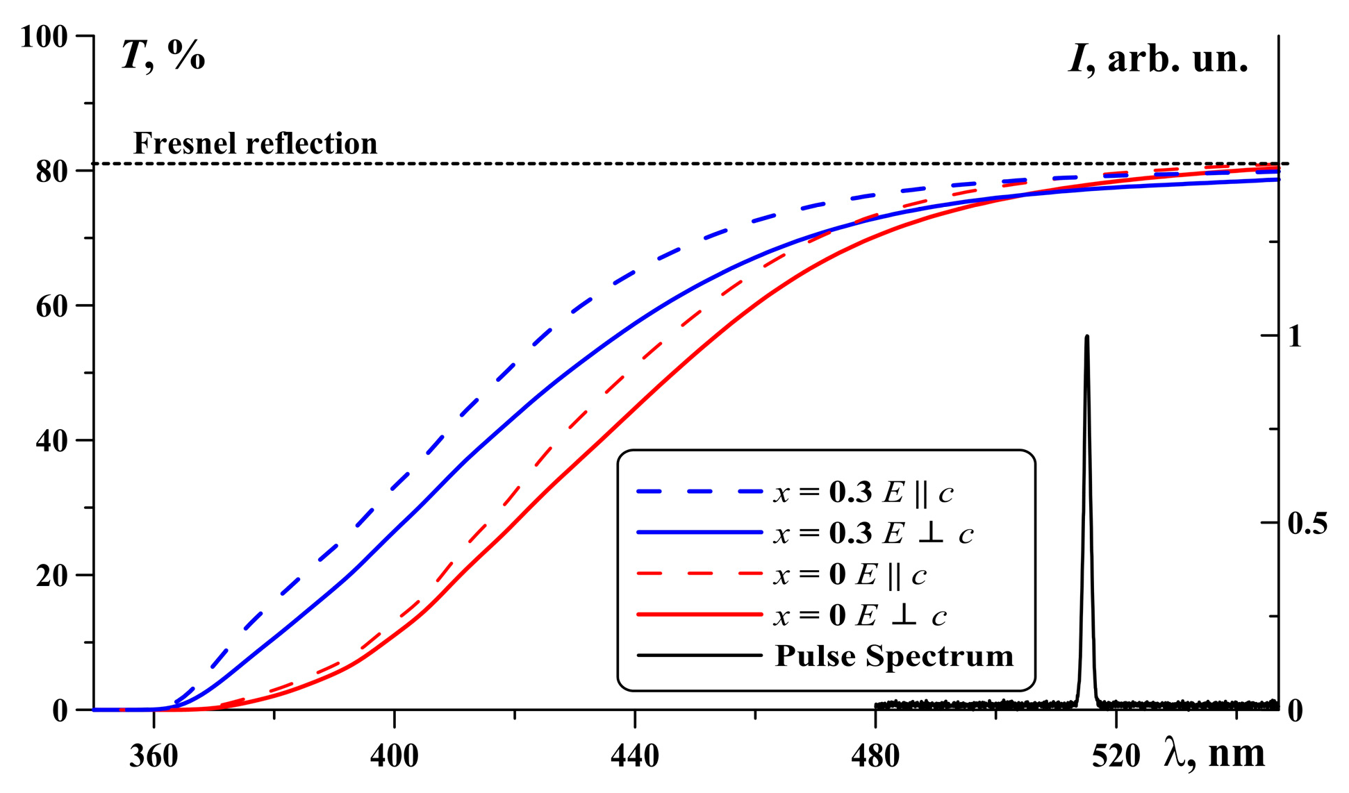
2. CSVO Crystals
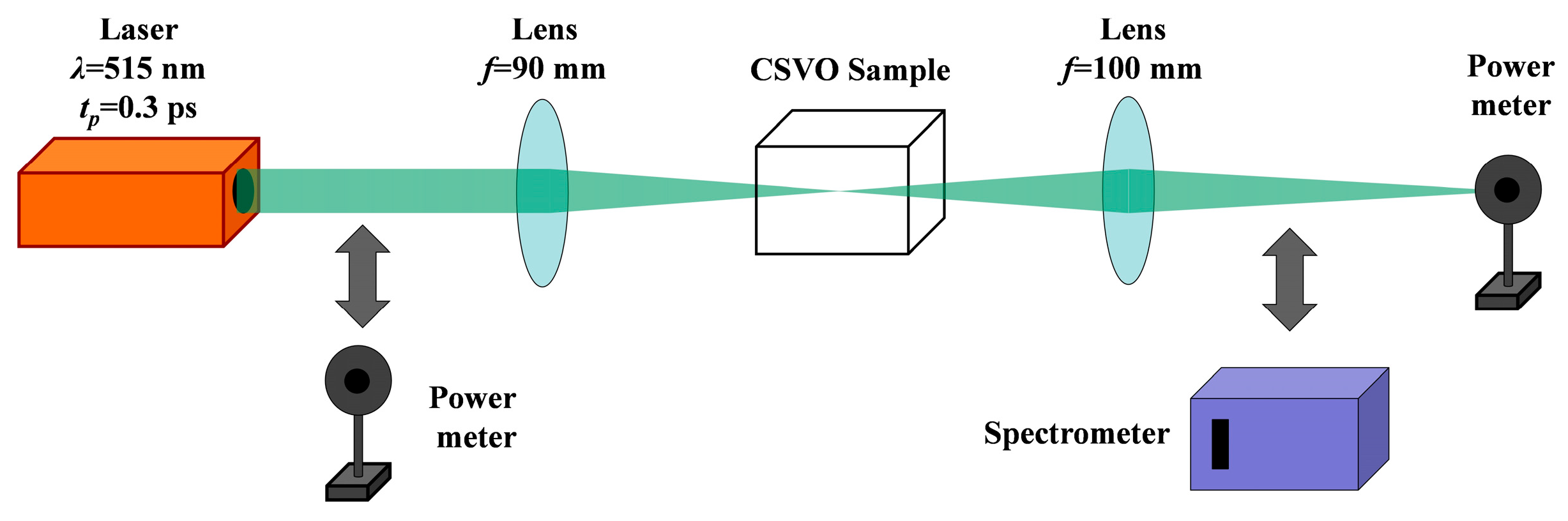
3. Experiment Set-Up
4. Data Processing Procedure
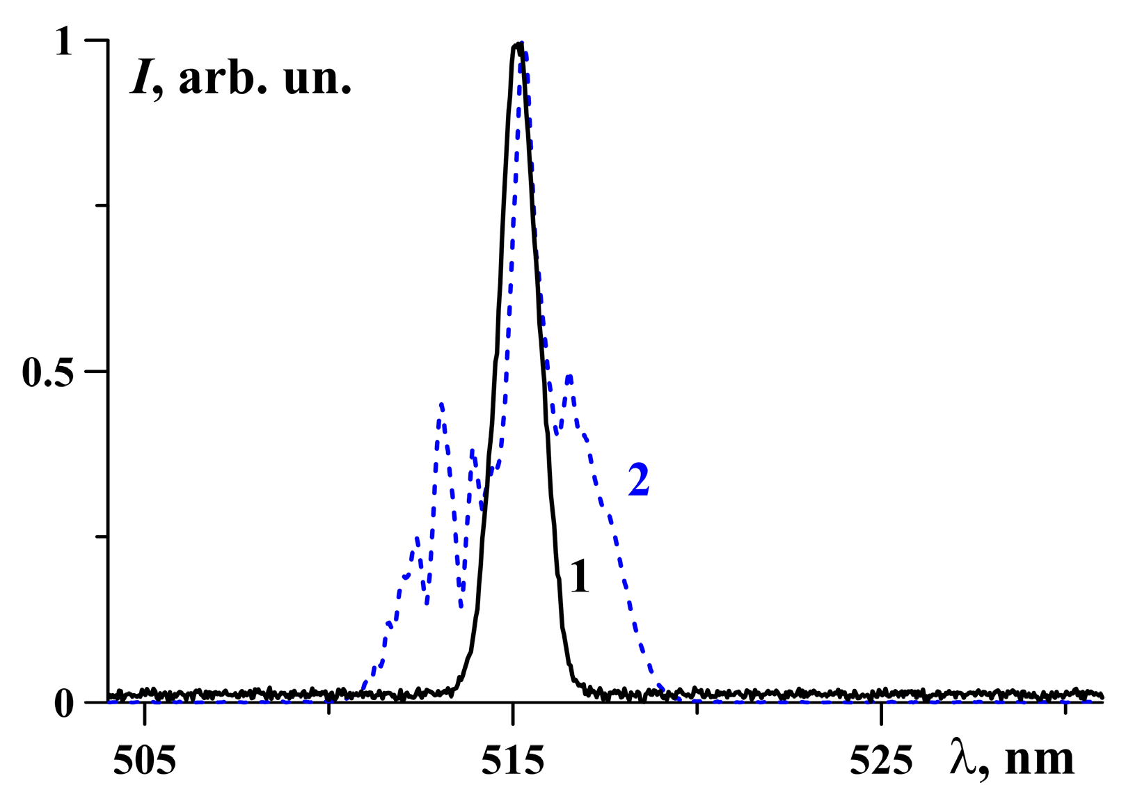
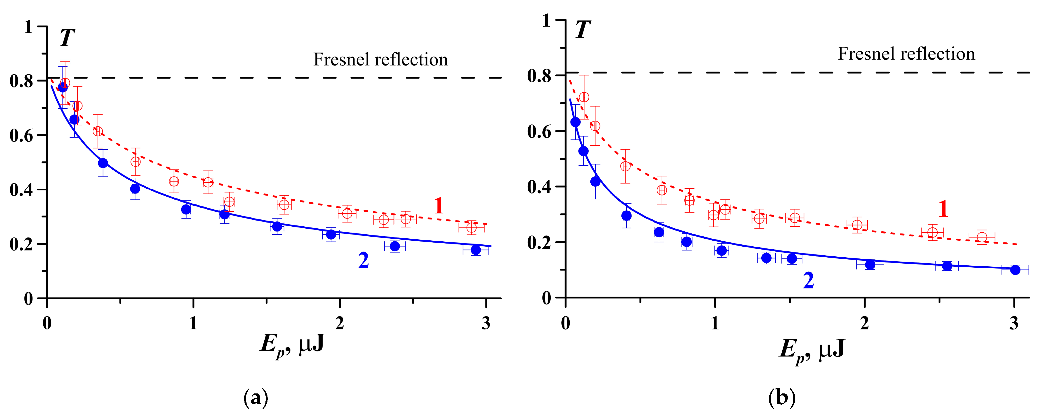
5. Experimental Results
6. Conclusions
Author Contributions
Funding
Institutional Review Board Statement
Informed Consent Statement
Data Availability Statement
Conflicts of Interest
References
- Chen, Z.; Wang, D.; Liu, L.; Yuan, F.; Huang, Y.; Zhang, L.; Lin, Z. Cr3+ doped Ca3(VO4)2: A new tunable laser crystal. J. Alloys Compd. 2022, 938, 168651. [Google Scholar] [CrossRef]
- Ivleva, L.I.; Dunaeva, E.E.; Voronina, I.S.; Doroshenko, M.E.; Papashvili, A.G. Ca3(VO4)2:Tm3+—A new crystalline medium for 2-μm lasers. J. Cryst. Growth 2018, 501, 18–21. [Google Scholar] [CrossRef]
- Bechthold, P.S.; Liebertz, J.; Deserno, U. Linear and nonlinear optical properties of Ca3(VO4)2. Opt. Commun. 1978, 27, 393–398. [Google Scholar] [CrossRef]
- Frank, M.; Smetanin, S.N.; Jelínek, M.; Vyhlídal, D.; Ivleva, L.I.; Dunaeva, E.E.; Voronina, I.S.; Tereshchenko, D.P.; Shukshin, V.E.; Zverev, P.G.; et al. Stimulated Raman scattering in yttrium, gadolinium, and calcium orthovanadate crystals with single and combined frequency shifts under synchronous picosecond pumping for sub-picosecond or multi-wavelength generation around 1.2 µm. Crystals 2020, 10, 871. [Google Scholar] [CrossRef]
- Kinyaevskiy, I.O.; Kovalev, V.I.; Koribut, A.V.; Dunaeva, E.E.; Semin, N.S.; Ionin, A.A. Stimulated Raman scattering of 0.3-ps 515-nm laser pulses in Ca3(VO4)2 and Ca2.7Sr0.3(VO4)2 crystals. Opt. Spectrosk 2023, 131, 207. [Google Scholar]
- Dorbakova, N.G.; Baryshnikova, O.V.; Morozov, V.A.; Belik, A.A.; Katsuya, Y.; Tanaka, M.; Stefanovich, S.Y.; Lazoryak, B.I. Tuning of nonlinear optical and ferroelectric properties via the cationic composition of Ca9.5–1.5xBixCd(VO4)7 solid solutions. Mater. Des. 2017, 119, 515–523. [Google Scholar]
- Voronina, I.S.; Dunaeva, E.E.; Voronov, V.V.; Shukshin, V.E.; Smetanin, S.N.; Ivleva, L.I. Growth and characterization of (Ca1−xSrx)3(VO4)2 solid solutions: A search for the new materials for ultrafast Raman lasers. Opt. Mater. 2021, 111, 110642. [Google Scholar] [CrossRef]
- Li, C.; Yang, W.; Chang, Y. Raman scattering study of calcium orthovanadate crystal. Jap. J. Appl. Phys. 1985, 24, 508. [Google Scholar] [CrossRef]
- Chunaev, D.S.; Dunaeva, E.E.; Kravtsov, S.B.; Voronina, I.S.; Zverev, P.G. Two-photon Absorption in Ca3(VO4)2 Crystal. In Proceedings of the European Conference on Lasers and Electro-Optics 2021, Munich, Germany, 21–25 June 2021; p. ce_p_12. [Google Scholar]
- Kovalev, V.I.; Rus’kin, O.L.; Suvorov, M.B. Nonlinear absorption of randomly pulsating CO2 laser radiation in narrow-gap semiconductors. Sov. J. Quantum Electron. 1991, 21, 1346. [Google Scholar] [CrossRef]
- Parhi, P.; Manivannan, V.; Kohli, S.; Mccurdy, P. Synthesis and characterization of M3V2O8 (M = Ca, Sr and Ba) by a solid-state metathesis approach. Bull. Mater. Sci. 2008, 31, 885. [Google Scholar] [CrossRef]
- Shen, Y.R. The Principles of Nonlinear Optics; Wiley: New York, NY, USA, 1984. [Google Scholar]
- Von der Linde, D. Experimental study of single picosecond light pulses. IEEE J. Quantum Electron. 1972, 8, 328. [Google Scholar] [CrossRef]
- Bredikhin, V.I.; Galanin, M.D.; Genkin, V.N. Two-photon absorption and spectroscopy. Sov. Phys. Usp. 1973, 16, 299–321. [Google Scholar] [CrossRef]
- Keldysh, L.V. Ionization in the field of a strong electromagnetic wave. Sov. Phys. JETP 1965, 20, 1307. [Google Scholar]
- Basov, N.G.; Grasyuk, A.Z.; Zubarev, I.G.; Katulin, V.A.; Krokhin, O.N. Semiconductor quantum generator with two-photon optical excitation. Sov. Phys. JETP 1966, 23, 366. [Google Scholar]
- Logvinov, I.N.; Perel’man, N.F. Theory of many-photon transitions in insulating crystals. Sov. Phys.-Solid State 1980, 22, 372. [Google Scholar]
- Kinyaevskiy, I.O.; Koribut, A.V.; Grudtsyn, Y.V.; Seleznev, L.V.; Kovalev, V.I.; Pushkarev, D.V.; Dunaeva, E.E.; Ionin, A.A. Frequency down-conversion of a chirped Ti: Sapphire laser pulse with BaWO4 Raman shifter and second-order nonlinear crystal. Laser Phys. Lett. 2022, 19, 095403. [Google Scholar] [CrossRef]


Disclaimer/Publisher’s Note: The statements, opinions and data contained in all publications are solely those of the individual author(s) and contributor(s) and not of MDPI and/or the editor(s). MDPI and/or the editor(s) disclaim responsibility for any injury to people or property resulting from any ideas, methods, instructions or products referred to in the content. |
© 2023 by the authors. Licensee MDPI, Basel, Switzerland. This article is an open access article distributed under the terms and conditions of the Creative Commons Attribution (CC BY) license (https://creativecommons.org/licenses/by/4.0/).
Share and Cite
Kinyaevskiy, I.O.; Kovalev, V.I.; Semin, N.S.; Danilov, P.A.; Kudryashov, S.I.; Koribut, A.V.; Dunaeva, E.E. Two-Photon Absorption in Ca3(VO4)2 and Ca2.7Sr0.3(VO4)2 Crystals. Photonics 2023, 10, 466. https://doi.org/10.3390/photonics10040466
Kinyaevskiy IO, Kovalev VI, Semin NS, Danilov PA, Kudryashov SI, Koribut AV, Dunaeva EE. Two-Photon Absorption in Ca3(VO4)2 and Ca2.7Sr0.3(VO4)2 Crystals. Photonics. 2023; 10(4):466. https://doi.org/10.3390/photonics10040466
Chicago/Turabian StyleKinyaevskiy, Igor O., Valery I. Kovalev, Nikita S. Semin, Pavel A. Danilov, Sergey I. Kudryashov, Andrey V. Koribut, and Elizaveta E. Dunaeva. 2023. "Two-Photon Absorption in Ca3(VO4)2 and Ca2.7Sr0.3(VO4)2 Crystals" Photonics 10, no. 4: 466. https://doi.org/10.3390/photonics10040466