Article

Spiral Shaped Photonic Crystal Fiber-Based Surface Plasmon Resonance Biosensor for Cancer Cell Detection

1
Optoelectronics and Photonics Research Lab, Department of Electronics and Communication Engineering, Manipal University Jaipur, Jaipur 303007, India
2
Quantum Research Group, School of Chemistry and Physics, University of KwaZulu-Natal, Durban 4001, South Africa
3
Department of Communication Lines, Povolzhskiy State University of Telecommunications and Informatics, 23 Lev Tolstoy Street, 443100 Samara, Russia
4
JSC “Scientific Production Association State Optical Institute Named after Vavilov S.I.”, 36/1, Babushkin Street, 192171 Saint Petersburg, Russia
5
Department of Photonics and Communication Links, Saint Petersburg State University of Telecommunications Named after M.A. Bonch-Bruevich, 22 Bolshevikov Avenue, 193232 Saint Petersburg, Russia
6
Department of Radiophotonics and Microwave Technologies, Kazan National Research State University, Named after A.N. Tupolev-KAI, 10, Karl Marx Street, 420111 Kazan, Russia
7
Division of Quantum Physics and Quantum Information, University of Science and Technology of China, 99 Xiupu Road Pudong District, Shanghai 200093, China
8
Department of ECE, Malaviya National Institute of Technology Jaipur (MNIT), Jaipur 302017, India
*
Author to whom correspondence should be addressed.
Photonics 2023, 10(3), 230; https://doi.org/10.3390/photonics10030230
Submission received: 22 December 2022 / Revised: 27 January 2023 / Accepted: 17 February 2023 / Published: 21 February 2023
(This article belongs to the Special Issue Adaptive Optics and Its Applications)

Abstract

:
This work presents the design and simulation of an all-optical sensor for detection of cancer cells. The proposed device is based on the surface plasmon resonance effect on a spiral shaped photonic crystal fiber structure. The finite element method (FEM) based simulations are carried out for the different cancer cells, such as HELA, Basal, Jurkat, and MDA-MB-231, MCF7, and PC12 detection. The sensor has shown the maximum sensitivity of −289 RIU−1 for the refractive index of the detection of breast cancer cell with the resolution of 2.33 × 10⁻⁴. The sensor is effective for the refractive index range of 1.36 to 1.401.The structure is based on spiral shaped photonic crystal fiber, and has shown promising linear sensing response to support the practical feasibility of the device. The proposed sensor design is effective in detecting cervical cancer, skin cancer, blood cancer, breast cancer type 1, breast cancer type 2, and adrenal gland cancer.

1. Introduction

Photonics biosensing platforms combine high sensitivity, selectivity, and durability, due to which they have tremendous advantages in the detection of many diseases, such as cancer, diabetes, malaria, etc. Recent surveys have revealed that more than ten million people lost their lives due to cancer in 2020, accounting for one out of six deaths worldwide [1,2]. Cancer is an epigenetic disorder which leads to unregulated cell growth, causing the formation of tumors in the human body [3]. Various types of cancer categories are Basal cell cancer, lung cancer, breast cancer, leukemia, prostate cancer, blood cancer, cervical cancer etc. [4]. Early detection of cancerous cells increases the chances of survival of patients and better response to treatment. Optical biosensors have the potential for fast and precise detection, reliable imaging of cancer cells, monitoring of onto-genesis of cancer cells, and determination of the effectiveness of anti-cancer chemotherapy agents. Numerous methods, such as Raman Spectroscopy [5,6,7] and surface-enhanced Raman Spectroscopy [8] have been investigated to detect cancer cells which employ Photonic Crystal Fiber (PCF)-based Surface Plasmon Resonance (SPR) biosensors. New designs, such as Hollow Core PCF (HC-PCF) [8,9], dual-core [10], and twin core PCF (TC-PCF) [11], have been used for effective biosensing. A dual-parameter SPR-PCF sensor is also designed that can measure both temperature and the magnetic field simultaneously. On either side of the fiber core, there are two elliptical channels in this sensor [12]. Many PCF-based SPR biosensors sense variations in Refractive Index (RI) with various optical parameters, and are classified as refractive-index-based biosensors. Due to flexibility of designing, scalable, and efficient birefringence [13,14,15] ease of dispersion tailoring, and various nonlinear properties [16,17,18], PCFs can be easily tailored to acquire the desired responses as sensing elements. Due to this, immense interest has developed in the usage of PCF for SPR-based biosensing [19]. Traditional prism-based SPR sensing difficulties, such as bulky design, accurate incident angle, and a huge number of adjustable mechanical components, which restrict the diverse range of applications, are all remedied by PCF SPR imaging technologies [20]. SPR is an optical phenomenon which is manifested by the collective oscillation of surface plasmons (i.e., free electrons). These electrons are found on the boundary of metal and dielectric surfaces. Any small change in RI due to an unknown analyte causes the shift of peak in the loss spectrum. When the wavelength of the surface electrons matches that of the incoming photons, resonance occurs. Here, at reverberation (resonance) frequency, Surface Plasmon Polariton (SPP) [21] interacts with the permeable center. Thus, the electric field at the core is leaked towards the metal, the dielectric medium and the corresponding mode are called SPP mode. This SPP mode is hyper sensitive in nature to even small changes in the analyte’s RI [22]. SPR based PCF sensors have applications in DNA detecting [23], gas sensing [24], monitoring of water sample [25], and early detection of cancer [26]. The SPR technology is used in accordance with various schemes, such as prism emerged [27], Mach Zehnder interferometer [28], but there are optomechanical issues in SPR technology, such as complex and bulky system, oblique incidence required, and limited analysis throughput. These limitations can be overcome with PCF- based biosensors In PCF-SPR sensors, plasmonic materials perform a vital role in the design and development of sensors. Different types of plasmonic materials, such as silver [29], zinc oxide [30], gold [31,32], titanium dioxide [33], and graphene [34,35], are used for designing and fabrication of SPR biosensors due to their unique properties. In this paper, gold is selected as a plasmonic material for designing PCF-based SPR biosensors as it is immune to oxidation in a wet environment, and it is very widely used in various biosensing applications. It is necessary that metal coating should be consistent. The proposed design is externally metal-coated PCF-SPR sensor which makes it feasible for fabrication. [36]. The refractive index component was used to analyze cancer cells from the cervical (HeLa), adrenal gland (PC12), blood (Jurkat), and breast (MCF-7 and MDA-MB-231) types. The suggested study is innovative because it proposes a model of a highly sensitive PCF- SPR based biosensor for the separation of malignant cells from healthy cells using correct FEM-based computational techniques. The article has been structured as follows: Section 2 comprised the basic principles and mathematical method involved in the sensor design. Section 3 includes all the details regarding the obtained results, and their comparison with the existing literature, followed by a conclusion.

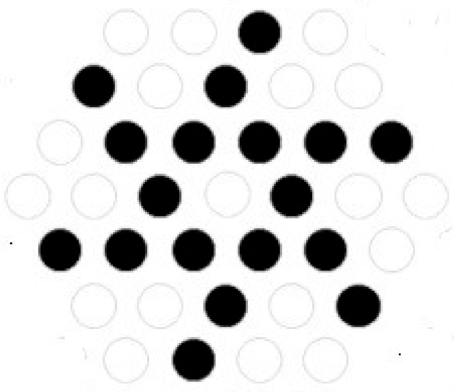
2. Biosensor Design and Geometry

The cross-section schematic of the proposed spiral shaped photonic crystal fiber based surface plasmon resonance biosensor is shown in Figure 1. The sensor design is based on a PCF made up of silica (SiO2) with circular air holes in spiral shaped cladding. This structure can be realized by stacking the silica capillaries (shown as black circles) and rods (shown as white circles) in a conventional hexagonal lattice, as shown in Figure 2. This stack can then be drawn into a fiber using the stack-and-draw technique. Such a hexagonal lattice-based design of the spiral PCF has been recently fabricated [37]. The distance between any two air holes pitch Λ is 2.3 µm, and the hole diameter d is 1 µm. To construct a solid core for light confinement, one air hole is removed from the first layer of hexagonal lattice. This configuration is suitable for producing surface plasmons. A 25 nm thick gold film is deposited over the PCF as a plasmonic material for effective sensing. This can be implemented using the chemical vapor deposition (CVD) process. A 2.6 µm cancer cell identification layer is placed above the plasmonic layer. Finally, to shield the PCF’s interior structure from outer air disturbances, a “perfectly matched layer (PML)” of thickness 0.3 µm is placed above the cancer cell detecting layer. The Sellmeier Equation (1) used for calculating RI of silica is as follows [38]:
    n 2 λ = 1 + B 1 λ 2 λ 2 C 1 B 2 λ 2 λ 2 C 2 B 3 λ 2 λ 2 C 3
Here, n represents the refractive index of silica while λ denotes the operating wavelength.
For silica, Sellmeier coefficients B1, B2, B3, C1, C2, and C3 are taken as 0.696166300, 0.407942600, 0.897479400, 0.00467914826, 0.0135120631, and 97.9340025, respectively. The Drude Lorentz Model, which is depicted in (2), is used to calculate the value of Gold’s dielectric constant [39].
  ε A u = ε ω D 2 ω ω + j y D Δ ε . Ω L 2 ω 2 Ω L 2 + j Γ L ω
Here, ϵAu is the permittivity of gold, ϵ is the permittivity of gold at high frequency, ω = 2πc/λ (angular frequency), where c is the velocity of light, ω D = 2113.6 THz × 2π (plasma frequency), y D = 15.92 THz × 2π (damping frequency), and ∆ϵ = 1.09 (weighting factor).
L = 650.07 2π THz (Lorentz oscillator strength) and Γ L = 104.86 × 2π THz (Spectral width). The modal analysis of the proposed PCF design was carried out with COMSOL Multiphysics 5.3 employing the Full-Vectorial Finite Element Method (FV-FEM) with perfectly matched layers (PML). Figure 3a–c shows electric fields in X-polarized, Y-polarized fundamental and SPP modes, respectively.
When the effective indices of plasmon and core modes are comparable, confinement losses are computed to attain peak values at the same wavelength. The variations at this peak wavelength are detected using the amplitude interrogation method. As a result, calculating confinement losses is extremely important for these sensor’s performance. Here, Equation (3) is utilized to calculate the confinement losses [40].
α d b c m = 8.686 × k 0 × I m n eff × 10 4
k0 = 2π/λ is defined as a wave number, λ is the operating wavelength. The sensor’s resolution determines the degree of analyte RI detection. The proposed PCF-SPR biosensor’s resolution (R) was calculated using Equation (4) [41]:
R = n a λ min λ p e a k  
Here, ∆na is the change in analyte RI, ∆λpeak is the difference in resonance peak shift and ∆λmin is the instrumental peak wavelength.
In PCF-based SPR sensors, sensitivity is a crucial metric. An amplitude approach can be used instead of the spectral method to detect the analyte refractive index variations. Unlike the spectral approach, this method detects changes by studying only specific wave-lengths rather than a broad range of wavelengths. Amplitude Sensitivity can be calculated using Equation (5) [42]:
      S A λ R I U 1 = 1 α λ , n a α λ , n a n a
where ∂na is the variation in the analyte’s refractive index, ∂α(λ, na) is the variation in propagation losses with na at wavelength λ, and α(λ, na) is the confinement loss for analyte’s refractive index, na at wavelength λ.

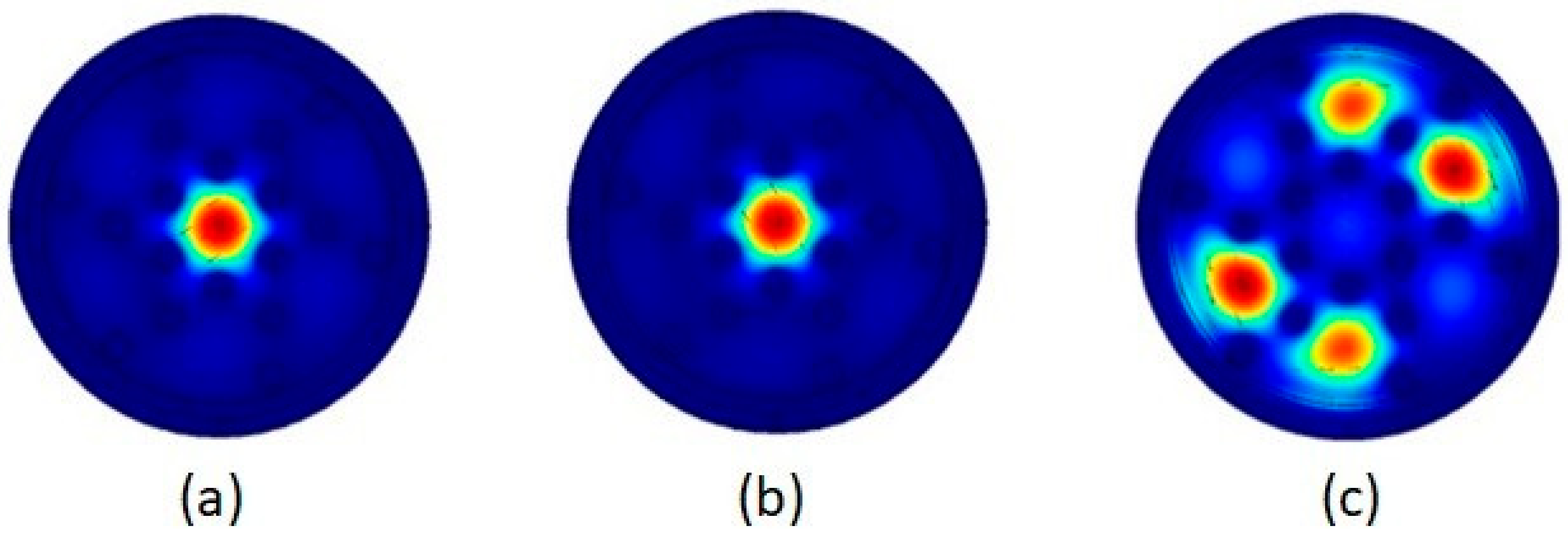
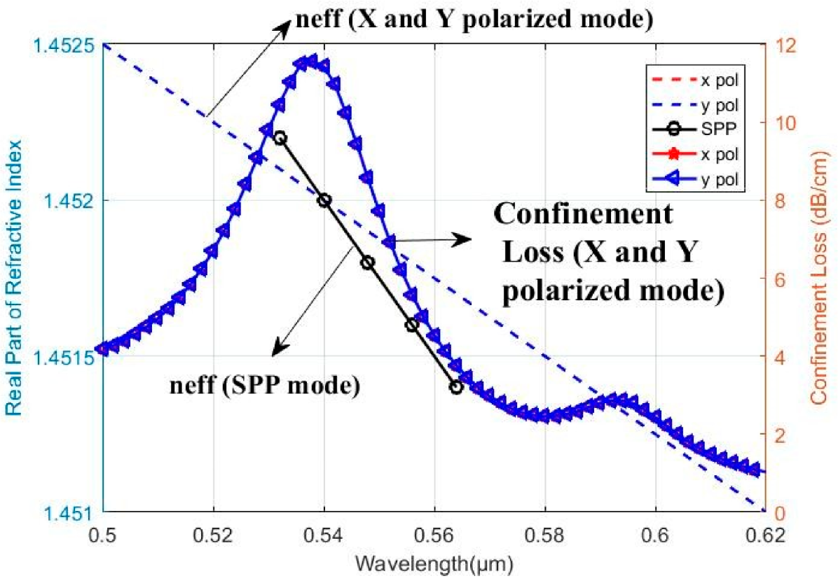
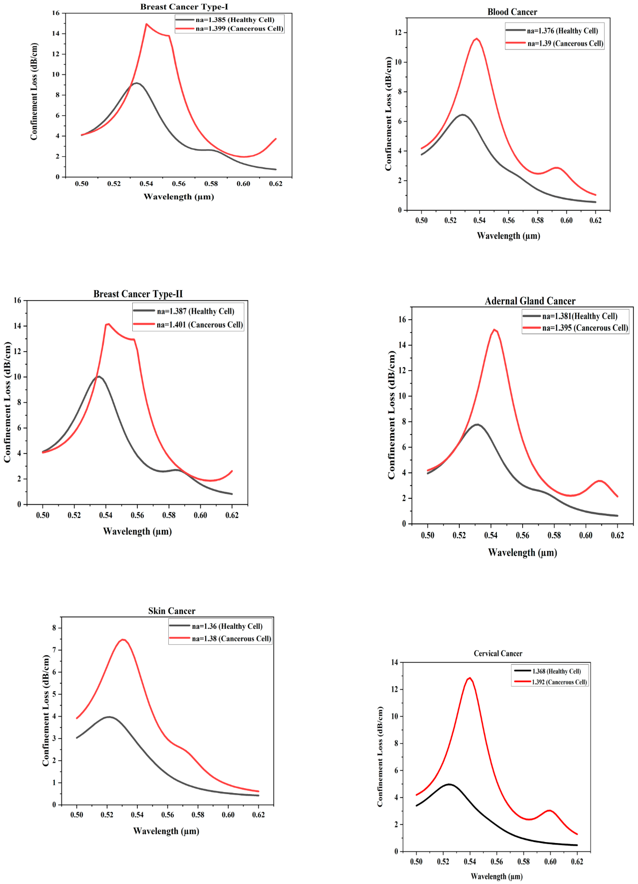
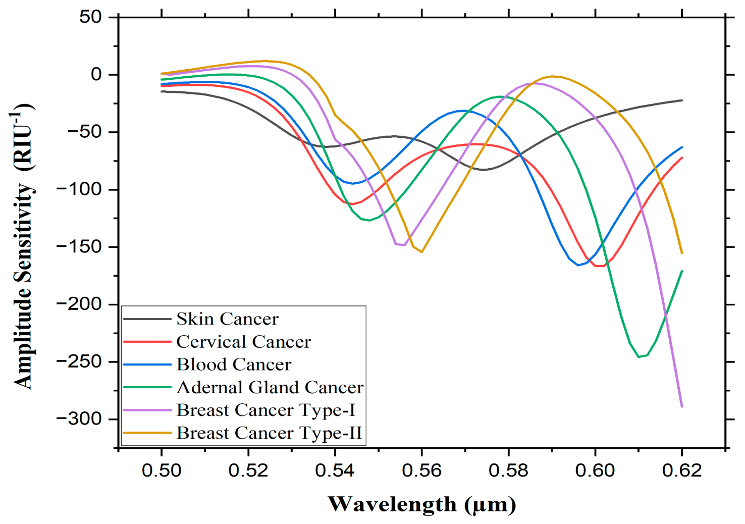
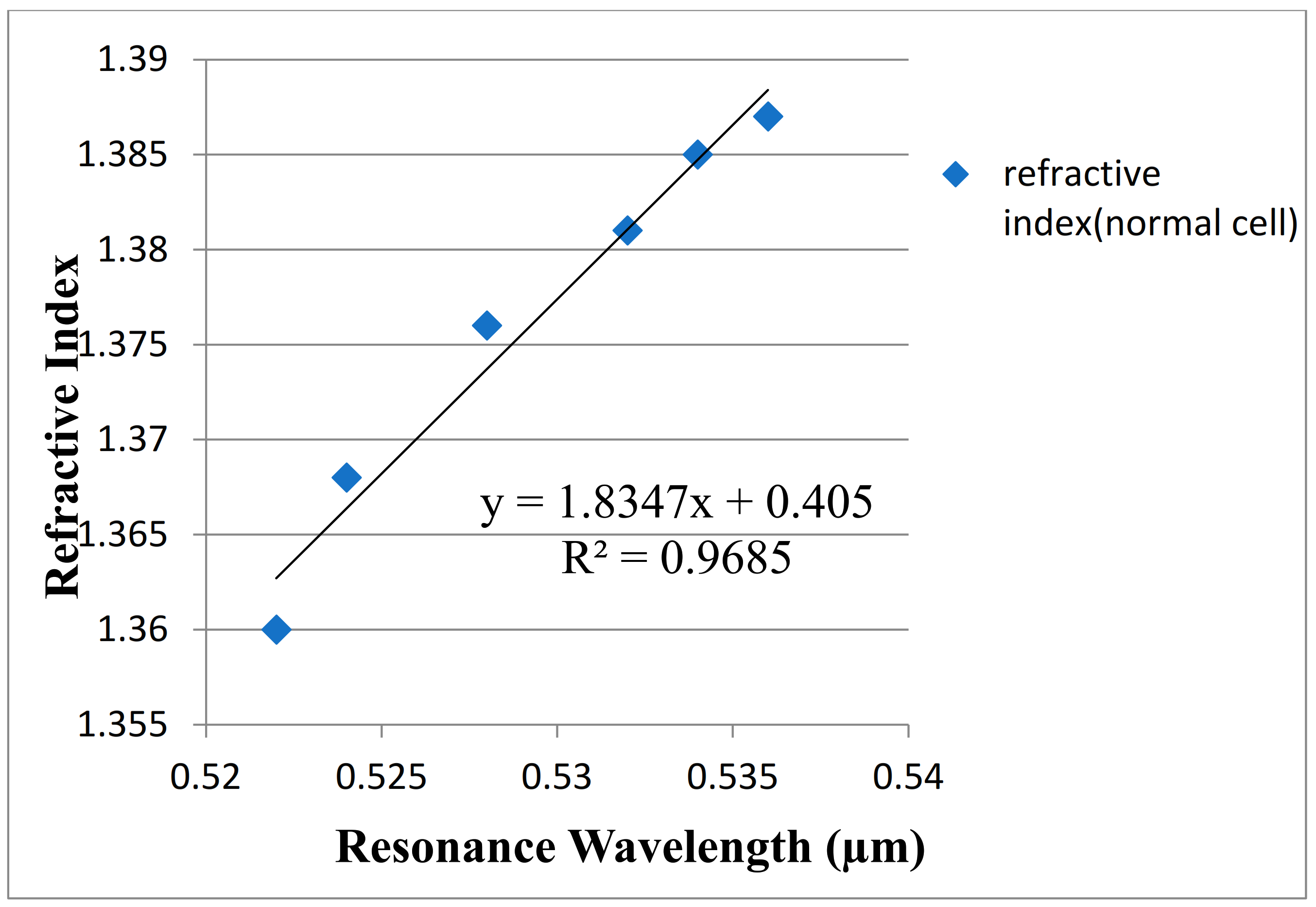
3. Results and Discussion

The sensing mechanism of the proposed sensor is based on the difference in wavelength of resonance peak for different analyte refractive index, na corresponding to cancerous cells and healthy cells. The proposed biosensor seeks to identify several cancer kinds, such as cervical cancer, skin cancer, blood cancer, breast cancer-type 1, breast cancer-type 2, and adrenal gland cancer has HELA, Basal, Jurkat, MDA-MB- 231, and MCF7 and PC12respectively as cancer cells. The changes in the refractive index of normal (healthy) and cancer-affected cells are shown in Table 1.
Figure 4 shows the resonance condition for the Jurkat (cancerous) cell. At 540 nm wavelength, the intersections of the efficient indices of the fundamental core mode (blue-dotted) and plasmon mode (black line) can be seen. At that wavelength, the propagation losses are at their peak due to the high-energy transfer from the fundamental core mode to the plasmon mode [50,51,52]. This is confirmation of the occurrence of a resonance phenomenon. Here, the values for the X-polarized fundamental mode and Y-polarized fundamental modes are the same, so the graph overlaps in both modes.
The confinement loss vs. wavelength for all cancer types considered in this investigation is shown in Figure 5a–f. The black and red line represents normal (healthy) and cancer-affected cells, respectively. When the normal cell in the analyte channel switched places with malignant cells, the confinement loss increases, and the peak shifted to longer wavelength, and similar behavior was observed for all types of cells. This is due to the strong metal-dielectric interaction, which is affected by the high value of analyte RI. The main basis of the amplitude interrogation approach is shifting in those peaks along the wavelength. The value of amplitude sensitivity and resolution for different cancerous cell is shown in Table 2.
Equations (4) and (5) are employed to calculate the proposed biosensor’s resolution and amplitude sensitivity, respectively. The highest amplitude sensitivity value is obtained as −289 RIU−1 for Breast Cancer (Type-1) (MDA-MB-231 cell) with 2 × 10⁻⁵ RIU resolution value as shown in Figure 6.

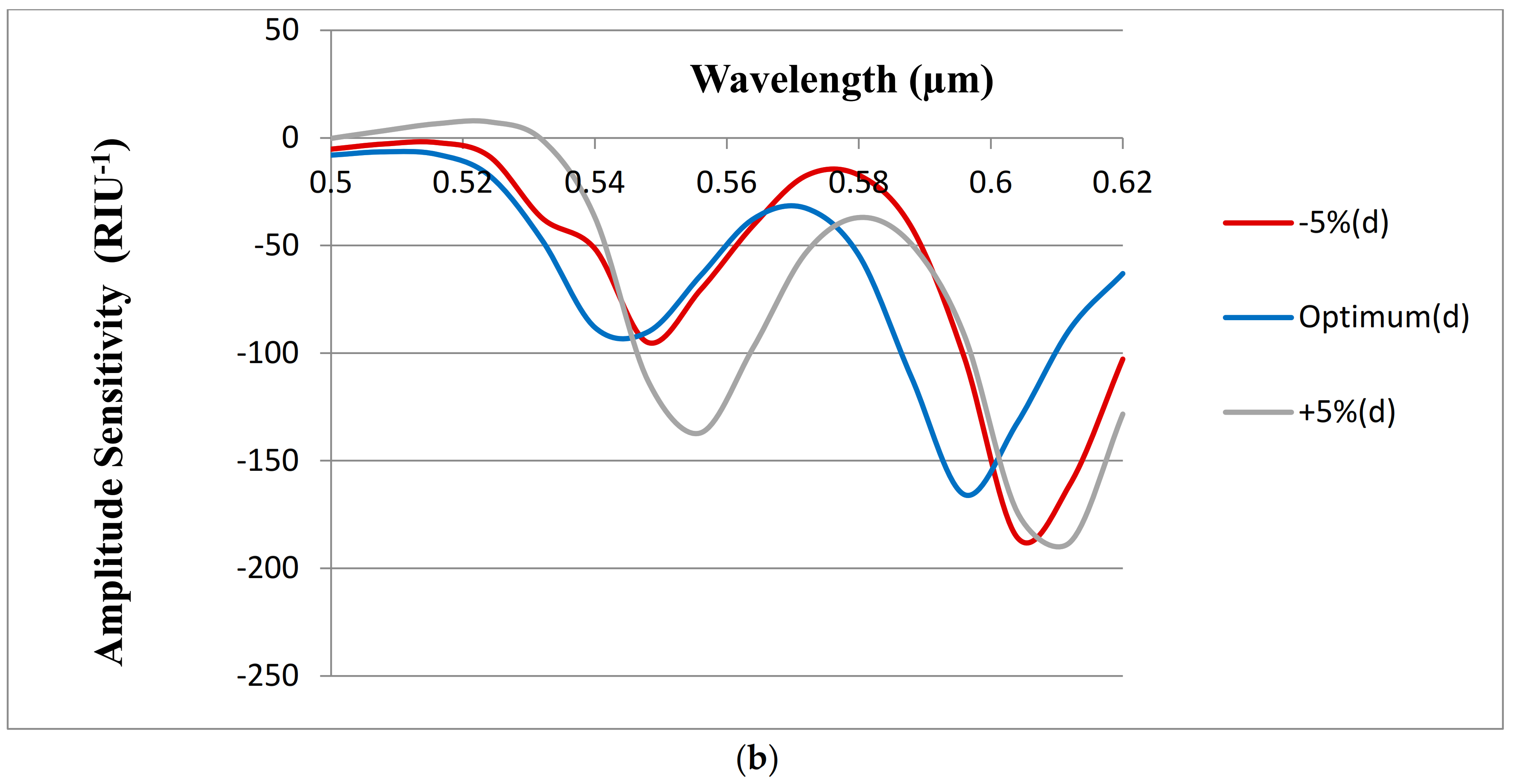
Changes of ± 5% in tg and d for Fabrication Tolerance Assessment

In order to proof the fabrication feasibility of the proposed design, a tolerance study [53] is made, as shown Figure 7. Figure 7 shows the sensor’s Amplitude sensitivity also varies within a tolerance of ±5% of the tg (gold layer) and d (air hole diameter) compared to that obtained using the optimized value of tg and d. It may be seen from Figure 7 that within a tolerance of ±5%, a slight shift occurs in the resonance wavelengths of the optimized parameters.
The linearity response is an important aspect of sensor design. For sensor applications, a high linearity response is a desirable metric to evaluate sensor performance. The linear fitting curve for the resonance wavelengths (for healthy cell) and for analyte refractive index ranging from 1.36 to 1.39 is shown in Figure 8. For such a Y-polarized fundamental mode, Equation (6) gives the relevant linear fitting model. The slope of this equation is used to compute the average sensitivity for various refractive index values. R2 is used to determine the sensor’s linear response. A high R2 value (0.9685) is estimated for the suggested biosensor, as shown in Figure 8. As a result, the presented biosensor has a satisfactory linear sensing response, indicating that the sensor design has significant potential.
y = 1.8347x + 0.405
As a result, compared to the previous approaches, the proposed sensor has shown promising characteristics. Table 3 tabulates the results of previously published designs in comparisons to the proposed work. The comparison is based on the sensor’s sensitivity, detection range, refractive indices, and resolution. Noticeably the presented PCF-based SPR biosensor has shown good results when evaluating sensor performance.
The suggested biosensor has a simple design, an external sensing approach, and a coating with plasmonic material. When compared to existing sensors, the suggested biosensor has produced appropriate sensing parameters. The given biosensor can accurately recognize a variety of malignant and healthy cells, which is advantageous in biomedical applications involving cancer exposure. This spiral shaped biosensor design can be utilized with visible light range (500–620 nm) sources, which proved the practical feasibility of the structure.

4. Conclusions

Spiral shaped PCF-based SPR biosensor for cancer cell detection is proposed in this paper. The numerical analysis of the sensor characteristics is done, and the same has shown promising results with gold coating as a plasmonic material. The device has shown high sensitivity of −289 RIU−1 for the refractive index of the detection of breast cancer cells with the resolution of 2.33 × 10⁻⁴,which is more than the previously published work. The Spiral PCF design is based on a hexagonal lattice, so it can be fabricated using a conventional stack and draw process and yields high amplitude sensitivity. The proposed sensor can be used for the detection of different types of cancer cells at an early stage. This spiral shaped biosensor design can be utilized with visible light range (500 nm–620 nm) sources, proving the structure’s practical feasibility.

Author Contributions

Conceptualization, S.M., A.S., Y.I., F.P., A.V.B., G.S. and M.T.; Methodology, S.M., A.S., A.V.B., O.G.M., V.V.D., J.Y. and M.T.; Investigation, S.M.; Writing—original draft preparation, S.M.; Writing—review and editing, A.S., Y.I., F.P., A.V.B., O.G.M., V.V.D., J.Y., G.S. and M.T.; Supervision, M.T. All authors have read and agreed to the published version of the manuscript.

Funding

This research was funded by RFBR, DST, NSFC, and NRF according to the research project 19-57-80016 BRICSt. This research was funded by DST/ICD/BRICS/Call-3/Manish Tiwari-QuSaF/2019.

Institutional Review Board Statement

Not applicable.

Informed Consent Statement

Not applicable.

Data Availability Statement

Not applicable.

Acknowledgments

The authors are thankful to Manipal University Jaipur for providing the various facilities during the research work. The authors are especially thankful to Optoelectronics MEMS lab and SAIF facilities for necessary support.

Conflicts of Interest

The author has no conflict of interest.

References

  1. Cancer-World Health Organization (WHO) 2018. Available online: https://www.who.int/news-room/fact-sheets/detail/cancer (accessed on 1 January 2022).
  2. Dujon, A.M.; Ujvari, B.; Thomas, F. Cancer risk landscapes: A framework to study cancer in ecosystems. Sci. Total Environ. 2021, 763, 142955. [Google Scholar] [CrossRef] [PubMed]
  3. Mishra, G.P.; Kumar, D.; Chaudhary, V.S.; Murmu, G. Cancer cell detection by a heart-shaped dual-core photonic crystal fiber sensor. Appl. Opt. 2020, 59, 10321. [Google Scholar] [CrossRef] [PubMed]
  4. Abdelghaffar, M.; Gamal, Y.; Soliman, W.; Badr, Y.; Hameed, M.F.O.; Obayya, S.S. Early cancer detection by plasmonic pcf sensor. In Proceedings of the 2022 International Conference on Numerical Simulation of Optoelectronic Devices (NUSOD), Politecnico di Torino, Italy, 12–16 September 2022; pp. 147–148. [Google Scholar]
  5. Wang, J.; Lin, W.; Cao, E.; Xu, X.; Liang, W.; Zhang, X. Surface plasmon resonance sensors on raman and fluorescence spectroscopy. Sensors 2017, 17, 2719. [Google Scholar] [CrossRef] [PubMed] [Green Version]
  6. Liu, S.; Li, L.; Chen, Z.; Chen, N.; Dai, Z.; Huang, J.; Lu, B. Surface-enhanced Raman spectroscopy measurement of cancerous cells with optical fiber sensor. Chin. Opt. Lett. 2013, 12, S13001. [Google Scholar]
  7. Chen, C.; Dong, N.; Huang, J.; Wang, Z.; Wang, J. Microscopic optical nonlinearities and transient carrier dynamics in indium selenide nanosheet. Opt. Express 2022, 30, 17967–17979. [Google Scholar] [CrossRef]
  8. Khetani, A.; Momenpour, A.; Alarcon, E.I.; Anis, H. Hollow core photonic crystal fiber for monitoring leukemia cells using surface enhanced raman scattering (SERS). Biomed. Opt. Express 2015, 6, 4599. [Google Scholar] [CrossRef] [Green Version]
  9. Russell, P.; Hölzer, P.; Chang, W.; Abdolvand, A.; Travers, J.C. Hollow-core photonic crystal fibres for gas-based nonlinear optics. Nat. Photon 2014, 8, 278–286. [Google Scholar] [CrossRef]
  10. Fang, L.-M.; Chen, H.-M. Double-core D-type photonic crystal fiber refractive indexsensor based on grid coating. Proc. SPIE 2019, 11191, 13. [Google Scholar]
  11. Mollah, M.A.; Yousufali, M.; Ankan, I.M.; Rahman, M.M.; Sarker, H.; Chakrabarti, K. Twin core photonic crystal fiber refractive index sensor for early detection of blood cancer. Sens. Bio-Sens. Res. 2020, 29, 100344. [Google Scholar] [CrossRef]
  12. Wang, D.; Yi, Z.; Ma, G.; Dai, B.; Yang, J.; Zhang, J.; Yu, Y.; Liu, C.; Wu, X.; Bian, Q. Two-channel photonic crystal fiber based on surface plasmon resonance for magnetic field and temperature dual-parameter sensing. Phys. Chem. Chem. Phys. 2022, 24, 21233–21241. [Google Scholar] [CrossRef]
  13. Fatema, S.; Absar, R.; Reja, M.I.; Akhtar, J. Effect of core infiltration in the birefringence of photonic crystal fiber. In Proceedings of the 2017 IEEE International Conference on Telecommunications and Photonics (ICTP), Dhaka, Bangladesh, 26–28 December 2017; pp. 1–5. [Google Scholar]
  14. Fatema, S.; Absar, R.; Reja, M.I.; Akhtar, J. Effect of soft glass rod infiltration in the core of photonic crystal fiber. J. Opt. Commun. 2020, 41, 371–383. [Google Scholar] [CrossRef]
  15. Monro, T.M.; Kiang, K.M.; Lee, J.H.; Frampton, K.; Yusoff, Z.; Moore, R.; Tucknott, J.; Hewak, D.W.; Rutt, H.N.; Richardson, D.J. High nonlinearity extruded single-mode holey optical fibers. In Proceedings of the Optical Fiber Communication Conference and Exhibit, Anaheim, CA, USA, 17–22 March 2002; p. FA1. [Google Scholar]
  16. Shafkat, A.; Reja, M.I.; Miah, M.J.; Fatema, S.; Absar, R.; Akhtar, J. Numerical exploration of external sensing scheme based photonic crystal fiber surface plasmonic sensor with different noble plasmonic materials and their alloys. Optik 2021, 231, 166418. [Google Scholar] [CrossRef]
  17. Maidi, A.M.; Yakasai, I.; Abas, P.E.; Nauman, M.M.; Apong, R.A.; Kaijage, S.; Begum, F. Design and simulation of photonic crystal fiber for liquid sensing. Photonics 2021, 8, 16. [Google Scholar] [CrossRef]
  18. Kalra, S.; Vyas, S.; Tiwari, M.; Ismail, Y.; Yupapin, P.; Ali, J.; Singh, G. Investiga- tion of as2s3-borosilicate chalcogenide glass-based dispersion- engineered photonic crystal fibre for broadband supercontinuum generation in the mid-ir region. J. Mod. Opt. 2020, 67, 920–926. [Google Scholar] [CrossRef]
  19. Wang, H.; Liao, M.; Xiao, H.; Han, X.; Jiang, Y.; Tan, J.; Zhang, P.; Shao, J.; Tian, Y.; Yang, J. High sensitivity temperature sensor based on a PDMS-assisted bow-shaped fiber structure. Opt. Commun. 2021, 481, 126536. [Google Scholar] [CrossRef]
  20. Rifat, A.A.; Haider, F.; Ahmed, R.; Mahdiraji, G.A.; Adikan, F.M.; Miroshnichenko, A.E. Highly sensitive selectively coated photonic crystal fiber-based plasmonic sensor. Opt. Lett. 2018, 43, 891–894. [Google Scholar] [CrossRef]
  21. Ramola, A.; Marwaha, A.; Singh, S. Design and investigation of a dedicated PCF SPR biosensor for CANCER exposure employing external sensing. Appl. Phys. A 2021, 127, 643. [Google Scholar] [CrossRef]
  22. Yan, X.; Wang, Y.; Cheng, T.; Li, S. Photonic crystal fiber SPR liquid sensor based on elliptical detective channel. Micromachines 2021, 12, 408. [Google Scholar] [CrossRef]
  23. Goodrich, T.T.; Lee, H.J.; Corn, R.M. Direct detection of genomic DNA by enzymatically amplified SPR imaging measurements of RNA microarrays. J. Am. Chem. Soc. 2004, 126, 4086–4087. [Google Scholar] [CrossRef]
  24. Nooke, A.; Beck, U.; Hertwig, A.; Krause, A.; Krüger, H.; Lohse, V.; Negendank, D.; Steinbach, J. On the application of gold based SPR sensors for the detection of hazardous gases. Sens. Actuators B Chem. 2010, 149, 194–198. [Google Scholar] [CrossRef]
  25. Mouvet, C.; Harris, R.D.; Maciag, C.; Luff, B.J.; Wilkinson, J.S.; Piehler, J.; Brecht, A.; Gauglitz, G.; Abuknesha, R.; Ismail, G. Determination of simazine in water samples by waveguide surface plasmon resonance. Anal. Chim. Acta 1997, 338, 109–117. [Google Scholar] [CrossRef]
  26. Eid, M.M.A.; Rashed, A.N.Z.; Bulbul, A.A.M.; Podder, E. Mono-rectangular core photonic crystal fiber (MRC-PCF) for skin and blood cancer detection. Plasmonics 2020, 16, 717–727. [Google Scholar] [CrossRef]
  27. Modeling_And_Simulation_Of_Surface_Plasmonic_Resonance_In_Photonic_Crystal_Fiber. Available online: http://studentsrepo.um.edu.my/8794/ (accessed on 1 March 2022).
  28. Kashif, M.; Bakar, A.A.A.; Hashim, F.H. Analysing surface plasmon resonance phase sensor based on mach-zehnder interferometer technique using glycerin. Opt. Commun. 2016, 380, 419–424. [Google Scholar] [CrossRef]
  29. Momota, M.R.; Hasan, M.R. Hollow-core silver coated photonic crystal fiber plasmonic sensor. Opt. Mater. 2018, 76, 287–294. [Google Scholar] [CrossRef]
  30. Wieduwilt, T.; Tuniz, A.; Linzen, S.; Goerke, S.; Dellith, J.; Hübner, U.; Schmidt, M.A. Ultrathin niobium nanofilms on fiber optical tapers—A new route towards low-loss hybrid plasmonic modes. Sci. Rep. 2015, 5, 17060. [Google Scholar] [CrossRef] [Green Version]
  31. Haque, E.; Mahmuda, S.; Hossain, M.A.; Hai, N.H.; Namihira, Y.; Ahmed, F. Highly sensitive dual-core pcf based plasmonic refractive index sensor for low refractive index detection. IEEE Photonics J. 2019, 11, 1–9. [Google Scholar] [CrossRef]
  32. Rifat, A.A.; Ahmed, R.; Yetisen, A.K.; Butt, H.; Sabouri, A.; Mahdiraji, G.A.; Yun, S.H.; Adikan, F.R.M. Photonic crystal fiber based plasmonic sensors. Sens. Actuators B Chem. 2017, 243, 311–325. [Google Scholar] [CrossRef]
  33. Shafkat, A.; Rashed, A.N.Z.; El-Hageen, H.M.; Alatwi, A.M. The effects of adding different adhesive layers with a microstructure fiber sensor based on surface plasmon resonance: A numerical study. Plasmonics 2021, 16, 819–832. [Google Scholar] [CrossRef]
  34. Suvarnaphaet, P.; Pechprasarn, S. Graphene-based materials for biosensors: A review. Sensors 2017, 17, 2161. [Google Scholar] [CrossRef] [Green Version]
  35. Yang, H.; Liu, M.; Chen, Y.; Guo, L.; Xiao, G.; Liu, H.; Li, J.; Yuan, L. Highly Sensitive Graphene-Au Coated Plasmon Resonance PCF Sensor. Sensors 2021, 21, 818. [Google Scholar] [CrossRef]
  36. Rahman, K.M.M.; Alam, M.S.; Islam, M.A. Gold-coated surface plasmon resonance photonic crystal fiber sensor in near-infrared region. Results Opt. 2022, 7, 100223. [Google Scholar] [CrossRef]
  37. Bourdine, A.V.; Barashkin, A.Y.; Burdin, V.A.; Dashkov, M.V.; Demidov, V.V.; Dukelskii, K.V.; Evtushenko, A.S.; Ismail, Y.; Khokhlov, A.V.; Kuznetsov, A.A.; et al. Twisted silica microstructured optical fiber with equiangular spiral six-ray geometry. Fibers 2021, 9, 27. [Google Scholar] [CrossRef]
  38. Islam, M.R.; Iftekher, A.N.M.; Hasan, K.R.; Nayen, M.J.; Islam, S.B.; Hossain, A.; Mustafa, Z.; Tahsin, T. Design and numerical analysis of a gold-coated photonic crystal fiber based refractive index sensor. Opt. Quantum Electron. 2021, 53, 112. [Google Scholar] [CrossRef]
  39. Hasan, M.R.; Akter, S.; Rifat, A.A.; Rana, S.; Ahmed, K.; Ahmed, R.; Subbaraman, H.; Abbott, D. Spiral photonic crystal fiber-based dual-polarizedsurface plasmon resonance biosensor. IEEE Sens. J. 2018, 18, 133–140. [Google Scholar] [CrossRef]
  40. Otupiri, R.; Akowuah, E.K.; Haxha, S. Multi-channel spr biosensor based on pcf for multi-analyte sensing applications. Opt. Express 2015, 23, 15716–15727. [Google Scholar] [CrossRef] [Green Version]
  41. Wang, Y.; Jiang, G.; Yu, Z.; Wang, Q.; Jiang, X. Trapezium-shaped groovephotonic crystal fiber plasmon sensor for low refractive index detection. Sens. Bio-Sens. Res. 2021, 34, 100452. [Google Scholar] [CrossRef]
  42. Yasli, A. Cancer detection with surface plasmon resonance-based photonic crystal fiber biosensor. Plasmonics 2021, 16, 1605–1612. [Google Scholar] [CrossRef]
  43. Haider, F.; Aoni, R.A.; Ahmed, R.; Miroshnichenko, A.E. Highly amplitude-sensitive photonic-crystal-fiber-based plasmonic sensor. J. Opt. Soc. Am. B 2018, 35, 2816–2821. [Google Scholar] [CrossRef]
  44. Zhang, Z.; Liu, C.; Liu, X.; Feng, Y.; Wang, S.; Shen, T.; Wei, H. Double-sided polishing photonic crystal fiber biosensor based on surface plasmon resonance for cancer single cell detection. J. Opt. Soc. Am. B 2022, 39, 1050–1058. [Google Scholar] [CrossRef]
  45. Sharma, P.; Sharan, P.; Deshmukh, P. A photonic crystal sensor for analysis and detection of cancer cells. In Proceedings of the 2015 International Conference on Pervasive Computing (ICPC), Pune, India, 8–10 January 2015; pp. 1–5. [Google Scholar]
  46. Thomson, E.A. Mit Radar Research Used to Treat Breast Cancer Enters Phase Iitrials; Mitnews: Cambridge, MA, USA, 2001. [Google Scholar]
  47. Ramanujam, N.R.; Amiri, I.S.; Taya, S.A.; Olyaee, S.; Udaiyakumar, R.; PasumponPandian, A.; Wilson, K.S.J.; Mahalakshmi, P.; Yupapin, P.P. Enhanced sensitivity ofcancer cell using one dimensional nano composite material coated photonic crystal. Microsyst. Technol. 2018, 25, 189–196. [Google Scholar] [CrossRef]
  48. Ayyanar, N.; Raja, G.T.; Sharma, M.; Kumar, D.S. Photonic crystal fiber-based refractive index sensor for early detection of cancer. IEEE Sens. J. 2018, 18, 7093–7099. [Google Scholar] [CrossRef]
  49. Sharan, P.; Bharadwaj, S.M.; Gudagunti, F.D.; Deshmukh, P. Design and modelling of photonic sensor for cancer cell detection. In Proceedings of the 2014 International Conference on the IMpact of E-Technology on US (IMPETUS), Banglore, India, 10–11 January 2014; pp. 20–24. [Google Scholar]
  50. Jabin, M.A.; Ahmed, K.; Rana, M.J.; Paul, B.K.; Islam, M.; Vigneswaran, D.; Uddin, M.S. Surface plasmon resonance based titanium coated biosensor for cancer cell detection. IEEE Photonics J. 2019, 11, 1–10. [Google Scholar] [CrossRef]
  51. Wu, H.; Song, Y.; Sun, M.; Wang, Q. Simulation of High-Performance Surface Plasmon Resonance Sensor Based on D-Shaped Dual Channel Photonic Crystal Fiber for Temperature Sensing. Materials 2022, 16, 37. [Google Scholar] [CrossRef] [PubMed]
  52. Sarker, H.; Alam, F.; Khan, M.R.; Mollah, M.A.; Hasan, M.L.; Rafi, A.B.M.S. Designing highly sensitive exposed core surface plasmon resonance biosensors. Opt. Mater. Express 2022, 12, 1977–1990. [Google Scholar] [CrossRef]
  53. Saber, A.M.; Hameed, M.F.O.; El-Azab, J.; Amer, R.Y.; Ismail, T.; Obayya, S.S.A. Plasmonic photonic crystal fiber sensor for optical partial discharge detection. Opt. Quant. Electron. 2022, 54, 433. [Google Scholar] [CrossRef]
  54. Hossain, M.B.; Mahendiran, T.V.; Abdulrazak, L.F.; Mehedi, I.M.; Hossain, M.A.; Rana, M.M. Numerical analysis of gold coating based quasi d-shape dual core PCF SPR sensor. Opt. Quantum Electron. 2020, 52, 446. [Google Scholar] [CrossRef]
  55. Mostufa, S.; Akib, T.B.A.; Rana, M.M.; Islam, M.R. Highly Sensitive TiO2/Au/Graphene Layer-Based Surface Plasmon Resonance Biosensor for Cancer Detection. Biosensors 2022, 12, 603. [Google Scholar] [CrossRef]
Figure 1. Cross-section schematic of proposed PCF-SPR biosensor.
Photonics 10 00230 g001
Figure 2. A scheme to make a stack of silica rods and capillaries.
Photonics 10 00230 g002
Figure 3. (a) X-polarized core mode (b) Y- polarized core mode (c) SPP mode.
Photonics 10 00230 g003
Figure 4. The resonance behavior of proposed PCF-SPR biosensor for Jurkat (cancerous) cell.
Photonics 10 00230 g004
Figure 5. Confinement loss properties of the proposed PCF-SPR biosensor for different kinds of normal and malignant cells.
Photonics 10 00230 g005
Figure 6. The amplitude sensitivity of proposed PCF-SPR biosensor.
Photonics 10 00230 g006
Figure 7. Fabrication tolerance analysis of the proposed sensor within a tolerance of ±5% of the (a) tg, and (b) d.
Photonics 10 00230 g007aPhotonics 10 00230 g007b
Figure 8. Linear fitting characteristics of proposed PCF-SPR biosensor.
Photonics 10 00230 g008
Table 1. The difference in refractive index among normal and cancer affected cells.
Type of CancerType of CellR.I. of Normal CellR.I. of Cancer
Affected Cell
References
SkinBasal1.3601.380[43,44]
BreastMDA-MB-2311.3851.399[45,46]
BreastMCF-71.3871.401[45,46]
BloodJurkat1.3761.390[47,48]
Adrenal GlandPC121.3811.395[49]
CervicalHela1.3681.392[49]
Table 2. Proposed PCF-SPR biosensor’s sensitivity analysis.
Type of CancerType of CellR.I. Change CellResolutionAmplitude Sensitivity (RIU−1)
SkinBasal1.360–1.3802 × 10⁻⁵−83
BreastMCF-71.387–1.4012.33 × 10−4−154.5
BloodJurkat1.376–1.391.4 × 10−4−165.9
CervicalHela1.368–1.3921.5 × 10−5−166.7
Adrenal GlandPC121.381–1.3951.4 × 10−4−245.5
BreastMDA-MB-2311.385–1.3992.33 × 110−4−289
Table 3. Comparison between the proposed PCF-SPR biosensor’s design, wavelength range, RI range and sensitivity to existing sensors.
Wavelength Range (nm)Sensor’s DesignR.I. RangeAmplitude
Sensitivity
Sensor’s ResolutionRef.
480–650D-shape1.33–1.37216 RIU−14.6 × 10⁻⁵[40]
1080–1560Quasi D-shape
Dual core Circular lattice
1.42–1.46230 RIU−1NA[53]
580–720Circular lattice PCF sensor hybrid1.33–1.36266 RIU−13.75 × 10⁻⁵[54]
633TiO2/Au/graphene layer-based (SPR)1.36–1.401292.86 deg/RIUNA[55]
500–620Spiral shape1.36–1.401289 RIU−12.33 × 10⁻⁴This work
Disclaimer/Publisher’s Note: The statements, opinions and data contained in all publications are solely those of the individual author(s) and contributor(s) and not of MDPI and/or the editor(s). MDPI and/or the editor(s) disclaim responsibility for any injury to people or property resulting from any ideas, methods, instructions or products referred to in the content.

