Radiophotoluminescence Phenomenon of CaF2 Ceramics Doped with Li


Abstract
:1. Introduction
2. Materials and Methods
2.1. Sample
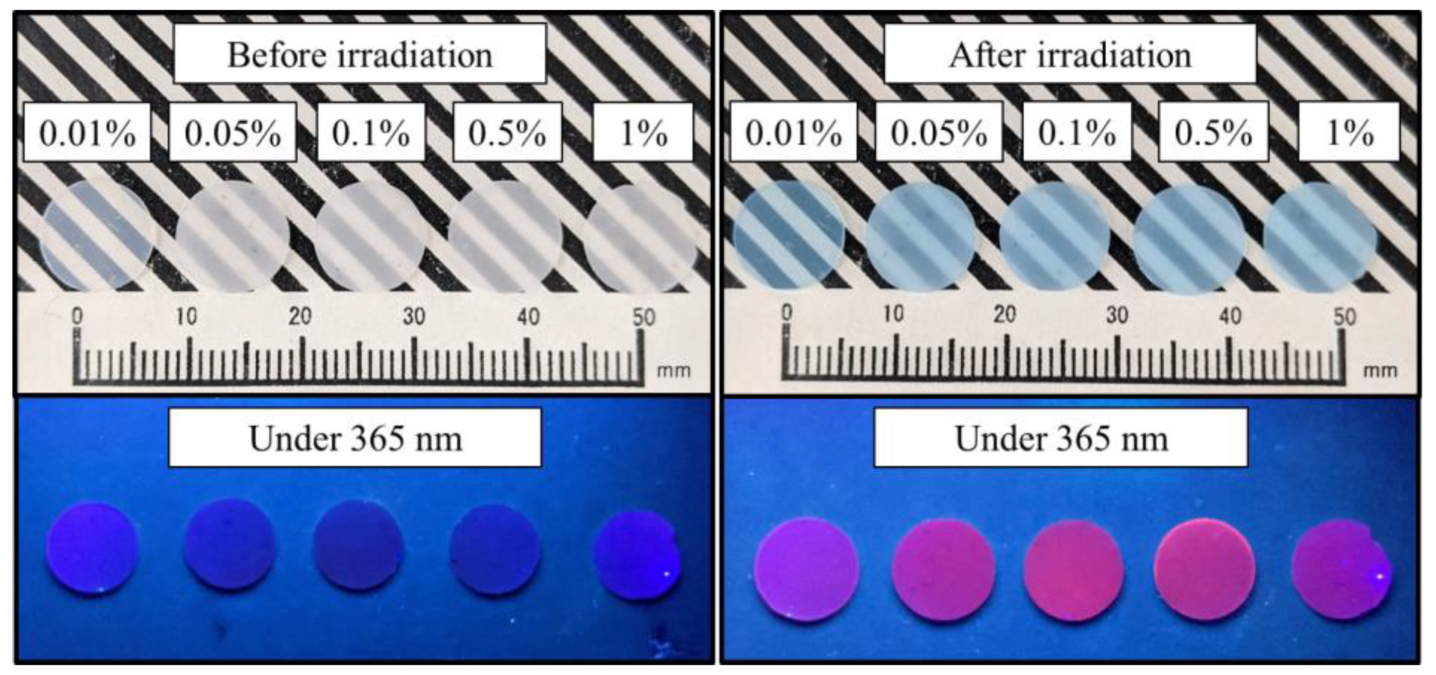
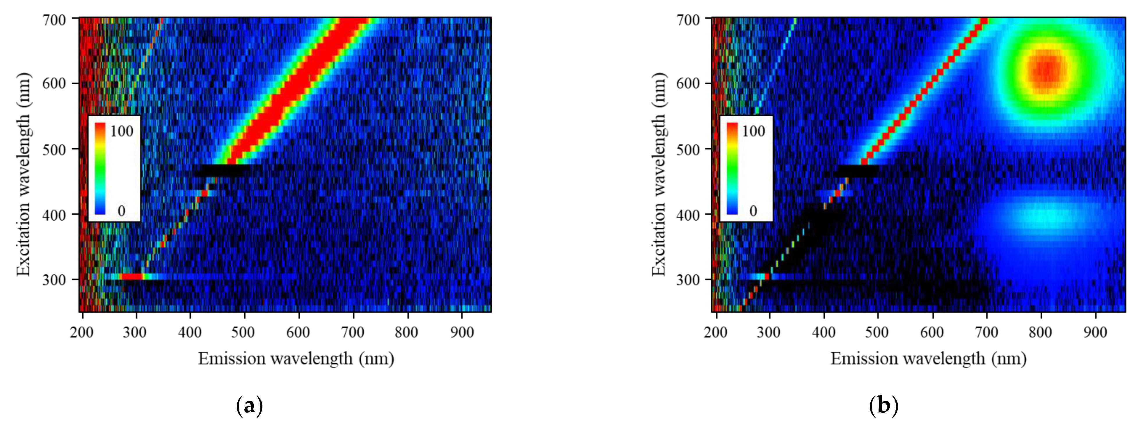
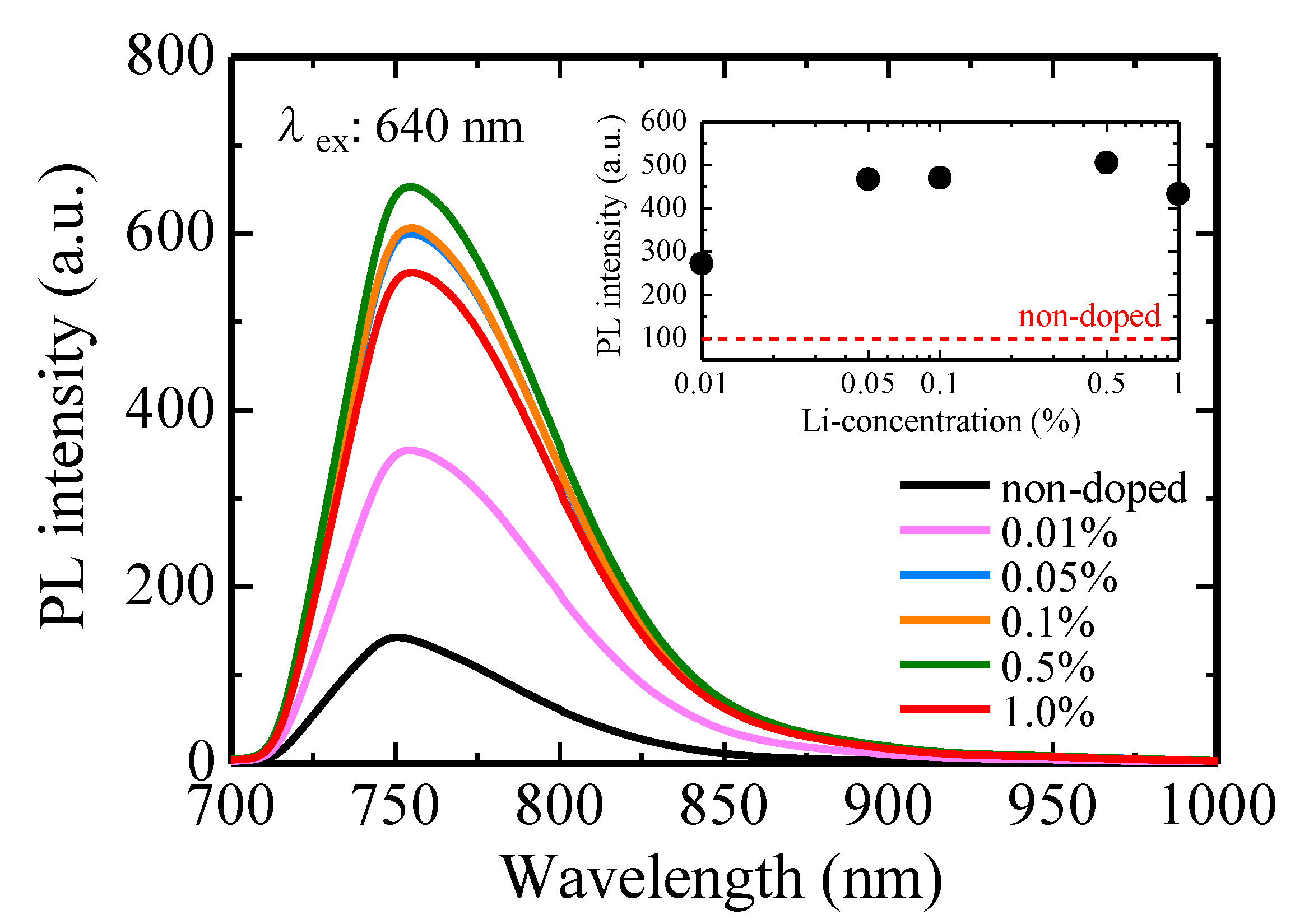
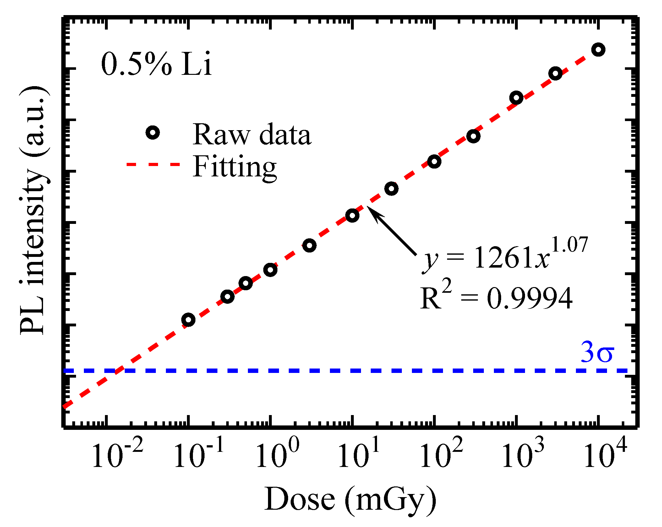
2.2. RPL Properties
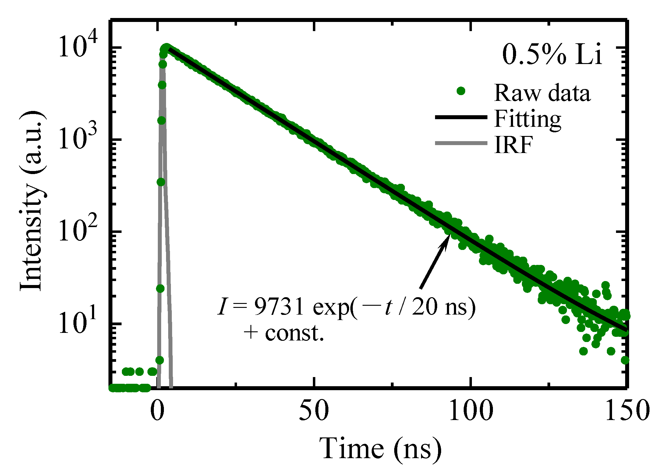
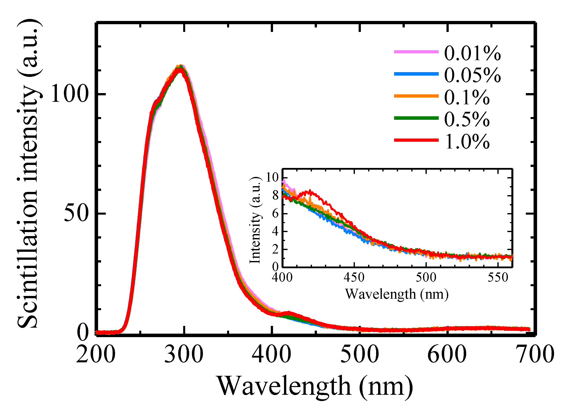
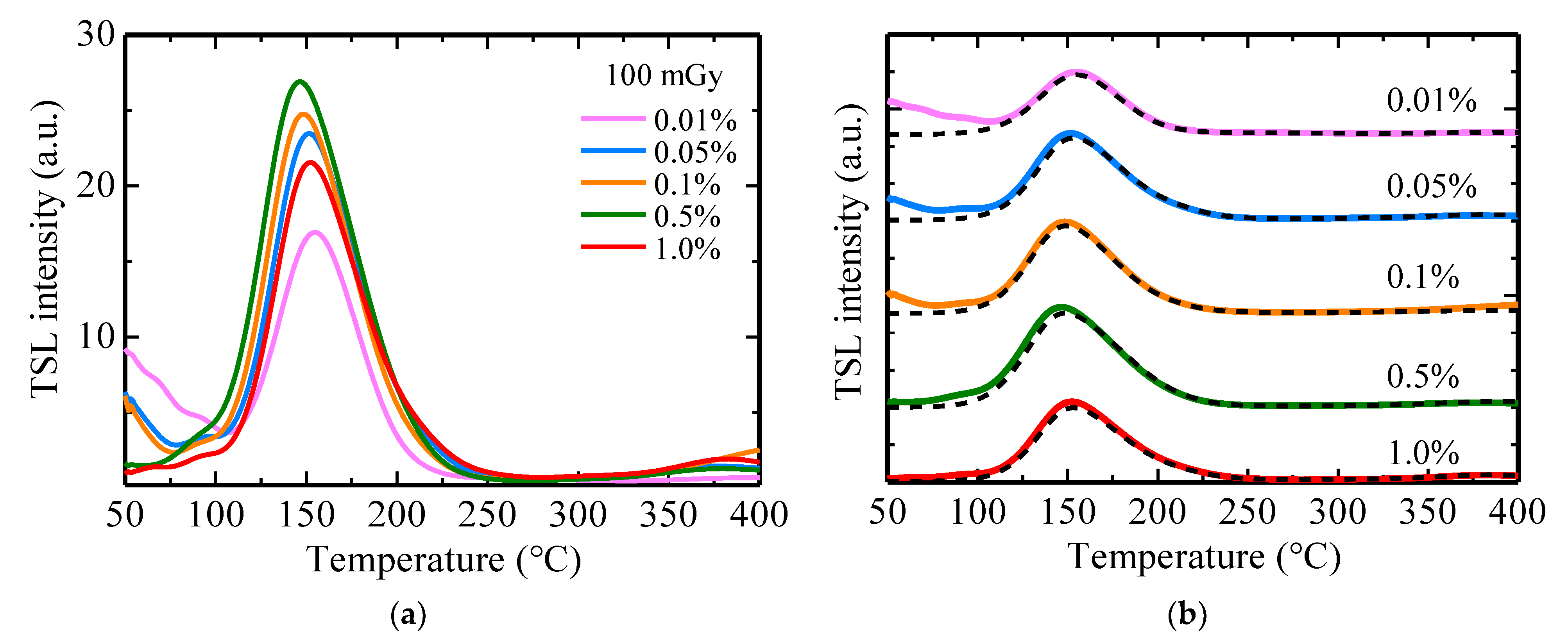
2.3. Scintillation and TSL Properties
3. Conclusions
Author Contributions
Funding
Institutional Review Board Statement
Informed Consent Statement
Data Availability Statement
Conflicts of Interest
References
- Okada, G. Novel Radio-Photoluminescence Materials and Applications. J. Ceram. Soc. Jpn. 2021, 129, 419–424. [Google Scholar] [CrossRef]
- Bos, A.J.J. Theory of Thermoluminescence. Radiat. Meas. 2006, 41, 45–56. [Google Scholar] [CrossRef]
- McKeever, S.W.S. Optically Stimulated Luminescence: A Brief Overview. Radiat. Meas. 2011, 46, 1336–1341. [Google Scholar] [CrossRef]
- Bhatt, B.C.; Kulkarni, M.S. Worldwide Status of Personnel Monitoring Using Thermoluminescent (TL), Optically Stimulated Luminescent (OSL) and Radiophotoluminescent (RPL) Dosimeters. Int. J. Lumin. Appl. 2013, 3, 6–10. [Google Scholar]
- Knežević, Ž.; Stolarczyk, L.; Bessieres, I.; Bordy, J.M.; Miljanić, S.; Olko, P. Photon Dosimetry Methods Outside the Target Volume in Radiation Therapy: Optically Stimulated Luminescence (OSL), Thermoluminescence (TL) and Radiophotoluminescence (RPL) Dosimetry. Radiat. Meas. 2013, 57, 9–18. [Google Scholar] [CrossRef]
- Kurobori, T.; Miyamoto, Y.; Maruyama, Y.; Yamamoto, T.; Sasaki, T. A Comparative Study of Optical and Radiative Characteristics of X-Ray-Induced Luminescent Defects in Ag-Doped Glass and LiF Thin Films and Their Applications in 2-D Imaging. Nucl. Instrum. Methods Phys. Res. Sect. B Beam Interact. Mater. Atoms 2014, 326, 76–80. [Google Scholar] [CrossRef] [Green Version]
- Kurobori, T.; Zheng, W.; Miyamoto, Y.; Nanto, H.; Yamamoto, T. The Role of Silver in the Radiophotoluminescent Properties in Silver-Activated Phosphate Glass and Sodium Chloride Crystal. Opt. Mater. 2010, 32, 1231–1236. [Google Scholar] [CrossRef] [Green Version]
- Yamamoto, T.; Maki, D.; Sato, F.; Miyamoto, Y.; Nanto, H.; Iida, T. The Recent Investigations of Radiophotoluminescence and Its Application. Radiat. Meas. 2011, 46, 1554–1559. [Google Scholar] [CrossRef]
- Miyamoto, Y.; Yamamoto, T.; Kinoshita, K.; Koyama, S.; Takei, Y.; Nanto, H.; Shimotsuma, Y.; Sakakura, M.; Miura, K.; Hirao, K. Emission Mechanism of Radiophotoluminescence in Ag-Doped Phosphate Glass. Radiat. Meas. 2010, 45, 546–549. [Google Scholar] [CrossRef]
- Kawamoto, H.; Koshimizu, M.; Fujimoto, Y.; Asai, K. Formation of Radiophotoluminescence Centers at Room Temperature in Ag-Doped Alkali Halides. Jpn. J. Appl. Phys. 2019, 58, 062004. [Google Scholar] [CrossRef]
- Sholom, S.; McKeever, S.W.S. High-Dose Dosimetry with Ag-Doped Phosphate Glass: Applicability Test with Different Techniques. Radiat. Meas. 2020, 132, 106263. [Google Scholar] [CrossRef]
- McKeever, S.W.S.; Sholom, S.; Shrestha, N. Observations Regarding the Build-up Effect in Radiophotoluminescence of Silver-Doped Phosphate Glasses. Radiat. Meas. 2019, 123, 13–20. [Google Scholar] [CrossRef]
- Hashikawa, R.; Fujii, Y.; Kinomura, A.; Saito, T.; Okada, A.; Wakasugi, T.; Kadono, K. Radiophotoluminescence Phenomenon in Copper-Doped Aluminoborosilicate Glass. J. Am. Ceram. Soc. 2019, 102, 1642–1651. [Google Scholar] [CrossRef]
- Asada, S.; Okada, G.; Kato, T.; Nakamura, F.; Kawano, N.; Kawaguchi, N.; Yanagida, T. Eu-Doped Ca2SiO4 as a New Radio-Photoluminescence Phosphor. Chem. Lett. 2018, 47, 59–61. [Google Scholar] [CrossRef]
- Calvert, R.L.; Danby, R.J. Thermoluminescence and Radiophotoluminescence from Eu-and Sm-doped CaSO4. Phys. Status Solidi 1984, 83, 597–604. [Google Scholar] [CrossRef]
- Kohara, Y.; Okada, G.; Tsuyumoto, I.; Kusano, E.; Nanto, H. Radiation-Induced Reduction of Eu3+ Doped in SrAl2O4. Mater. Lett. 2021, 303, 130502. [Google Scholar] [CrossRef]
- Dhoble, S.J.; Nagpure, I.M.; Dhoble, N.S.; Molina, P. Effect of Bi Ion on Eu2+ → Eu3+ Conversion in CaF2:Eu Phosphors for RPL Dosimetry. J. Mater. Sci. 2011, 46, 7253–7261. [Google Scholar] [CrossRef]
- Schuyt, J.J.; Williams, G.V.M. Radiation-Induced Changes in the Optical Properties of NaMgF3(Sm): Observation of Resettable Sm Radio-Photoluminescence. Mater. Res. Bull. 2018, 106, 455–458. [Google Scholar] [CrossRef]
- Okada, G.; Vahedi, S.; Morrell, B.; Koughia, C.; Belev, G.; Wysokinski, T.; Chapman, D.; Varoy, C.; Edgar, A.; Kasap, S. Examination of the Dynamic Range of Sm-Doped Glasses for High-Dose and High-Resolution Dosimetric Applications in Microbeam Radiation Therapy at the Canadian Synchrotron. Opt. Mater. 2013, 35, 1976–1980. [Google Scholar] [CrossRef]
- Chicilo, F.; Okada, G.; Belev, G.; Chapman, D.; Edgar, A.; Curry, R.J.; Kasap, S. Instrumentation for High-Dose, High-Resolution Dosimetry for Microbeam Radiation Therapy Using Samarium-Doped Fluoroaluminate and Fluorophosphate Glass Plates. Meas. Sci. Technol. 2020, 31, 015201. [Google Scholar] [CrossRef]
- Eller, S.A.; Ahmed, M.F.; Bartz, J.A.; Akselrod, M.S.; Denis, G.; Yukihara, E.G. Radiophotoluminescence Properties of Al2O3:C,Mg Crystals. Radiat. Meas. 2013, 56, 179–182. [Google Scholar] [CrossRef]
- Akselrod, M.S.; Sykora, G.J. Fluorescent Nuclear Track Detector Technology—A New Way to Do Passive Solid State Dosimetry. Radiat. Meas. 2011, 46, 1671–1679. [Google Scholar] [CrossRef]
- Akselrod, M.S.; Akselrod, A.E. New Al2O3:C,Mg Crystals for Radiophotoluminescent Dosimetry and Optical Imaging. Radiat. Prot. Dosim. 2006, 119, 218–221. [Google Scholar] [CrossRef]
- Knežević, Z.; Beck, N.; Milković, D.; Miljanić, S.; Ranogajec-Komor, M. Characterisation of RPL and TL Dosimetry Systems and Comparison in Medical Dosimetry Applications. Radiat. Meas. 2011, 46, 1582–1585. [Google Scholar] [CrossRef]
- Nakamura, F.; Kato, T.; Okada, G.; Kawaguchi, N.; Fukuda, K.; Yanagida, T. Scintillation, TSL and RPL Properties of MgF2 Transparent Ceramic and Single Crystal. Ceram. Int. 2017, 43, 7211–7215. [Google Scholar] [CrossRef]
- Nakamura, F.; Kato, T.; Okada, G.; Kawano, N.; Kawaguchi, N.; Yanagida, T. Radio-Photoluminescence in Non-Doped K2CO3 Ceramics. Mater. Lett. 2018, 211, 100–102. [Google Scholar] [CrossRef]
- Nakamura, F.; Kato, T.; Nakauchi, D.; Okada, G.; Kawano, N.; Kawaguchi, N.; Yanagida, T. Non-Doped Na2CO3 Ceramics as a New Radio-Photoluminescence (RPL) Material. Chem. Lett. 2017, 46, 1383–1385. [Google Scholar] [CrossRef]
- Dhopte, S.M.; Muthal, P.L.; Kondawar, V.K. Luminescence in CaF2:Eu. J. Lumin. 1992, 54, 95–101. [Google Scholar] [CrossRef]
- Bos, A.; Dielhof, J. The Analysis of Thermoluminescent Glow Peaks in CaF2:Tm (TLD-300). Radiat. Prot. Dosim. 1991, 37, 231–239. [Google Scholar] [CrossRef] [Green Version]
- Danilkin, M.; Lust, A.; Kerikmäe, M.; Seeman, V.; Mändar, H.; Must, M. CaF2:Mn Extreme Dosimeter: Effects of Mn Concentration on Thermoluminescence Mechanisms and Properties. Radiat. Meas. 2006, 41, 677–681. [Google Scholar] [CrossRef]
- Necmeddin Yazici, A.; Chen, R.; Solak, S.; Yegingil, Z. The Analysis of Thermoluminescent Glow Peaks of CaF2: Dy (TLD-200) after β-Irradiation. J. Phys. D. Appl. Phys. 2002, 35, 2526–2535. [Google Scholar] [CrossRef] [Green Version]
- Zahedifar, M.; Sadeghi, E. Synthesis and Dosimetric Properties of the Novel Thermoluminescent CaF2:Tm Nanoparticles. Radiat. Phys. Chem. 2012, 81, 1856–1861. [Google Scholar] [CrossRef]
- Sunta, C.M. Associated Luminescence Centres and Traps in the Thermoluminescence of CaF2:Dy (TLD-200). J. Phys. D. Appl. Phys. 1977, 10, 47–51. [Google Scholar] [CrossRef]
- Topaksu, M.; Correcher, V.; Garcia-guinea, J. Luminescence Emission of Natural Fluorite and Synthetic CaF2:Mn. Radiat. Phys. Chem. 2016, 119, 151–156. [Google Scholar] [CrossRef] [Green Version]
- Bakshi, A.K.; Dhabekar, B.; Rawat, N.S.; Singh, S.G.; Joshi, V.J.; Kumar, V. Study on TL and OSL Characteristics of Indigenously Developed CaF2:Mn Phosphor. Nucl. Instrum. Methods Phys. Res. Sect. B Beam Interact. Mater. Atoms 2009, 267, 548–553. [Google Scholar] [CrossRef]
- Asfora, V.; De Barros, V.S.M.; Da Silva, R.J.G.; Vasconcelos, D.A.A.; Nobre, B.S.; Yamato, M.E.; Khoury, H.J.; Oliveira, R.A.; Azevedo, W.M. Optically Stimulated Luminescence of CaF2:Tm. Radiat. Meas. 2016, 85, 73–77. [Google Scholar] [CrossRef]
- Nakamura, F.; Kato, T.; Okada, G.; Kawaguchi, N.; Fukuda, K.; Yanagida, T. Scintillation and Dosimeter Properties of CaF2 Transparent Ceramic Doped with Eu2+. Ceram. Int. 2017, 43, 604–609. [Google Scholar] [CrossRef] [Green Version]
- Kato, T.; Nakauchi, D.; Kawaguchi, N.; Yanagida, T. Radio-Photoluminescence Phenomenon in Non-Doped CaF2 Ceramic. Mater. Lett. 2020, 270, 127688. [Google Scholar] [CrossRef]
- Kato, T.; Nakauchi, D.; Kawaguchi, N.; Yanagida, T. Radio-Photoluminescence Properties of CaF2 Transparent and Opaque Ceramics. Curr. Appl. Phys. 2020, 20, 1195–1200. [Google Scholar] [CrossRef]
- Tijero, J.M.G.; Jaque, F. Thermal and Optical Properties of the FA and (F2+)A Centers in Na-Doped CaF2 Crystals. Phys. Rev. B 1990, 41, 3832–3836. [Google Scholar] [CrossRef]
- Komine, N.; Sakuma, S.; Shiozawa, M.; Mizugaki, T.; Sato, E. Influence of Sodium Impurities on ArF Excimer-Laser-Induced Absorption in CaF2 Crystals. Appl. Opt. 2000, 39, 3925–3930. [Google Scholar] [CrossRef]
- Kato, T.; Okada, G.; Nakauchi, D.; Kawaguchi, N.; Yanagida, T. Na-Concentration Dependence on Radiophotoluminescence Properties of CaF2. Solid State Sci. 2022, 128, 106892. [Google Scholar] [CrossRef]
- Yanagida, T.; Kamada, K.; Fujimoto, Y.; Yagi, H.; Yanagitani, T. Comparative Study of Ceramic and Single Crystal Ce:GAGG Scintillator. Opt. Mater. 2013, 35, 2480–2485. [Google Scholar] [CrossRef]
- Yanagida, T.; Fujimoto, Y.; Kawaguchi, N.; Yanagida, S. Dosimeter Properties of AlN. J. Ceram. Soc. Japan 2013, 121, 988–991. [Google Scholar] [CrossRef] [Green Version]
- Kato, T.; Shiratori, D.; Nakauchi, D.; Kawaguchi, N.; Yanagida, T. Evaluation of Quantum Yields and Thermally Stimulated Luminescence Glow Curves of Ag-Doped Phosphate Glasses. Jpn. J. Appl. Phys. 2020, 59, 112001. [Google Scholar] [CrossRef]
- Cramer, L.P.; Cumby, T.D.; Leraas, J.A.; Langford, S.C.; Dickinson, J.T. Effect of Surface Treatments on Self-Trapped Exciton Luminescence in Single-Crystal CaF2. J. Appl. Phys. 2005, 97, 103533. [Google Scholar] [CrossRef]
- Antonyak, O.T.; Vistovskyy, V.V.; Zhyshkovych, A.V.; Kravchuk, I.M. Defect Luminescence in CaF2 Nanoparticles. J. Lumin. 2015, 167, 249–253. [Google Scholar] [CrossRef]


Disclaimer/Publisher’s Note: The statements, opinions and data contained in all publications are solely those of the individual author(s) and contributor(s) and not of MDPI and/or the editor(s). MDPI and/or the editor(s) disclaim responsibility for any injury to people or property resulting from any ideas, methods, instructions or products referred to in the content. |
© 2023 by the authors. Licensee MDPI, Basel, Switzerland. This article is an open access article distributed under the terms and conditions of the Creative Commons Attribution (CC BY) license (https://creativecommons.org/licenses/by/4.0/).
Share and Cite
Kato, T.; Nakauchi, D.; Kawaguchi, N.; Yanagida, T. Radiophotoluminescence Phenomenon of CaF2 Ceramics Doped with Li. Photonics 2023, 10, 211. https://doi.org/10.3390/photonics10020211
Kato T, Nakauchi D, Kawaguchi N, Yanagida T. Radiophotoluminescence Phenomenon of CaF2 Ceramics Doped with Li. Photonics. 2023; 10(2):211. https://doi.org/10.3390/photonics10020211
Chicago/Turabian StyleKato, Takumi, Daisuke Nakauchi, Noriaki Kawaguchi, and Takayuki Yanagida. 2023. "Radiophotoluminescence Phenomenon of CaF2 Ceramics Doped with Li" Photonics 10, no. 2: 211. https://doi.org/10.3390/photonics10020211