Lensless Imaging via Blind Ptychography Modulation and Wavefront Separation

Abstract
:1. Introduction
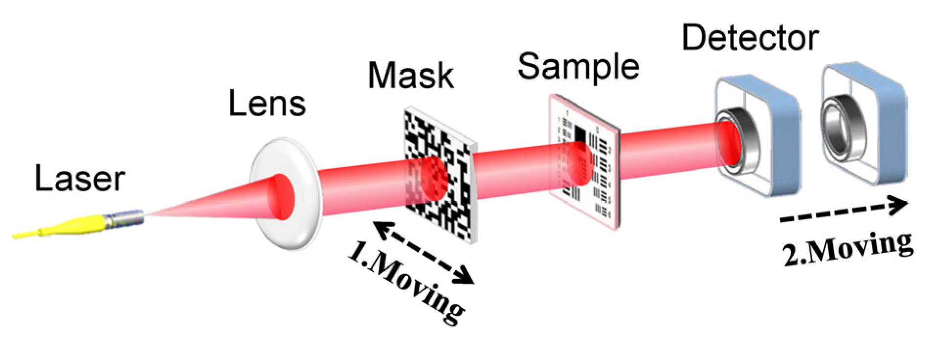
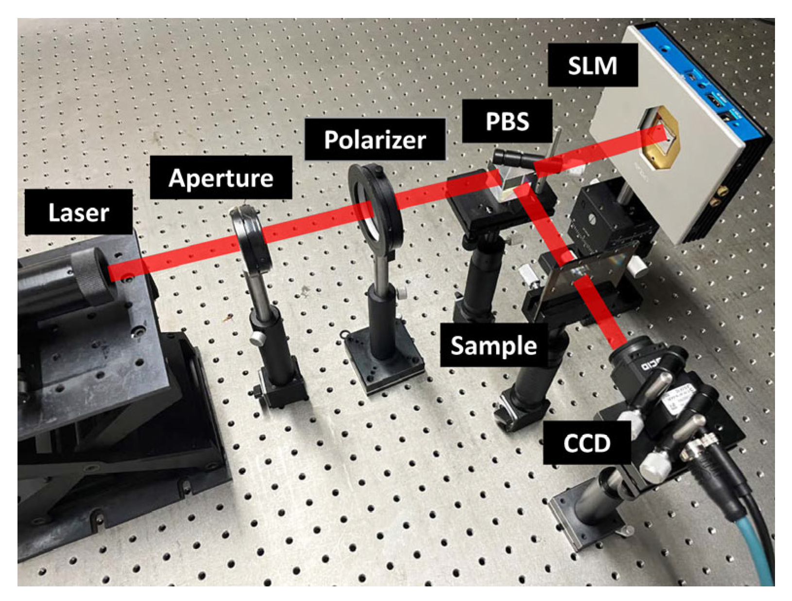
2. Methods
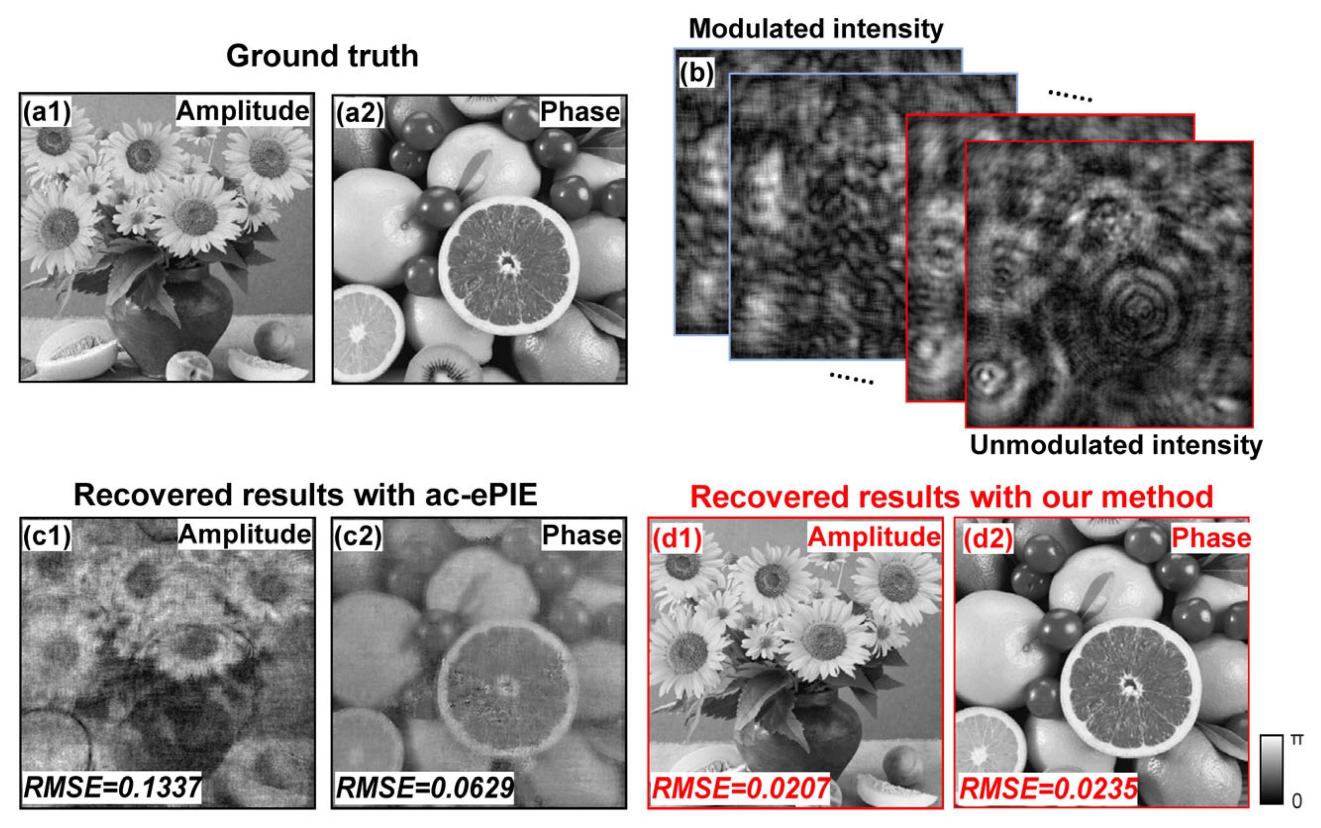
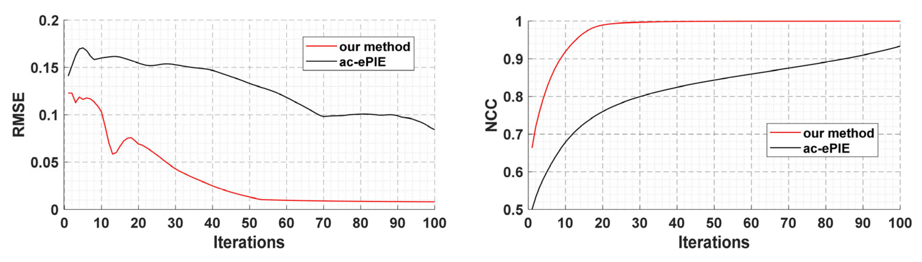
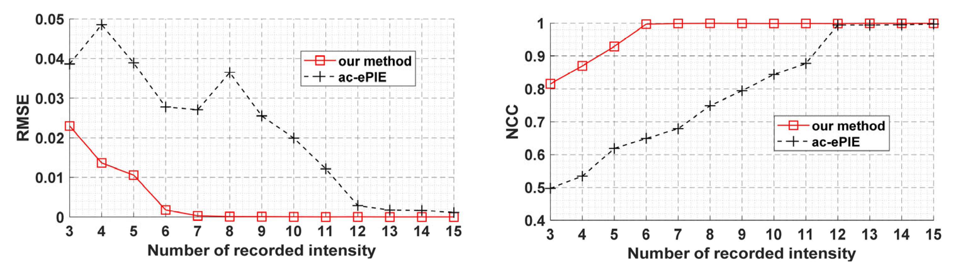
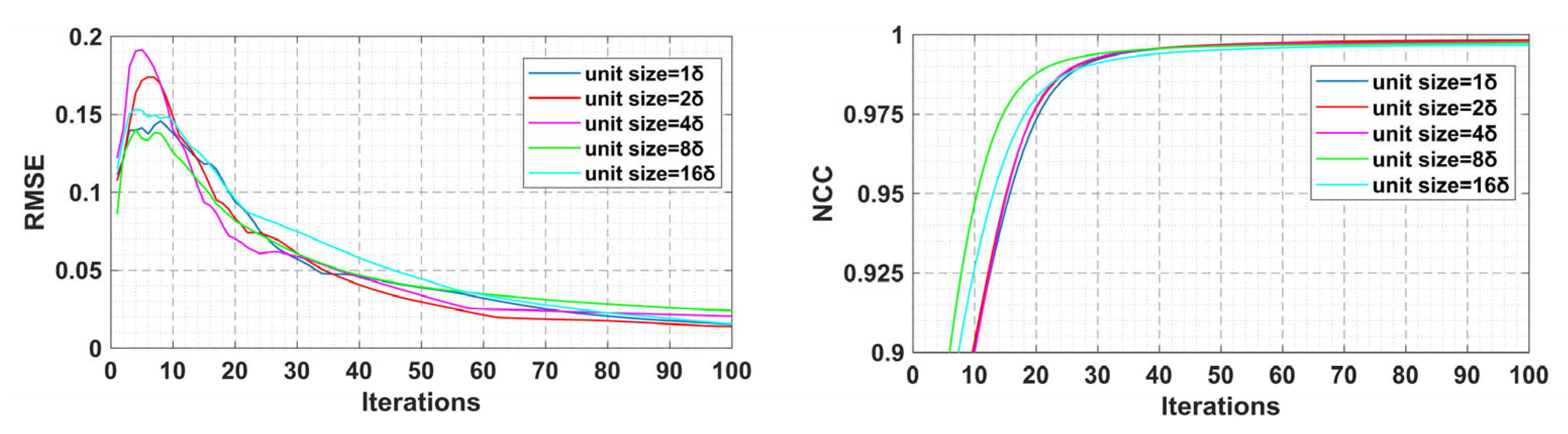
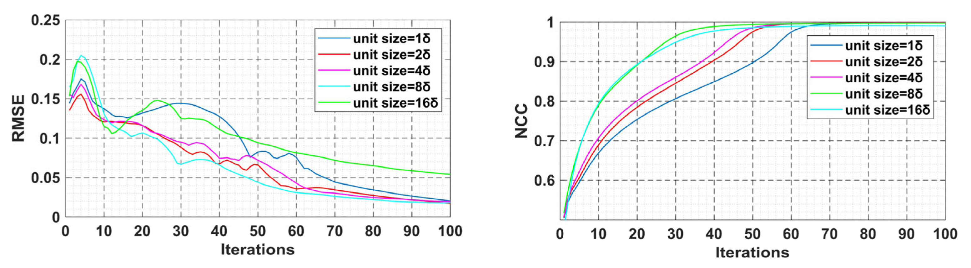
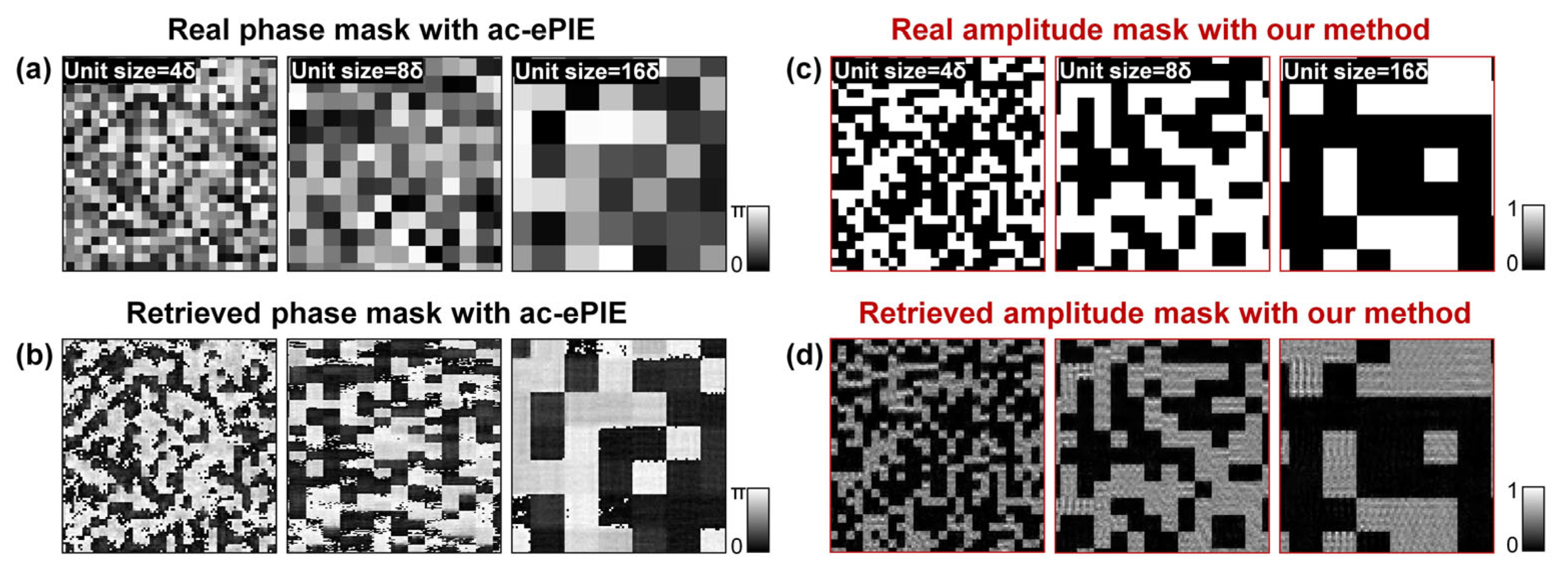
3. Simulation
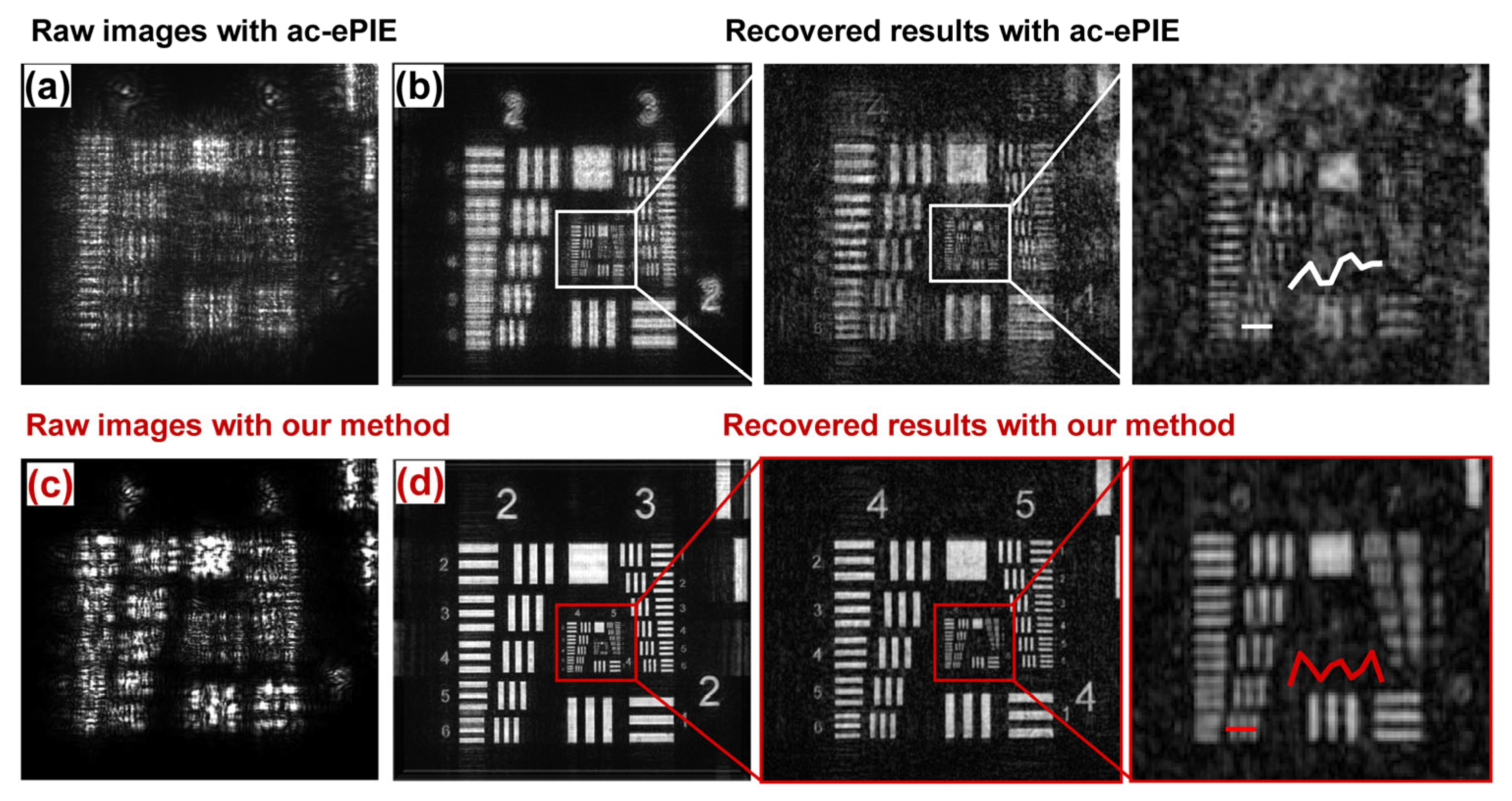
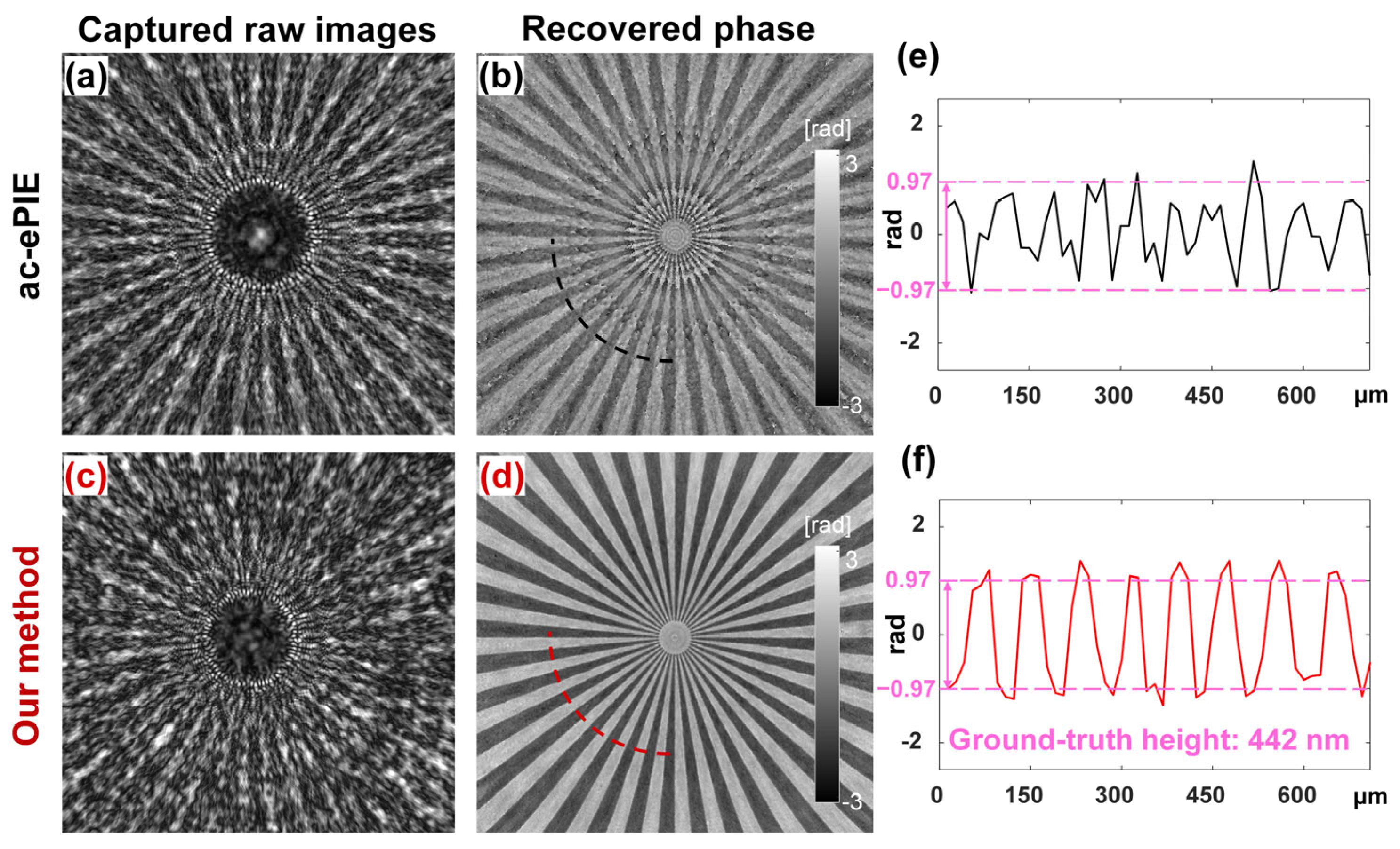
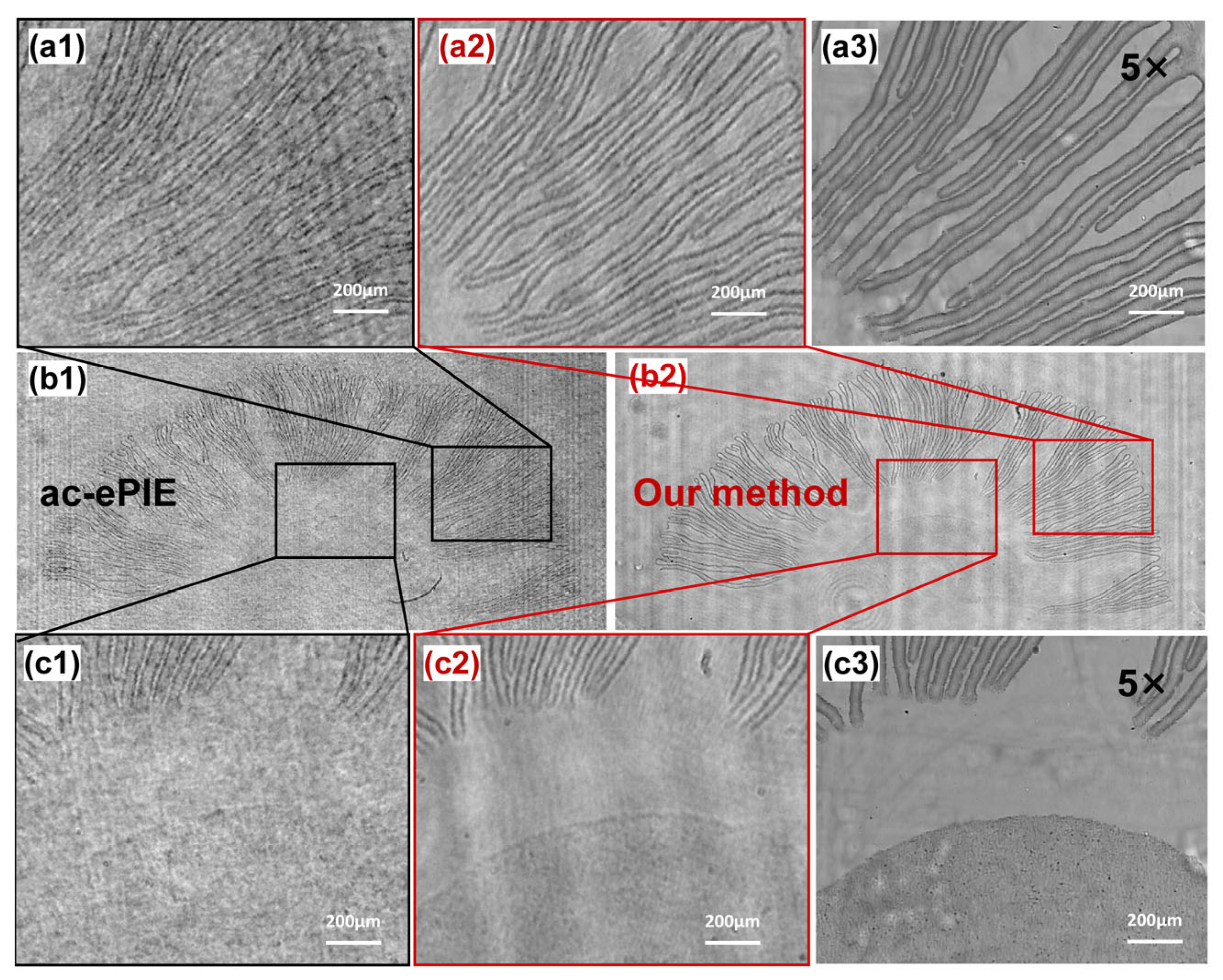
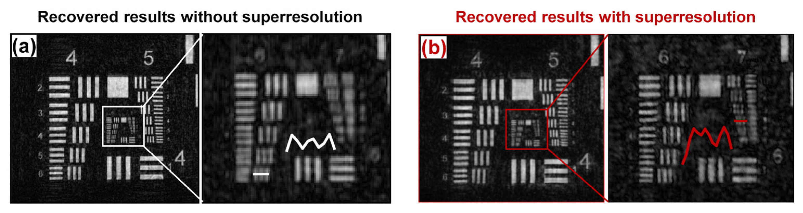
4. Experiments
5. Discussion
6. Conclusions
Author Contributions
Funding
Conflicts of Interest
References
- Takazawa, S.; Kang, J.; Abe, M.; Uematsu, H.; Ishiguro, N.; Takahashi, Y. Demonstration of single-frame coherent X-ray diffraction imaging using triangular aperture: Towards dynamic nanoimaging of extended objects. Opt. Express 2021, 29, 14394–14402. [Google Scholar] [CrossRef] [PubMed]
- Kang, J.; Takazawa, S.; Ishiguro, N.; Takahashi, Y. Single-frame coherent diffraction imaging of extended objects using triangular aperture. Opt. Express 2021, 29, 1441–1453. [Google Scholar] [CrossRef] [PubMed]
- He, X.; Veetil, S.P.; Jiang, Z.; Kong, Y.; Wang, S.; Liu, C. High-speed coherent diffraction imaging by varying curvature of illumination with a focus tunable lens. Opt. Express 2020, 28, 25655–25663. [Google Scholar] [CrossRef] [PubMed]
- Almoro, P.F.; Pedrini, G.; Gundu, P.N.; Osten, W.; Hanson, S.G. Phase microscopy of technical and biological samples through random phase modulation with a diffuser. Opt. Lett. 2010, 35, 1028–1030. [Google Scholar] [CrossRef]
- Jiang, H.; Song, C.; Chen, C.-C.; Xu, R.; Raines, K.S.; Fahimian, B.P.; Lu, C.-H.; Lee, T.-K.; Nakashima, A.; Urano, J.; et al. Quantitative 3D imaging of whole, unstained cells by using X-ray diffraction microscopy. Proc. Natl. Acad. Sci. USA 2010, 107, 11234–11239. [Google Scholar] [CrossRef]
- Kocsis, P.; Shevkunov, I.; Katkovnik, V.; Egiazarian, K. Single exposure lensless subpixel phase imaging: Optical system design, modeling, and experimental study. Opt. Express 2020, 28, 4625–4637. [Google Scholar] [CrossRef]
- Rodenburg, J.M.; Faulkner, H.M.L. A phase retrieval algorithm for shifting illumination. Appl. Phys. Lett. 2004, 85, 4795–4797. [Google Scholar] [CrossRef]
- Maiden, A.; Johnson, D.; Li, P. Further improvements to the ptychographical iterative engine. Optica 2017, 4, 736–745. [Google Scholar] [CrossRef]
- Li, M.; Bian, L.; Zheng, G.; Maiden, A.; Liu, Y.; Li, Y.; Suo, J.; Dai, Q.; Zhang, J. Single-pixel ptychography. Opt. Lett. 2021, 46, 1624–1627. [Google Scholar] [CrossRef]
- Sun, A.; He, X.; Kong, Y.; Cui, H.; Song, X.; Xue, L.; Wang, S.; Liu, C. Ultra-high speed digital micro-mirror device based ptychographic iterative engine method. Biomed. Opt. Express 2017, 8, 3155–3162. [Google Scholar] [CrossRef] [Green Version]
- Maiden, A.M.; Rodenburg, J.M. An improved ptychographical phase retrieval algorithm for diffractive imaging. Ultramicroscopy 2009, 109, 1256–1262. [Google Scholar] [CrossRef]
- Baksh, P.D.; Ostril, M.; Miszczak, M.; Pooley, C.; Brocklesby, W.S. Quantitative and correlative extreme ultraviolet coherent imaging of mouse hippocampal neurons at high resolution. Sci. Adv. 2020, 6, eazz3025. [Google Scholar] [CrossRef]
- Tanksalvala, M.; Porter, C.L.; Esashi, Y.; Wang, B.; Jenkins, N.W.; Zhang, Z.; Miley, G.P.; Knobloch, J.L.; McBennett, B.; Horiguchi, N.; et al. Nondestructive, high-resolution, chemically specific 3D nanostructure characterization using phase-sensitive EUV imaging reflectometry. Sci. Adv. 2021, 7, eadb9667. [Google Scholar] [CrossRef]
- Eschen, W.; Loetgering, L.; Schuster, V.; Kals, R.; Kirsche, A.; Berthold, L.; Steinert, M.; Pertsch, T.; Gross, H.; Krause, M.; et al. Material-specific high-resolution table-top extreme ultraviolet microscopy. Light Sci. Appl. 2022, 11, 117. [Google Scholar] [CrossRef]
- Brooks, N.J.; Wang, B.; Binnie, L.; Tanksalvala, M.; Esashi, Y.; Knobloch, J.L.; Nguyen, Q.L.D. Temporal and spectral multiplexing for EUV multibeam ptychography with a high harmonic light source. Opt. Express 2022, 30, 30331–30346. [Google Scholar] [CrossRef]
- Wang, B.; Brooks, N.J.; Johnsen, P.C.; Jenkins, N.W.; Esashi, Y.; Binnie, Y.; Tanksalvala, M.; Kapteyn, H.C.; Margaret, M. Murnane High-fidelity ptychographic imaging of highly periodic structures enabled by vortex high harmonic beams. arXiv 2023, arXiv:2301.05563. [Google Scholar]
- Abregana, T.J.T.; Almoro, P.F. Phase retrieval by amplitude modulation using digital micromirror device. Opt. Lasers Eng. 2022, 150, 106851. [Google Scholar] [CrossRef]
- Jiang, S.; Zhu, J.; Song, P.; Guo, C.; Bian, Z.; Wang, R.; Huang, Y.; Wang, S.; Zhang, H.; Zheng, G. Wide-field, high-resolution lensless on-chip microscopy via near-field blind ptychographic modulation. Lab Chip 2020, 20, 1058–1065. [Google Scholar] [CrossRef]
- Zhang, H.; Bian, Z.; Jiang, S.; Liu, J.; Song, P.; Zheng, G. Field-portable quantitative lensless microscopy based on translated speckle illumination and sub-sampled ptychographic phase retrieval. Opt. Lett. 2019, 44, 1976–1979. [Google Scholar] [CrossRef]
- Zhang, Z.; Zhou, Y.; Jiang, S.; Guo, K.; Hoshino, K.; Zhong, J.; Suo, J.; Dai, Q.; Zheng, G. Invited article: Mask-modulated lensless imaging with multi-angle illuminations. APL Photonics 2018, 3, 060803. [Google Scholar] [CrossRef]
- Lu, C.; Zhou, Y.; Guo, Y.; Jiang, S.; Zhang, Z.; Zheng, G.; Zhong, J. Mask-modulated lensless imaging via translated structured illumination. Opt. Express 2021, 29, 12491–12501. [Google Scholar] [CrossRef] [PubMed]
- Zhang, F.; Pedrini, G.; Osten, W. Phase retrieval of arbitrary complex-valued fields through aperture plane modulation. Phys. Rev. A 2007, 75, 43805. [Google Scholar] [CrossRef]
- Wen, X.; Geng, Y.; Zhou, X.; Tan, J.; Liu, S.; Tan, C.; Liu, Z. Ptychography imaging by 1-D scanning with a diffuser. Opt. Express 2020, 28, 22658–22668. [Google Scholar] [CrossRef] [PubMed]
- Kocsis, P.; Shevkunov, I.; Katkovnik, V.; Rekola, H.; Egiazarian, K. Single-shot pixel super-resolution phase imaging by wavefront separation approach. Opt. Express 2021, 29, 43662–43678. [Google Scholar] [CrossRef]
- Shen, C.; Tan, J.; Wei, C.; Liu, Z. Coherent diffraction imaging by moving a lens. Opt. Express 2016, 24, 16520–16529. [Google Scholar] [CrossRef]
- Xu, C.; Pang, H.; Cao, A.X.; Deng, Q.L.; Yang, H. Phase retrieval by random binary amplitude modulation and ptychography principle. Opt. Express 2022, 30, 14505–14517. [Google Scholar] [CrossRef]
- Xu, C.; Pang, H.; Cao, A.X.; Deng, Q.L. Enhanced multiple-plane phase retrieval using a transmission grating. Opt. Lasers Eng. 2022, 149, 106810. [Google Scholar] [CrossRef]
- Guo, C.; Li, Q.; Wei, C.; Tan, J.; Liu, S.; Liu, Z. Axial multi-image phase retrieval under tilt illumination. Sci. Rep. 2017, 7, 7562. [Google Scholar] [CrossRef]
- Mainden, A.M.; Humphry, M.J.; Zhang, F.C.; Raodenburg, J.M. Superresolution imaging via ptychography. J. Opt. Soc. Am. A 2011, 28, 604–612. [Google Scholar] [CrossRef]


Disclaimer/Publisher’s Note: The statements, opinions and data contained in all publications are solely those of the individual author(s) and contributor(s) and not of MDPI and/or the editor(s). MDPI and/or the editor(s) disclaim responsibility for any injury to people or property resulting from any ideas, methods, instructions or products referred to in the content. |
© 2023 by the authors. Licensee MDPI, Basel, Switzerland. This article is an open access article distributed under the terms and conditions of the Creative Commons Attribution (CC BY) license (https://creativecommons.org/licenses/by/4.0/).
Share and Cite
Xu, C.; Pang, H.; Cao, A.; Deng, Q.; Hu, S.; Yang, H. Lensless Imaging via Blind Ptychography Modulation and Wavefront Separation. Photonics 2023, 10, 191. https://doi.org/10.3390/photonics10020191
Xu C, Pang H, Cao A, Deng Q, Hu S, Yang H. Lensless Imaging via Blind Ptychography Modulation and Wavefront Separation. Photonics. 2023; 10(2):191. https://doi.org/10.3390/photonics10020191
Chicago/Turabian StyleXu, Cheng, Hui Pang, Axiu Cao, Qiling Deng, Song Hu, and Huajun Yang. 2023. "Lensless Imaging via Blind Ptychography Modulation and Wavefront Separation" Photonics 10, no. 2: 191. https://doi.org/10.3390/photonics10020191