Article

Polarization-Sensitive Nonlinear Optical Interaction of Ultrashort Laser Pulses with HPHT Diamond

by
George K. Krasin
,
Yulia S. Gulina
*,
Evgeny V. Kuzmin
,
Victor P. Martovitskii
and
Sergey I. Kudryashov
Lebedev Physical Institute, Russian Academy of Sciences, Leninskiy Prospekt 53, 119991 Moscow, Russia
*
Author to whom correspondence should be addressed.
Photonics 2023, 10(2), 106; https://doi.org/10.3390/photonics10020106
Submission received: 10 December 2022 / Revised: 14 January 2023 / Accepted: 17 January 2023 / Published: 19 January 2023
(This article belongs to the Special Issue Women’s Special Issue Series: Photonics)

Abstract

:
The filamentation of focused 300 fs laser pulses with variable polarization azimuth in bulk of synthetic HPHT diamond demonstrates the possibility of polarization-dependent bandgap control for crystal dielectric photoexcitation. This directly affects the value of the filamentation threshold power, which exhibits the distinct dependence on the polarization azimuth angle. The nonlinear photoluminescence yield, when focusing ultrashort laser pulses with variable polarization in bulk of the synthetic diamond, indicates different polarization-dependent regimes in the dynamics of electron-hole plasma formation, arising due to different processes of photoexcitation and recombination of free carriers during the filamentation process. Thus, at the onset of the filamentation process, at relatively low intensities, the photoluminescence yield rate depends on polarization azimuth controlling bandgap, while at high intensities the resulting dense absorbing plasma exhibits isotropy with respect to laser radiation polarization, and photoluminescence yield weakly depends on polarization azimuth.

1. Introduction

The parameters of the strong electromagnetic wave can change during its propagation inside of solid media due to the action of nonlinear optical effects [1]. Among the parameters of the laser radiation that affect the nonlinear optical interaction in bulk crystalline dielectrics the laser polarization along with the wavelength and energy (intensity) could be rather important. The first results demonstrated as early as 1956 by P. P. Feofilov using photoluminescence (PL) in crystals with cubic crystal system (diamond, fluorite)—an optically isotropic medium in linear approximation—showed that such crystals exhibit anisotropic properties under photoexcitation by polarized continuous radiation [2]. This anisotropy is a result of the individual optical centers’ orientation in crystals relative to selected directions defined as optical axes [2,3]. It can be argued that, with the polarization of the laser radiation, one can control the photoionization rate through the bandgap along the corresponding directions in the Brillouin zone [4]. The influence of ultrashort laser pulses polarization on the processes of nonlinear optical interaction with crystalline materials was demonstrated recently. For example, it was reported in [5,6] that photoionization rate in wide-bandgap crystals differed for ultrashort pulses with different polarization azimuths. The high-harmonic generation and spectroscopy of crystals of quartz, MgO and Si was revealed to be sensitive to the polarization of the excitation fields [7,8]. Azimuthal dependences of phase transitions excited by femtosecond laser radiation in VO2 thin films deposited on various substrates, including sapphire crystals (R and C planes), were shown [9]. The dependence of the probing pulse transmittance on the polarization azimuth of the pump pulse in the LiF crystal was also obtained [10].
The study of the effect of polarization on the filamentation process [11] is of particular interest. In itself the process of filament channels formation during the propagation of ultrashort laser pulses in condensed media is well studied. Despite the fact that this process is influenced by many factors, it can be described by the action of two central nonlinear physical effects [1]. On the one hand, the laser beam is affected by the optical Kerr effect, which counteracts diffraction and leads to an increase in intensity in the paraxial region [11,12]. This intensity increase leads to a local change of the refractive index of the media, which in turn leads to even stronger self-focusing of the laser beam. On the other hand, intensity is capped due to the multiphoton absorption, which is followed by plasma formation, lowering the local refractive index and defocusing the laser beam [13,14,15,16]. Setting a dynamic balance between Kerr’s self-focusing and plasma defocusing results in a filamentation channel. Given the fact that laser polarization determines the direction in the Brillouin zone and its corresponding bandgap for photoexcitation of the dielectric media, we can say that it allows the selection of the bandgap along which the nonlinear optical interaction of ultrashort laser pulses with crystalline dielectrics will occur [17].
Previous studies have revealed the polarization dependences of the natural diamond surface ablation [18,19], and primary dependences of PL in natural and synthetic diamond crystals [19,20]. The circular and linear polarizations were compared as a part of the study of pulse-width-dependent critical power for the self-focusing of ultrashort laser pulses [21]. However, until now the processes of the nonlinear in-bulk interaction of ultrashort laser pulses with crystalline dielectric materials, leading to the formation of filament channels and affecting the efficiency of energy deposition in the modified area, have not been studied depending on the angle of laser polarization. Therefore, identifying the relationship between the above nonlinear optical effects with the polarization azimuth of radiation and the bandgap of a crystalline dielectric material is a relevant, but still unresolved, scientific problem.
This paper presents a study of nonlinear optical effects arising during interaction in bulk synthetic diamond of crystallographic orientation (110) with femtosecond laser pulses with variable polarization azimuth in the (pre)filamentation regime.

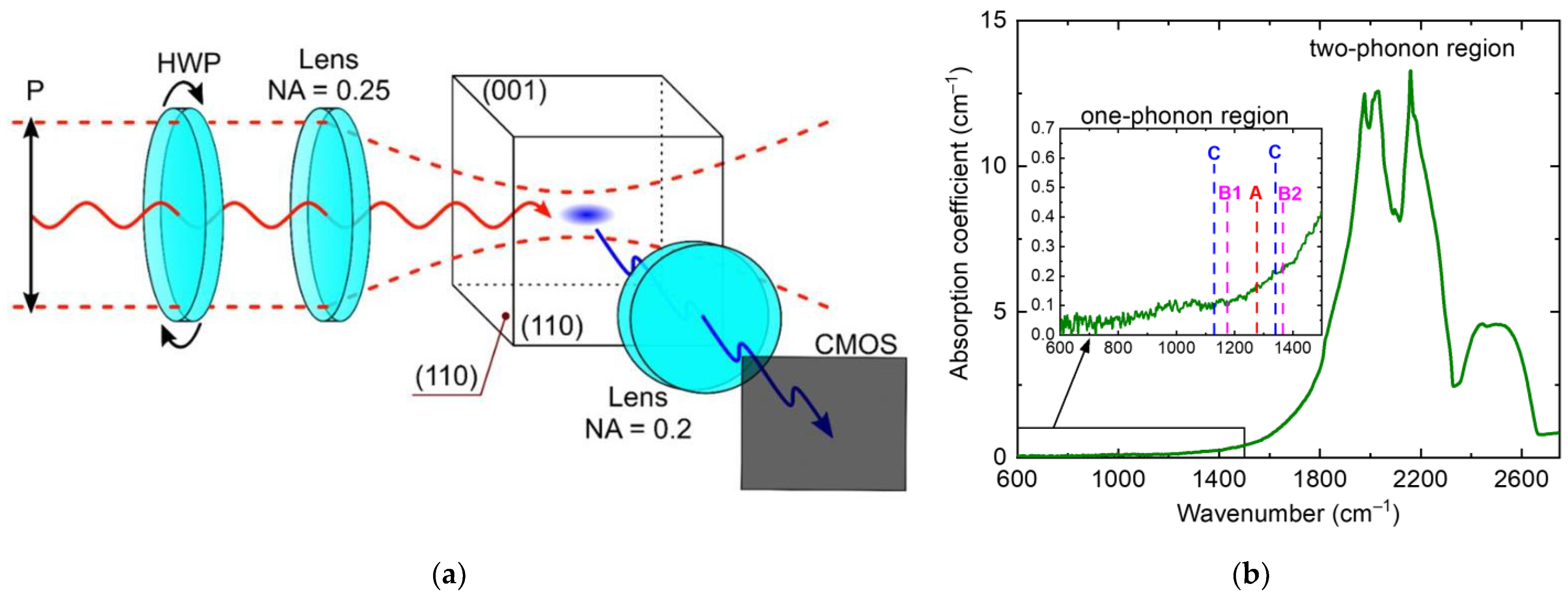
2. Materials and Methods

The femtosecond laser Satsuma (Amplitude Systemes, France) was used as a source of linearly polarized radiation. The laser pulses were focused in bulk of the HPHT synthetic type IIA diamond with dimensions of 2 × 2 × 1 mm3 using a 0.25-NA (Numerical Aperture) microscope objective (beam waist in the focal plane for the 1/e energy level was w 0 3.75 μm for the 1030 nm and w 0 1.9 μm for 515 nm). During the multi-pulse exposure by ultrashort pulses, with a duration of ~300 fs at wavelengths of 1030 nm and 515 nm with a repetition rate of 100 kHz, the formation of extended luminous channels was observed in the rear focal plane of this microscope objective. The micro images of the luminous tracks were captured at a right angle on a color CCD-camera using a 0.2-NA quartz/fluorite microscope objective (see Figure 1a). The change of the linear polarization azimuth of the exciting laser radiation was carried out using a half-wave plate, put right before the microscope objective. During the experiments the half-wave plate was rotated on the 180° around its axis, which led to the turn of the polarization azimuth on the angle of 360°.
The preliminary characterization of the sample was carried out on a X’Pert PRO MRD diffractometer (PANalytical BV, Netherlands), where the main crystal faces and axes were determined. In the experiment the ultrashort laser pulses were focused in bulk of the sample via (110) face at a normal angle, while the luminous tracks were captured at a right angle via another (110) face. The optical absorption spectrum of the sample was obtained using an IR spectrometer VERTEX 70v (Bruker, Karlsruhe, Germany). The obtained spectral characteristic shown in the Figure 1b demonstrates the absence of the nitrogen impurity absorption bands (<1 ppm). In the following experiments the polarization azimuth of 0° coincided with the direction [001] in the diamond (110) face, while azimuth of 90° corresponded to the direction [110].

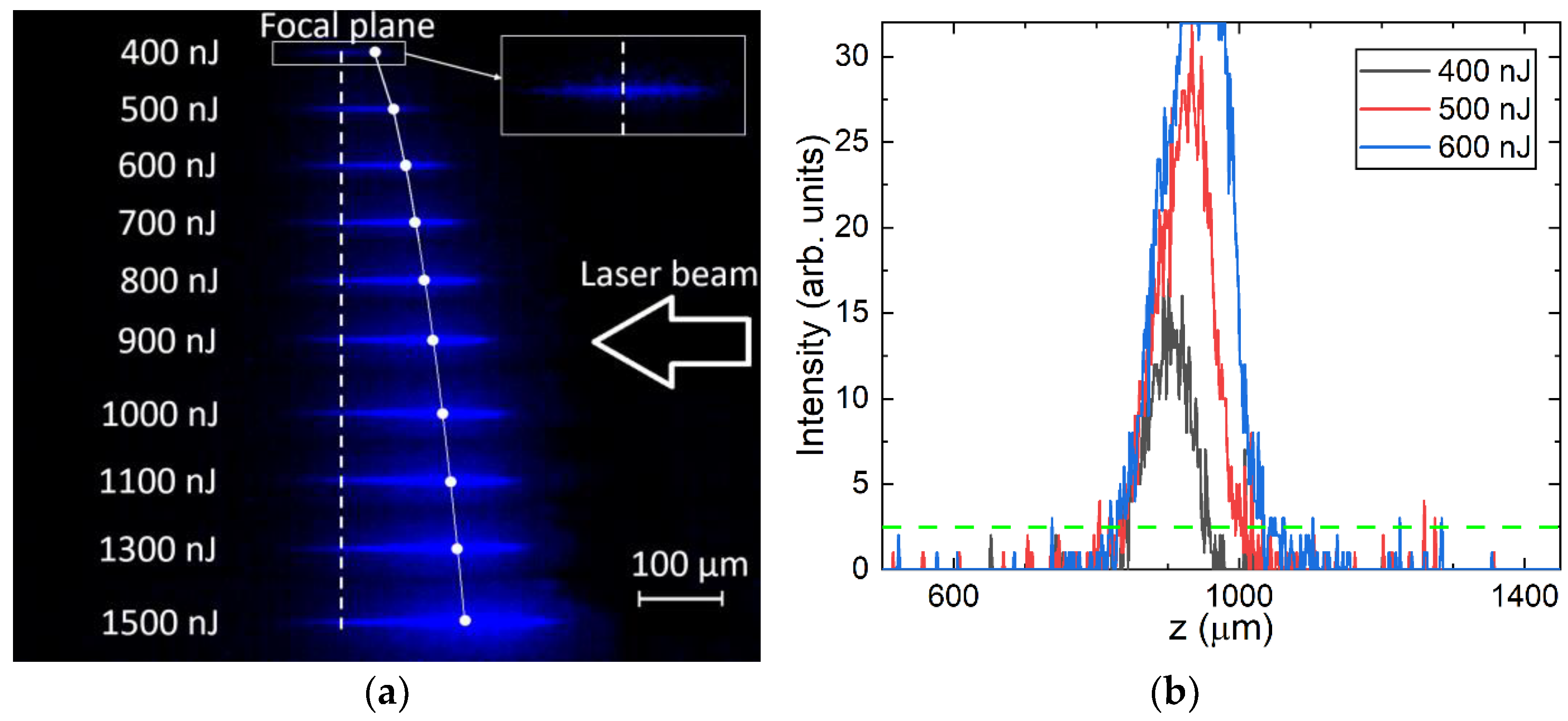
3. Experimental Results

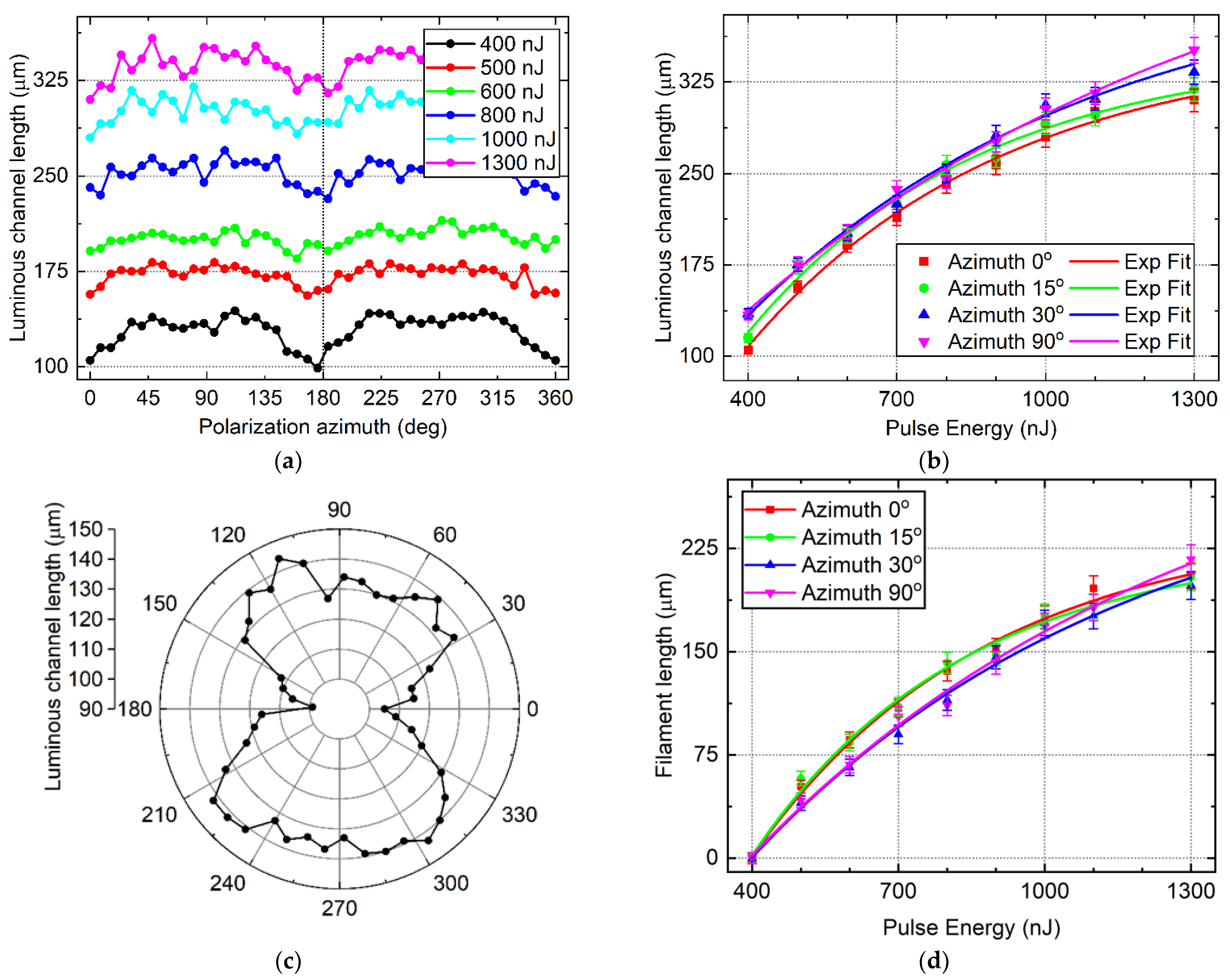
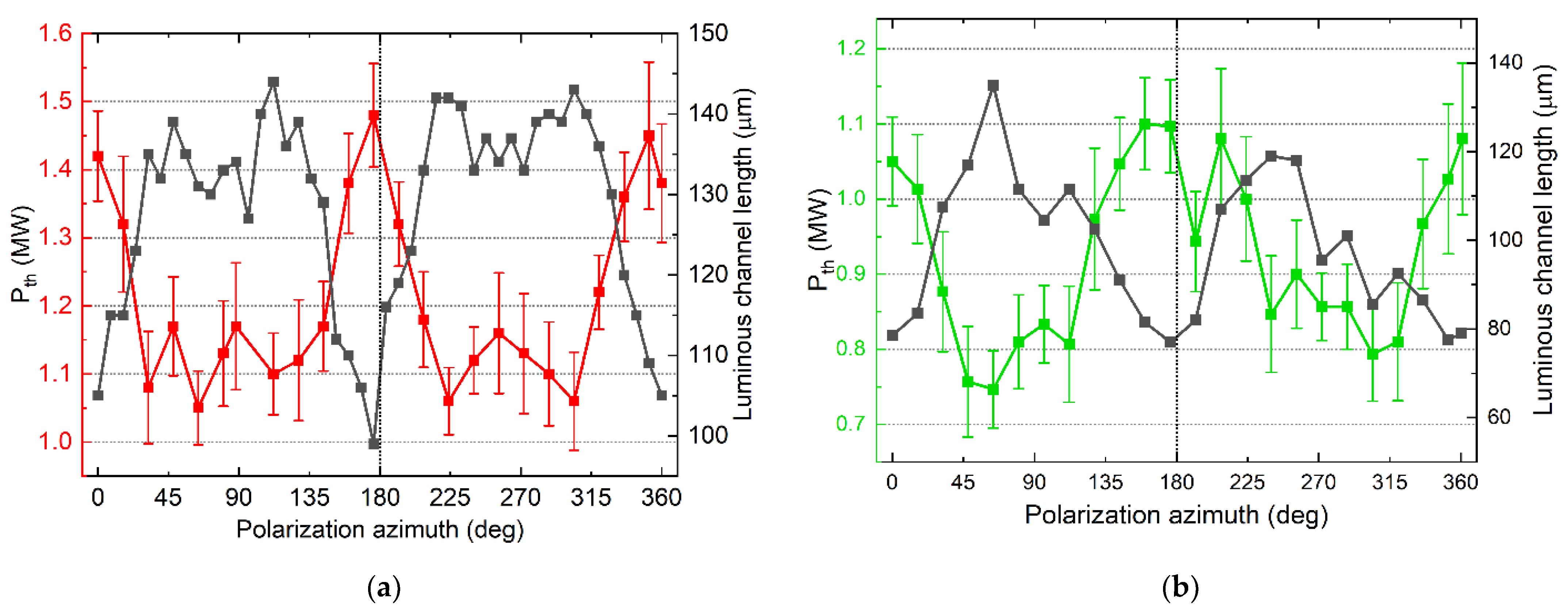
3.1. Luminous Channels Length vs. Polarization

The micro images of the luminous spatial channels formed under the action of the ultrashort laser pump are shown in Figure 2a. The luminous tracks appeared around the central position of the linear focus z f 900 μm at pulse energies E ≥ 400 nJ, and, with a further increase in energy, they began to asymmetrically elongate from the geometric focus towards the incident radiation. The dashed line shows the focal plane, while the solid line shows the onset of the nonlinear focus. Figure 2b shows the cross sections of a few first luminous channels through the point of the maximum intensity. The luminous channel length was calculated using one intensity level (dashed line Figure 2b), which was determined based on the 2σ condition in the associated region to exclude the noise.
From the experimental data for the known Gaussian intensity distribution around focus plane, the theoretical onset of the nonlinear focus (end of linear focus position) can be calculated for the used range of pulse energies. Using the following equation to calculate the Gaussian beam width at a given position from the focus plane f′
w ( z ) = w 0 1 + ( z f z R ) 2 ,
one can calculate the intensity distributions along z axis from the known beam width and pulse energy. Then using one level of intensity, one can obtain theoretical onset of the nonlinear focus or the place where the filamentation process should begin theoretically (see Figure 2a, white solid line) [21].
Figure 3 clearly demonstrates the dependence of the luminous channel length on the direction in which photoexcitation by ultrashort laser pulses is carried out, which is specified by the radiation polarization azimuth. So, from Figure 3a, it can be seen that the modulation of the channel length decreased with increasing laser pulse energy from ~35% at 400 nJ to ~15% at 600 nJ, after which it slowly decreased to around 10% at 1500 nJ. Observed in Figure 3b, the values of the luminous channel lengths on the pulse energy for some values of the polarization azimuth also confirmed this dependence. The obtained data points on Figure 3b exhibit saturating dependences (solid lines on the graph). The Figure 3c for the luminous channel length at pulse energy E = 400 nJ plotted in polar coordinates demonstrates the second degree of symmetry, which coincides with the degree of symmetry of the (110) crystal face.
Accounting for the area of the linear focusing of the Gaussian beam, one can calculate the filament length from the length of the luminous channel. Considering that the luminous channel for pulse energy E = 400 nJ has a symmetrical cross-section in the Figure 2b, we can assume that in this regime there was no filamentation involved and substitute its length from all of the following luminous channels to calculate the filament length as L f i l a m e n t = L c h a n n e l L 400 n J [21]. The obtained dependences of the filament length on the pulse energy for different polarization azimuths are shown on the Figure 3d.

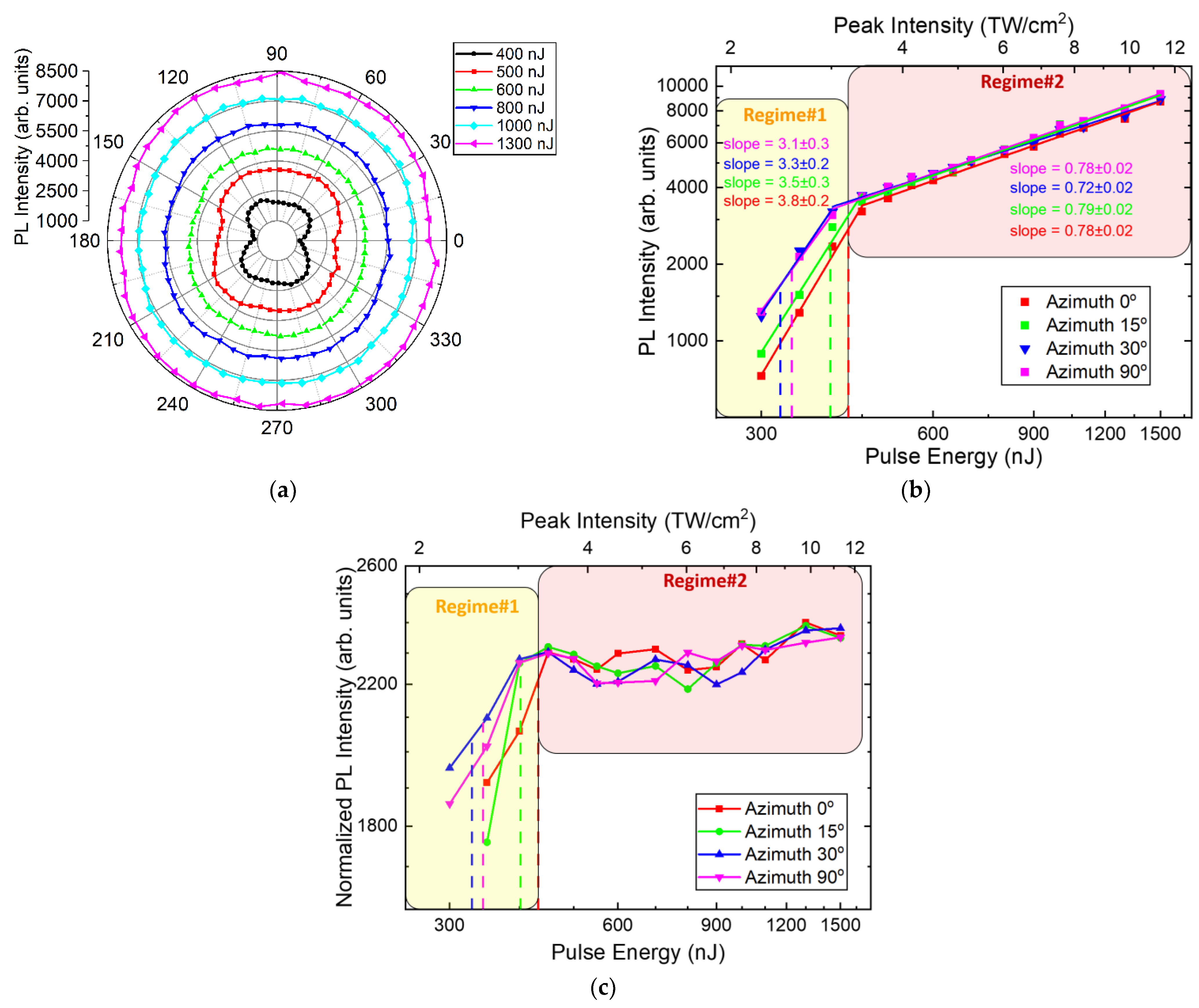
3.2. PL Intensity vs. Polarization

The dependences of the integral PL intensity of the observed channels on the pulse energy for different polarization azimuths are shown in Figure 4. The Figure 4a shows the dependence of the integrated PL intensity on the polarization azimuth, which demonstrated the modulation of the intensity value with a change in the polarization of laser radiation. As the energy of laser pulses increased, the dependence remained, although it became less pronounced, becoming indistinguishable after the level of 500 nJ. Plotting the dependencies of the PL intensity on the pulse energy for the same azimuth in Cartesian coordinates using more points, we could demonstrate this effect of reducing the modulation of the PL signal with increasing fluence. A similar effect was previously demonstrated for polarization-dependent ablation in cubic crystals [22].
It is known that bends in the slopes in the pump power-dependent stationary PL output can be identified as transition points between different characteristic regimes, apparently representing some features of ultrafast inter-band photoexcitation accompanying the dynamics of free charge carriers and the filamentation process [23,24]. In addition, when the graphs of the dependence of PL intensity on the pulse energy are normalized on the lengths of filaments (see Figure 4c), we find that with an increase in the intensity of ultrashort laser pulses in the registered filaments, there was no increase in the PL yield and that pulse energy went into the channel elongation, which is in good agreement with the well-known fact about the intensity clamping in the process filamentation [25,26].

3.3. Filamentation Threshold Power vs. Polarization

The onset of the filamentation process in our research was identified as a visible asymmetric elongating of the luminescent focal channels with increasing laser pulse energy. Based on the analysis of the difference between the Rayleigh length characterizing the linear focusing and the luminescent channel length, similarly to the technique outlined in [18], the threshold power of the filamentation onset was determined. Plots of the obtained dependences of the threshold power and of filament channels length on the polarization azimuth for the pulse energy E = 400 nJ (1.33 MW) are shown in Figure 5.
The filamentation threshold power for 1030 nm laser pulses varied between 1.05 and 1.5 MW, which is almost one and a half times the modulation of P t h (see Figure 5a). The same dependence was obtained for the 515 nm pulses where the range of P t h values varied from 0.75 to 1.1 MW (see Figure 5b). In both cases, the modulation period of P t h values were 180°, which agreed with the symmetry of (110) crystal face through which the laser-matter interaction took place.
The observed power-dependent increase in filament length due to a nonlinear change of focus usually occurs when a dynamic balance between Kerr self-focusing, diffraction, and plasma defocusing is established. In the diffraction approximation the filament length via the semi-empirical Marburger formula [11] could be related to the threshold power by an expression [27]
L ( P ) f 2 0.367 L D F ( P P t h 0.852 ) ,
here L D F denotes the Rayleigh length.
The Equation (2) shows an inverse dependence of the filament channel length on the threshold power P t h , that is clearly demonstrated by the experimental data shown in Figure 5. The maximum value of the threshold power corresponds to the filament minimum length.

4. Discussion

Although radiative recombination and the related PL is rather slow and low-rate process during photoexcitation of synthetic and natural diamonds at high laser intensities, it can be effectively used as an indicator of electron-hole plasma (EHP) density ρ ( t ) , produced via different photogeneration and free carriers’ recombination processes
Φ β ρ ( t ) 2 d t ,
here β—the electron radiative recombination coefficient.
The EHP density depended on the seed processes rate, which are multiphoton and tunnel ionization. The kinetic rate equation for the EHP density could be written in the common form [28,29,30]
d ρ ( t ) d t = W P I ( I ( t ) ) + W A V ( I ( t ) , ρ ( t ) ) W R ( ρ ( t ) , t ) ,
where W P I is the photoionization rate, W A V is the avalanche ionization rate, W R is the electron recombination rate.
The Keldysh theory describing the interaction of intense laser radiation with matter acted as a fundamental basis for modeling EHP density growth based on photoionization processes. Within this theory, multiphoton and tunnel photoionization are set as two limiting regimes of the same physical phenomenon—ionization in an oscillating field of electromagnetic radiation. To determine the interaction regime of light field with the matter the Keldysh parameter was used [31,32]
γ K = ω e m e * c n 0 ε 0 E g I ,
here e denotes the electron charge, m e * denotes the electron effective mass, ω is the electromagnetic radiation frequency, n 0 is the linear refractive index of the medium, c is the speed of light in a vacuum, E g denotes the bandgap, ε 0 is the vacuum permittivity, I denotes the electromagnetic radiation intensity.
At low laser intensities, multiphoton ionization is the dominant process with the value of the Keldysh parameter of γ K > 1.5 [33]. The number of photons m required to excite an electron from the valence into the conduction band was determined by the material bandgap E g
m ω > E g
Tunnel ionization occurs in particularly intense laser fields with laser pulse durations τ < 10 fs or intensities I > 10 14 W/cm2 [29]. In strong laser fields an oscillating potential barrier arises through which bound electrons can tunnel from the valence to the conduction band. This regime is given by the value of the Keldysh parameter 1.5 < γ K .
For the case of intense laser radiation interaction with dielectric materials, including diamond considered in this paper, the value of the Keldysh parameter is γ K > 1.5 , and multiphoton photoionization process will dominate. Since the onset of the filamentation process with inevitable formation of plasma was considered (regime# 1 in Figure 4b), Auger recombination would be the process to compensate the multiphoton ionization. In [34] it is shown that the photoionization process rate based on the Keldysh photoionization theory can be written as
W M P I ( t ) β m I m ,
where the multiphoton absorption coefficient defined as
β m = ω 9 π ( m e * ω ) 3 2 ( e 2 8 ω 2 m e * c ε 0 ) m exp ( 2 m ) ( n 0 E g ) m 1 / E g m
In this case, the kinetic rate Equation (4) will be as follows
d ρ ( t ) d t = I m ( t ) E g m γ ρ 3 ( t ) ,
here γ is the Auger recombination coefficient.
The Auger recombination coefficient according to the research of the authors [35,36] was related by an inverse power relation to the bandgap γ 1 / E g 4 . When dynamic equilibrium was established, we can express from Equation (9) the dependence of the EHP density on the intensity and the bandgap
I m ( t ) E g m = ρ 3 ( t ) E g 4 ,       ρ ( t ) I ( t ) m 3 E g 4 m 3
and the PL intensity was proportional to
Φ β ρ ( t ) 2 d t I ( t ) 2 m 3 E g 2 ( 4 m ) 3
In the investigated synthetic diamond with ultra-low impurity content, the inter-band EHP photoexcitation presumably occurred in a pure carbon-carrier matrix [37]. Therefore, the analysis of PL yield in such sample paved the way to understanding the fundamental basis of nonlinear inter-band photoexcitation processes in diamonds in general [38]. Diamond is an indirect semiconductor with the bandgap E g , i 5.4 eV. However, in the multiphoton photoionization regime, direct transitions dominate over indirect ones and the first direct bandgap E g 7.2 eV is located near the Γ-point of the Brillouin zone [6,39]. Therefore, to excite an electron from the valence to the conduction band through the direct band gap during 1030 nm laser radiation photoexcitation (quantum energy ℏω ≈ 1.2 eV), m = 6 photons were needed. In this case, the PL yield intensity was proportional to Φ I ( t ) 4 E g 4 / 3 , which corresponded to the curve slope for 0 deg polarization azimuth (Slope ≈ 3.8), obtained from the experimental data. It is known that the polarization of laser radiation strongly affects the multiphoton ionization rate in diamond [6] and electron density respectively due to the crystal symmetry and Brillouin zone structure. Taking into account the dependence of semiconductors direct bandgap at the Γ-point on the dynamically photoexcited conduction electron density [35], it could be assumed that the polarization azimuth variation leads to electron-phonon bandgap renormalization [40,41]. This can result in a reduction of diamond direct bandgap by about 0.7 eV [42]. Consequently, in the synthetic diamond sample under study, the number of photons required for direct charge carriers transfer from conduction to valence zone was reduced to m = 5 when the polarization azimuth changed. This led to variation of the PL intensity curve slope in the range from ≈3 to ≈3.8, which is clearly demonstrated for regime#1 in Figure 4b.
In the case of high intensities at which a stable filamentation process was established, a dense plasma was present in the material and absorbed the incident radiation. Therefore, the dominant ionization mechanism was replaced by an avalanche one and the process counteracting it was Auger recombination (regime#2 in Figure 4b). The kinetic rate Equation (4) can be written in the form [43,44]
d ρ ( t ) d t = α I ρ ( t ) γ ρ 3 ( t ) ,
here α is the avalanche ionization coefficient.
According to the Drude avalanche ionization model, the avalanche ionization coefficient can be defined as [45,46]
α = σ E g ,     σ = e 2 c m e * n 0 ε 0 τ c 1 + ω 2 τ c 2 ,
here σ denotes the absorption cross section, τ c is the mean collision time, which could be estimated as [47]
τ c = 16 π ε 0 2 m e * ( 0.1 E g ) 3 2 e 4 ρ ( t )
Based on Equations (13) and (14), it could be shown that α E g / ρ ( t ) .
The EHP density dependence on the laser intensity and the bandgap derived from Equation (12) was as follows
E g ρ ( t ) I ( t ) ρ ( t ) = ρ 3 ( t ) / E g 4 ,       ρ ( t ) I ( t ) 1 / 3 E g 3 / 2
and the PL intensity was proportional to
Φ β ρ ( t ) 2 d t I ( t ) 2 / 3 E g 3
Thus, at steady-state filamentation process at high intensities, the PL yield rate was independent of the polarization-controlled change in the bandgap, which was consistent with the experimentally obtained data. The slopes of experimental curves were 0.72–0.79 ± 0.02, close to the power of 2/3 obtained in Equation (16). Presumably, this was due to the isotropic absorption properties of formed dense plasma in relation to the laser radiation polarization.

5. Conclusions

The investigation of filamentation of ultrashort laser pulses with variable polarization azimuth in bulk of synthetic HPHT diamond has shown that laser radiation polarization allows to control corresponding bandgap for crystal dielectric photoexcitation. This directly affects the value of the threshold power of the filamentation onset. It was shown that, during the focusing of 1030 nm laser pulses with 300 fs duration in bulk of synthetic diamond, the threshold power varied in the range from 1.05 to 1.5 MW with a period of 180°, and for the 515 nm pulses the range was 0.75–1.1 MW. This modulation of the filamentation threshold power value was consistent with the (110) face symmetry through which the photoexcitation was carried out. Similar dependence was demonstrated by the PL yield, however, with the increase in pulse energy, the PL intensity modulation decreased and was almost indistinguishable at energies exceeding E = 500 nJ.
The nonlinear PL yield when focusing ultrashort laser pulses with variable polarization in bulk of the synthetic diamond indicates different polarization-dependent regimes in the dynamics of EHP formation arising due to different processes of photoexcitation and recombination of free carriers during the filamentation process. Thus, at the onset of the filamentation process at relatively low intensities, the PL yield rate depends on polarization azimuth controlling bandgap, while at high intensities the resulting dense absorbing plasma exhibits isotropy with respect to laser radiation polarization and PL yield does not depend on polarization azimuth.

Author Contributions

Conceptualization, G.K.K., Y.S.G. and S.I.K.; methodology, G.K.K., Y.S.G. and S.I.K.; software, G.K.K. and E.V.K.; validation, G.K.K., Y.S.G. and S.I.K.; formal analysis, G.K.K., Y.S.G. and V.P.M.; investigation, G.K.K. and Y.S.G.; resources, V.P.M.; data curation, G.K.K. and E.V.K.; writing—original draft preparation, G.K.K. and Y.S.G.; writing—review and editing, G.K.K., Y.S.G. and S.I.K.; visualization, G.K.K. and E.V.K.; supervision, S.I.K.; project administration, S.I.K.; funding acquisition, S.I.K. All authors have read and agreed to the published version of the manuscript.

Funding

This research was financially supported by Russian Science Foundation, grant number 21-79-30063.

Institutional Review Board Statement

Not applicable.

Informed Consent Statement

Not applicable.

Data Availability Statement

The data presented in this study are available on request from the corresponding author.

Conflicts of Interest

The authors declare no conflict of interest.

References

  1. Boyd, R.W. Nonlinear Optics, 3rd ed.; Academic Press: Boston, MA, USA, 1992. [Google Scholar]
  2. Feofilov, P.P. Polarized luminescence in cubic crystals. Usp. Fiz. Nauk 1956, 58, 69–84. [Google Scholar] [CrossRef]
  3. Elliott, R.J.; Matthew, I.G.; Mitchell, E.W.J. The polarization of luminescence in diamond. Philos. Mag. 1958, 3, 360–369. [Google Scholar] [CrossRef]
  4. Temnov, V.V.; Sokolowski-Tinten, K.; Zhou, P.; El-Khamhawy, A.; von der Linde, D. Multiphoton Ionization in Dielectrics: Comparison of Circular and Linear Polarization. Phys. Rev. Lett. 2006, 97, 237403. [Google Scholar] [CrossRef] [PubMed]
  5. Gertsvolf, M.; Jean-Ruel, H.; Rajeev, P.P.; Klug, D.D.; Rayner, D.M.; Corkum, P.B. Orientation-Dependent Multiphoton Ionization in Wide Band Gap Crystals. Phys. Rev. Lett. 2008, 101, 243001. [Google Scholar] [CrossRef]
  6. Kozák, M.; Otobe, T.; Zukerstein, M.; Trojánek, F.; Malý, P. Anisotropy and polarization dependence of multiphoton charge carrier generation rate in diamond. Phys. Rev. B 2019, 99, 104305. [Google Scholar] [CrossRef]
  7. Heinrich, T.; Taucer, M.; Kfir, O.; Corkum, P.B.; Staudte, A.; Ropers, C.; Sivis, M. Chiral high-harmonic generation and spectroscopy on solid surfaces using polarization-tailored strong fields. Nat. Commun. 2021, 12, 3723. [Google Scholar] [CrossRef]
  8. Suthar, P.; Trojánek, F.; Malý, P.; Derrien, T.J.-Y.; Kozák, M. Role of Van Hove singularities and effective mass anisotropy in polarization-resolved high harmonic spectroscopy of silicon. Commun. Phys. 2022, 5, 288. [Google Scholar] [CrossRef]
  9. Liu, H.; Lysenko, S.; Vikhnin, V.; Rua, A.; Fernandez, F. Azimuth Dependence of Light-Induced Ultrafast Insulator-to-Metal Phase Transition in VO2 Thin Film. ECS Trans. 2006, 3, 37–49. [Google Scholar] [CrossRef]
  10. Dachraoui, H.; Oberer, C.; Heinzmann, U. Femtosecond crystallographic experiment in wide-bandgap LiF crystal. Opt. Express 2011, 19, 2797–2804. [Google Scholar] [CrossRef] [Green Version]
  11. Couairon, A.; Mysyrowicz, A. Femtosecond filamentation in transparent media. Phys. Rep. 2007, 441, 47–189. [Google Scholar] [CrossRef]
  12. Chekalin, S.V.; Kandidov, V.P. From self-focusing light beams to femtosecond laser pulse filamentation. Phys.-Usp. 2013, 56, 123–140. [Google Scholar] [CrossRef]
  13. Kosareva, O.; Daigle, J.-F.; Panov, N.; Wang, T.; Hosseini, S.; Yuan, S.; Roy, G.; Makarov, V.; Chin, S.L. Arrest of self-focusing collapse in femtosecond air filaments: Higher order Kerr or plasma defocusing? Opt. Lett. 2011, 36, 1035–1037. [Google Scholar] [CrossRef] [PubMed] [Green Version]
  14. Martynovich, E.F.; Dresviansky, V.P.; Kuznetsov, A.V.; Kuzakov, A.S.; Popov, A.A.; Alekseev, S.V.; Losev, V.F.; Ratakhin, A.N.; Bagayev, S.N. Simulation of filamentation of single femtosecond laser pulses in LiF. Laser Phys. 2014, 24, 074001. [Google Scholar] [CrossRef]
  15. Li, Q.; Perrie, W.; Li, Z.; Edwardson, S.P.; Dearden, G. Two-photon absorption and stimulated emission in poly-crystalline Zinc Selenide with femtosecond laser excitation. Opto-Electron. Adv. 2022, 5, 210036. [Google Scholar] [CrossRef]
  16. Chen, L.; Hong, M. Functional nonlinear optical nanoparticles synthesized by laser ablation. Opto-Electron. Sci. 2022, 1, 210007. [Google Scholar] [CrossRef]
  17. Zukerstein, M.; Trojánek, F.; Rezek, B.; Šobáň, Z.; Kozák, M.; Malý, P. Coherent phonon dynamics in diamond detected via multiphoton absorption. Appl. Phys. Lett. 2019, 115, 161104. [Google Scholar] [CrossRef]
  18. Krasin, G.; Kovalev, M.; Danilov, P.; Stsepuro, N.; Oleynichuk, E.; Bibicheva, S.; Martovitskii, V.; Kudryashov, S. Ablation of (111) and (001) Crystal Plates by Ultrashort Laser Pulses with Rotated Linear Polarization. JETP Lett. 2021, 114, 117–123. [Google Scholar] [CrossRef]
  19. Krasin, G.K.; Kovalev, M.S.; Kudryashov, S.I.; Danilov, P.A.; Martovitskii, V.P.; Gritsenko, I.V.; Podlesnykh, I.M.; Khmelnitskii, R.A.; Kuzmin, E.V.; Gulina, Y.S.; et al. Polarization-dependent near-IR ultrashort-pulse laser ablation of natural diamond surfaces. Appl. Surf. Sci. 2022, 595, 153549. [Google Scholar] [CrossRef]
  20. Krasin, G.K.; Stsepuro, N.G.; Martovitsky, V.P.; Kovalev, M.S. Polarization-dependent filamentation of femtosecond laser pulses in synthetic diamond. Opt. Spectrosc. 2022, 132, 418–421. [Google Scholar] [CrossRef]
  21. Kudryashov, S.I.; Danilov, P.A.; Kuzmin, E.V.; Gulina, Y.S.; Rupasov, A.E.; Krasin, G.K.; Zubarev, I.G.; Levchenko, A.O.; Kovalev, M.S.; Pakholchuk, P.P.; et al. Pulse-width-dependent critical power for self-focusing of ultrashort laser pulses in bulk dielectrics. Opt. Lett. 2022, 47, 3487–3490. [Google Scholar] [CrossRef]
  22. Li, X.; Rong, W.; Jiang, L.; Zhang, K.; Li, C.; Cao, Q.; Zhang, G.; Lu, Y. Generation and elimination of polarization-dependent ablation of cubic crystals by femtosecond laser radiation. Opt. Express 2014, 22, 30170–30176. [Google Scholar] [CrossRef] [PubMed]
  23. Kudryashov, S.; Danilov, P.; Smirnov, N.; Levchenko, A.; Kovalev, M.; Gulina, Y.; Kovalchuk, O.; Ionin, A. Femtosecond-laser-excited luminescence of the A-band in natural diamond and its thermal control. Opt. Mater. Express 2021, 11, 2505–2513. [Google Scholar] [CrossRef]
  24. Kudryashov, S.I.; Khmelnitskii, R.A.; Danilov, P.A.; Smirnov, N.A.; Levchenko, A.O.; Kovalchuk, O.E.; Uspenskaya, M.V.; Oleynichuk, E.A.; Kovalev, M.S. Broadband and fine-structured luminescence in diamond facilitated by femtosecond laser driven electron impact and injection of “vacancy-interstitial” pairs. Opt. Lett. 2021, 46, 1438–1441. [Google Scholar] [CrossRef] [PubMed]
  25. Liu, W.; Petit, S.; Becker, A.; Aközbek, N.; Bowden, C.M.; Chin, S.L. Intensity clamping of a femtosecond laser pulse in condensed matter. Opt. Commun. 2002, 202, 189–197. [Google Scholar] [CrossRef]
  26. Kandidov, V.P.; Fedorov, V.Y.; Tverskoi, O.V.; Kosareva, O.G.; Chin, S.L. Intensity clamping in the filament of femtosecond laser radiation. Quant. Electron. 2011, 41, 382–386. [Google Scholar] [CrossRef]
  27. Kudryashov, S.I.; Levchenko, A.O.; Danilov, P.A.; Smirnov, N.A.; Ionin, A.A. IR femtosecond laser micro-filaments in diamond visualized by inter-band UV photoluminescence. Opt. Lett. 2020, 45, 2026–2029. [Google Scholar] [CrossRef]
  28. Gruzdev, V.E. Fundamental mechanisms of laser damage of dielectric crystals by ultrashort pulse: Ionization dynamics for the Keldysh model. Opt. Eng. 2014, 53, 122515. [Google Scholar] [CrossRef]
  29. Mao, S.; Quéré, F.; Guizard, S.; Mao, X.; Russo, R.E.; Petite, G.; Martin, P. Dynamics of femtosecond laser interactions with dielectrics. Appl. Phys. A 2004, 79, 1695–1709. [Google Scholar] [CrossRef]
  30. Stuart, B.C.; Feit, M.D.; Rubenchik, A.M.; Shore, B.W.; Perry, M.D. Laser-Induced Damage in Dielectrics with Nanosecond to Subpicosecond Pulses. Phys. Rev. Lett. 1995, 74, 2248–2251. [Google Scholar] [CrossRef] [Green Version]
  31. Keldysh, L.V. Ionization in the Field of a Strong Electromagnetic Wave. Sov. Phys. JETP 1965, 20, 1307–1314. [Google Scholar]
  32. Schaffer, C.B.; Brodeur, A.; Mazur, E. Laser-induced breakdown and damage in bulk transparent materials induced by tightly focused femtosecond laser pulses. Meas. Sci. Technol. 2001, 12, 1784–1794. [Google Scholar] [CrossRef]
  33. Eaton, S.M.; Cerullo, G.; Osellame, R. Fundamentals of Femtosecond Laser Modification of Bulk Dielectrics. In Femtosecond Laser Micromachining; Osellame, R., Cerullo, G., Ramponi, R., Eds.; Springer: Berlin/Heidelberg, Germany, 2012; pp. 3–18. [Google Scholar]
  34. Mero, M.; Liu, J.; Rudolph, W.; Ristau, D.; Starke, K. Scaling laws of femtosecond laser pulse induced breakdown in oxide films. Phys. Rev. B 2005, 71, 115109. [Google Scholar] [CrossRef]
  35. Apostolova, T.T.; Ionin, A.A.; Kudryashov, S.I.; Seleznev, L.V.; Sinitsyn, D.V. Self-limited ionization in bandgap renormalized GaAs at high femtosecond laser intensities. Opt. Eng. 2012, 51, 121808. [Google Scholar] [CrossRef]
  36. Ionin, A.A.; Kudryashov, S.I.; Makarov, S.V.; Saltuganov, P.N.; Seleznev, L.V.; Sinitsyn, D.V.; Sharipov, A.R. Ultrafast electron dynamics on the silicon surface excited by an intense femtosecond laser pulse. JETP Lett. 2012, 96, 375–379. [Google Scholar] [CrossRef]
  37. Thonke, K.; Schliesing, R.; Teofilov, N.; Zacharias, H.; Sauer, R.; Zaitsev, A.M.; Kanda, H.; Anthony, T.R. Electron–hole drops in synthetic diamond. Diam. Relat. Mater. 2000, 9, 428–431. [Google Scholar] [CrossRef]
  38. Kudryashov, S.I.; Danilov, P.A.; Smirnov, N.A.; Stsepuro, N.G.; Rupasov, A.E.; Khmelnitskii, R.A.; Oleynichuk, E.A.; Kuzmin, E.V.; Levchenko, A.O.; Gulina, Y.S.; et al. Signatures of ultrafast electronic and atomistic dynamics in bulk photoluminescence of CVD and natural diamonds excited by ultrashort laser pulses of variable pulsewidth. Appl. Surf. Sci. 2022, 575, 151736. [Google Scholar] [CrossRef]
  39. Zaitsev, A.M. Optical Properties of Diamond: A Data Handbook; Springer: Berlin/Heidelberg, Germany, 2013. [Google Scholar]
  40. Antonius, G.; Poncé, S.; Boulanger, P.; Côté, M.; Gonze, X. Many-body effects on the zero-point renormalization of the band structure. Phys. Rev. Lett. 2014, 112, 215501. [Google Scholar] [CrossRef]
  41. Giustino, F.; Louie, S.G.; Cohen, M.L. Electron-phonon renormalization of the direct band gap of diamond. Phys. Rev. Lett. 2010, 105, 265501. [Google Scholar] [CrossRef] [PubMed]
  42. Ramírez, R.; Herrero, C.P.; Hernández, E.R. Path-integral molecular dynamics simulation of diamond. Phys. Rev. B 2006, 73, 245202. [Google Scholar] [CrossRef] [Green Version]
  43. Kudryashov, S.; Danilov, P.; Rupasov, A.; Khonina, S.; Nalimov, A.; Ionin, A.; Krasin, G.; Kovalev, M. Energy deposition parameters revealed in the transition from 3D to 1D femtosecond laser ablation of fluorite at high-NA focusing. Opt. Mater. Express 2020, 10, 3291–3305. [Google Scholar] [CrossRef]
  44. Krasin, G.K.; Kudryashov, S.I.; Danilov, P.A.; Smirnov, N.A.; Levchenko, A.O.; Kovalev, M.S. Ultrashort-laser electron–hole plasma and intragap states in diamond. Eur. Phys. J. D 2021, 75, 221. [Google Scholar] [CrossRef]
  45. Jing, X.; Tian, Y.; Zhang, J.; Chen, S.; Jin, Y.; Shao, J.; Fan, Z. Modeling validity of femtosecond laser breakdown in wide bandgap dielectrics. Appl. Surf. Sci. 2012, 258, 4741–4749. [Google Scholar] [CrossRef]
  46. Jürgens, P.; Jupé, M.; Gyamfi, M.; Ristau, D. Ultrafast polychromatic ionization of dielectric solids. In Proceedings of the SPIE Laser Damage 2016, Boulder, CO, USA, 6 December 2016. [Google Scholar] [CrossRef] [Green Version]
  47. Starke, K.; Ristau, D.; Welling, H.; Amotchkina, T.V.; Trubetskov, M.; Tikhonravov, A.A.; Chirkin, A.S. Investigations in the nonlinear behavior of dielectrics by using ultrashort pulses. In Proceedings of the XXXV Annual Symposium on Optical Materials for High Power Lasers: Boulder Damage Symposium, Boulder, CO, USA, 10 June 2004; Volume 5273, pp. 501–514. [Google Scholar] [CrossRef]
Figure 1. (a) Schematic diagram of the experiment; (b) optical absorption spectrum of the HPHT diamond sample, inset shows the mid-IR absorption spectra with marked positions of the main optical defects.
Photonics 10 00106 g001
Figure 2. (a) Microimages of the luminous channels with linear focal plane (dashed white line) and nonlinear focus onset (solid white line); (b) the cross sections for first few selected luminous channels. Green dashed line shows the intensity level used to determine the luminous channel length.
Photonics 10 00106 g002
Figure 3. (a) Dependences of the luminous channel length on the polarization azimuth; (b) on the energy with exponential fits of the data; (c) polar plot of the luminous channel length for an energy of 400 nJ; (d) Dependences of the filament length on the polarization azimuth. Luminous channel length error bars: 5%.
Photonics 10 00106 g003
Figure 4. (a) Dependence of the integral PL intensity on the polarization azimuth in polar coordinates; (b) Dependence of the integral PL intensity on the pulse energy; (c) the same dependence normalized on the filament length for the corresponding pulse energy and azimuth. The dashed lines indicate the threshold energies characterizing the onset of the filamentation process. PL intensity error bars: 5%.
Photonics 10 00106 g004
Figure 5. Filamentation threshold power on polarization azimuth dependencies for (a) 1030 nm; (b) and 515 nm wavelengths. The black lines show the luminous filament channel lengths as a function of the polarization azimuth for comparison with error bars: 5%.
Photonics 10 00106 g005
Disclaimer/Publisher’s Note: The statements, opinions and data contained in all publications are solely those of the individual author(s) and contributor(s) and not of MDPI and/or the editor(s). MDPI and/or the editor(s) disclaim responsibility for any injury to people or property resulting from any ideas, methods, instructions or products referred to in the content.

