Article

Investigation of Imidazolium-Based Ionic Liquids as Additives for the Separation of Urinary Biogenic Amines via Capillary Electrophoresis

1
Department of Pharmaceutical Chemistry, Medical University of Gdańsk, Hallera 107, 80-416 Gdańsk, Poland
2
Department of Analytical Chemistry, Medical University of Gdańsk, Hallera 107, 80-416 Gdańsk, Poland
*
Author to whom correspondence should be addressed.
Separations 2023, 10(2), 116; https://doi.org/10.3390/separations10020116
Submission received: 5 January 2023 / Revised: 23 January 2023 / Accepted: 30 January 2023 / Published: 7 February 2023
(This article belongs to the Special Issue Ionic Liquids in Separation Technology)

Abstract

:
Ionic liquids (ILs), such as imidazoles, can be used to prevent the sorption of analytes onto the walls of the capillary. Prior works have confirmed that coating the capillary wall with a cationic layer can increase its surface stability, thereby improving the repeatability of the separation process. In this study, micellar electrokinetic chromatography (MEKC) is employed to evaluate how two ILs with different anions—namely, 1-hexyl-3-methylimidazolium chloride [HMIM+Cl] and 1-hexyl-3-methylimidazolium tetrafluoroborate [HMIM+BF4]—affect the separation efficiency for biogenic amines (BAs) such as metanephrine (M), normetanephrine (NM), vanilmandelic acid (VMA), and homovanillic acid (HVA) in urine samples. To this end, solid-phase extraction (SPE) is employed using different sample pH values, with the results demonstrating that HVA and VMA is easily extracted at a sample pH of 5.5, while a sample pH of 9.0 facilitated the extraction of M and NM. In the applied SPE protocol, selected analytes were isolated from urine samples using hydrophilic–lipophilic-balanced (HLB) columns and eluted with methanol (MeOH). The validation data confirmed the method’s linearity (R2 > 0.996) for all analytes within the range of 0.25–10 µg/mL. The applicability of the optimized SPE-MEKC-UV method was confirmed by employing it to quantify clinically relevant BAs in real urine samples from pediatric neuroblastoma (NBL) patients.

1. Introduction

Capillary electrophoresis (CE) is an important separation technique, as its high separation efficiency, short analysis times, capacity for automation, and low sample and solvent consumption make it an appealing alternative to high-performance liquid chromatography (HPLC) or gas chromatography (GC) [1,2]. However, the short optical light path used in CE limits its sensitivity, which is a notable disadvantage. Fortunately, various sensitive detection techniques, including laser-induced fluorescence (LIF), UV absorbance, mass spectrometry (MS), and electrochemical (EC), can be coupled with the CE system to improve its detection sensitivity [2]. Furthermore, on-line preconcentration methods are also frequently applied to improve the sensitivity of the CE analysis and to decrease the limits of detection (LODs) for the studied analytes [2,3]. The use of ionic liquids (ILs) to create a dynamic quartz capillary wall coating is a novel and promising approach for CE analysis, as it can enhance the analyte migration time repeatability and the method’s separation efficiency [4]. In such applications, imidazolium-based ILs are most commonly employed during CE separation [4,5].
ILs are organic salts that are composed of ions (cations and organic or inorganic anions) and behave as liquids below 100 °C, with melting points near room temperature. ILs are some of the most promising available synthetic chemicals, as they offer numerous unique benefits compared to conventional organic solvents, including low vapor pressure, strong dissolving capacity, low viscosity, non-flammability, and high thermal stability [1,6,7,8,9]. One of the more attractive properties of ILs is that they can be easily synthesized for specific application by selecting the appropriate cationic or anionic components. As a result, ILs are considered to be designable solvents [10]. Moreover, ILs are widely applied in organic chemistry [7], where they have been used in HPLC, GC [8,11], and CE [5,7,12] methods, as well as in other analytical areas. On the other hand, the “green” properties of ILs—or lack thereof—have been criticized in reports, which have highlighted their toxicity to the environment. Hence, ILs should be used in minimal but effective amounts [13]. Nevertheless, ILs play an important role in separation science, as they can be successfully mixed with many of the other solvents used in CE [12]. ILs are suitable for use as background electrolyte (BGE) additives in CE, because the cation building IL can be adsorbed onto the capillary wall, and can modify the electroosmotic flow (EOF) [7] and block silanol hydroxyl groups from settling, thus preventing the sorption of basic analytes [5]. In addition, possible interactions (i.e., hydrophobic, ion dipole, π–π interactions, and the formation of hydrogen bonds) between ILs and the analytes can affect their efficiency and the selectivity of the separation. Bessonova et al. reported a number of key outcomes from the use of imidazole-based ILs as BGE additives. In such applications, they observed that imidazole-based ILs can participate in a dynamic capillary modification, function as an ion-pair agent, contribute to the formation of a pseudostationary phase (modify the micelles and microemulsions), provide additional interactions with analytes, and/or act as a chiral selector in the separation of enantiomers [5]. The use of ILs in online preconcentration processes in capillary zone electrophoresis (CZE) and micellar electrokinetic chromatography (MEKC) (e.g., such as sweeping and stacking) significantly increased the method’s sensitivity in the determination of both hydrophobic (polyphenols; steroid hormones) and hydrophilic (catecholamines; amino acids) biologically active substances [5]. The application of ILs to modify the capillary wall was first reported by Qin et al. during the separation of DNA fragments [14]. Subsequent results published by Bessonova et al. revealed that the application of ILs to create a covalent coating based on N-alkylimidazole results in the increased efficiency and peak symmetry for E, NorE, NM, and DA [15]. Furthermore, experiments described by Kolobova et al. demonstrated that the addition of ILs can improve separation efficiency and facilitate greater electrostatic repulsion of metal ions and positively charged imidazolium cations from the modified capillary wall [16]. Moreover, chiral ILs (CILs) have also been used as BGE additives in the enantiomeric separation of selected drugs (i.e., duloxetine, ketoconazole, sulconazole, and citalopram) [1,17]. In a previous work, we examined how 12 ILs containing cations with an imidazole ring with different alkyl substituents in position 1 and different anions impacted different endogenous compounds [4]. In particular, the effect of these ILs as modifiers of the electrophoretic system was examined for the analysis of seven BAs: dihydroxyphenylglicol (DHPG), VMA, 3-metoxy-4-hydroxyphenyl glycol (MHPG), HVA, NM, M, and dihydroxyphenylacetic acid (DOPAC). It was observed that the imidazole cations that form the ILs are adsorbed onto the silica capillary wall, which precludes the sorption of positively charged ions, such as BAs, due to electrostatic repulsion. This reaction provides good peak symmetry and positively influences the separation efficiency for the targeted compounds. However, this effect depends on the specific substituent at the 1-position on the imidazole ring and the type of anions forming the IL. The results revealed that anions such as [Cl], [BF4], and [MeSO4] dissolve very well in the BGEs, and their ability to stabilize and dynamically modify the silica capillary wall surface increases alongside the chain length at the 1-position of the imidazole ring. In addition, the IL containing a hexyl substituent (i.e., 1-hexyl-3-methylimidazolium chloride [HMIM+Cl], 1-hexyl-3-methylimidazolium tetrafluoroborate [HMIM+BF4]) stabilizes the inner wall of the silica capillary and ensures a highly repeatable analyte migration time. Moreover, imidazole-based ILs are recognized as electrophoretic flow modifiers (EPF), as they dynamically modify the quartz capillary wall, which in turn ensures better stability for the EOF [4]. Based on the above results, the present study examines how the addition of a dynamic double coating of [HMIM+Cl] and [HMIM+BF4] to the BGE at levels below the critical micelle concentration (CMC) influences the determination of VMA, HVA, NM, and M in real urine samples. To the best of our knowledge, this work is the first to investigate such an approach.
The use of biogenic amines (BAs) as signaling molecules can play a crucial role in nervous system functioning, as these compounds are effective at facilitating signal transmission in the neuronal synapses [18]. From a clinical point of view, BAs are an important group of endogenous compounds because they influence numerous physiological processes, in addition to acting as neuromodulators and neurotransmitters in the regulation of biological processes throughout the body, from neuropsychiatric to cardiovascular functions [19]. In this context, the diagnosis of NETs and the analysis of pathological and physiological conditions in the body can be achieved via the quantitative analysis of BAs in biological samples [20]. Moreover, elevated BA levels in urine samples can indicate the formation of the neuroblastoma (NBL) or pheochromocytoma and paraganglioma (PPGL) [21,22,23], as well as Alzheimer’s disease or Hashimoto’s thyroiditis [19,20].
NBLs account for around 15% of all pediatric oncological deaths. Although these tumors are typically characterized by the excessive production of BAs, they tend to be more heterogeneous with respect to histopathological appearance, location, and clinical signs [16]. Moreover, NBL is the most common type of extracranial solid tumor in pediatric patients, accounting for 7–10% of childhood malignancies. The biochemical investigation of BA-secreting tumors, such as PPGL and NBL, entails measuring the above-mentioned BAs and their metabolites—metanephrines (i.e., M, NM, and 3-MT) [23].
Urinary levels of VMA and HVA, which are the end products of NA and DA metabolisms and are considered clinical biomarkers of NBL, can be measured using either single spot urine samples or a 24 h urine collection to support NBL diagnosis and follow up [21]. In comparison to healthy subjects, patients suffering from NBL produce significant amounts of VMA and HVA (micromolar concentrations) and excrete it in their urine, which allows for easy sample collection and measurement. The biochemical detection of HVA and VMA in urine samples enables the diagnosis of NBL with high specificity (>99%) and sensitivity (66–100%) when these two markers are combined [24]. In addition, the measurement of free plasma concentrations of MNs or urinary-fractionated MNs is also a first-line test for the diagnosis and monitoring of PPGL.
Different approaches have been reported for the isolation of BAs from biological matrices. For instance, microextraction with packed sorbent (MEPS) and hydrophilic interaction liquid chromatography-tandem mass spectrometry (HILIC-MS/MS) have been applied to quantitate free concentrations of MNs in plasma [22]. Elsewhere, Lefeuvre et al. developed a method consisting of solid-phase extraction (SPE) and LC separation coupled with high-resolution mass spectrometry (LC-HRMS) to measure the levels of epinephrine (E), norepinephrine (NorE), dopamine (DA), and the total methylated metabolites (NM, M, and 3-methoxytyramine (3-MT)) in urine samples from patients with PPGL and NBL [23]. Other options for the extraction of BAs include liquid–liquid extraction (LLE) with ethyl acetate or dichloromethane [20], or the ultrasound-assisted ionic liquid dispersive liquid–liquid microextraction (UA-IL-DLLME) with [BMIM+PF6] as the extraction phase and ACN as the disperser solvent [25]. Conversely, some authors have proposed approaches in which the urine sample is only diluted with water or ammonium acetate buffer [21,23]. It should be highlighted that, in the analysis of biological matrices, urine sampling is significantly more straightforward compared to the sampling of blood or biological tissue. Furthermore, since metabolites in the blood are excreted into the urine, urine is an appropriate matrix for metabolite analysis. To measure free concentrations of M and NM in urine samples, an acid hydrolysis step is generally required to convert the sulfate-conjugated metabolites into their free form. This has led some authors to suggest that the incorporation of a hydrolysis process is essential for the analysis of BAs in urine samples [23]. On the other hand, sulfate-conjugated BAs are created by specific sulfotransferase isoenzymes found at higher concentrations in the gastrointestinal tract. Thus, the level of conjugated BAs will be affected by the food in the subject’s gastrointestinal tract. For this reason, researchers have suggested that, compared to the analysis of sulfate-conjugated BAs, the measurements of free BAs in urine samples are more beneficial in diagnostic protocols wherein the patient is not subject to any dietary restrictions [26].
To summarize, there are very little published data regarding the application of ILs as BGE additives in the electrophoretic analysis of BAs in complex biological samples [19]. Therefore, the main goal of this study is to employ an MEKC-based technique to optimize a methodology for monitoring the levels of selected BAs in urine samples. To this end, we build on the previous results reported by Kaczmarczyk et al. [4] and apply two very promising ILs (i.e., [HMIM+Cl] and [HMIM+BF4]) as separation buffer additives during the determination of M, NM, VMA, and HVA. To isolate the analytes from the tested samples, SPE with an HLB extraction phase will be employed. Finally, the applicability of the developed SPE-MEKC-UV method will be confirmed by employing it to analyze real urine samples obtained from patients diagnosed with NBL. Although only a relatively small number of patients are examined in this study, it is worth emphasizing that the present work is the promising, analytical, and diagnostic approach for the quantification of HVA, VMA, M, and NM, which simultaneously utilizes [HMIM+Cl] and [HMIM+BF4] as BGE additives and dynamic capillary coating agents during the electrophoretic separation.

2. Materials and Methods

2.1. Chemical Reagents

BAs, including M, NM, HVA, and VMA (all >98% purity), along with sodium dodecyl sulfate (SDS) and acetonitrile (ACN), were purchased from Sigma-Aldrich (Darmstadt, Germany). Similarly, SurineTM Negative Urine Control (artificial urine) and boric acid were also acquired from Sigma-Aldrich (St. Louis, MO, USA). Sodium tetraborate decahydrate (borax), 1-hexyl-3-methylimidazolium tetrafluoroborate [HMIM+BF4], 1-hexyl-3-methylimidazolium chloride [HMIM+Cl], and ammonia (25%) were obtained from Merck (Darmstadt, Germany), while methanol (MeOH), acetone, and hydrochloric acid (HCl) were supplied by POCh (Gliwice, Poland). The Capillary Regenerator Basic Wash (0.1 M sodium hydroxide) was purchased from Beckman Coulter (Fullerton, CA, USA), and the HLB cartridges were purchased from Supelco/Sigma-Aldrich (St. Louis, MI, USA). All chemicals used in this work were of analytical grade and were applied without further purification. The deionized water employed in all experiments was obtained using Milli-Q equipment (Millipore, Bedford, MA, USA).

2.2. Analytical Equipment

Analytical separation was carried out on a P/ACE MDQ Capillary Electrophoresis System (Beckman Coulter, Fullerton, CA, USA) equipped with an automatic sample dispenser and a UV/VIS-DAD detector working at a wavelength of 200 nm. The device was also equipped with a capillary thermostat system, which used coolant to maintain a constant temperature during electrophoretic separation. Data acquisition was performed using 32 Karat 8.0 software (Beckmann, Fullerton, CA, USA). Sample preparation was conducted using an SPE system (Agilent Vac Elut SPS 24 Manifold, Santa Clara, United States), with sample-evaporation being performed using a CentriVap vacuum concentrator (Labconco®, Kansas City, MO, USA).

2.3. Preparation of Standard and Working Solutions of BAs

Stock solutions were prepared by weighing 1.0 mg of each analyte on an electronic scale (Ohaus, PA, USA) and then dissolving them in 1 mL of MeOH with mixing on an MS3 Basic shaker (IKA®, Wilmington, NC, USA) for 2 min. The stock standard solutions were placed in a sealed opaque container to protect them from light and stored at −20 °C until use. New stock solutions were prepared every two months, while working solutions were prepared daily just before use by appropriately diluting the stock solution with deionized water. The working solutions were prepared at concentrations of 1 µg/mL and 10 µg/mL for each analyte, and were stored at 4 °C in a closed container for a maximum of 6 h.

2.4. Instrumentation and Separation Conditions

Separation was performed using a CE system. An uncoated fused silica capillary (75 µm i.d. × 60.2 cm length) was maintained at 25 (±0.1) °C to ensure that the obtained results had good reproducibility. After a hydrodynamic injection for 20 s (at 0.5 psi = 3.45 kPa), a voltage of 22 kV was applied, generating a current of 48 µA. To ensure the repeatability of the analyses, the new capillary was subjected to a conditioning procedure consisting of the following steps: an initial rinse with 0.1 M NaOH for 5 min at 20 psi; a second rinse with deionized Milli-Q water for 10 min at 20 psi; and one final rinse with a background electrolyte (BGE) for 5 min at 20 psi. Prior to analysis, the capillary was rinsed with 0.1 M NaOH for 1 min at 20 psi, followed by additional rinsing with deionized Milli-Q water and BGE for 5 min at 20 psi. Between each run, the capillary was rinsed with 0.1 M NaOH for 1 min at 20 psi, followed by rinsing with deionized Mili-Q water and with BGE. At the end of each day, the capillary was washed with 0.1 M NaOH for 1 min at 20 psi and again with deionized Milli-Q water for 4 min at 20 psi. The BGEs consisted of 5 mM sodium tetraborate decahydrate, 50 mM SDS, 15% MeOH (v/v), 150 mM boric acid, and 1 mM of IL (either [HMIM+BF4] or [HMIM+Cl]). The BGE’s pH value was adjusted to 7.27 using 1 M NaOH. The pH values of the separation buffers and other solutions were also measured using a pH meter (Mettler Toledo, Warsaw, Poland).

2.5. Biological Sample Collection and Pretreatment

2.5.1. Sample Storage and Source

Twenty-four hour urine samples were collected from eight patients who had been diagnosed with NBL. All patients were hospitalized in the Department of Pediatrics, Hematology, and Oncology, at the University Clinical Center, Gdansk, Poland. The study was approved by the Local Ethics Committee of the Medical University of Gdańsk (No. NKBBN/93-626/2021). All aspects of the experiment were explained in detail to the patients’ guardians, and their informed consent was obtained in writing (manually signed) after they had confirmed that they fully understood the experiment. For at least three days prior to the sample collection, patients followed a set diet that excluded certain products correlated with increased BA levels in urine, including bananas, vanilla, chocolate, cocoa, citrus fruits, and coffee. Urine samples were collected in tubes filled with 100 µL of 6 M HCl (for every 10 mL of urine) and stored in the dark at −20 °C until analysis.

2.5.2. The Preparation of Human Urine Samples

Directly prior to sample preparation, the pH values of the artificial and real urine were adjusted to 5.5 and 9.0 by adding 1 M HCl or 5% ammonia solution, respectively. This step was performed, as modifying the sample pH enables the selective isolation of analytes based on their pKa value. For example, adjusting the pH of urine samples to 5.5 makes it easy to extract HVA and VMA (pKa value, respectively, −4.9 and −4.1), while increasing the sample pH to 9.0 makes it easier to extract M and NM (pKa value 9.25 and 9.06, respectively).
Artificial urine spiked with the working standard solutions of the analytes of interest was used to optimize the separation conditions and to prepare the calibration samples. For the separation optimization experiments, the analyzed BAs were spiked in the samples at a concentration of 10 µg/mL, while the calibration samples were prepared by spiking the analytes in the samples at concentrations of 0.25, 0.5, 0.75, 1, 5, and 10 µg/mL.

2.5.3. Sample Pretreatment Procedure

After achieving the appropriate pH value (5.5 or 9.0), ballast substances were removed from the urine samples (see Section 2.5.2). To this end, 1 mL of ACN was added as a deproteinizing reagent to 1 mL of the urine sample, which was then shaken for 10 min at 350 rpm followed by centrifugation for an additional 10 min at 12,000 rpm. Next, 1.8 mL of supernatant was collected and transferred to a clean glass tube, and the ACN phase was evaporated down to a volume of 1 mL at 45 °C for 1 h. Prior to the SPE procedure, 1 mL of water was added to the sample.
The SPE experiments were conducted using HLB cartridges that had been previously activated with 1 mL of MeOH and 1 mL of ultrapure water. The pretreated urine samples were loaded into the SPE column, washed with 1 mL of an MeOH:water (1:9 (v/v)) mixture, and dried in a vacuum for 5 min. The analytes were then eluted into clean glass tubes using 1 mL of MeOH. After elution, the samples were fully evaporated at 45 °C in a CentriVap vacuum concentrator (Labconco®, Kansas City, MO, USA), with the resultant residues being reconstituted in 50 µL of 2 mM sodium tetraborate and injected into the capillary for analysis via an elaborated MEKC method.

2.6. Validation Procedure

The developed SPE-MEKC-UV method was validated in accordance with the guidelines set forth by the Food and Drug Administration (FDA) and the International Conference of Harmonization of Technical Requirements for the Registration of Pharmaceuticals for Human Use (ICH) with respect to selectivity, linearity, limits of detection (LODs), limits of quantification (LOQs), precision, accuracy, and extraction recovery of VMA, HVA, M, and NM in the urine samples [27,28].

3. Results

3.1. The Separation Buffer Composition

In optimizing the entire analytical procedure, factors such as buffer composition, the concentration of the buffer components, and the chemical structure and pKa values of the analytes were considered. The simultaneous determination of the selected analytes required the use of the MEKC mode due to their different pKa values (data in Section 2.5.2) and amphoteric nature. This approach enables greater separation for amphoteric and hydrophobic analytes due to the specific properties of the micelles. In addition, the ionization of the analytes is dependent on selecting the optimal pH for the separation buffer. In this study, borate-based BGE was selected because catechol compounds, such as BAs, can become charged in a weakly alkaline electrolyte, which allows adequate electrophoretic mobility to be obtained. To increase the separation efficiency of all analytes, the addition of anionic surfactants (SDS) and an organic modifier (MeOH) was investigated, with the best results being achieved using a mixture of 5 mM sodium tetraborate, 150 mM boric acid, and 50 mM SDS and MeOH (15%, v/v) (apparent pH 7.27). Under these MEKC conditions, VMA and HVA were present as cations and did not interact with the hydrophobic interior of the SDS micelles, and they also possessed shorter migration times (tm) compared to NM and M, which were present as anions. The effect of BGEs without the addition of ILs on the separation efficiency for the analyzed BAs is described and discussed in detail in a previous publication by our group [29].
Regardless of the optimized separation conditions, the poor sensitivity and reproducibility of the analyte migration times is a common inconvenience during the electrophoretic analysis. One potential solution for improving these limitations during simultaneous separation is to use ILs as a BGE additive. The solution evaluated in this work is predicated on the addition of two selected ILs (i.e., [HMIM+Cl] and [HMIM+BF4]) at a concentration of 1 mM to the separation buffer in order to form a stable charge on the inner surface of the capillary wall. This results in the capillary wall becoming dynamically coated with a layer of cationic ions from the ILs during separation. Similar solutions for other groups of analytes have been previously published in the literature [5].

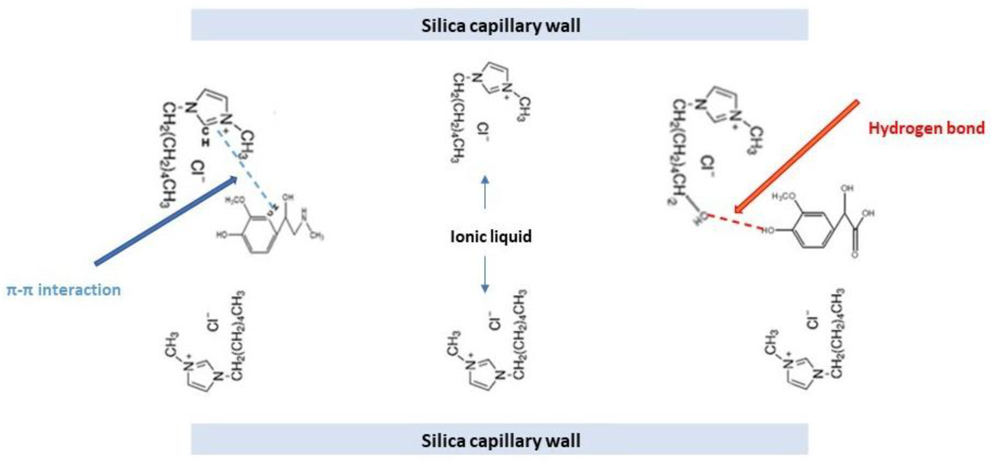
3.2. Dynamic Double Coating Effect of Examined ILs as Additives to Separation Buffer

As mentioned in the introduction, imidazole-based ILs, which were used as BGE additives in this work, perform numerous different functions during CE analysis, including participating in dynamic capillary modification, acting as ion-pair agents, contributing to the formation of the pseudostationary phase (modifiers micelles and microemulsions), providing additional interactions with analytes, and acting as chiral selectors in the separation of enantiomers. According to the literature, enriching the separation buffer with ILs at concentrations lower than the CMC value (<12.15 mM) dynamic modifies the fused silica capillary wall by creating a stable positive charge on the capillary surface, which ultimately increases the separation efficiency, as it prevents the adsorption of basic compounds. When the IL concentration is higher than the CMC value, another mechanism that improves the separation efficiency is induced. Here, the method’s selectivity improves as a result of the interactions between the analyzed compounds and the hydrophobic inner cavity in the formed micelle, as well as the π–π interactions with the imidazole ring [5]. Furthermore, a comparison between ILs and the traditionally used cationic surfactants indicated that the use of ILs can significantly increase the method’s sensitivity for various analytes [10].
The miscibility of ILs with a wide range of solvents makes them useful in CE as components in the separation buffer and as EOF modifiers due to their good electrical conductivity and slightly more viscous properties compared to organic solvents. In effect, low IL concentrations can be sufficient to significantly improve the electrophoretic separation. It should be highlighted that both the cations and anions of ILs may change the migration behavior of analytes, although the activity of the IL cations has a greater impact on the resolution in CE. In modifying the ionic strength of the BGE, the IL cations are able to change the EOF, which influences the migration times of the analytes and can improve the method’s separation efficiency. In addition, ILs influence the adsorption of IL cations on the capillary’s inner surface, which can reduce, or even reverse, the EOF. Both of the above-mentioned mechanisms improve the resolution of analytes. Thus, imidazolium-based ILs are widely recognized as electrophoretic flow modifiers that facilitate the dynamic modification of the capillary walls, which in turn ensures the stability of the EOF [4]. A diagram depicting the interaction between the examined ILs, the capillary wall, and analytes is shown in Figure 1.
In their review, Ali and co-workers present data summarizing the applications of ILs in CE for the analysis of a wide range of compounds [12]. It is worth noting that, among the 25 examined applications published between 2003 and 2021, only one focused on BAs. Furthermore, in CE, it is necessary to determine the separation mechanism when using ILs as a component of the BGEs. In general, the inclusion complexes are formed between the ILs and the separated molecules [2]. In their work, Yang et al. [30] and Zhang et al. [31] applied molecular modeling to establish the separation mechanism, observing multiple interactions between stereoisomers and hydroxy acid chiral ILs or spirocyclic ILs (hydrogen bonding, dipole–dipole interactions, π–π, host–guest inclusions, charge–charge, and hydrophobic interactions) [30,31].
Presently, the study of how dynamic coatings impact the capillary wall is a trend in CE research. As mentioned in the introduction, the addition of imidazolium-based ILs to the BGE ensures the positive charge of the silica capillary wall coating via the formation of a double layer. In this study, MEKC was applied to investigate how the double coating formed by adding [HMIM+Cl] and [HMIM+BF4] to the BGE at a concentration of 1 mM, impacted the migration times and separation efficiencies for HVA, VMA, NM, and M. As reported in our previous paper, adding ILs to the separation buffer at levels lower than the CMC value may result in interactions with the silica capillary wall (via hydrogen bonding) and with the analytes, thus affecting their electrophoretic mobility [4]. The results of this study demonstrated that adding the IL with a chloride anion at a concentration of 1 mM provided a better migration time repeatability. A comparison of the repeatability of the migration times (tm) attained for both tested ILs is presented in Table 1. As can be observed, the addition of [HMIM+Cl] to the BGE resulted in better tm repeatability compared to the addition of [HMIM+BF4]. The relative standard deviations (RSD) of the migration times achieved with [HMIM+Cl] were 0.209% for VMA, 0.336% for HVA, 0.156% for NM, and 0.121% for M; for [HMIM+BF4], these figures were 0.366% for VMA, 0.345% for HVA, 0.319% for NM, and 0.326% for M. These results may be related to [HMIM+Cl]’s slightly better solubility in the separation buffer. Although both anions enable good IL solubility in the BGE, the Cl anion has higher water miscibility than the BF4 anion, which affects the BGE’s viscosity and, consequently, the electro-osmotic flow (EOF) and the electrophoretic mobility of the analytes.
Therefore, [HMIM+Cl] was used to modify the inner wall of the capillary during the validation step and for the determination of the selected BAs in real urine samples. The results of the validation experiments demonstrated that, compared to [HMIM+BF4], adding [HMIM+Cl] to the BGE had a more beneficial effect on the migration times and peak heights of the tested compounds, thus confirming the validity of this IL and its positive impact on the separation efficiency for the targeted analytes (Table 2).

3.3. Optimization of Sample Preparation Procedure

Both the data in the literature and the results of our previous experiments indicated that small amounts of proteins, bile dyes, and nitrogenous metabolism products may be present in biological samples and can interfere with analyte signals [32]. Therefore, in this study, the sample matrix was first purified of ballast substances via the addition of ACN, followed by the application of SPE for the isolation and preconcentration of BAs. In SPE, the pH value of the sample matrix must be close to the pKa values of the targeted analytes to ensure their appropriate adsorption onto the selected extraction phase (SPE column).
In our study, ACN was first applied to deproteinize the urine samples, which was followed by the extraction of BAs via SPE with HLB cartridges and desorption with MeOH to isolate the analytes. Adjusting the sample’s pH to achieve the appropriate acidity or alkalinity positively influenced the adsorption of the analyzed compounds onto the HLB columns. HVA and VMA (pKa values of −4.9 and −4.1, respectively) were easily extracted from the samples at a pH of 5.5, whereas increasing the sample pH to 9.0 favored the extraction of M and NM (pKa values of 9.25 and 9.06, respectively). Additionally, the sample volume was increased through the addition of water to extend the time required for the sample to pass through the sorbent. This approach enabled longer absorption times, and thus, more efficient extraction results for the analytes of interest. The proposed method met the validation criteria, with the absolute recovery values for all analytes ranging between 91.7 and 120.0%, which aligns with the results of the previous studies [20,23,24].

3.4. Validation of the Developed Method

The proposed method was validated using artificial urine samples in accordance with ICH and FDA guidelines on the bioanalytical method validation [27,28]. A total of 1 mL of urine was used for both the calibration samples and the quality control (QC) samples. The urine samples were enriched with analytes at concentration levels of 10.0, 5.0, 1.0, 0.75, 0.5, and 0.25 µg/mL, while the urinary QCs were spiked with HVA, VMA, M, and NM at concentrations of 0.25, 1.0, and 10.0 µg/mL to obtain the low (LQC), middle (MQC), and high (HQC) concentration QC samples, respectively. Next, the samples were deproteinized and subjected to protein ballast removal, followed by SPE and quantitative analysis via the described MEKC method. Using the obtained data, six calibration curves were prepared, with the results being presented in Table 2 and in Figure 2.
The method’s selectivity was verified by applying it to analyze blank and spiked urine samples for the peak co-migration. The results of these tests indicated that the urine sample ballast did not interfere with the analyte signals.
An analytical procedure’s linearity is determined by its ability to produce results that are directly proportional to the concentration (amount) of analyte in the sample. To evaluate the proposed method’s linearity, six independent calibration curves were prepared within the same day. Validation concentrations ranging from 0.25–10 µg/mL and correlation coefficients (R2) higher than 0.996 were observed for all compounds of interest, thus indicating that the proposed method offered good linearity in the considered concentration range (Table 3).
The method’s LODs, LOQs, and precision were also evaluated based on six independent replicate calibration curves. The LOD was defined as the lowest measurable concentration at which the peak height could be calculated as three times the baseline noise; the LOQ was defined as the lowest concentration that could be detected with precision, as expressed by an RSD below 15%; and the method’s accuracy was determined as the percent of analytes detectable at nominal concentrations ranging from 80–120% and with a signal-to-noise ratio greater than 10. The LOD values obtained for HVA, VMA, M, and NM were 0.08 µg/mL, while the LOQ values were 0.25 µg/mL.
The method’s precision, expressed as RSD (%), was estimated by carrying out six independent assays of the tested urine samples during the same day (intra-day precision) and over five different days in the laboratory (intermediate precision) (Table 4). The intra-day precisions were below 12.57% for VMA, 8.82% for HVA, 9.35% for M, and 8.24% for NM, while the inter-day precision for all analytes was below 8.98%. The accuracies, expressed as the percentage of the ratio of the found to nominated concentrations of analyzed BAs, ranged between 94.62 and 100.89% for VMA, 93.68 and 105.91% for HVA, 98.49 and 103.64.% for M, and 97.25 to 103.45% for NM.
The absolute recoveries (AR) for each analyzed BA were determined at three concentration levels (10, 1.00, and 0.25 µg/mL) with six replicates being performed for each concentration. The ARs were assessed by comparing the peak highs obtained via extractions from human urine samples with those obtained from non-extracted urine samples containing the same concentrations of the compounds of interest. The results indicated mean absolute recoveries of greater than 91.7% for the targeted analytes (Table 3). These findings confirm that the proposed extraction procedure enables the evaluation of BA levels in human urine samples.
Additionally, a dilution study was performed to investigate whether the developed MEKC method can be applied to analyze samples at concentrations exceeding the upper limit of linearity after dilution. To this end, the calibration samples were diluted at concentrations of 15 and 20 µg/mL (each in six repetitions) and analyzed using the electrophoretic conditions described in Section 2.4. The results of these tests confirmed that the proposed MEKC method offered a precision of <5% and accuracy within the range of 95.42–104.56%. Thus, the developed method can be applied to analyze previously diluted samples that are present at concentrations beyond the upper limit of linearity. If the concentration of the tested analytes in the sample was above the method’s linearity range (concentrations between 10 and 20 µg/mL), the tested sample was diluted by 2-folds in 2 mM of sodium tetraborate (pH 8.7 [29]) prior to the MEKC analysis.
Therefore, the obtained validation results confirmed that the proposed SPE-MECK method satisfies the criteria for the bioanalytical method validation recommended by FDA and ICH guidelines [27,28].

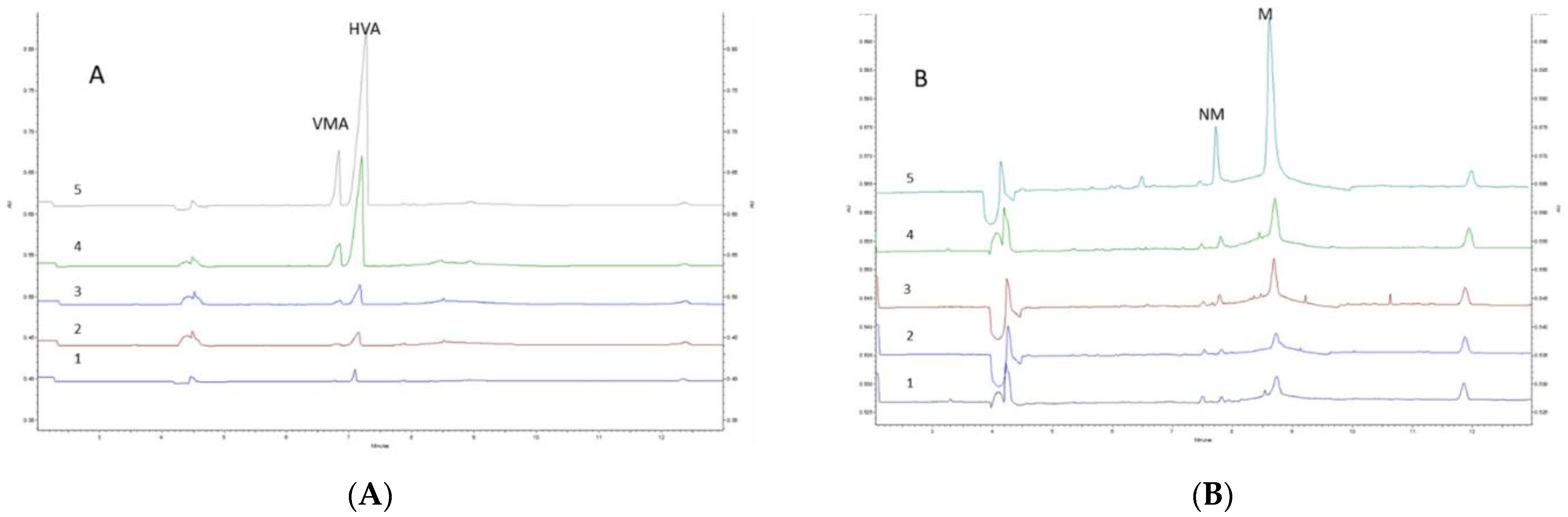
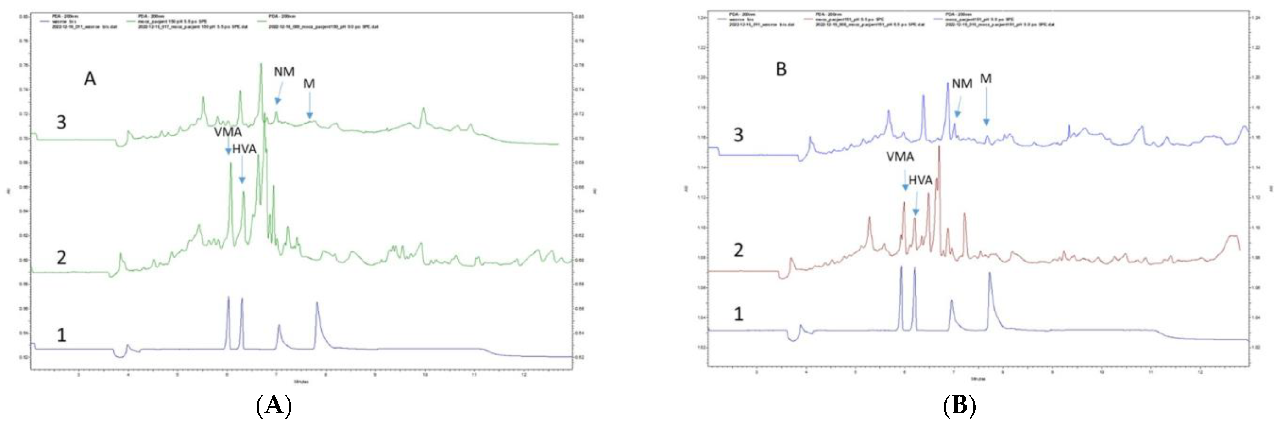
3.5. Application of the Developed Method in Real Urine Samples

The proposed IL-modified MEKC method was coupled with the DAD detection and applied for the analysis of real urine samples from eight pediatric patients with NBL. Figure 3A,B show the electropherograms obtained for Patient 5 and Patient 6 at two urine pH values (5.5 and 9.0), while Table 5 presents the measured free levels of VMA, HVA, M, and NM in the 8 urine samples. The results revealed concentrations ranging between 1.89–11.22 µg/mL for VMA, 1.48–16.27 µg/mL for HVA, 0.49–9.08 µg/mL for NM, and 0.16–12.27 µg/mL for M. Interestingly, elevated levels of both markers were detected in Patient 5, with VMA at 11.22 µg/mL and HVA at 13.77 µg/mL. The highest concentration of HVA (16.27 µg/mL) was determined in the urine samples from Patient 7, which also contained elevated values for NM (9.08 µg/mL) and M (8.47 µg/mL). Conversely, the highest level of M (12.27 µg/mL) was determined in the urine samples from Patient 1. Additionally, it is worth highlighting that the literature indicates that, after 12 months of age, the patient’s age at the time of diagnosis and a low VMA/HVA ratio are both associated with unfavorable prognoses [33].
Owing to the great variability in the concentration of markers, the VMA/HVA ratio was also defined. This parameter has a great diagnostic importance for NBL, as it enables more accurate estimates of the patient’s survival outlook and disease prognosis. Our data demonstrated that the VMA/HVA ratio varied from 0.33 to 1.27 for the studied oncologic patients, whereas the literature indicates that the average VMA/HVA ratio is equal to 3 for healthy controls [34]. Furthermore, the literature also indicates that the physiological ranges of M and NM in pediatric patients (0–18 years) are 0.045–0.202 µg/mL and 0.07–0.393 µg/mL, respectively [25]; however, the levels of M and NM in the studied group of pediatric patients exceeded these physiological ranges, which suggests that these two metanephrines could be used as potential markers of NBL.
Overall, the developed SPE-MEKC method enabled the determination of VMA, HVA, M, and NM levels in urine samples from pediatric NBL patients. The results of this research also demonstrate that the selected BAs can be useful biomarkers in the diagnosis of different types of NETs, including NBL. We intend to build on these findings in a future study by confirming the developed method’s performance using a larger group of patients.

4. Conclusions

The universality of ILs allows researchers to consider the use of different ionic combinations as a solution to the analytical limitations of CE-based techniques. This paper presented a comprehensive study examining the effectiveness of two alkylimidazolium IL structures as BGE additives for use in the determination of biogenic amines via an MECK technique. Since the selected ILs contained different anions and cations, this work was able to provide deep insights into how their structures influenced the dynamic coating of the capillary walls. Specifically, the results indicated that alkylimidazolium ILs containing anions such as [Cl] or [BF4] positively influenced the EOF parameters. The hexyl substituent in the imidazolium ring cation (in [HMIM+Cl] and [HMIM+BF4]) stabilized the inner wall of the silica capillary and ensured a high migration time repeatability for the BAs, especially for ILs with Cl anions. These findings clearly demonstrate the usefulness of [HMIM+Cl] in the analysis of VMA, HVA, M, and NM in urine samples. In addition, the experiments performed in this study illuminated how the sample’s pH value impacts the determination of BAs when ILs are added to the BGE. The obtained results demonstrated that a pH of 5.5 provided a better extraction efficiency for HVA and VMA, whereas increasing the sample pH to 9.0 improved the extraction of M and NM. Moreover, all the results relating to the use of ILs enabled the development of an analytical method for the determination of BAs in urine samples. The validation parameters for all the tested analytes fell within a range of 0.25–10 µg/mL, thus confirming the proposed method’s adherence to FDA and ICH criteria. Furthermore, the application of the proposed SPE-MEKC-UV method for the analysis of eight real urine samples from pediatric NBL patients confirmed its suitability for the routine monitoring of BAs. Finally, the concentration levels measured for the tested analytes (significantly elevated for M and NM, and a low VMA/HVA ratio) proved the necessity of monitoring BAs in biofluids from cancer patients and provided new data for use in future clinical and pharmacokinetic studies.

Author Contributions

Conceptualization, I.O. and N.K.; methodology, N.K., I.O. and N.T.; software, I.O., N.K. and P.K.; validation, N.K., I.O. and A.P.; formal analysis, I.O., N.K. and A.P.; investigation, I.O., N.K. and A.R.; resources, A.P. and N.K.; data curation, I.O.; writing—original draft preparation, N.K., I.O. and N.T.; writing—review and editing, N.K., I.O., A.P., P.K. and A.R.; visualization, I.O. and N.K.; supervision, I.O.; project administration, I.O. and T.B.; funding acquisition, I.O. and T.B. All authors have read and agreed to the published version of the manuscript.

Funding

Publication of this article was supported by the project, POWR.03.02.00-00-I014/17-00, co-financed by the European Union through the European Social Fund under the Operational Programme Knowledge Education Development 2014–2020.

Institutional Review Board Statement

The study was approved by the Local Ethics Committee of the Medical University of Gdańsk (No. NKBBN/93-626/2021 approved on 8 July 2021).

Informed Consent Statement

Informed consent was obtained from all subjects involved in the study.

Data Availability Statement

Not applicable.

Conflicts of Interest

The authors declare no conflict of interest.

References

  1. Zhang, Y.; Du, Y.; Yu, T.; Feng, Z.; Chen, J. Investigation of dextrin-based synergistic system with chiral ionic liquids as additives for enantiomeric separation in capillary electrophoresis. J. Pharm. Biomed. Anal. 2019, 164, 413–420. [Google Scholar] [CrossRef]
  2. Wang, X.; Li, K.; Yao, L.; Wang, C.; Schepdael, A. Recent advances in vitamins analysis by capillary electrophoresis. J. Pharm. Biomed. Anal. 2018, 147, 278–287. [Google Scholar] [CrossRef] [PubMed]
  3. Cheng, M.; Chen, Z. Recent advances in screening of enzymes inhibitors based on capillary electrophoresis. J. Pharm. Anal. 2018, 8, 226–233. [Google Scholar] [CrossRef] [PubMed]
  4. Kaczmarczyk, N.; Ciżewska, J.; Treder, N.; Miękus, N.; Plenis, A.; Kowalski, P.; Roszkowska, A.; Bączek, T.; Olędzka, I. The critical evaluation of the effects of imidazolium-based ionic liquids on the separation efficiency of selected biogenic amines and their metabolites during MEKC analysis. Talanta 2022, 238, 122997. [Google Scholar] [CrossRef]
  5. Bessonova, E.A.; Kartsova, L.A.; Moskvichev, D.O. Ionic Liquids in Electrophoretic Separation and Preconcentration Processes. J. Anal. Chem. 2021, 76, 1111–1118. [Google Scholar] [CrossRef]
  6. Mohamed, A.H.; Noorhisham, N.A.; Bakar, K.; Yahaya, N.; Mohamad, S.; Kamaruzaman, S.; Osman, H. Synthesis of imidazolium-based poly(ionic liquids) with diverse substituents and their applications in dispersive solid-phase extraction. Microchem. J. 2022, 178, 107363. [Google Scholar] [CrossRef]
  7. Ma, X.; Li, J.; Li, X.; Feng, Z.; Yang, X.; Liu, J.; Du, Y. L-Histidinium Chiral Ionic Liquid Functionalized β-Cyclodextrin as Chiral Selector in Capillary Electrophoresis. J. Chromatogr. Sci. 2021, 59, 388–395. [Google Scholar] [CrossRef]
  8. Guo, Y.; Yin, Z.; Sun, Y.; Yu, H. Separation and indirect ultraviolet detection of common fluorine-containing anions by ionic liquids in reversed-phase chromatography. J. Liq. Chromatogr. Relat. Technol. 2020, 43, 597–603. [Google Scholar] [CrossRef]
  9. Treder, N.; Bączek, T.; Wychodnik, K.; Rogowska, J.; Wolska, L.; Plenis, A. The influence of ionic liquids on the effectiveness of analytical methods used in the monitoring of human and veterinary pharmaceuticals in biological and environmental samples—Trends and perspectives. Molecules 2020, 25, 286. [Google Scholar] [CrossRef]
  10. Dharaskar, S.A.; Varma, M.N.; Shende, D.Z.; Yoo, C.K.; Wasewar, K.L. Synthesis, characterization and application of 1-butyl-3 methylimidazolium chloride as green material for extractive desulfurization of liquid fuel. Sci. World J. 2013, 2013, 395274. [Google Scholar] [CrossRef]
  11. Calabuig-Hernández, S.; Peris-García, E.; García-Alvarez-Coque, M.C.; Ruiz-Angel, M.J. Suitability of 1-hexyl-3-methylimidazolium ionic liquids for the analysis of pharmaceutical formulations containing tricyclic antidepressants. J. Chromatogr. A 2018, 1559, 118–127. [Google Scholar] [CrossRef] [PubMed]
  12. Ali, I.; Suhail, M.; Locatelli, M.; Ali, S.; Aboul-Enein, H.Y. Role of Ionic Liquids in Capillary Electrophoresis. Analytica 2022, 3, 236–250. [Google Scholar] [CrossRef]
  13. Flieger, J.; Flieger, M. Ionic liquids toxicity—Benefits and threats. Int. J. Mol. Sci. 2020, 21, 6267. [Google Scholar] [CrossRef] [PubMed]
  14. Qin, W.; Li, S.F.Y. Electrophoresis of DNA in ionic liquid coated capillary. Analyst 2003, 128, 37–41. [Google Scholar] [CrossRef] [PubMed]
  15. Bessonova, E.; Kartsova, L.; Gallyamova, V. Ionic liquids based on imidazole for online concentration of catecholamines in capillary electrophoresis. J. Sep. Sci. 2017, 40, 2304–2311. [Google Scholar] [CrossRef]
  16. Kolobova, E.; Kartsova, L.; Kravchenko, A.; Bessonova, E. Imidazolium ionic liquids as dynamic and covalent modifiers of electrophoretic systems for determination of catecholamines. Talanta 2018, 188, 183–191. [Google Scholar] [CrossRef]
  17. Hussain, A.; AlAjmi, M.F.; Hussain, I.; Ali, I. Future of Ionic Liquids for Chiral Separations in High-Performance Liquid Chromatography and Capillary Electrophoresis. Crit. Rev. Anal. Chem. 2019, 49, 289–305. [Google Scholar] [CrossRef]
  18. Jha, R.R.; Singh, C.; Pant, A.B.; Patel, D.K. Ionic liquid-based ultrasound assisted liquid-liquid microextraction for simultaneous determination of 15 neurotransmitters in rat brain, plasma and cell samples. Anal. Chim. Acta 2018, 1005, 43–53. [Google Scholar] [CrossRef] [PubMed]
  19. Kossakowska, N.; Olędzka, I.; Kowalik, A.; Miękus, N.; Kowalski, P.; Plenis, A.; Bień, E.; Kaczorowska, A.; Krawczyk, M.A.; Adamkiewicz-Drożyńska, E.; et al. Application of SPME supported by ionic liquids for the determination of biogenic amines by MEKC in clinical practice. J. Pharm. Biomed. Anal. 2019, 173, 24–30. [Google Scholar] [CrossRef] [PubMed]
  20. Lee, Y.R.; Lew, B.L.; Sim, W.Y.; Hong, J.; Chung, B.C. Serial hydrolysis for the simultaneous analysis of catecholamines and steroids in the urine of patients with alopecia areata. Molecules 2021, 26, 2734. [Google Scholar] [CrossRef] [PubMed]
  21. Grouzmann, E.; Centeno, C.; Eugster, P.J. Quantification of vanillylmandelic acid, homovanilic acid and 5-hydroxyindoleacetic acid in urine using a dilute-and-shoot and ultra-high pressure liquid chromatography tandem mass spectrometry method. Clin. Chem. Lab. Med. 2018, 56, 1533–1541. [Google Scholar] [CrossRef] [PubMed]
  22. Xiong, X.; Zhang, Y.; Zhao, R. Quantitative Measurement of Plasma Free Metanephrines by a Simple and Cost-Effective Microextraction Packed Sorbent with Porous Graphitic Carbon and Liquid Chromatography-Tandem Mass Spectrometry. J. Anal. Methods Chem. 2021, 2021, 8821276. [Google Scholar] [CrossRef] [PubMed]
  23. Lefeuvre, S.; Bois-Maublanc, J.; Mongeois, E.; Policarpo, V.; Farmaux, L.; Francia, T.; Billaud, E.M.; Got, L. Quantitation using HRMS: A new tool for rapid, specific and sensitive determination of catecholamines and deconjugated methanephrines metanephrines in urine. J. Chromatogr. B Anal. Technol. Biomed. Life Sci. 2021, 1166, 122391. [Google Scholar] [CrossRef] [PubMed]
  24. Hwang, N.; Chong, E.; Oh, H.; Cho, H.W.; Lee, J.W.; Sung, K.W.; Lee, S. Application of an lc–ms/ms method for the simultaneous quantification of homovanillic acid and vanillylmandelic acid for the diagnosis and follow-up of neuroblastoma in 357 patients. Molecules 2021, 26, 3470. [Google Scholar] [CrossRef]
  25. Zhou, G.S.; Yuan, Y.-C.; Yin, Y.; Tang, Y.-P.; Xu, R.-J.; Liu, Y.; Chen, P.-D.; Yin, L.; Duan, J.-A. Hydrophilic interaction chromatography combined with ultrasound-assisted ionic liquid dispersive liquid–liquid microextraction for determination of underivatized neurotransmitters in dementia patients’ urine samples. Anal. Chim. Acta 2020, 1107, 74–84. [Google Scholar] [CrossRef] [PubMed]
  26. Jong, W.H.A.; Eisenhofer, G.; Post, W.J.; Muskiet, F.A.J.; Vries, E.G.E.; Kema, I.P. Dietary influences on plasma and urinary metanephrines: Implications for diagnosis of catecholamine-producing tumors. J. Clin. Endocrinol. Metab. 2009, 94, 2841–2849. [Google Scholar] [CrossRef]
  27. FDA and CDER. Bioanalytical Method Validation Guidance for Industry Biopharmaceutics Bioanalytical Method Validation Guidance for Industry Biopharmaceutics Contains Nonbinding Recommendations. 2018. Available online: http://www.fda.gov/AnimalVeterinary/GuidanceComplianceEnforcement/GuidanceforIndustry/default.htm (accessed on 22 May 2018).
  28. European Medicines Agency. 2** Committee for Medicinal Products for Human Use (CHMP) Guideline on Bioanalytical Method Validation. Available online: www.ema.europa.eu/contact (accessed on 21 July 2011).
  29. Miękus, N.; Plenis, A.; Rudnicka, M.; Kossakowska, N.; Olędzka, I.; Kowalski, P.; Bączek, T. Extraction and preconcentration of compounds from the L-tyrosine metabolic pathway prior to their micellar electrokinetic chromatography separation. J. Chromatogr. A 2020, 1620, 461032. [Google Scholar] [CrossRef]
  30. Yang, X.; Du, Y.; Feng, Z.; Liu, Z.; Li, J. Establishment and molecular modeling study of maltodextrin-based synergistic enantioseparation systems with two new hydroxy acid chiral ionic liquids as additives in capillary electrophoresis. J. Chromatogr. A 2018, 1559, 170–177. [Google Scholar] [CrossRef]
  31. Zhang, Y.; Du, S.; Feng, Z.; Du, Y.; Yan, Z. Evaluation of synergistic enantioseparation systems with chiral spirocyclic ionic liquids as additives by capillary electrophoresis. Anal. Bioanal. Cem. 2016, 408, 2543–2555. [Google Scholar] [CrossRef]
  32. Woo, H.I.; Yang, J.S.; Oh, H.J.; Cho, Y.Y.; Kim, J.H.; Park, H.-D.; Lee, S.-Y. A simple and rapid analytical method based on solid-phase extraction and liquid chromatography-tandem mass spectrometry for the simultaneous determination of free catecholamines and metanephrines in urine and its application to routine clinical analysis. Clin. Biochem. 2016, 49, 573–579. [Google Scholar] [CrossRef]
  33. Strenger, V.; Kerbl, R.; Dornbusch, H.J.; Ladenstein, R.; Ambros, P.F.; Ambros, I.M.; Urban, C. Diagnostic and prognostic impact of urinary catecholamines in neuroblastoma patients. Pediatr. Blood Cancer 2007, 48, 504–509. [Google Scholar] [CrossRef] [PubMed]
  34. Miekus, N.; Kowalski, P.; Olędzka, I.; Plenis, A.; Bień, E.; Miękus, A.; Krawczyk, M.; Adamkiewicz-Drożyńska, E.; Bączek, T. Cyclodextrin-modified MEKC method for quantification of selected acidic metabolites of catecholamines in the presence of various biogenic amines. Application to diagnosis of neuroblastoma. J. Chromatogr. B Anal. Technol. Biomed. Life Sci. 2015, 1003, 27–34. [Google Scholar] [CrossRef] [PubMed]
Figure 1. Schematic of separation within the capillary containing [HMIM+Cl] and possible interaction with analytes.
Separations 10 00116 g001
Figure 2. Electropherograms of artificial urine samples spiked with analytes obtained after SPE-HLB at a sample pH 5.5 (A) and 9.0 (B). Concentration of analytes within the method’s linearity: 0.25 µg/mL (1), 0.5 µg/mL (2), 0.75 µg/mL (3), 5 µg/mL (4), and 10 µg/mL (5). Separation conditions: applied voltage—25 kV; capillary—60.2 cm and 75 µm i.d.; λ = 200 nm; injection time—15 s. The BGE consisted of 5 mM sodium tetraborate decahydrate, 50 mM SDS, 15% (v/v) MeOH, 150 mM boric acid, and 1 mM of [HMIM+Cl].
Separations 10 00116 g002
Figure 3. Electropherograms of urine samples from real patients after the SPE-HLB procedure: (A) Patient No. 5 and (B) Patient No. 6. (1) Standards of analytes in deionized water at a concentration of 10 µg/mL without SPE, (2) urine sample prepared at pH 5.5, and (3) urine sample prepared at pH 9.0. Separation conditions: applied voltage—25 kV; capillary—60.2 cm and 75 µm i.d.; λ = 200 nm; injection time—15 s. The BGE consisted of 5 mM sodium tetraborate decahydrate, 50 mM SDS, 15% (v/v) MeOH, 150 mM boric acid, and 1 mM of [HMIM+Cl].
Separations 10 00116 g003
Table 1. Migration times obtained from the analysis of the targeted BAs at a concentration of 10 µg/mL using BGE with the addition of ILs (n = 3).
BGE with ILMigration Time [Min]
VMAHVANMM
[HMIM+ Cl]
AVG6.536.867.638.52
SD0.0140.0230.0120.010
RSD0.2090.3360.1560.121
[HMIM+ BF4]
AVG6.616.947.097.83
SD0.0240.0240.0230.025
RSD0.3660.3450.3190.326
Table 2. Migration times and peak heights obtained during the analysis of the tested compounds using BGE without the addition of IL and with the addition of [HMIM+CL] (n = 3).
BGE without ILBGE with [HMIM+CL]
Migration Time [Min]HeightMigration Time [Min]Height
VMAAVGSDRSDAVGSDRSDVMAAVGSDRSDAVGSDRSD
48,917.00702.941.44 50,807411.150.81
28,806.00806.722.80 29,006.33540.731.86
5627.00271.764.83 6221.00204.543.29
6.660.111.714157.67375.979.04 6.750.091.384956.67283.155.71
2940.00342.3511.64 3317.67257.287.75
1371.33108.577.92 1516.0075.674.99
HVA HVA
44,152.003073.966.96 46,098.672797.486.07
23,941.332134.918.92 23,340.332053.318.80
6.990.121.734357.33432.779.93 7.090.111.505364.67264.974.94
3184.67401.2212.60 3720.00380.2310.22
2276.67361.6015.88 2194.0096.324.39
1047.00142.2013.58 1120.0091.708.19
NM NM
16,061.33653.484.07 18,309.00622.213.40
8001.00873.0910.91 8225.33511.646.22
8.210.101.191541.00200.1612.99 8.030.060.711658.67167.1310.08
1251.6745.893.67 1247.3312.280.98
764.6752.226.83 914.3335.113.84
360.3334.929.69 473.6736.817.77
M M
25,352.331608.506.34 25,782.001047.614.06
12,222.001597.2313.07 13,023.671133.568.70
9.280.111.202206.00363.6716.49 9.020.070.752688.33251.229.34
1714.33147.758.62 2088.67132.636.35
1072.33109.4010.20 1538.0085.215.54
503.6743.868.71 621.3345.187.27
Table 3. Quantitative performance of the proposed SPE-MEKC-UV method.
ParametersVMAHVANMM
Linearity [µg/mL]0.25–100.25–100.25–100.25–10
LOQ [µg/mL]0.250.250.250.25
LOD [µg/mL]0.080.080.080.08
R20.99630.99950.99820.9997
Regression equation y = 5113.5x + 1123.1y = 4637.5x + 80.25y = 1782.6x − 120.04y = 2664.2 + 3.03
AR [%]91.7–116.39101.30–117.97112.61–120.0091.83–113.76
Table 4. Inter- and intra-day accuracies and precisions for the studied BAs.
AnalytesNominal Concentration µg/mLIntra-Day AssayInter-Day Assay
Measured Concentration
µg/mL
CV (%)Measured Concentration
µg/mL
CV (%)
VMA109.781.7410.091.66
11.004.011/003.12
0.250.2212.570.248.98
HVA1010.123.3010.202.34
11.074.831.065.72
0.250.228.820.236.93
NM1010.353.2010.343.29
11.018.041.027.00
0.250.258.240.245.04
M1010.057.5810.265.20
11.019.351.046.76
0.250.245.100.253.22
Table 5. Measurements of free concentrations of VMA, HVA, M, and NM in urine samples from 8 pediatric cancer patients.
NoDiagnosisConcentration Found in Urine Sample (µg/mL)
pH 5.5pH 9.0
VMAHVAVMA/HVA
Ratio
NMM
1Neuroendocrine tumors, NBL1.891.481.274.4512.27 *
2n.mn.mn.m.n.mn.m
36.8112.41 *0.545.20n.m
43.5010.470.330.490.16
511.22 *13.77 *0.81n.on.m
63.957.000.563.270.87
7n.m16.27 *n.m.9.088.47
8n.mn.mn.m.1.400.66
Median3.9511.440.563.860.87
Min1.891.480.330.490.16
Max11.2216.271.279.0812.27
n.m.—not marked, concentration of the analyte in the urine sample below the method, LOD. *—Concentration above the high range of linearity determined after 2-fold dilution of the examined urine sample (see Section 3.4).
Disclaimer/Publisher’s Note: The statements, opinions and data contained in all publications are solely those of the individual author(s) and contributor(s) and not of MDPI and/or the editor(s). MDPI and/or the editor(s) disclaim responsibility for any injury to people or property resulting from any ideas, methods, instructions or products referred to in the content.

