Next Issue
Volume 9, September
Previous Issue
Volume 9, March


Dermatopathology, Volume 9, Issue 2 (June 2022) – 14 articles

Cover Story (view full-size image):

Malignant melanoma (MM) is derived through malignant transformation of pigment-producing cells in human skin. Highly differentiated melanoma cells usually maintain their pigment-producing function and, hence, immunohistochemically express melanocytic markers.

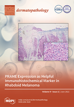
Rhabdoid melanoma, a rare, dedifferentiated variant of MM, is characterized by the potential loss of conventional melanocytic markers.  Since rhabdoid differentiation is associated with poor prognosis, immunohistochemical clues for accurate diagnosis are highly desirable. A relatively new marker molecule is PRAME (preferentially expressed antigen in melanoma), an antigen that is expressed in multiple malignant tumors, including MM.

In four new cases of primary rhabdoid melanomas, consistent nuclear positivity for PRAME underlines a possible immunohistochemical clue in the diagnosis of this rare entity. View this paper

  • Issues are regarded as officially published after their release is announced to the table of contents alert mailing list.
  • You may sign up for e-mail alerts to receive table of contents of newly released issues.
  • PDF is the official format for papers published in both, html and pdf forms. To view the papers in pdf format, click on the "PDF Full-text" link, and use the free Adobe Reader to open them.
Order results
Result details
Section
Select all
Export citation of selected articles as:
7 pages, 3318 KiB  
Case Report
Subungual Exostosis Presenting as a Pyogenic Granuloma-like Lesion with Reactive Myofibroblastic Proliferation in Two Young Women
by Rosanna Fox, Effie Katsarma, Nick Tiffin and Manuraj Singh
Dermatopathology 2022, 9(2), 196-202; https://doi.org/10.3390/dermatopathology9020024 - 20 Jun 2022
Viewed by 5736
Abstract
Subungual exostosis (SE) is a well-recognised benign proliferation of the distal phalanx most often seen in young adults and affecting the big toe. Possible triggers include previous trauma and chronic irritation or infection. We describe two atypical cases of SE in two young [...] Read more.
Subungual exostosis (SE) is a well-recognised benign proliferation of the distal phalanx most often seen in young adults and affecting the big toe. Possible triggers include previous trauma and chronic irritation or infection. We describe two atypical cases of SE in two young women presenting with pyogenic granuloma-like lesions clinically. Diagnostic biopsies were performed to confirm the diagnosis and excluded amelanotic melanoma. However, histology unexpectedly revealed reactive myofibroblastic proliferations mimicking nodular fasciitis overlying the SE. Given the atypical clinical presentation, the diagnosis was initially missed or not considered in both patients. They highlight two important points; the first is that SEs may present with pyogenic granuloma-like lesions clinically and that histological analysis is then required to exclude malignancy, particularly amelanotic melanoma. Secondly, that the histology will show a reactive myofibroblastic proliferation and if the sample is relatively superficial and pathologists are not aware of this potential reaction pattern, the underlying diagnosis of SE may be missed. Full article
(This article belongs to the Section Clinico-Pathological Correlation in Dermatopathology)
Show Figures

Figure 1

13 pages, 1317 KiB  
Article
An Up-to-Date Correlation of Epidemiological and Histopathological Characteristics of Basal Cell Carcinoma of the Skin in a County Hospital in Romania
by Iuliu Gabriel Cocuz, Maria Elena Cocuz, Adrian-Horațiu Sabău, Raluca Niculescu, Andreea Cătălina Tinca, Vlad Vunvulea, Corina Eugenia Budin, Mihaela Cornelia Șincu, Maria Cătălina Popelea and Ovidiu Simion Cotoi
Dermatopathology 2022, 9(2), 183-195; https://doi.org/10.3390/dermatopathology9020023 - 18 Jun 2022
Cited by 1 | Viewed by 2094
Abstract
Background and Objectives: Basal carcinoma of the skin (BCC) is part of the nonmelanoma skin cancer (NMSC) family and is the most frequently occurring type of skin cancer in humans. A combination of clinical and histopathological approaches is necessary in order to [...] Read more.
Background and Objectives: Basal carcinoma of the skin (BCC) is part of the nonmelanoma skin cancer (NMSC) family and is the most frequently occurring type of skin cancer in humans. A combination of clinical and histopathological approaches is necessary in order to establish the best treatment regime for patients who have been diagnosed with this type of cancer. The objective of the present study was to establish the statistical value of prediction for certain sociodemographic characteristics (age category and environment of origin) and histopathological parameters of the subjects that could be related to the incidence of diagnosis with certain histopathological subtypes of BCC. Materials and Methods: In order to verify the veracity of the established research hypotheses, we conducted a retrospective study based on the histopathological reports of 216 patients who were treated at the Pathology Department of Mureș Clinical County Hospital. Results: Cystic BCC is higher in patients who are older than 71 years of age, and the superficial multicentric and keratotic subtypes are more frequently diagnosed in urban areas. Patients who have been diagnosed with the superficial multicentric BCC subtype are not usually very old in contrast to the patients who tend to be diagnosed with the cystic BCC subtype. The nodular BCC subtype is positively associated with ulceration (p = 0.004); the superficial multicentric BCC subtype is positively associated with intra- and peritumoral inflammatory infiltrate (p = 0.022, p = 0.034) and negatively associated with ulceration (r = −0.218, p = 0.001). The infiltrative BCC subtype is positively associated with ulceration (p = 0.021), and the keratotic BCC subtype is positively associated with peritumoral inflammatory infiltrate (p = 0.02). Conclusions: Depending on each patient’s epidemiological and sociodemographic data, a pattern can be established regarding the appropriate clinical and treatment approaches for that patient, which can be supported based on the implications of the histopathological diagnostic. This can lead to an improvement in the patient’s quality of life and increased satisfaction with the provided medical services. Full article
(This article belongs to the Section Clinico-Pathological Correlation in Dermatopathology)
Show Figures

Figure 1

11 pages, 8670 KiB  
Article
Spongiotic Pattern in Pemphigus: A Retrospective Observational Single-Center Study
by Ivan Arni C. Preclaro and Yu-Hung Wu
Dermatopathology 2022, 9(2), 172-182; https://doi.org/10.3390/dermatopathology9020022 - 20 May 2022
Cited by 3 | Viewed by 5690
Abstract
Pemphigus is a chronic blistering disorder caused by autoantibodies that target desmosomal proteins in the epidermis. Acantholysis may be absent, and pemphigus may present only with spongiosis and vesiculation, thereby leading to a misdiagnosis of eczema. Herein, we conducted a retrospective, observational, single-center [...] Read more.
Pemphigus is a chronic blistering disorder caused by autoantibodies that target desmosomal proteins in the epidermis. Acantholysis may be absent, and pemphigus may present only with spongiosis and vesiculation, thereby leading to a misdiagnosis of eczema. Herein, we conducted a retrospective, observational, single-center study to establish a pattern of spongiosis in cases of pemphigus confirmed by direct immunofluorescence. Immunopathologically diagnosed pemphigus specimens from 2001 to 2020 were retrieved, and specimens with spongiosis were analyzed for the following features: vesiculation, acantholysis, spongiosis, inflammatory cells in the epidermis, and inflammation in the dermis. Cases of spongiotic dermatitis were used as control. Out of 99 immunopathologically diagnosed pemphigus specimens, 41 samples with spongiosis were identified. About one quarter of the specimens did not have acantholysis. Spongiosis in the middle to lower thirds of the perilesional epidermis (p = 0.030), exocytosis with either neutrophils or eosinophils (p = 0.016), dermal infiltrates composed of lymphocytes, eosinophils, and neutrophils (p = 0.012), and absence of Langerhans cell microabscesses (p < 0.001) were more common in pemphigus than control. Spongiosis in pemphigus may mimic eczema in patients without acantholysis. The subtle histological findings in this study provide diagnostic clues and suggest that further immunofluorescence should be performed to confirm pemphigus diagnosis. Full article
(This article belongs to the Special Issue Dermatopathology in Asia)
Show Figures

Figure 1

8 pages, 4053 KiB  
Review
Drug-Induced vs. Viral Maculopapular Exanthem—Resolving the Dilemma
by Sujay Khandpur and Rhea Ahuja
Dermatopathology 2022, 9(2), 164-171; https://doi.org/10.3390/dermatopathology9020021 - 07 May 2022
Cited by 3 | Viewed by 11084
Abstract
Maculopapular exanthem is a commonly encountered presentation in routine clinical practice, and differentiation between its two most common etiologies, i.e., viral- and drug-induced, often poses a diagnostic dilemma. Clinical, hematological and biochemical investigations are seldom reliable in distinguishing between a drug reaction and [...] Read more.
Maculopapular exanthem is a commonly encountered presentation in routine clinical practice, and differentiation between its two most common etiologies, i.e., viral- and drug-induced, often poses a diagnostic dilemma. Clinical, hematological and biochemical investigations are seldom reliable in distinguishing between a drug reaction and a viral exanthem. Certain key histopathological features such as the presence of a moderate degree of spongiosis, extensive basal cell damage with multiple necrotic keratinocytes and dermal infiltrate rich in eosinophils or lymphocytes and histiocytes may favor a drug exanthem, while distinctive epidermal cytopathic changes and lymphocytic vasculitis point towards a viral etiology. Similarly, notable immunohistochemical markers such as IL-5, eotaxin and FAS ligand may support a diagnosis of a drug-induced maculopapular eruption. Histopathological and immunohistochemical evaluations may help in distinguishing between the two etiologies when faced with a clinical overlap, especially in patients on multiple essential drugs when drug withdrawal and rechallenge is not feasible. Full article
(This article belongs to the Special Issue Dermatopathology in Asia)
Show Figures

Figure 1

6 pages, 2510 KiB  
Case Report
Scar Endometriosis: A Rare Cause of Abdominal Pain
by Rohit Nepali, Santosh Upadhyaya Kafle, Tarun Pradhan and Jiba Nath Dhamala
Dermatopathology 2022, 9(2), 158-163; https://doi.org/10.3390/dermatopathology9020020 - 05 May 2022
Cited by 2 | Viewed by 5710
Abstract
Scar endometriosis or incisional endometriosis is the presence of endometrial tissues with glands in the previous incision or scar. Its overall estimated incidence after post-cesarean and post-hysterectomy is 0.03–0.4% and 1.08–2%, respectively. The patient presents with non-specific symptoms such as cyclical abdominal pain [...] Read more.
Scar endometriosis or incisional endometriosis is the presence of endometrial tissues with glands in the previous incision or scar. Its overall estimated incidence after post-cesarean and post-hysterectomy is 0.03–0.4% and 1.08–2%, respectively. The patient presents with non-specific symptoms such as cyclical abdominal pain at the site of a previous surgical incision and scar and an abdominal lump with a cyclical increment in size, which is tender. The diagnosis is made only after the surgical excision with confirmation by histopathological analysis. We present the case of a 31-year-old female complaining of cyclical abdominal pain and a lump on the right side of a Pfannenstiel incision for five months. She had undergone two Lower Segment Caesarean Sections (LSCSs); the last surgery was eight months prior. Surgical excision was planned with the corresponding clinical features and radiological data. After the surgical excision, the sample was sent for histopathological examination, and scar endometriosis was diagnosed. Full article
(This article belongs to the Special Issue Dermatopathology in Asia)
Show Figures

Figure 1

10 pages, 88121 KiB  
Article
PRAME Expression as Helpful Immunohistochemical Marker in Rhabdoid Melanoma
by Valerie Glutsch, Marion Wobser, Bastian Schilling, Anja Gesierich, Matthias Goebeler and Hermann Kneitz
Dermatopathology 2022, 9(2), 148-157; https://doi.org/10.3390/dermatopathology9020019 - 02 May 2022
Cited by 6 | Viewed by 3337
Abstract
Background: Rhabdoid melanoma is a rare variant of malignant melanoma with characteristic cytomorphologic features. Due to the potential loss of conventional melanocytic markers, histopathologic diagnosis is often challenging. We hypothesize that immunostaining for PReferentially expressed Antigen in MElanoma (PRAME) might have the potential [...] Read more.
Background: Rhabdoid melanoma is a rare variant of malignant melanoma with characteristic cytomorphologic features. Due to the potential loss of conventional melanocytic markers, histopathologic diagnosis is often challenging. We hypothesize that immunostaining for PReferentially expressed Antigen in MElanoma (PRAME) might have the potential to uncover the melanocytic origin of these dedifferentiated tumors. Methods: Four cases of rhabdoid primary melanomas were assessed by immunohistochemistry for expression of PRAME and conventional melanocytic markers. Immunohistochemical expression patterns were analyzed in the rhabdoid primaries and, if available, associated metastases. Results: All four cases of rhabdoid primary melanomas showed a strong nuclear positivity for PRAME, while the expression of conventional melanocytic markers S100, MART-1, SOX-10 and HMB-45 was variable between the analyzed cases. Conclusions: In summary, we report four cases of rhabdoid primary melanoma with high to intermediate expression of PRAME despite the partial and variable loss of other melanocytic markers. Hence, PRAME might facilitate the recognition of this highly aggressive entity to avoid misdiagnosis due to histopathologic pitfalls. Full article
Show Figures

Figure 1

5 pages, 1930 KiB  
Case Report
Primary Cutaneous Gamma-Delta T-Cell Lymphoma Initially Diagnosed as Subcutaneous Panniculitis-like T-Cell Lymphoma with Dermatomyositis
by Chika Hirata, Kozo Nakai, Yusuke Kurasawa, Naoki Maekawa, Shuichi Kuniyuki, Keiko Yamagami, Masahiko Ohsawa and Daisuke Tsuruta
Dermatopathology 2022, 9(2), 143-147; https://doi.org/10.3390/dermatopathology9020018 - 29 Apr 2022
Cited by 1 | Viewed by 2940
Abstract
Primary cutaneous gamma-delta T-cell lymphoma (CGD-TCL) is a rare cutaneous lymphoma. Panniculitis-like T-cell lymphoma (SPTCL) has a better prognosis than CGD-TCL. SPTCL is sometimes associated with autoimmune disease. A 64-year-old Japanese female with a history of dermatomyositis presented with subcutaneous nodules on the [...] Read more.
Primary cutaneous gamma-delta T-cell lymphoma (CGD-TCL) is a rare cutaneous lymphoma. Panniculitis-like T-cell lymphoma (SPTCL) has a better prognosis than CGD-TCL. SPTCL is sometimes associated with autoimmune disease. A 64-year-old Japanese female with a history of dermatomyositis presented with subcutaneous nodules on the upper extremities and exacerbated dermatomyositis. A skin biopsy showed lobular panniculitis, a vacuolar interface change, and a dermal mucin deposit. Fat cells rimmed by neoplastic cells, fat necrosis, and karyorrhexis were observed. The atypical lymphoid cells showed CD3+, CD4−, CD8+, granzyme B+, CD20−, and CD56−. Polymerase chain reaction analysis demonstrated a T-cell receptor rearrangement. The patient was initially diagnosed with SPTCL, so the dose of prednisone was raised from 7.5 to 50 mg daily (1 mg/kg). After one month, erythematous nodules regressed, and muscle symptoms improved. Subsequently, prednisone was tapered, and cyclosporin A was added. After one year, the patient remained symptom-free and continued taking 7.5 mg prednisone and 100 mg cyclosporin A daily. Afterward, we immunostained skin samples with antibodies against TCR-ß and δ and found positive TCR-δ and negative TCR-ß. Therefore, we corrected the diagnosis to CGD-TCL, although the clinical course and the presence of dermatomyositis were reminiscent of SPTCL. Full article
(This article belongs to the Special Issue Dermatopathology in Asia)
Show Figures

Figure 1

7 pages, 7941 KiB  
Commentary
Conceptual Evolution and Current Approach to Spitz Tumors
by Carmelo Urso, Vincenzo De Giorgi and Daniela Massi
Dermatopathology 2022, 9(2), 136-142; https://doi.org/10.3390/dermatopathology9020017 - 26 Apr 2022
Cited by 2 | Viewed by 3668
Abstract
Over the past several decades, the study of Spitz neoplasms has flourished, with expanded conceptualization and refined terminology, providing a framework for the assessment and classification of Spitz nevi, atypical Spitz Tumors, and Spitz melanoma. Cancer genomics have generated concepts such as driver [...] Read more.
Over the past several decades, the study of Spitz neoplasms has flourished, with expanded conceptualization and refined terminology, providing a framework for the assessment and classification of Spitz nevi, atypical Spitz Tumors, and Spitz melanoma. Cancer genomics have generated concepts such as driver and passenger genes and clonal evolution, which can be applied to Spitz tumors. Herein, we provide a historical perspective, followed by a summary of current knowledge and clinical approaches for these challenging tumors. Full article
(This article belongs to the Special Issue In Memory of Raffaele Gianotti)
Show Figures

Figure 1

5 pages, 2415 KiB  
Case Report
“Lichenoid Granulomatous Pattern” in a Case of Lupus Vulgaris
by Chirag Desai and Ismail Shaikh
Dermatopathology 2022, 9(2), 131-135; https://doi.org/10.3390/dermatopathology9020016 - 20 Apr 2022
Viewed by 2466
Abstract
Lupus vulgaris is a one of the most common skin infections in the Indian subcontinent. Even today, it often creates a diagnostic dilemma for both clinicians and histopathologists. We describe a case of lupus vulgaris that showed lichenoid granulomatous inflammation in the dermis. [...] Read more.
Lupus vulgaris is a one of the most common skin infections in the Indian subcontinent. Even today, it often creates a diagnostic dilemma for both clinicians and histopathologists. We describe a case of lupus vulgaris that showed lichenoid granulomatous inflammation in the dermis. This pattern is not uncommon, but is rarely described in the literature as newer modalities currently take precedence in diagnosis. Our aim is to make clinicians and dermatopathologists aware of this pattern of inflammation seen in this common infection. Full article
(This article belongs to the Special Issue Dermatopathology in Asia)
Show Figures

Figure 1

9 pages, 2189 KiB  
Case Report
Aggressive Cutaneous Squamous Cell Carcinomas Following Treatment for Graft-versus-Host Disease: A Case Report and Review of Risk Factors
by Gehan A. Pendlebury, Michelle A. Bongiorno and Jeffrey N. Lackey
Dermatopathology 2022, 9(2), 122-130; https://doi.org/10.3390/dermatopathology9020015 - 31 Mar 2022
Cited by 2 | Viewed by 4733
Abstract
A 19-year-old female with a history of pre-B cell acute lymphocytic leukemia (ALL) presented with two aggressive cutaneous squamous cell carcinomas (C-SCC) in the right hand. The patient was diagnosed with pre-B cell ALL at four years of age. She underwent chemotherapy with [...] Read more.
A 19-year-old female with a history of pre-B cell acute lymphocytic leukemia (ALL) presented with two aggressive cutaneous squamous cell carcinomas (C-SCC) in the right hand. The patient was diagnosed with pre-B cell ALL at four years of age. She underwent chemotherapy with initial remission. However, recurrence of the pre-B cell ALL required an unrelated allogeneic cord hematopoietic stem cell transplant (alloHSCT). Post-transplant, the patient developed Graft-Versus-Host Disease (GVHD), which was treated with immunosuppressant therapy for six years until resolution. Fourteen years following the transplant, the patient developed a morbilliform drug eruption secondary to clindamycin. She consequently received prednisone treatment. During the treatment period, the patient developed a new ulcerated and tender nodule on the dorsal aspect of her right hand. Further histopathological biopsy confirmed the diagnosis of C-SCC, which required excision. Ten months following the excision, the patient developed an additional C-SCC nodule on the same right hand, separated by 2.6 cm from the prior C-SCC. She was referred for a ray resection procedure. This case illustrates a patient with multiple risk factors that may have contributed to the continued development of C-SCC. Such risk factors include: a prolonged course of immunosuppressant medications and voriconazole treatment. Additional research is needed to investigate the etiologies and risks of C-SCC development in patients who require a transplant and long-duration immunosuppressive therapy. Full article
Show Figures

Figure 1

11 pages, 1901 KiB  
Review
Histopathologic Features of Maculopapular Drug Eruption
by Madison Ernst and Alessio Giubellino
Dermatopathology 2022, 9(2), 111-121; https://doi.org/10.3390/dermatopathology9020014 - 30 Mar 2022
Cited by 6 | Viewed by 4717
Abstract
Background: Cutaneous adverse drug reaction (CADR) is common in both inpatient and outpatient clinical settings and has been associated with a large variety of medications. Drug reactions represent a significant burden to the healthcare system due to increased hospital stay durations and associated [...] Read more.
Background: Cutaneous adverse drug reaction (CADR) is common in both inpatient and outpatient clinical settings and has been associated with a large variety of medications. Drug reactions represent a significant burden to the healthcare system due to increased hospital stay durations and associated costs. Moreover, some of these reactions may be life-threatening. The most common clinical manifestation of a CADR is a maculopapular drug eruption (MDE). Due to its many clinical mimics and associations with a variety of histopathologic patterns, maculopapular drug eruption is difficult to definitively diagnose from both a clinical and histopathological perspective. Summary: We reviewed the clinical and histopathologic features of 327 cases of MDE from several studies in the literature and summarized characteristic histopathologic findings and their frequencies of occurrence. We found that the most common and suggestive histopathologic features of MDE were epidermal spongiosis, lymphocytic infiltrate, and occasional necrotic keratinocytes; interface change at the DEJ; superficial perivascular and interstitial lymphocytic infiltrate with or without eosinophils and neutrophils in the mid-to-deep dermis and mild papillary dermal edema; and dilation of superficial vessels. The presence of multiple histopathologic patterns within the same tissue specimen is also suggestive of MDE. This review and analysis suggest that a biopsy may improve the diagnostic accuracy by both establishing common and uncommon features associated with MDE and reviewing features that help to exclude other causes of maculopapular eruption. Key Message: Histopathologic criteria for the diagnosis of MDE, while not entirely specific, may aid in establishing a differential that includes a drug eruption. Thus, a biopsy can be a helpful diagnostic tool when MDE is suspected by demonstrating findings suggestive of MDE or by ruling out clinical mimics. However, biopsy results cannot be used in isolation as clinical-pathologic correlation is paramount in MDE. Full article
Show Figures

Figure 1

11 pages, 1598 KiB  
Review
Balloon Cell Melanoma: Presentation of Four Cases with a Comprehensive Review of the Literature
by Gerardo Cazzato, Eliano Cascardi, Anna Colagrande, Antonietta Cimmino, Giuseppe Ingravallo, Lucia Lospalluti, Paolo Romita, Aurora Demarco, Francesca Arezzo, Vera Loizzi, Miriam Dellino, Irma Trilli, Emilio Bellitti, Paola Parente, Teresa Lettini, Caterina Foti, Gennaro Cormio, Eugenio Maiorano and Leonardo Resta
Dermatopathology 2022, 9(2), 100-110; https://doi.org/10.3390/dermatopathology9020013 - 28 Mar 2022
Cited by 4 | Viewed by 3461
Abstract
Background: balloon cell melanoma represents less than 1% of all histological forms of malignant melanoma and represents a diagnostic challenge for the dermatopathologist. Methods: in this paper we present our cases of BCM found in our daily practice from 1 January 2008 to [...] Read more.
Background: balloon cell melanoma represents less than 1% of all histological forms of malignant melanoma and represents a diagnostic challenge for the dermatopathologist. Methods: in this paper we present our cases of BCM found in our daily practice from 1 January 2008 to 31 December 2021, and we conduct a review of the literature relating to this entity in the period from the first description, 1970, to early 2022. Results: four cases of melanoma balloon cell have been extrapolated from our electronic database, while in the review of the literature we have identified 115 cases of patients with primary and/or metastatic BCM. Conclusions: we believe that future studies with numerous case series are essential not only to increase the knowledge of the pathophysiology of this neoplasm but also to correctly evaluate the response of BCM patients to new oncological therapies. Full article
Show Figures

Figure 1

6 pages, 2778 KiB  
Case Report
A Case of Adult-Onset Eccrine Angiomatous Hamartoma—The Comparison with Epithelioid Hemangioma
by Mai Nishimura, Yoshiaki Matsushima, Yasuo Nakai, Koji Habe, Akinobu Hayashi and Keiichi Yamanaka
Dermatopathology 2022, 9(2), 94-99; https://doi.org/10.3390/dermatopathology9020012 - 25 Mar 2022
Viewed by 2966
Abstract
Eccrine angiomatous hamartoma (EAH) is a relatively rare benign skin disease characterized by the proliferation of eccrine sweat glands associated with capillary hemangioma and the proliferation of other skin elements such as adipose tissue, hair, and epidermis. The onset of the disease is [...] Read more.
Eccrine angiomatous hamartoma (EAH) is a relatively rare benign skin disease characterized by the proliferation of eccrine sweat glands associated with capillary hemangioma and the proliferation of other skin elements such as adipose tissue, hair, and epidermis. The onset of the disease is usually at birth or in childhood and tends to occur in the extremities of females, but it occurred in an adult male in this case. The patient was a 72-year-old man with a 12 × 12 mm light brown, elastic, slightly firm skin nodule on the flexor aspect of his right forearm. A biopsy revealed enlargement of blood vessels, sweat glands, sweat ducts, and erector spongiosum with both lumen dilation and narrowing, leading to the diagnosis of EAH. The histopathological features of EAH include a marked proliferation of microvessels, epithelial-like changes in vascular endothelial cells (such as enlarged nuclei), and infiltration of inflammatory cells, mainly lymphocytes and plasma cells. In adult-onset cases, EAH can be clinically difficult to distinguish from epithelioid hemangioma (EH), which differs in the predominance of microvascular proliferation and the presence of eosinophils in the infiltrating inflammatory cells. It can also be distinguished from EAH by the negative results of S100 and anti-EMA in immunohistological staining. In the current cases, we were able to differentiate the two cases from characteristic findings on HE staining. Full article
Show Figures

Figure 1

12 pages, 2452 KiB  
Article
Quantification of Immunohistochemically Stained Cells in Skin Biopsies
by Thomas Emmanuel, Mikkel Bo Brent, Lars Iversen and Claus Johansen
Dermatopathology 2022, 9(2), 82-93; https://doi.org/10.3390/dermatopathology9020011 - 24 Mar 2022
Cited by 1 | Viewed by 3609
Abstract
Immunohistochemical quantification of inflammatory cells in skin biopsies is a valuable tool for diagnosing skin diseases and assessing treatment response. The quantification of individual cells in biopsies is time-consuming, tedious, and difficult. In this study, we presented and compared two methods for the [...] Read more.
Immunohistochemical quantification of inflammatory cells in skin biopsies is a valuable tool for diagnosing skin diseases and assessing treatment response. The quantification of individual cells in biopsies is time-consuming, tedious, and difficult. In this study, we presented and compared two methods for the quantification of CD8+ T cells in skin biopsies from patients with psoriasis using both commercial software (Adobe Photoshop) and open-source software (Qupath). In addition, we provided a detailed, step-by-step description of both methods. The methods are scalable by replacing the CD8 antibody with other antibodies to target different cells. Moreover, we investigated the correlation between quantifying CD8+ cells normalized to area or epidermal length and cell classifications, compared cell classifications in QuPath with threshold classifications in Photoshop, and analyzed the impact of data normalization to epidermal length or area on inflammatory cell densities in skin biopsies from patients with psoriasis. We found a satisfactory correlation between normalizing data to epidermal length and area for psoriasis skin. However, when non-lesional and lesional skin samples were compared, a significant underestimation of inflammatory cell density was found when data were normalized to area instead of epidermal length. Finally, Bland–Altman plots comparing Qupath and Photoshop to quantify inflammatory cell density demonstrated a good agreement between the two methods. Full article
Show Figures

Figure 1

Previous Issue
Next Issue
Back to TopTop