Next Issue
Volume 8, June
Previous Issue
Volume 7, December


Dermatopathology, Volume 8, Issue 1 (March 2021) – 11 articles

Cover Story (view full-size image): 
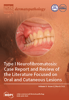
Neurofibromatosis type I (NF1) or von Recklinghausen’s disease is a very common genetic disease caused by the mutation of a tumor suppressor gene. Its principal clinical manifestation is benign cutaneous and oral neurofibromas that rarely undergo malignant transformation into a malignant peripheral nerve sheath tumor. Other dermatological clinical signs as well as ocular and skeletal abnormalities are also part of its diagnosis criteria. As NF1 has almost 100% penetrance, with a variable phenotypic expression, we believe it necessary for clinicians to be acquainted with oral alterations and include regular oral cavity examination during follow-up visits to those patients. In this study, we present a literature review of the oral and cutaneous manifestations of NF1 and describe a clinical case of an NF1 patient who presents cutaneous and oral lesions. View this paper.
  • Issues are regarded as officially published after their release is announced to the table of contents alert mailing list.
  • You may sign up for e-mail alerts to receive table of contents of newly released issues.
  • PDF is the official format for papers published in both, html and pdf forms. To view the papers in pdf format, click on the "PDF Full-text" link, and use the free Adobe Reader to open them.
Order results
Result details
Section
Select all
Export citation of selected articles as:
15 pages, 2345 KiB  
Article
Screening for Melanoma and Other Skin Cancer Shows a Higher Early Melanoma Incidence: Social Educational Program “Life Fear-Free”
by Lev Demidov, Igor Samoylenko, Nina Vand, Igor Utyashev, Irina Shubina and Igor Sinelnikov
Dermatopathology 2021, 8(1), 54-68; https://doi.org/10.3390/dermatopathology8010011 - 15 Mar 2021
Viewed by 2618
Abstract
Background: The screening program Life Fear-Free (LFF) aimed at early diagnosis of cutaneous melanoma (CM) was introduced in Samara, Chelyabinsk, Yekaterinburg, and Krasnodar (Russia) in 2019. Objectives: To analyze the impact of the program on early CM and non-melanoma skin cancer (NMSC) detection. [...] Read more.
Background: The screening program Life Fear-Free (LFF) aimed at early diagnosis of cutaneous melanoma (CM) was introduced in Samara, Chelyabinsk, Yekaterinburg, and Krasnodar (Russia) in 2019. Objectives: To analyze the impact of the program on early CM and non-melanoma skin cancer (NMSC) detection. Methods: According to the social educational campaign, people were informed about CM risk factors and symptoms and were invited for skin examination. The program planned to involve 3200 participants in total. Participants with suspicious lesions were invited for excisional biopsy. Results: 3143 participants, including 75.4% women, were examined for skin lesions. The average age of the participants was 43.7 years. Mostly skin phototypes II and III were registered (48.2% and 41.0%, respectively); 3 patients had CM, 15 had basal cell carcinoma, and 1 had Bowen’s disease, which were confirmed histologically. All detected melanomas had Breslow’s thickness of 1 mm. Conclusion: The participants showed high interest in early skin cancer detection programs. The incidence rate of CM and NMSCs among the program participants was higher than in general public. The early disease grade was proven for the detected CMs and NMSCs. The study has shown that it is important to continue such programs. Full article
Show Figures

Figure 1

5 pages, 716 KiB  
Case Report
Hair Shaft Examination: A Practical Tool to Diagnose Griscelli Syndrome
by Trinidad Montero-Vilchez, Alexandra Remon-Love, Jesús Tercedor-Sánchez and Salvador Arias-Santiago
Dermatopathology 2021, 8(1), 49-53; https://doi.org/10.3390/dermatopathology8010010 - 09 Mar 2021
Cited by 3 | Viewed by 2759
Abstract
Griscelli syndrome (GS) is a rare disease that is characterized by silvery hair and fair skin. It is included in congenital grey hair syndromes, a rare group of autosomal recessive disorders characterized by silvery grey hair and severe multisystem disorders, such as immune [...] Read more.
Griscelli syndrome (GS) is a rare disease that is characterized by silvery hair and fair skin. It is included in congenital grey hair syndromes, a rare group of autosomal recessive disorders characterized by silvery grey hair and severe multisystem disorders, such as immune system impairment, defects in immunological function, ocular and skeletal alterations, and nervous system defects. Herein, we report a rare case of GS type 1 and highlight the importance of a dermatological and hair examination to make an early diagnosis of these life-threatening diseases. Full article
(This article belongs to the Section Pediatric Dermatopathology)
Show Figures

Figure 1

4 pages, 10801 KiB  
Case Report
Petrified Ears: A Clue for Adrenal Insufficiency
by Sebastiano Recalcati and Fabrizio Fantini
Dermatopathology 2021, 8(1), 45-48; https://doi.org/10.3390/dermatopathology8010009 - 06 Mar 2021
Cited by 3 | Viewed by 3424
Abstract
Petrified ears is an uncommon clinical entity. It describes auricular cartilage hardening, due usually to ectopic calcification or, less commonly, ossification. The most common causes are frostbite and mechanical trauma. However, endocrinopathies have also been reported to cause ectopic calcium deposition through an [...] Read more.
Petrified ears is an uncommon clinical entity. It describes auricular cartilage hardening, due usually to ectopic calcification or, less commonly, ossification. The most common causes are frostbite and mechanical trauma. However, endocrinopathies have also been reported to cause ectopic calcium deposition through an unknown mechanism. Addison’s disease is the systemic disease most frequently associated, but the exact pathogenesis remains unclear. Patients are usually asymptomatic, and the diagnosis is made incidentally when rigid helices are noted on palpation and can be confirmed by radiological imaging. A biopsy can also help to differentiate between calcification and ossification. The presence of this condition may be a useful clinical sign, which in some cases precedes the development of endocrinopathies by many years. We report on a case, and we review the current literature. Full article
Show Figures

Figure 1

5 pages, 7095 KiB  
Case Report
Advanced Cutaneous Leiomyosarcoma of the Forearm
by Gerardo Cazzato, Maria Chiara Sergi, Sara Sablone, Anna Colagrande, Teresa Lettini, Francesco Fanelli, Umberto Orsini and Giuseppe Ingravallo
Dermatopathology 2021, 8(1), 40-44; https://doi.org/10.3390/dermatopathology8010008 - 27 Feb 2021
Cited by 5 | Viewed by 2625
Abstract
Leiomyosarcoma is a malignant smooth muscle neoplasm, which is traditionally divided into superficial and deep tumors. Superficial leiomyosarcomas are quite rare entities, accounting for approximately 7% of soft tissue neoplasms and 0.04% of all cancers. Here we describe a rare case of advanced [...] Read more.
Leiomyosarcoma is a malignant smooth muscle neoplasm, which is traditionally divided into superficial and deep tumors. Superficial leiomyosarcomas are quite rare entities, accounting for approximately 7% of soft tissue neoplasms and 0.04% of all cancers. Here we describe a rare case of advanced primary cutaneous leiomyosarcoma (PCL) in a 93-year-old woman, highlighting the considerable size of the lesion and the correct surgical and oncological management. The clinical story began about 4 years ago, and the neoplasia was treated only with local radiotherapy, but the patient suffered from a dramatic volumetric increase of the right arm sarcoma one year ago. Then, an amputation of the limb was performed without following adjuvant chemotherapy. Currently, she does not show signs of recurrence and is in good shape. Full article
Show Figures

Figure 1

3 pages, 518 KiB  
Case Report
Anti-PD-1-Induced Hidradenitis Suppurativa
by Alexia Maillard, Damien Pastor and Rastine Merat
Dermatopathology 2021, 8(1), 37-39; https://doi.org/10.3390/dermatopathology8010007 - 25 Feb 2021
Cited by 9 | Viewed by 3239
Abstract
Mucocutaneous adverse events are commonly observed under immune checkpoint inhibitors (ICIs) therapy. Here, we report the case of a 43-year-old male patient with a stage IIIC melanoma disease who developed hidradenitis suppurativa (HS) three months after the beginning of an anti-PD-1 (nivolumab) adjuvant [...] Read more.
Mucocutaneous adverse events are commonly observed under immune checkpoint inhibitors (ICIs) therapy. Here, we report the case of a 43-year-old male patient with a stage IIIC melanoma disease who developed hidradenitis suppurativa (HS) three months after the beginning of an anti-PD-1 (nivolumab) adjuvant therapy. The patient had no comorbidities other than obesity and severe acne during adolescence. After an unsuccessful course of lymecycline while he was still treated with nivolumab, he gradually improved under zinc gluconate therapy and, more importantly, after nivolumab cessation. HS is a recurrent follicular inflammatory disease in the apocrine gland-bearing areas of the body often associated with obesity, metabolic syndrome, tobacco smoking, inflammatory bowel diseases, psoriasis, and arthritis. In our patient, the latency period between drug initiation and onset of HS symptoms and the improvement after immunotherapy discontinuation, argued strongly in favor of an anti-PD-1-induced HS. Anti-PD-1 therapies often trigger T cells-mediated adverse events that mimic Th17-mediated inflammatory and neutrophilic diseases. We suggest that HS, as other pustular skin reactions and ICIs-induced neutrophilic colitis, can be part of the anti-PD-1 mucocutaneous adverse event spectrum. Full article
Show Figures

Figure 1

4 pages, 538 KiB  
Case Report
A Case of Bullous Rash Apparently Triggered by Meningococcal and Rotavirus Vaccines in an Infant: Focus on Infantile Bullous Pemphigoid
by Iria Neri, Valeria Evangelista, Alba Guglielmo, Andrea Sechi and Annalucia Virdi
Dermatopathology 2021, 8(1), 33-36; https://doi.org/10.3390/dermatopathology8010006 - 23 Feb 2021
Cited by 4 | Viewed by 2387
Abstract
Bullous pemphigoid (BP) is an autoimmune bullous disease and is a rare condition in childhood. Acquired tense acral bullae and fixed urticarial annular lesions on the trunk are diagnostic clues of infantile BP. Diagnosis is supported by immunosorbent assay (IgG anti-BP180 and BP230) [...] Read more.
Bullous pemphigoid (BP) is an autoimmune bullous disease and is a rare condition in childhood. Acquired tense acral bullae and fixed urticarial annular lesions on the trunk are diagnostic clues of infantile BP. Diagnosis is supported by immunosorbent assay (IgG anti-BP180 and BP230) and direct immunofluorescence (linear deposition of IgG at the dermo-epidermal junction). Topical and/or systemic corticosteroids are the first-line treatment. The prognosis is good with a self-limited clinical course. Differential diagnoses include impetigo and other bullous diseases in children, such as dermatitis herpetiformis, linear IgA bullous dermatosis and erythema multiforme. The etiopathogenesis is still unknown, and the role of antigen stimuli such as infections, drugs and vaccination is still debated. Full article
Show Figures

Figure 1

4 pages, 968 KiB  
Case Report
Primary Dermal Melanoma: A Rare Clinicopathological Variant Mimicking Metastatic Melanoma
by Oriana Simonetti, Elisa Molinelli, Valerio Brisigotti, Donatella Brancorsini, Davide Talevi and Annamaria Offidani
Dermatopathology 2021, 8(1), 29-32; https://doi.org/10.3390/dermatopathology8010005 - 01 Feb 2021
Cited by 3 | Viewed by 3454
Abstract
Primary dermal melanoma (PDM) is a rare distinct variant of cutaneous melanoma, predominantly occurring on the extremities of young or middle-aged adults. In comparison to conventional melanoma, PDM is characterized by unexpectedly prolonged survival and long-term survival. Thus, correct identification of this variant [...] Read more.
Primary dermal melanoma (PDM) is a rare distinct variant of cutaneous melanoma, predominantly occurring on the extremities of young or middle-aged adults. In comparison to conventional melanoma, PDM is characterized by unexpectedly prolonged survival and long-term survival. Thus, correct identification of this variant is crucial to avoid potential misdiagnosis and establish correct treatment and follow-up. In addition, no consensus and specific guidelines exist on the management of this peculiar subtype of cutaneous melanoma. Full article
(This article belongs to the Section Clinico-Pathological Correlation in Dermatopathology)
Show Figures

Figure 1

4 pages, 2358 KiB  
Case Report
Atypical Fibroxanthoma-Like Amelanotic Melanoma: A Diagnostic Challenge
by Gerardo Cazzato, Anna Colagrande, Antonella Cimmino, Giovanni Liguori, Teresa Lettini, Gabriella Serio, Giuseppe Ingravallo and Andrea Marzullo
Dermatopathology 2021, 8(1), 25-28; https://doi.org/10.3390/dermatopathology8010004 - 12 Jan 2021
Cited by 6 | Viewed by 3184
Abstract
Atypical fibroxanthoma-like amelanotic melanoma is a very rare variant of melanoma that can, if not correctly recognized and framed, lead to diagnostic errors that can potentially cause problems of extreme relevance to patients. Correct knowledge of this entity and the execution of adequate [...] Read more.
Atypical fibroxanthoma-like amelanotic melanoma is a very rare variant of melanoma that can, if not correctly recognized and framed, lead to diagnostic errors that can potentially cause problems of extreme relevance to patients. Correct knowledge of this entity and the execution of adequate immunohistochemical investigations are the basic conditions for the correct management of this lesion. We report on a case of atypical fibroxanthoma-like amelanotic melanoma, which clinically simulated a fibrohistiocytic lesion, and which created differential diagnostic problems, and finally, we conduct a short review of the literature. Full article
(This article belongs to the Section Clinico-Pathological Correlation in Dermatopathology)
Show Figures

Figure 1

8 pages, 4340 KiB  
Case Report
Type I Neurofibromatosis: Case Report and Review of the Literature Focused on Oral and Cutaneous Lesions
by Samanta Buchholzer, Raùl Verdeja and Tommaso Lombardi
Dermatopathology 2021, 8(1), 17-24; https://doi.org/10.3390/dermatopathology8010003 - 07 Jan 2021
Cited by 5 | Viewed by 6249
Abstract
Neurofibromatosis type 1 (NF1) is a common genetic disease whose dermatological lesions are at the forefront of its development. Cutaneous manifestations include café au lait spots, intertriginous freckling, and neurofibromas which appear during childhood and adolescence and are part of the clinical criteria [...] Read more.
Neurofibromatosis type 1 (NF1) is a common genetic disease whose dermatological lesions are at the forefront of its development. Cutaneous manifestations include café au lait spots, intertriginous freckling, and neurofibromas which appear during childhood and adolescence and are part of the clinical criteria to diagnose NF1. However, it is only recently that oral manifestations have been highlighted in many studies as frequently associated to NF1. This article aims to review oral and cutaneous manifestations related to NF1 and to report a case of a 51-year-old male with skin and oral neurofibromas related to NF1. Our patient presented with lesions on the gingiva, a rare localization that takes a hypertrophic form mimicking other various pathological conditions. Although not frequent, malignant transformation in NF1, especially regarding plexiform neurofibromas, is well established. Patients with NF1 therefore have regular follow-ups based on clinical examination, as sarcomatous transformation brings an extremely poor prognosis, recurrences and distant metastasis being common. Full article
Show Figures

Figure 1

7 pages, 2688 KiB  
Case Report
Scrotal Lymphangiectasia with Penile Elephantiasis in Underlying Lymphatic Filariasis—Challenging the Diagnostic Mind! A Case Report
by Tejas Vishwanath, Angela Nagpal, Sunil Ghate and Aseem Sharma
Dermatopathology 2021, 8(1), 10-16; https://doi.org/10.3390/dermatopathology8010002 - 01 Jan 2021
Cited by 2 | Viewed by 8911
Abstract
Background: A plethora of diseases manifest as acquired genital lymphangiectasias which clinically manifest as superficial vesicles. They range from infections such as tuberculosis to connective tissue diseases such as scleroderma and even malignancy. Amongst infectious etiologies, lymphatic filariasis leads as the cause for [...] Read more.
Background: A plethora of diseases manifest as acquired genital lymphangiectasias which clinically manifest as superficial vesicles. They range from infections such as tuberculosis to connective tissue diseases such as scleroderma and even malignancy. Amongst infectious etiologies, lymphatic filariasis leads as the cause for lymphatic obstruction. Despite this, acquired lymphangiectasias due to this cause are not commonly reported. An unusual case of acquired scrotal lymphangiectasia secondary to filariasis is detailed in this paper with dermoscopic and histologic findings. Methods: A 65-year-old male farmer presented with multiple, asymptomatic vesicles over the scrotum with thickened scrotal and penile skin that had occurred for six years. He gave past history of intermittent fever and milky urine, was diagnosed with filariasis and treated with diethylcarbamazine for a year, four years previously. Systemic complaints abated but the peno-scrotal lesions did not. Results: Polarized dermoscopy revealed multiple skin-colored nodules and translucent pale blue lacunae over the scrotum. A few radially arranged linear irregular vessels were noted over the nodules. On histopathology, multiple ectatic lymphatics were noted in the mid and upper dermis with acanthosis and superficial perivascular lymphocytes. Peripheral smear revealed eosinophils; however, microfilariae could not be detected despite repeated diethylcarbamazine provocation and night smears being taken. The findings were compatible with acquired scrotal lymphangiectasia secondary to treated lymphatic filariasis. Local hygiene was advised; however, procedural treatments were refused by the patient. Conclusion: Herein, we report an unusual case of acquired scrotal lymphangiectasia of the scrotum secondary to treated lymphatic filariasis. Very few similar reports exist. To the best of our knowledge, dermoscopic features of this condition have not been elucidated before. This case, detailing an uncommon manifestation of a common disease (filariasis), demonstrates the importance of careful history taking and examination. This was especially so in the present case since only circumstantial evidence of filariasis was noted in investigations. There is a need to heighten awareness of this unusual condition amongst physicians especially if the patient hails from an area endemic for filariasis. Full article
Show Figures

Figure 1

9 pages, 3857 KiB  
Case Report
Low-Grade Myofibroblastic Sarcoma of the Oral Cavity: A Report of Three Cases Illustrating an Emerging Disease in Children
by Primali Rukmal Jayasooriya, Chamara Athukorala, Manjula Attygalla, Balapuwaduge Ranjit Rigobert Nihal Mendis and Tommaso Lombardi
Dermatopathology 2021, 8(1), 1-9; https://doi.org/10.3390/dermatopathology8010001 - 01 Jan 2021
Cited by 6 | Viewed by 3346
Abstract
Low-grade myofibroblastic sarcoma (LGMS) is a mesenchymal tumor of myofibroblasts that occurs more frequently in adults. A series of three cases is presented to illustrate that LGMS may also occur within the oral cavity in children and adolescents. The first case (Case 1) [...] Read more.
Low-grade myofibroblastic sarcoma (LGMS) is a mesenchymal tumor of myofibroblasts that occurs more frequently in adults. A series of three cases is presented to illustrate that LGMS may also occur within the oral cavity in children and adolescents. The first case (Case 1) occurred intra-osseously in the mandible, while the remaining two presented as gingival swellings and were purely restricted to soft tissue (Cases 2 and 3). The intra-osseous lesion arose in a 7-year-old girl, whereas the gingival lesions were observed in a 12-year-old girl (Case 2) and a 13-year-old boy (Case 3). Histopathologically, all cases were composed of spindle shaped cells arranged into long fascicles showing mild to moderate degree of nuclear atypia. Ki-67 (MIB-1) proliferation activity was relatively low, amounting to 3–5% in all cases. Immunohistochemically, all cases showed smooth muscle actin (SMA) positivity in spindle cells, while desmin, beta catenin, cytokeratin, and CD34 were negative, resulting in a diagnosis of LGMS. In conclusion, current series of three cases of LGMSs that occurred in the oral cavity in a child and two adolescent patients is presented to highlight an emerging disease that requires additional data for further characterization. Full article
Show Figures

Figure 1

Previous Issue
Next Issue
Back to TopTop