Article

Evaluation of Rate of Adhesion of Lactobacillus namurensis Strain GYP-74 to Porous Fine Ceramics

1
Department of Marine Biology and Environmental Sciences, Faculty of Agriculture, University of Miyazaki, Miyazaki 889-2192, Japan
2
Center for Collaborative Research & Community Cooperation, University of Miyazaki, Miyazaki 889-2192, Japan
3
Department of Mechanical Systems Engineering, University of Miyazaki, Miyazaki 889-2192, Japan
4
Kuroki Kensetsu Co. Ltd., Miyazaki 883-0015, Japan
5
Kimura Tsukemono Miyazaki Kougyou Co. Ltd., Miyazaki 889-1403, Japan
*
Author to whom correspondence should be addressed.
Processes 2021, 9(4), 658; https://doi.org/10.3390/pr9040658
Submission received: 22 February 2021 / Revised: 28 March 2021 / Accepted: 5 April 2021 / Published: 9 April 2021
(This article belongs to the Special Issue Novel Processes for Development of Fermentation-Based Products)

Abstract

:
This study aimed to evaluate the ratio of adherence of lactic acid bacteria (LAB) to porous fine ceramics in order to develop a novel LAB-rich pickle container for the production of functional fermented vegetables. Some LAB were isolated from the salted rice bran used for pickling (Nukadoko in Japanese). These isolates were classified in Lactobacillus namurensis by phylogenetic analysis. Some pottery-shard (PS) samples were prepared by varying the mixing rate of polyacetal (POM) resin to clay (0–30% (v/v)) and the burning temperature (1000 °C or 1100 °C). A test of the adherence of strain GYP-74 to the PSs was performed. The results showed that the adherence rate was significantly higher in the PSs burned at 1100 °C as compared with those burned at 1000 °C. A pore distribution analysis showed that pore sizes of less than a few µm and pore sizes in the range of a few µm to a few hundred µm were mainly distributed in the PSs without and with POM, respectively. X-ray diffraction analysis showed that both PSs with and without POM contained quartz and hematite. The PSs burned at 1000 °C and 1100 °C specifically contained microcline and mullite, respectively. This study revealed the basal information regarding what makes PSs adequate for LAB adhesion.

1. Introduction

Miyazaki Prefecture, Japan, set forth the food business promotion plan in 2013 and set the efficient utilization of local foods including vegetables with added value as a basic objective. Under this plan, the creation of novel local industry in the field of fisheries is expected to occur based on an industry–academia–government collaboration. In Miyazaki, the production of white radish is ranked fifth in Japan, and fermented radishes in a salted rice bran bed (Nukazuke in Japanese) are a very well-known food. Nukazuke is fermented by various microbes in salted rice bran (Nukadoko in Japanese). The storage of the Nukadoko depends on natural fermentation by microbes. In other words, Nukadoko produces the complex microflora. It has been reported that the bacterial flora in Nukadoko mainly contains lactic acid bacteria (LAB) [1,2]. Through the fermentation of LAB, a fermented flavor which is mainly based on the formation of volatile organic acids was formed and coupled with an umami flavor. In rice bran products, LAB contributes to the taste and long-term preservation. Surprisingly, Nukadoko can be kept without spoilage for 100 years [1].
Eating fermented food containing sufficient LAB cells is considered to be the ingestion of “probiotics”. Some species of LABs have been identified as probiotics for various host animals including aquacultured species [3,4,5]. Generally, probiotics are defined as live microbial feed additives that improve the microbial condition of the host animal’s gastrointestinal tract [6]. Probiotics include components of microbial cells that stimulate the immune system against pathogens [7]. Probiotics can improve the health of the host in mammalians, including humans, and in aquaculture species [8,9]. The effects of probiotics in humans have been reported to include improvement in the resistance to infectious disease, the modification of blood lipid profiles and blood pressure, growth enhancement for undernourished young children, the prevention of vaginal infections and inflammatory bowel diseases, inhibition of diarrhea, and protection against Helicobacter pylori colonization of the stomach [10,11,12,13,14]. In addition, some LAB species produce functional and bioactive substances such as γ-amino butyric acids (GABA). GABA is synthesized from L-glutamic acids by L-glutamate decarboxylase [15] and provides beneficial effects in humans such as stress relief and decreasing the blood pressure [16].
This evidence indicates that the ingestion of fermented foods such as Nukazuke is good for human health. Thus, we planned to develop a specific fermentation container to enrich the LAB cells to efficiently and sufficiently supply LAB to consumers. The goal was to develop a container with a surface to which LAB can easily adhere. Bacterial adhesion to a solid surface is the first step in the production of extracellular polymeric substances (EPS) and the formation of biofilm and colonies [17,18]. EPS are especially well known to be an important factor in bacterial adhesion in bridging between the cell surface and the surface of the materials. Generally, bacteria easily or willingly adhere to rough surfaces or to the insides of pores. It is well known that surface roughness is an important factor for the attachment of bacterial cells to surfaces prior to biofilm formation. In the first step, bacteria migrate towards the materials, and the bacterial cells move to the surface of the materials by their motility or by passive moving based on the Brownian motion [19,20,21]. In the next step, bacterial cells move close to the surface, and irreversible binding between cells and surface occurs. Similarly, the bacterial attachment is very complex due to the hydrophobicity and chemistry of the surface, flow conditions, and cell structures [20,22,23,24]. In case of Staphylococcus aureus, the presence of nanoscale roughness on a steel surface induced an increase in bacterial attachment to a steel surface [25]. In the field of wastewater treatment, selection of materials is a key factor for induction of initial formation of bacterial biofilms. In the case of four bacterial strains (Alcaligenes faecalis, Cytophaga hutchinsonii, Bacillus subtilis and Escherichia coli), carbon fiber is considered as one of the support materials for biofilms for wastewater treatment [26]. Flagella are known to be a bacterial adhesive factor. Song et al. reported that the flagella play a role in increasing the contact with the surface [27]. On the other hand, research on protecting against the adherence of harmful bacterial by reducing the contact area has been carried out. These findings indicated that the bacterial adhesion to materials, such as the container used for bacterial fermentation, can be increased by controlling the roughness and pore size of the surface and by enlarging the contact area.
This study aimed to examine the characteristics of pottery that are adequate for LAB adhesion. The adherence ratio of the LAB isolated from the salted rice bran bed was evaluated with pottery shards (PS) with different resin sizes and contents, and that were fired at different temperatures. In this study, especially, we focused on porous ceramics as the material and the relationship between the pore sizes and the adherence ratio of LAB.

2. Materials and Methods

2.1. Isolation of LAB

The salted rice bran used for pickling (Nukadoko) maintained by Kimura Tsukemono Miyazaki Kougyo Co. Ltd., Miyazaki, Japan, was used for the isolation of LAB. Samples to isolate LAB were suspended in a sterile physiological saline solution (0.85% NaCl (w/v)), shaken vigorously and left for 10 min at room temperature. The solution, except for precipitate, was serially diluted in a physiological saline solution, and the diluted samples were plated on de Man, Rogosa and Sharpe (MRS) agar plate medium consisting of 2 g glucose, 0.5 g yeast extract, 1 g peptone, 1 g meat extract, 0.5 g sodium acetate, 0.2 g ammonium citrate, 10 mg MgSO4·7H2O, 5 mg MnSO4·5H2O, 0.2 g K2HPO4·7H2O, 2 mL tween 80 (50 mg/mL distilled water), 1.5 g agar and 100 mL distilled water (pH 6.5). These plates were incubated at 28 °C for 24–48 h.
The white colonies were randomly picked up and streaked on Glucose-Yeast-Peptone (GYP) agar consisting of 1 g glucose, 1 g yeast extract, 0.5 g peptone, 0.2 g meat extract, 0.2 g sodium acetate, 0.5 mL salt solution (MgSO4·7H2O 40 mg, MnSO4·5H2O 20 mg, FeSO4·7H2O 2 mg, NaCl2 mg per 1 mL distilled water), 1 mL tween 80 (50 mg/mL distilled water), 1.5 g agar and 100 mL distilled water (pH 6.8) containing 0.5% CaCO3 (w/v) as an indicator for acid production. These plates were incubated at 28 °C for 24–72 h. This study has been approved by the research ethics committee in the Department of Marine Biology and Environmental Sciences, University of Miyazaki.

2.2. DNA Extraction of Strain GYP-74 and Amplification of 16S rRNA Gene by Polymerase Chain Reaction (PCR)

Total genomic DNA was extracted from cultured cells of strain GYP-74 in GYP liquid medium according to the method of Vural and Ozgun with a slight modification [28]. The cells suspended in 400 µL of Tris-EDTA (TE) buffer (10 mM Tris-HCl (pH 8.0) and 1 mM Ethylenediaminetetraacetic acid (EDAT)) were mixed with 20 µL protease K (6 mg/mL) and 80 µL sodium dodecyl sulfate (5%, w/v) at 60 °C for 2 h. The cell lysates were subsequently treated with phenol/chloroform and precipitated with 99.5% ethanol solution and 3M (w/v) sodium acetate solution. The DNA pellet was subsequently washed with 70% ethanol solution. The DNA pellet was dried in a vacuum desiccator and re-suspended in 50 µL of TE buffer. The solution was used as a template DNA solution for polymerase chain reaction (PCR) assay.
The 16S rRNA gene was amplified using the universal primers pair: forward primer 27f (5′-AGAGTTTGATCCTGGCTCAG-3′) and reverse primer 1525r (5′-AAAGGAGGTGAT CCAGCC-3′). The PCR mixture contained 2 µL of 10 × Ex Taq buffer, 0.4 µL deoxynucleoside triphosphate (10 mM), 0.2 µL of each primer (1 µM in final concentration), 1 µL of template DNA solution (1 ng/µL) and 0.1 µL of Ex Taq DNA polymerase (5 U/µL) (Takara Bio Inc., Shiga, Japan). PCR was performed in a Takara PCR Thermal Cycler (Takara Bio Inc., Japan) with the following PCR protocol: DNA denaturation for 2 min at 95 °C (1 cycle), 30 cycles of 1 min at 95 °C (denaturation), 1 min at 55 °C (annealing) and 1 min at 72 °C (extension), and final extension for 10 min at 72 °C (1 cycle). The PCR products were purified by the polyethylene glycol precipitation method [29]. Sequencing was performed with an Applied Biosystems 3730xl DNA analyzer (Thermo Fisher Scientific Inc., Yokohama, Japan).

2.3. Basic Local Alignment Search Tool (BLAST) Analysis and Construction of Phylogenetic Tree

The nucleotide sequence of 16S rRNA of strain GYP-74 was deposited in the database of the National Center for Biotechnology Information (NCBI). The closest known relatives of the partial 16S rRNA gene sequence were searched using the BLAST method [30] and the sequence data set of 16S rRNA of LAB including strain GYP-74 were aligned together using CLUSTALX version 1.83 [31]. Pairwise evolutionary distances were calculated according to the model of Kimura 2-parameters [32], and the phylogenetic trees were constructed by the neighbor-joining statistical method [33] using Mega version 7.0 [34]. Bootstrap values were set at 1000 replications.

2.4. Adherent Test of Strain GYP-74 to PS and Morphological Observation with a Scanning Electron Microscope (SEM)

Strain GYP-74 was selected based on phylogenetic analysis and growth rate, and used for further experiments. PS was prepared for the adherence test. Clay was fired at 1000 °C or 1100 °C with or without polyacetal (POM; pore size 0.2 mm or 0.5mm, blending amount 0, 10, 20 and 30%). PS was prepared by cutting the fired clay.
Thirteen PS samples (#1–#13) were prepared as follows: #1 only clay (without resin), 1000 °C; #2 only clay (without resin), 1100 °C; #3 10% resin, particle size < 0.2 mm, 1000 °C; #4 20% resin, particle size < 0.2 mm, 1000 °C; #5 10% resin, particle size < 0.2 mm, 1100 °C; #6 20% resin, particle size < 0.2 mm, 1100 °C; #7 30% resin, particle size < 0.2 mm, 1100 °C; #8 10% resin, particle size 0.2–0.5 mm, 1000 °C; #9 20% resin, particle size 0.2–0.5 mm, 1000 °C; #10 30% resin, particle size 0.2–0.5 mm, 1000 °C; #11 10% resin, particle size 0.2–0.5 mm, 1100 °C; #12 20% resin, particle size 0.2–0.5 mm, 1100 °C; and #13 30% resin, particle size 0.2–0.5 mm, 1100 °C.
Each PS (approx. 5 × 5 mm) was weighed and added to a test tube containing 1/100 strength MRS broth. As a control, a silicon shard was used. These test tubes were sterilized by autoclaving, and strain GYP-74 pre-cultured in MRS broth was added to the test tubes at 105 cfu/mL. The test tubes were incubated at 27 °C for 45 h. After incubation, each PS was aseptically collected and washed with 5 mL of sterile 0.85% (w/v) NaCl solution three times. The washed PS was crushed well in a mortar using a pestle, and serially diluted with sterile 0.85% (w/v) NaCl solution. Each dilution was inoculated on an MRS agar plate medium and incubated at 30 °C for 72 h. The colony number on the plate was counted to calculate the cells adherent to PS.
The remaining PS was fixed with glutaraldehyde solution and dehydrated with ethanol solutions as described above. The samples were mounted on an aluminum SEM stub with carbon conductive tape. The cells attached to the surface of PS and to the pores were observed and photographed with an SEM (SU3500, Hitachi, Tokyo, Japan). The cells of strain GYP-74 cultured in MRS medium were also observed with an SEM to confirm the cell morphology.

2.5. Analysis of the Pore Distribution Using a Mercury Porosimeter

The pore distributions were analyzed using a mercury porosimeter (AutoPore IV 9500, Micromeritics Instrument Corporation, Norcross, GA, USA). The various PSs were fired into a circular cylindrical shape. The diameter and height were about 10 and 15 mm, respectively.

2.6. X-ray Diffraction Analysis of the PS

The various PSs were powdered and measured using an X-ray diffractometer (X’Part PRO MRD, PANalytical, Almelo, The Netherlands) with CuKα radiation to identify the crystalline form. The range of the measured angle was from 10 to 70 degrees. The crystal form was identified by comparing it with the obtained diffraction pattern and the powder diffraction file database released by the International Center for Diffraction Data.

2.7. Statistical Analysis

Experimental data were analyzed using one way analysis of variance followed by the Tukey-Kramer test (BellCurve for Excel, Social Survey Research Information Co., Ltd., Tokyo, Japan).

3. Results and Discussion

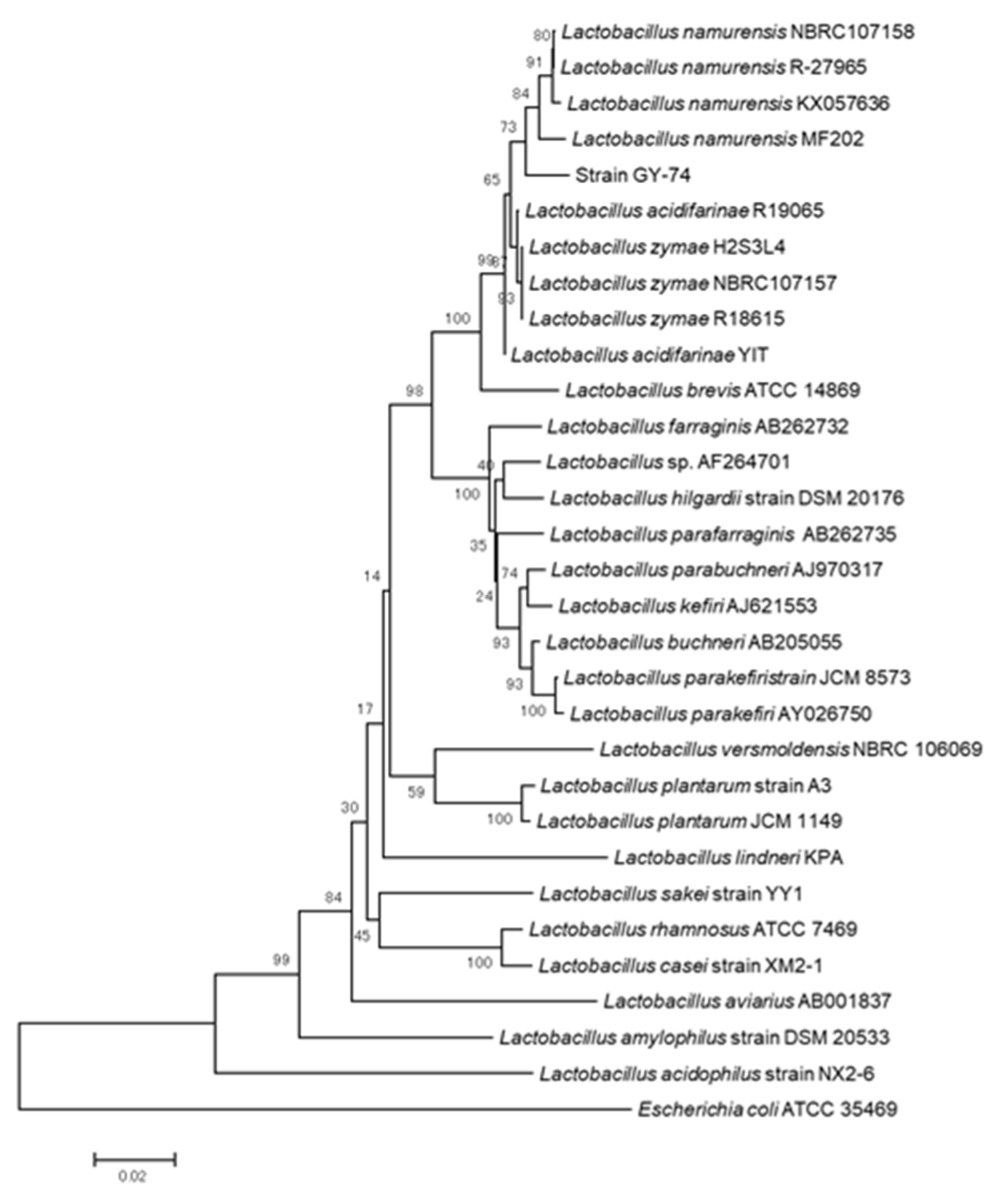
3.1. Isolation of LAB and Identification Based on Morphological Observation, BLAST Research and Phylogenetic Analysis

Twenty strains were isolated from the salted rice bran bed. A partial length of the 16S rRNA gene nucleotide sequence (1449 bp) of strain GYP-74 was deposited in the data bank with accession number LC327006. Figure 1 shows the result of phylogenetic analysis. The 16S rRNA gene sequence of strain GYP-74 used in the adherence test was located in the cluster of Lactobacillus namurensis. BLAST searches also confirmed the identities (on the basis of the highest score obtained) as L. namurensis NBRC 107158. The morphological observation with an SEM showed that the cell form of strain GYP-74 was rod-shaped (Figure 2).
Nakayama et al. reported that two species, L. namurensis and L. acetotolerans were the dominant LAB in Nukadoko [2]. Sakamoto et al. reported that the doubling time was significantly different between L. namurensis (6.2 h) and L. acetotolerancs (92.9 h), and that it was difficult to culture L. acetotolerancs [1]. Some studies reported that various fermented foods by LAB such as Nukazuke with L. namurensis as a dominant LAB, fermented milk and fish sauce showed a blood pressure-reducing effect (angiotensin converting enzyme inhibitory effect) and antioxidant activities [35,36,37]. Enrichment of some LAB to porous ceramics has potential for the improvement of lifestyle-related diseases.

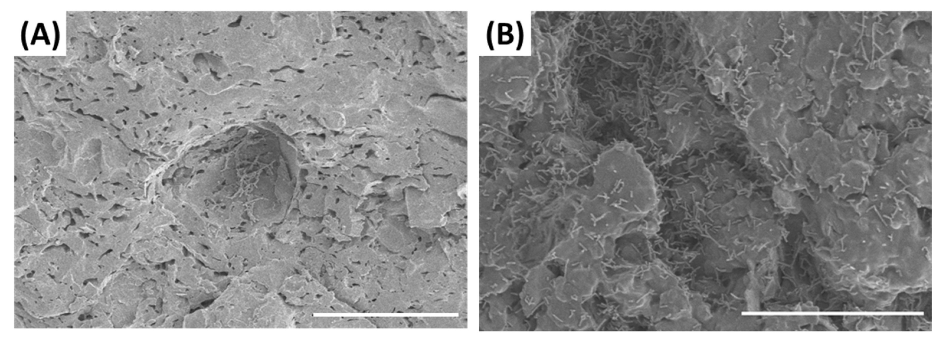
3.2. Adherent Test of Strain GYP-74 to PS

Figure 3 shows the number of cells that were adherent to the PS. In the control, no adherent cells were observed. In the case of PS without POM or with POM with the same pore size, the adherent cell numbers in PS burned at 1100 °C were significantly higher than those in PS burned at 1000 °C. The blending quantity of POM in PS influenced the adherence ratio of strain GYP-74. In the case of a 0.2 mm pore size fired at 1100 °C, the adherent ratio was significantly higher at the 30% POM-blending quantity (#7) than for other groups (#5 and #6). In the case with a 0.5 mm pore size at 1100 °C, the adherence ratio was significantly lower at the 30% POM-blending quantity (#13) than for other groups (#11 and #12). The highest adherence ratio was observed among all groups at 0.2-mm pore size, a 1100 °C firing temperature and a 30% POM blending quantity (#7). Figure 4 shows pictures of the surface of the clay without resin fired at 1000 °C (#1) and the 30% POM resin-containing PS fired at 1100 °C by electron microscopic observation. Numerous cells were observed on the surface of #13 as compared with #1. In particular, the cells adhered locally to the hollow parts of the pores.

3.3. Pore Distribution Analysis

Figure 5 shows the result of an analysis of the pore distribution using a mercury porosimeter. In the case of PS without POM (only clay) fired at 1000 °C, the pore diameter distribution of the porous body was 0.4–50 μm, and mainly concentrated to 1 µm (Figure 5 #1). By increasing the firing temperature to 1100 °C, the peak value shifted to 4–5 µm (Figure 5 #2). A tendency of the peak value to increase with the increase in the burning temperature was observed in the case of PS with POM (Figure 5 #4, #6, #9 and #12). The peak value obviously increased to more than 10 µm with the addition of POM irrespective of the change in the firing temperatures. The results of the adherence test showed that the number of adherent cells was higher in the POM-containing PS fired at 1000 °C (Figure 3).
Yamamoto et al. described that the adherence ratio of beneficial bacteria to porous fine ceramics was significantly higher in ceramics having more than a 10 µm pore size as compared to those having a less-than-10 µm pore size [38]. This report agrees with our result. Pereira et al. evaluated the retention of bacterial biomass to four types of porous microcarriers (sepiolite, caly, pozzolana, and form glass) with different pore sizes, and found that the attached bacterial biomass was higher in sepiolite (pore size: cell size crevices) and clay (pore size: 10–100 µm) compared to pozzolana and foam glass (having a 10–300 µm and 20–1000 µm pore size, respectively) [39]. The present study and studies by Yamamoto et al. and Pereira et al. indicate that the adequate pore size for bacterial adhesion is from bacterial cell sizes to less than 100 µm. Rajab et al. reported that the cell length of L. delbrueckii subsp. lactis, L. paracasei, L. acidophilus, and L. rhamnosus was 5.2–7.6 µm, and significantly longer than other Lactobacillus strains, L. sakei subsp. sakei, L. reuteri and L. plantarum with 1.6–2.8 µm cell length [40]. In the present study, the cell length of L. namurensis was estimated to be approximately 2–10 µm by light-microscopic and SEM observation. Figure 3 and Figure 5 show that the adhered cell number was higher in the pottery shards having more than a few µm pore size, which indicates that cell size and pore size affected the cell adherence.

3.4. X-ray Diffraction Analysis

Figure 6 shows the results of X-ray diffraction analysis of the PS. In the case of PS fired at 1000 °C (#3), microcline was specifically formed. In the case of PS fired at 1100 °C (#5), mullite was specifically formed. The quarts and hematite were detected at both temperatures. Generally, a diameter of 10–200 µm is the pore size necessary for excellent microbial growth because fluids containing nutrients have to pass through pores to supply the energy to the bacteria. Kitaoka et al. reported that porous materials prepared from mud mainly consist of silica, and no difference in their chemical characteristics was found [41]. Therefore, it is considered that the space in the porous materials strongly influenced the bacterial adherence.
Probiotic studies have focused on the adherence to the gastrointestinal tract in host animals. The adherence mechanisms are related to the components present on the surface of the epithelium of the intestine such as mucin, collagens and fibronectin [42,43]. However, it is assumed that the mechanisms of adherence to porous materials are significantly different. The mucus layer which covers the intestinal epithelium of humans consists of two layers: the inner and outer layer. Basically, dominant bacteria in the intestine are detected in only the outer layer, and bacteria show the adherence to mucin which is a main component of mucus in the intestine. Adhesin is known as an adherence factor in bacterial cells which exhibits a receptor-bonding ability in mucin. Many LAB have the adhesion mechanism to intestinal mucus via glycans [44,45]. Holst et al. reported that L. plantarum has a mannose-specific adhesin [46]. The process of bacterial adhesion to intestinal mucus is mainly divided into three steps: (1) adhesion via adhesin, (2) aggregation among bacterial cells, and (3) colonization to the mucus of the intestine [47]. On the other hand, the adhesion mechanisms of bacteria to materials such as ceramics are significantly different to those done via glycan.
In the present study, we focus on the relationship between the pore sizes and the ratio of adherence. However, interactions between bacteria and material’s surface consist of very complex mechanisms, and bacteria can migrate towards a surface based on physical forces such as gravitational settling, hydrodynamic forces and Brownian motion, and/or their own motility with flagella [21]. Some studies reported that the surface roughness and surface wetting also affect the adherence of bacteria to the material surfaces. Katsikogianni and Missirlis reported that bacteria preferentially adhere to surface irregularities whose typical sizes are of the order of their own diameters [48]. Ammar et al. reconstructed the topography of the bacterial cell and substrate surfaces using Surface Element Integration (SEI) technique. They compared SEI predictions with those of a simple model for surface roughness, results indicated that three parameters linked with the surface roughness have an impact on the height of the energy barrier [49]. Some studies indicated that the cell surface hydrophobicity of S. aureus, known as a major human pathogen, is a main factor for its own adhesion ability, and it harbors the adhesion genes and adhesin genes [22,50]. Slullitel et al. reported that there is no superiority of ceramics against adhesion of biofilm-producing S. aureus and S. epidermidis, and E. coli [51]. Some reports, mainly related to dental implants, have indicated that the surface roughness and wettability influenced bacterial adhesion to materials [48,52]. It is generally known that bacteria prefer to grow on available surfaces of materials than in the aqueous phase [53], and Kirov indicated that this adherent process was related to bacterial chemotaxis and haptotaxis [54]. Taylor et al. found that the surface roughness of poly methyl methacrylate induced a significant increase in bacterial adhesion [55]. In further studies, the surface roughness and hydrophobicity should be measured to comprehensively understand the adhesion mechanism of strain GYP-74. Additionally, implementation of an adherence test with wide varieties of LAB will support the versatility of the fermentation container prepared with porous ceramics. The present study showed that the pore size and the distribution varied by controlling the burning temperature and ratio of the POM content, and the adherent ratio of strain GYP-74 also varied.

4. Conclusions

This study provided the preliminary data on the relationship between L. namurensis adherence and pore size distribution in porous fine ceramics. In this paper, we isolated L. namurensis strain GYP-74 from salted rice bran (Nukadoko). We report that the distribution of pore size and the crystal form in the PS can be controlled by arranging the firing temperatures. The adhesion rate of strain GYP-74 increased in the PS which had pore sizes greater than their own cell size. When we make the fermentation container to enrich LAB, considering the pore size and the cell size of target LAB makes the adhesion efficiency of LAB cells increase.

Author Contributions

Conceptualization, Y.T., H.F., N.K., M.K., T.K., A.H. and A.K.; methodology, Y.T., K.S. and H.K.; validation, Y.T., K.S. and H.K.; formal analysis, Y.T., K.S. and H.K.; resources, N.K., M.K. and A.K.; data curation, Y.T., K.S. and H.K.; writing—original draft preparation, Y.T.; funding acquisition, T.K. All authors have read and agreed to the published version of the manuscript.

Funding

This research was partially funded by Miyazaki Prefecture’s industry–academia–government collaboration R&D.

Institutional Review Board Statement

Not applicable.

Informed Consent Statement

Not applicable.

Data Availability Statement

The data presented in this study are available in the main text of the article.

Acknowledgments

This work was supported by Miyazaki Prefecture’s industry-academia-government collaboration R&D.

Conflicts of Interest

The authors declare no conflict of interest.

References

  1. Sakamoto, N.; Tanaka, S.; Sonomoto, K.; Nakayama, J. 16S rRNA pyrosequencing-based investigation of the bacterial community in nukadoko, a pickling bed of fermented rice bran. Int. J. Food Microbiol. 2011, 144, 352–359. [Google Scholar] [CrossRef]
  2. Nakayama, J.; Hoshiko, H.; Fukuda, M.; Tanaka, H.; Sakamoto, N.; Tanaka, S.; Ohue, K.; Sakai, K.; Sonomoto, K. Molecular monitoring of bacterial community structure in long-aged nukadoko: Pickling bed of fermented rice bran dominated by slow-growing lactobacilli. J. Biosci. Bioeng. 2007, 104, 481–489. [Google Scholar] [CrossRef]
  3. Guan, Q.; Xiong, T.; Xie, M. Influence of Probiotic Fermented Fruit and Vegetables on Human Health and the Related Industrial Development Trend. Engineering 2021, 7, 212–218. [Google Scholar] [CrossRef]
  4. Yang, Y.; Park, J.H.; Kim, I.H. Effects of probiotics containing (Lactobacillus planetarium) and chlortetracycline on growth performance, nutrient digestibility, fecal microflora, diarrhea score and fecal gas emission in weanling pigs. Livest. Sci. 2020, 241, 104186. [Google Scholar] [CrossRef]
  5. Kuebutornye, F.K.A.; Abarike, E.D.; Lu, Y. A review on the application of Bacillus as probiotics in aquaculture. Fish Shellfish Immunol. 2019, 87, 820–828. [Google Scholar] [CrossRef]
  6. Fuller, R. Probiotics in man and animals. J. Appl. Bacteriol. 1989, 66, 365–378. [Google Scholar] [PubMed]
  7. Salminen, S.; Isolauri, E.; Salminen, E. Clinical uses of probiotics for stabilizing the gut mucosal barrier: Successful strains and future challenge. Antonie Van Leeuuwenboek 1997, 70, 347–358. [Google Scholar] [CrossRef] [PubMed]
  8. Shu, Q.; Qu, F.; Gill, H.S. Probiotic treatment using Bifidobacterium lactis HNO19 reduces weanling diarrhea associated lymphoid tissue in neonatal swine. Immunol. Methods 2001, 241, 185–199. [Google Scholar]
  9. Sakai, M. Current research status of fish immunostimulants. Aquaculture 1998, 72, 63–92. [Google Scholar] [CrossRef]
  10. Anukam, K.C.; Osazuwa, E.; Osemene, G.I.; Ehigiagbe, F.; Bruce, A.W.; Reid, G. Clinical study comparing probiotic Lactobacillus GR-1 and RC-14 with metronidazole vaginal gel to treat symptomatic bacterial vaginosis. Microbes Infect. 2006, 8, 1–5. [Google Scholar] [CrossRef]
  11. Gionchetti, P.; Rizzello, F.; Venturi, A.; Brigidi, P.; Matteuzzi, D.; Bazzocchi, G.; Poggioli, G.; Miglioli, M.; Campieri, M. Oral bacteriotherapy as maintenance treatment in patients with chronic pouchitis: A doubleblind, placebo-controlled trial. Gastroenterology 2000, 119, 305–309. [Google Scholar] [CrossRef] [PubMed]
  12. Saran, S.; Gopalan, S.; Krishna, T.P. Use of fermented foods to combat stunting andfailure to thrive. Nutrition 2002, 18, 393–396. [Google Scholar] [CrossRef]
  13. Sazawal, S.; Hiremath, G.; Dhingra, U.; Malik, P.; Deb, S.; Black, R.E. Efficacy of probiotics in prevention of acute diarrhoea: A meta-analysis of masked, randomised, placebo-controlled trials. Lancet Infect. Dis. 2006, 6, 374–382. [Google Scholar] [CrossRef]
  14. Sheu, B.S.; Cheng, H.C.; Kao, A.W.; Wang, S.T.; Yang, Y.J.; Yang, H.B.; Wu, J.J. Pretreatment with Lactobacillus- and Bifidobacterium-containing yogurt can improve the efficacy of quadruple therapy in eradicating residual Helicobacter pylori infection after failed triple therapy. Am. J. Clin. Nutr. 2006, 83, 864–869. [Google Scholar] [CrossRef] [PubMed] [Green Version]
  15. Komatsuzaki, N.; Tsukahara, K.; Toyoshima, H.; Suzuki, T.; Shimizu, N.; Kimura, T. Effect of soaking and gaseous treatment on GABA content in germinated brown rice. J. Food Eng. 2007, 78, 556–560. [Google Scholar] [CrossRef]
  16. Chung, H.J.; Jang, S.H.; Cho, H.Y.; Lim, S.T. Effects of steeping and anaerobic treatment on GABA (γ-aminobutyric acid) content in germinated waxy hull-less barley. LWT Food Sci. Technol. 2009, 42, 1712–1716. [Google Scholar] [CrossRef]
  17. Hori, K.; Ishikawa, M. Microbial Adhesion to Surfaces Mediated by Bacterial Nanofibers. J. Environ. Biotechnol. 2010, 10, 3–7. [Google Scholar]
  18. Hori, K.; Matsumoto, S. Bacterial adhesion: From mechanism to control. Biochem. Eng. J. 2010, 48, 424–434. [Google Scholar] [CrossRef]
  19. Chaban, B.; Hughes, H.V.; Beeby, M. The flagellum in bacterial pathogens: For motility and a whole lot more. Semin. Cell Dev. Biol. 2015, 46, 91–103. [Google Scholar] [CrossRef] [Green Version]
  20. Beren, C.; Ellison, C.K.; Ducret, A.; Brun, Y.V. Bacterial adhesion at the single-cell level. Nat. Rev. Microbiol. 2018, 16, 616–627. [Google Scholar] [CrossRef]
  21. Morisaki, H.; Nagai, S.; Ohshima, H.; Ikemoto, E.; Kogure, K. The effect of motility and cell-surface polymers on bacterial attachment. Microbiology 1999, 145, 2797–2802. [Google Scholar] [CrossRef] [PubMed] [Green Version]
  22. Kouidhi, B.; Zmantar, T.; Hentati, H.; Bakhrouf, A. Cell surface hydrophobicity, biofilm formation, adhesives properties and molecular detection of adhesins genes in Staphylococcus aureus associated to dental caries. Microb. Pathog. 2010, 49, 14–22. [Google Scholar] [CrossRef] [PubMed]
  23. Poncin-Epaillard, F.; Herry, J.M.; Marmey, P.; Legeay, G.; Debarnot, D.; Bellon-Fontaine, M.N. Elaboration of highly hydrophobic polymeric surface—A potential strategy to reduce the adhesion of pathogenic bacteria? Mater. Sci. Eng. C 2013, 33, 1152–1161. [Google Scholar] [CrossRef]
  24. Hao, X.; Chen, S.; Qin, D.; Zhang, M.; Fan, J.; Wang, C.; Dong, M.; Zhang, J.; Cheng, F.; Guo, Z. Antifouling and antibacterial behaviors of capsaicin-based pH responsive smart coatings in marine environments. Mater. Sci. Eng. C 2020, 108, 110361. [Google Scholar] [CrossRef] [PubMed]
  25. Boyd, R.D.; Verran, J.; Jones, M.V.; Bhakoo, M. Use of the atomic force microscope to determine the effect of substratum surface topography on bacterial adhesion. Adv. Colloid Interface Sci. 2012, 179, 142–149. [Google Scholar] [CrossRef]
  26. Matsumoto, S.; Ohtaki, A.; Hori, K. Carbon fiber as an excellent support material for wastewater treatment biofilms. Environ. Sci. Technol. 2012, 46, 10175–10181. [Google Scholar] [CrossRef] [PubMed]
  27. Song, F.; Koo, H.; Ren, D. Effects of Material Properties on Bacterial Adhesion and Biofilm Formation. J. Dent. Res. 2015, 94, 1027–1035. [Google Scholar] [CrossRef]
  28. Vural, H.C.; Ozgun, D. An improving DNA isolation method for identification of anaerobic bacteria in human colostrum and faeces samples. J. Med. Genet. Genomics 2011, 3, 95–100. [Google Scholar]
  29. Schmitz, A.; Riesner, D. Purification of nucleic acids by selective precipitation with polyethylene glycol 6000. Anal. Biochem. 2006, 354, 311–313. [Google Scholar] [CrossRef]
  30. Saeedi, M.; Shahidi, F.; Mortazavi, S.A.; Milani, E.; Yazdi, F.T. Isolation and identification of lactic acid bacteria in winter salad (Local Pickle) during fermentation using 16S rRNA gene sequence analysis. J. Food. Saf. 2015, 35, 287–294. [Google Scholar] [CrossRef] [Green Version]
  31. Thompson, J.D.; Gibson, T.J.; Plewniak, F.; Higgins, D.G. The CLUSTAL X windows interface: Flexible strategies for multiple sequence alignment aided by quality analysis tools. Nucleic Acids Res. 1997, 25, 4876–4882. [Google Scholar] [CrossRef] [PubMed] [Green Version]
  32. Kimura, M. A simple method for estimating evolutionary rates of base substitutions through comparative studies of nucleotide sequences. J. Mol. Evol. 1980, 16, 111–120. [Google Scholar] [CrossRef]
  33. Saitou, N.; Nei, M. The Neighbor-joining method: A new method for reconstructing phylogenetic trees. Mol. Biol. Evol. 1987, 4, 406–425. [Google Scholar] [PubMed]
  34. Kumar, S.; Stecher, G.; Tamura, K. MEGA7: Molecular evolutionary genetics analysis version 7.0 for bigger datasets. Mol. Biol. Evol. 2016, 33, 1870–1874. [Google Scholar] [CrossRef] [PubMed] [Green Version]
  35. Oda, K.; Imanishi, T.; Yamane, Y.; Ueno, Y.; Mori, Y. Bio-functional pickles that reduce blood pressure of rats. Biosci. Ciotechnol. Biochem. 2014, 78, 882–890. [Google Scholar] [CrossRef]
  36. Okamoto, A.; Matsumoto, E.; Yasuhara, T.; Koizumi, Y.; Yanagida, F. Angiotensin I-converting enzyme inhibitory action of fish sauce. Food Sci. Technol. Int. Tokyo 1995, 1, 101–106. [Google Scholar] [CrossRef]
  37. Yokoyama, K.; Chiba, H.; Yoshikawa, M. Peptide inhibitors for angiotensin I-converting enzyme from thermolysin digest of dried bonito. Biosci. Biotechnol. Biochem. 1992, 56, 1541–1545. [Google Scholar] [CrossRef] [PubMed]
  38. Yamamoto, T.; Sakami, T.; Awai, K.; Shirakata, A. ZnO-based transparent conducting oxide films by reactive plasma deposition. Convertech 2004, 375, 68. [Google Scholar]
  39. Pereira, M.A.; Azeredo, J.; Mota, M.; Oliveira, R. Influence of physico-chemical properties of porous microcarriers on the adhesion of an anaerobic consortium. J. Ind. Microbiol. Biotechnol. 2000, 24, 181–186. [Google Scholar] [CrossRef] [Green Version]
  40. Rajab, S.; Tabandeh, F.; Shahraky, M.K. The effect of lactobacillus cell size on its probiotic characteristics. Anaerobe 2020, 62, 102103. [Google Scholar] [CrossRef] [PubMed]
  41. Kitaoka, S.; Wada, T.; Osa, N.; Inagaki, H. Improvement of steam oxidation resistance and flame impingement resistance of glass having carbon coating film with controlled interfaces. In Prospect of Surface Treatment and Modification Technology in Inorganic Materials -Metal, Ceramics, and Glass-; Kamijo, E., Suzuki, Y., Fujisawa, A., Eds.; CMC Publisher: Tokyo, Japan, 2015; pp. 251–291. [Google Scholar]
  42. Uchida, H.; Fujitani, K.; Kawai, Y.; Kitazawa, H.; Horii, A.; Shiiba, K.; Saito, K.; Saito, T. A new assay using surface plasmon response (SPR) to determine binding of the Lactobacillus acidophilus group to human colonic mucin. Biosci. Biotechnol. Biochem. 2004, 68, 1–7. [Google Scholar] [CrossRef] [PubMed] [Green Version]
  43. Horie, M.; Ishiyama, A.; Fujihira-Ukei, Y.; Sillanpaa, J.; Korhonen, T.K.; Toba, T. Inhibition of the adherence of Escherichia coli strains to basement membrane by Lactobacillus crispatus expressing an S-layer. J. Appl. Microbiol. 2002, 92, 396–403. [Google Scholar] [CrossRef] [PubMed] [Green Version]
  44. Johansson, M.E.V.; Holmén Larsson, J.M.; Hansson, G.C. The two mucus layers of colon are organized by the MUC2 mucin, whereas the outer layer is a legislator of host–microbial interactions. Proc. Natl. Acad. Sci. USA 2011, 108, 4659–4665. [Google Scholar] [CrossRef] [Green Version]
  45. Johansson, M.E.V.; Phillipson, M.; Petersson, J.; Velcich, A.; Holm, L.; Hansson, G.C. The inner of the two Muc2 mucin-dependent mucus layers in colon is devoid of bacteria. Proc. Natl. Acad. Sci. USA 2008, 105, 15064–15069. [Google Scholar] [CrossRef] [Green Version]
  46. Holst, B.; Glenting, J.; Holmstrøm, K.; Israelsen, H.; Vrang, A.; Antonsson, M.; Ahrné, S. Molecular Switch Controlling Expression of the Mannose-Specific Adhesin, Msa, in Lactobacillus plantarum. Appl. Environ. Microbiol. 2019, 85, e02954-18. [Google Scholar] [CrossRef] [Green Version]
  47. Nishiyama, K.; Mukai, T. Colonization Properties of Lactic Acid Bacteria to Mucosal Surface of the Intestinal Tract. Kagakutoseibutsu 2016, 54, 471–477. [Google Scholar]
  48. Katsikogianni, M.; Missirlis, Y.F. Concise review of mechanisms of bacterial adhesion to biomaterials and of techniques used in estimating bacteria–material interactions. Eur. Cell Mater. 2004, 8, 37–57. [Google Scholar] [CrossRef]
  49. Ammar, Y.; Swailes, D.; Bridgens, B.; Chen, J. Influence of surface roughness on the initial formation of biofilm. Surf. Coat. Technol. 2015, 284, 410–416. [Google Scholar] [CrossRef] [Green Version]
  50. Hamadi, F.; Latrache, H.; Mabrrouki, M.; El Ghmari, A.; Outzourhit, A.; Ellouali, M.; Chtaini, A. Effect of pH on distribution and adhesion of Staphylococcus aureus to glass. J. Adhes. Sci. Technol. 2005, 19, 73–85. [Google Scholar] [CrossRef]
  51. Slullitel, P.A.; Buttaro, M.A.; Greco, G.; Oñativia, J.I.; Sánchez, M.L.; Mc Loughlin, S.; García-Ávila, C.; Comba, F.; Zanotti, G.; Piccaluga, F. No lower bacterial adhesion for ceramics compared to otherbiomaterials: An in vitro analysis. Orthop. Traumatol. Surg. Res. 2018, 104, 439–443. [Google Scholar] [CrossRef] [PubMed]
  52. Teughels, W.; Van Assche, N.; Sliepen, I.; Quirynen, M. Effect of material characteristics and/or surface topography on biofilm development. Clin. Oral Implants Res. 2006, 17, 68–81. [Google Scholar] [CrossRef] [PubMed]
  53. Gottenbos, B.; Busscher, H.J.; van der Mei, H.C.; Nieuwenhuis, P. Pathogenesis and prevention of biomaterial centered infections. J. Mater. Sci. Mater. Med. 2002, 13, 717–722. [Google Scholar] [CrossRef] [PubMed]
  54. Kirov, S.M. Bacteria that express lateral fragella enable dissection of the multifunctional roles of fragella in pathogenesis. FEMS Microbiol. Lett. 2003, 224, 151–159. [Google Scholar] [CrossRef] [Green Version]
  55. Taylor, R.L.; Verran, J.; Lees, G.C.; Ward, A.J.P. The influence of substratum topography on bacterial adhesion to polymethyl methacrylate. J. Mater. Sci. Mater. Med. 1998, 9, 17–22. [Google Scholar] [CrossRef] [PubMed]
Figure 1. Phylogenetic tree was constructed by the neighbor-joining method based on the partial sequence of the 16S rRNA gene of strain GYP-74 and corresponding region in those for the authentic bacterial genes. Numbers at branches denote the bootstrap percentages of 1000 replicates. The scale at the bottom indicates the evolutionary distance of nucleotide substitutions per site.
Processes 09 00658 g001
Figure 2. Morphological observation of strain GYP-74 cultured in de Man, Rogosa and Sharpe (MRS) medium with a scanning electron microscope (scale bar, 5 µm).
Processes 09 00658 g002
Figure 3. The cell numbers of GYP-74 adhered to the pottery shards (PSs). Bars denoted by the different letters are significantly different (p > 0.05, Tukey-Kramer test). Values represent mean ± standard deviation (n = 3).
Processes 09 00658 g003
Figure 4. Observation of the surface of PSs (#1 and #13) with addition of strain GYP-74 with a scanning electron microscope (scale bar, 50 µm). (A) #1, (B) #13.
Processes 09 00658 g004
Figure 5. Distribution of pore size of PSs (#1, #2, #4, #6, #9 and #12). Values represent mean (n = 3).
Processes 09 00658 g005
Figure 6. X-ray diffraction pattern of PSs (#3 and #5).
Processes 09 00658 g006
Publisher’s Note: MDPI stays neutral with regard to jurisdictional claims in published maps and institutional affiliations.

