1. Introduction
A century ago, Ivar Bang first described dried blood spot (DBS) technology as an original method for collecting and storing samples of biological origin [
1]. Later, Robert Guthrie in 1963 introduced the DBS method for screening newborn genetic diseases [
2]. The test developed by Guthrie for the detection of phenylketonuria has been successfully introduced into clinical practice [
3]. The introduction of advanced analytical methods for the detection of molecular components of biosamples, including mass spectrometric methods, has expanded the testing capabilities and improved analysis performance [
4,
5].
The advantages of using DBS with respect to “raw” blood samples are: (1) No obligation for the specific storage conditions during transportation; (2) sustainability of molecular components in DBS samples for several years under conditions of deep cold [
6], which can be eluted with a high level of extraction by solvents for subsequent analysis; (3) in low contagiousness with infectious agents [
7]; (4) in a small amount of biomaterial (~ 20 μL) for analysis [
6]; and (5) in the absence of the need for specially trained personnel, since the sampling of biomaterial can be carried out outside laboratory conditions [
8].
Membrane carriers DBS provide opportunity for the long-term storage of biological samples and their transportation to analytical laboratories, as well as for creating biobanks [
9]. Today, the use of mass spectrometry for DBS analysis is becoming a routine method in biomedical and pharmaceutical laboratories.
In a study by Millington et al. [
10] used fast atomic bombardment (FAB) mass spectrometry for the quantitative determination of amino acid and acylcarnitine ester derivatives in DBS samples for the diagnostic screening of phenylketonuria and medium chain acyl-coenzyme A dehydrogenase deficiency in newborns. The advent of electrospray ionization has led to the development of a high-throughput multiple reaction monitoring (MRM) assay that detects low-concentration non-derivatized metabolites and allows for increased assay throughput. Many medical screening programs use a multiplex one-step approach to detect multiple analytes in an assay [
11]. DBS-based screening assays using mass spectrometry can detect phenylketonuria, medium chain acyl-coenzyme A dehydrogenase deficiency [
12], leucinosis [
13], homocystinuria [
14] and lysosomal storage diseases [
15].
The pharmaceutical industry uses DBS samples in research. Identification and quantitative analysis are currently performed for a number of drugs and their metabolites [
7]. Various techniques have been tried, including desorption electrospray ionization (DESI) [
16], paperspray ionization [
17], surface sealing sampling probe (SSSP) [
18] and liquid surface extraction analysis (LESA) [
19]. These methods have helped to increase the productivity of DBS analysis by eliminating the need for any pretreatment or lengthy elution steps.
Study [
20] used a new high-throughput method that profiled over 80 different N-glycans in DBS samples with linkage-specific sialic acid analysis using matrix-assisted laser desorption/ionization (MALDI) MS.
Review of the related literature shows that DBS technology is a widely used and actual method at the preanalytical stages of diagnosis [
9]. It is important that the biomaterial on DBS membrane carriers demonstrates resistance to long-term storage and does not require special storage conditions in comparison with samples of cells or blood plasma [
21].
In the present study, we analyzed the stability of DBS of blood plasma samples with variations in temperature (from −20 to 25 °C) and storage duration (up to 35 days) using time-of-flight mass spectrometric analysis (MALDI-TOF-MS). We used two protocols for preliminary preparation of DBS samples. In the first, the procedure of preliminary extraction of molecular components from the DBS of the sample into the solution was carried out, followed by trypsinolysis and MS analysis. In the second protocol, trypsinolysis of protein components was carried out directly in a membrane carrier, followed by elution and MS analysis of tryptic components. We considered the invariability of the mass spectrometric intensities of signals from the tryptic components at different temperature conditions and storage duration as a criterion for the stability of a biological sample. We propose to use the results obtained in the study as a recommendation for the maximum possible conditions (temperature regime and duration) of storage of blood samples, under which the protein composition is likely to remain stable.
2. Materials and Methods
2.1. Chemicals
Acetonitrile (Fischer Chemicals AG, Zurich, Switzerland), bicarbonate buffer (Sigma, St. Louis, MI, USA), trypsin (Promega, Madison, WI, USA), trifluoroacetic acid (Fischer Chemicals AG, Zurich, Switzerland), matrix (HCCA) (Bruker Daltonics, Bremen, Germany). Deionized water was obtained using the Milli-Q system (Millipore, Molsheim, France).
2.2. Demography
Peripheral venous blood samples from healthy females (n = 3; ages 44, 43, 45 years old) and samples from females with ovarian cancer (n = 3; ages 43, 42, 45 years old) were taken following an overnight fasting. Blood was collected in the pre-chilled tubes with EDTA, gently agitated and centrifuged at 4 °C and 2000 g for 10 min. The obtained plasma samples were collected, transferred into clean cryotubes and frozen upon necessity.
Written consents to participate in the study and to use their biological material were obtained from each subject. This study was approved by the independent local research ethics committee of the Institute of Urology and Reproductive Health (Sechenov University, Protocol No. 10–19 of 17 July 2019). Written informed consent was obtained from the patients and healthy volunteers authorizing their participation in the study and the use of their biological material. All the samples were deactivated before their use in the study to provide biological safety.
2.3. Sample Preparation
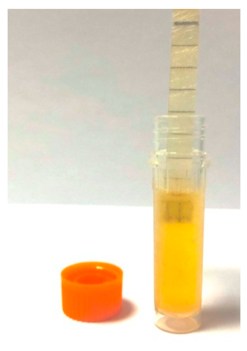
As a carrier of a dry sample of a biological fluid (blood plasma), a membrane strip made of porous hydrophilic fiberglass material 0.5 cm wide with an applied marking (Immunoved Ltd., Moscow, Russia) was used.
All samples were preserved using membrane support technology. A portion of the membrane (5 mm) was immersed in the sample and kept until the liquid strip was completely wetted and held for the time required to raise the liquid along the entire length of the membrane strip (
Figure 1).
Then the strip with the sample was dried in air at room temperature for 1.5–2 h. The dried samples were stored in tightly closed plastic bags. The samples were preserved for a long-term study (0, 7, 14, 21, 30 days) and at different temperature conditions (−20, +4, 25 °C). Since it is known that the presence of moisture during storage affects the properties of a biological material more than temperature [
22], DBS was stored in individual plastic bags with a desiccant. For analysis, parts of the membrane were cut in the form of a square (0.5 × 0.5 cm) using scissors.
2.4. Sample Preparation for MS Analysis
The DBS sample preparation for subsequent mass spectrometric analysis was carried out using two protocols.
The excised DBS sector was placed in an Eppendorf tube (V = 200 μL). Acetonitrile 200 μL was added to the test tube. Then, the procedure of ultrasonic processing of the DBS sample was performed for 10 min. Then the solution with the eluate was pipetted into clean labeled Eppendorf tubes (V = 200 μL). The eluate was dried on a vacuum evaporator at room temperature. The dry residue was dissolved with a buffer solution (20 μL of bicarbonate buffer, 5 μM trypsin) and hydrolysis was performed overnight. Interruption of hydrolysis was carried out using a 0.7% solution of trifluoroacetic acid (TFA). The hydrolyzate was dried on a vacuum evaporator at room temperature and then redissolved with a solution of 0.7% TFA in a volume of 20 μL.
The MTP AnchorChip 384 target was loaded with hydrolyzate in a volume of 1 μL with the addition of 1 μL of matrix (HCCA) and dried at room temperature.
The excised DBS sector was placed in an Eppendorf tube (V = 200 μL) with a buffer solution (20 μL bicarbonate buffer, 5 μM trypsin) and hydrolysis was performed overnight. The hydrolysis was interrupted using a 0.7% TFA solution. Then, the hydrolyzed DBS sample was sonicated in an ultrasonic bath for 3 min, after which it was transferred using a pipette into clean labeled Eppendorf tubes (V = 200 μL). The hydrolyzate was dried in a vacuum evaporator at room temperature, followed by reconstitution in a 20 µL 0.7% TFA solution.
The MTP AnchorChip 384 target was coated with a 1µl hydrolyzate supplemented with a 1 µL matrix (HCCA) and dried at room temperature.
2.5. MALDI-TOF-MS Analysis
Analysis was performed on an Autoflex III time-of-flight mass-spectrometer (Bruker, Bremen, Germany), equipped with nitrogen laser with emission wavelength of 337 nm. The instrument was calibrated in the reflector mode with voltage of 3.5–4.0 kV using the peptide calibration standard for the positive ions. Mass spectrum were recorded in a range of 750–3000 m/z with a pulse delay time of 200 ns. The peptide calibration standard comprised of (monoisotolic single-protonated ionic mass) bradykinin (757.3992 Da), angiotensin II (1046.5420 Da), angiotensin I (1296.6853 Da), R peptide (1347.7361 Da), bombesin (1619.8230 Da), renin (1758.9326 Da), ACTH fragment 1–17 (2093.0868 Da), ACTH fragment 18–39 (2465.1990 Da), and somatostatin (3147.4714 Da). The spectrum analysis excluded the peaks of trypsin matrix and autolysis. Samples were mixed with the excess of matrix (HCCA in 50% acetonitrile solution in 0.7% TFA) at the ratio ranging from 1:1000 to 1:10,000 and spotted onto MTP AnchorChip 384 target. Typically, up to 50,000 shots per sample were performed in an auto mode to accumulate the necessary mass spectrum collection. The mass spectra were processed with flexAnalysis 2.0 (Bruker, Germany). Mass spectrometric measurements were performed using the equipment of “Human Proteome” Core Facility (IBMC, Moscow, Russia).
2.6. Statistical Analysis
Statistical analysis was performed with R (R Core Team (2020). R: A language and environment for statistical computing. R Foundation for Statistical Computing, Vienna, Austria. URL
https://www.R-project.org/). MS spectra obtained with flexAnalysis 2.0 were further processed in batch mode through workflow published in GitHub (
https://github.com/sgibb/MALDIquant/blob/master/demo/workflow.R) using MALDIquant R package [
23]. For the routine data processing, raw mass spectra were imported following variance stabilization and smoothing. Baseline subtraction from the smoothed MALDI spectra was performed, followed-up by intensities normalization and spectra alignment. Peaks that meet criteria of signal-to-noise ratio exceeding 5 and frequency at least 0.25 were retained for consideration. Eventually, ten
m/
z-signatures were elected as confident indicators for monitoring of samples degradation at the certain storage conditions (initial and final day of storage at different temperature modes) [
23]. The normalized relative intensities were analyzed by two-sample Wilcoxon test for evaluating statistical differences between each two groups. A two-sided
p < 0.05 was considered as statistically significant.
3. Results and Discussion
Both protocols were invented by our group for the first time. The proteomic-centered samples preparation procedure that utilizes DBS can be performed by one of the offered ways. The first way suggests the digestion of the sample immediately on the surface of the membrane carrier. Another way supposes digestion of the sample following preliminary elution from the surface of DBS wherein the procedure of samples preparation addressed for the mass spectrometry analysis is being typical and described repeatedly in numerous publications including our authored papers [
24,
25,
26].
We analyzed the stability of 6 blood plasma samples using the DBS method and two protocols of preliminary sample preparation at different storage times (up to 35 days) and temperature conditions (from −20 to 25 °C). Evaluation of the stability of the samples under different storage conditions was carried out by identifying changes in the intensities of the mass-charge characteristics, detected in the MALDI-TOF-MS analysis.
As a result of the analysis of mass spectrometric measurements, 10 mass-charge characteristics were selected, which were detected in all measurements, regardless of the biological sample, temperature and storage duration (
Supplementary Materials, Figure S1). The mass-charging characteristics were considered significant changes in intensity for
p-value < 0.05.
As can be seen from
Figure S1 (Supplementary Materials), even with short storage periods (1–7 days) for DBS samples prepared according to Protocol 1, significant changes in the content of the selected components are observed at all selected temperature conditions. On the contrary, for Protocol 2, the content of components for temperature conditions of −20 and +4 °C remains unchanged during the entire storage period. For storage at a temperature of +25 °C, changes in the content of components are observed after 14 days of storage. The
p-values for the comparison groups 0 days versus 7, 14, and 35 days of storage are shown in
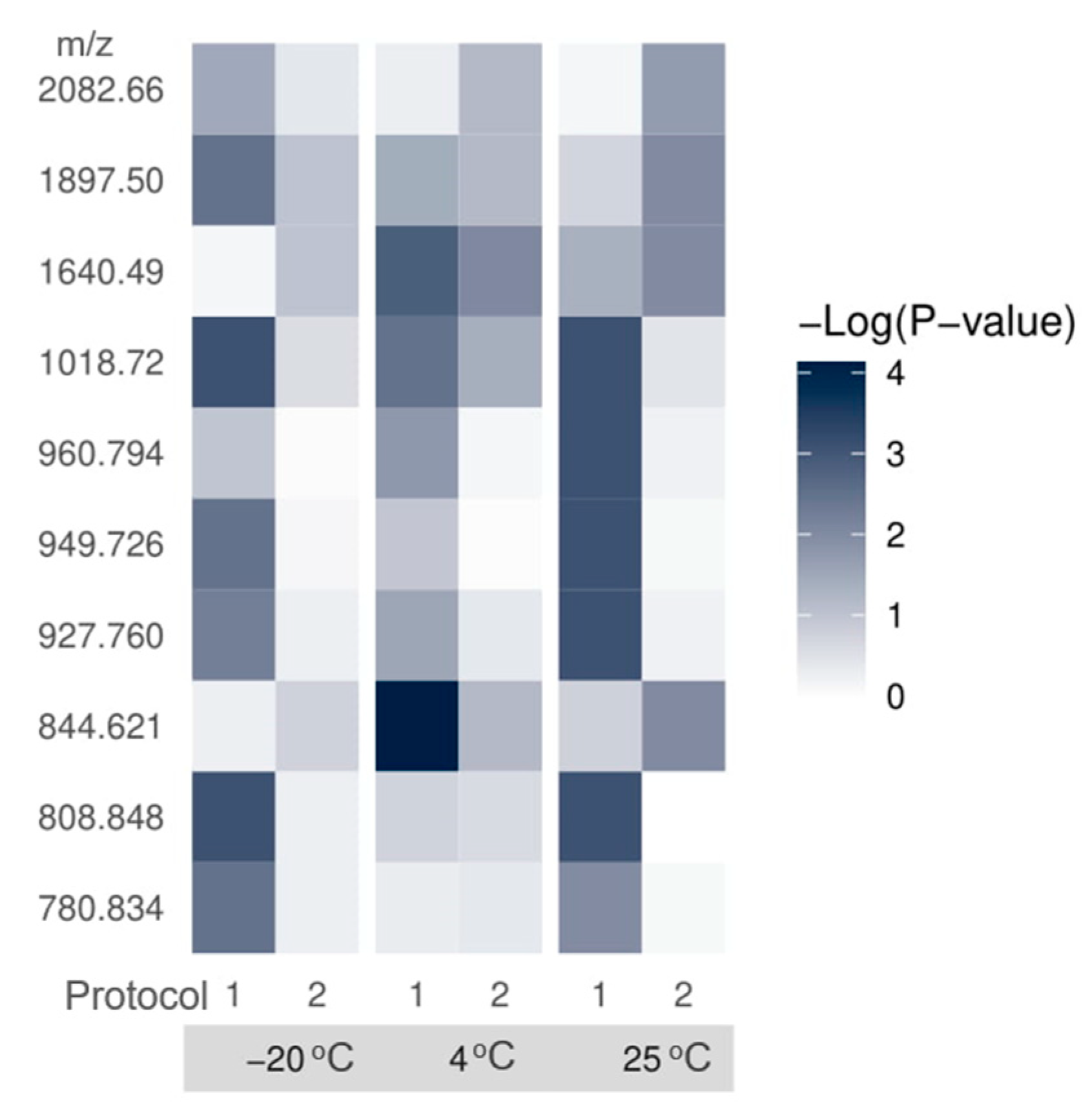
Table S1 (Supplementary Materials)Thus, we recommend using Protocol 2 for analytical measurements as procedures for preliminary preparation of DBS biosamples, which demonstrated the most stable result in our study (
Figure 2). Actually, we submit Protocol #2 as the most optimal choice for sample preparation referenced to mass spectrometry analysis (
Supplementary Materials, Figures S2 and S3). Nevertheless, we observed several
m/z-features that demonstrated mild instability during the specified storage time. Changes of the following measured and detected features (m/z = 780.834; m/z = 808.848; m/z = 949.726 and m/z = 960.794) were statistically insignificant. Notwithstanding, we did not exclude these data from the list of sample stability signatures because the list was populated and aligned for both protocols utilized in this study, and the exhausting data does not significantly impact the final results.
It should be admitted, that the variability of results, in the sense, is not limited to the measuring system. The latter can be neglected if several consequent replications are performed purposed to estimated intra- and inter-day accuracy. The larger input in variability is supplied with other steps of the sample custody and preparation. These include (1) departures from the standardized protocol of pre-analytical sample processing; (2) faults in mass spectrometry conditions that can be not optimal for measurement and detection, insufficient calibration of mass spectrometer; and (3) departures and flaws of data analysis rules. In this assay, we aware of the responsibility for each step in the designed study and attempted to neutralize the possible errors by means of technical and experimental replications and careful compliance with sample preparation protocols downstream mass spectrometry analysis.
We observed the expected alteration of the selected m/z-signatures caused by the influence of high-temperature custody conditions on the DBS samples stability. Insofar the presented study was designed as an application study, the obtained results admitted to justify the selected protocol of sample preparation and conditions which included six biological samples, three storage temperatures, and four time points of sampling. Each measurement has been performed in three replicates which gave a library of more than 400 spectra in total sufficient for the robust statistical data analysis. The obtained results would be beneficial for researchers who implicate in proteomic assays and schedule to utilize DBS for sample storage and transportation.
4. Conclusions
In recent years, the DBS approach has been widely used in various fields in clinical and biological research. In this study, we used a thin, labeled membrane made of porous hydrophilic glass fiber material. Unlike the traditionally used cellulose carriers, the glass fiber is characterized by numerous mechanical (high rigidity and resistance to deformations) and structural (solid stricture of fiber) advantages [
27]. In contrast to the hollow cellulose fibers, the integrity structure of glass fibers completely excludes possible penetration of the deposited sample and, thus, increases sample recovery during elution. Besides, the specific structure of glass fiber completely excludes a chromatography-like separation effect while absorbing biological fluid. Glass fiber membranes also provide an opportunity for quantitative analysis of blood samples which is hard to accomplish on the cellulose membranes due to the known effect of hematocrits [
27].
Most studies demonstrate long-term preservation of biomaterial components on membrane carriers, which confirms the wide applicability and reliability of this method. In the present study, we demonstrated the possibility of storing blood plasma samples on a membrane carrier for up to 35 days, which was a fiberglass strip. In our study, we have shown that the most appropriate method for preliminary preparation of DBS samples for subsequent MS analysis is procedures performed directly in a membrane carrier with subsequent extraction of components for mass spectrometric measurements. We have shown, using the example of tryptic components of proteins, that the composition of biosamples remains stable at −20 and + 4 °C for up to 35 days of storage, and at +25 °C for 14 days.
Despite its potential limitations, DBS samples are undoubtedly the most ethical and inexpensive way to collect, deliver and store biomaterial.

 ,
, 

