Article

Extraction Process, Identification of Fatty Acids, Tocopherols, Sterols and Phenolic Constituents, and Antioxidant Evaluation of Seed Oils from Five Fabaceae Species

1
Institute of Natural Products Chemistry, Vietnam Academy of Science and Technology (VAST), 18 Hoang Quoc Viet St., Caugiay Dist., Hanoi 100803, Vietnam
2
Graduate University of Science and Technology, Vietnam Academy of Science and Technology (VAST), 18 Hoang Quoc Viet St., Caugiay Dist., Hanoi 100803, Vietnam
3
Thai Nguyen University of Agriculture and Forestry, Thai Nguyen University, Thai Nguyen Province 23000, Vietnam
4
School of Natural Sciences Education, Vinh University, Vinh City 43100, Vietnam
5
NTT Hi-Tech Institute, Nguyen Tat Thanh University, Ho Chi Minh City 70000, Vietnam
6
Center of Excellence for Biochemistry and Natural Products, Nguyen Tat Thanh University, Ho Chi Minh City 23000, Vietnam
*
Authors to whom correspondence should be addressed.
Processes 2019, 7(7), 456; https://doi.org/10.3390/pr7070456
Submission received: 25 June 2019 / Revised: 10 July 2019 / Accepted: 11 July 2019 / Published: 16 July 2019
(This article belongs to the Special Issue Green Separation and Extraction Processes)

Abstract

:
The present study aimed to extract seed oils and characterize the chemical composition, including fatty acid profiles, tocopherols, sterols, and total phenolics of oils and extracts from five Fabaceae seeds: Glycine soja, Vigna angularis, Phaseolus lunatus, Phaseolus vulgarisand, and Phaseolus coccineus. The composition and content of all substance layers in total lipids of the extracted seed oils from five Fabaceae species contain: polar lipid (PL), sterol (ST), diacylglycerol (DG), triacylglycerol (TG), free fatty acid (FFA), and hydrocarbon and wax (HC + W). Antioxidant activity determined by the 2,2-diphenyl-1-picrylhydrazyl (DPPH) method was also estimated. Among these examined samples, Phaseolus vulgarisand and Phaseolus coccineus seed oils showed high content of α-linolenic acid (59.39% and 49.38%, respectively). Linoleic acid was abundantly found in Vigna angularis (49.01%). Ferunic and caffeic acid, γ-tocopherol, and β-sistosterol were the main ingredients present in the species studied. The V. angularis seed extract displayed significant antioxidant activity.

1. Introduction

Although the plant biodiversity in Vietnam has compelled agricultural potential and utilization of bio-based resource to gain in importance, investigations on the chemical composition and economic feasibility of plants, especially plant seeds, have been scarce. In addition, recent calls for sustainable sources of nutrition have also increased the need for more knowledge about the composition of wild plant seeds from Vietnam [1]. Among the many seeds that are widely distributed in Vietnam, seeds of beans figure prominently due to their economic, nutritional, and medicinal values [2]. A wide array of nutrients can be found in beans, such as minerals, proteins, amino acids, and fatty acids [3]. Among them, fatty acids fundamentally compose lipid molecules, hormones, and cell membranes, serve as an energy source for cells, and play a key role in energetic, metabolic, and structural activities [4]. While fatty acids constitute saturated and unsaturated fatty acids, the latter are the components of interest as they are considered to be health-beneficial.
The importance of unsaturated fatty acids is accentuated by the fact that the two polyunsaturated fatty acids, linoleic and linolenic acids, are essential and unobtainable, except by dietary means, in the human body. To be specific, linoleic acid, an unsaturated omega-6 fatty acid found in some specific plants, including pumpkin seeds, canola oil, soy beans, and flaxseeds, plays an important role for a wide range of human biological organs, including the nervous, skeletal, and reproductive systems, and allows them to function healthily [5]. In the human body, linoleic acid is converted into docosahexanoic acid (DHA), and eicosapentanoic acid (EPA), which are both responsible for the reduction of cholesterol and inflammation, enhancement of brain functions, and prevention of cancers and autoimmune conditions [6,7,8,9]. In addition, linolenic acid might contribute to improving cardiovascular health, as suggested by a previous large-scale study where linoleic acid consumption was found to confer moderate protection from heart disease. In fat cells treated with linolenic acid, it was found that the activity of eight different genes responsible for the promotion of cholesterol and triglyceride production is curbed. Furthermore, it is advisable that an inclusion of 5%–10% of omega-6 fatty acids in daily protein intake could lower the risk of heart disease [10].
For linoleic acid, the fatty acid is common in the seeds of many plant foods including safflower, sunflower, soybean, pine nuts, pecans, Brazil nuts, and corn oils. Linoleic acid also accounts for 85 to 90 percent of dietary omega-6 fatty acids. The ratio between omega-3 and omega-6 fatty acids is recommended to range from 2:1 to 4:1. However, due to the popularity and high omega-6 fatty acid content of safflower, sunflower, soybean, and corn oils in the standard Western diet, it is estimated that the consumption of omega-6 polyunsaturated fatty acids is 14 times higher than that of omega-3 fatty acids. To be specific, while the recommended daily intake of linoleic acid ranges from 1 to 2 g, one tablespoon of Flaxseeds, canola oil, or walnuts have already contributed 2.2, 1.3, and 0.3 g of linoleic acid [11,12,13].
Despite the nutritional potentials of seeds and their derived products, records on the chemical composition of extracts from seeds grown in Vietnam have been lacking. In this study, five seeds in the Fabaceae family, one of the most consumed and widely produced seed species in Vietnam, were characterized for fatty acids, tocopherols, sterols, and phenolic constituents using chromatographic and spectrometric methods. In addition, antioxidant determination by the 2,2-diphenyl-1-picrylhydrazyl (DPPH) method was also performed.

2. Materials and Methods

2.1. Plant Seed Materials

The five Fabaceae seeds: Glycine soja (M7), Vigna angularis (M12), Phaseolus lunatus (M9), Phaseolus vulgaris (M14), and Phaseolus coccineus (M10) were collected from Phu Yen, Vietnam in 2018. Samples were stored at −4 °C at the Department of Organic Biochemistry, Institute of Natural Products Chemistry, Vietnam Academy of Science and Technology. Samples were botanically identified by Dr. Nguyen Quoc Binh (Department of Biology, Vietnam National Museum of Nature, Vietnam Academy of Science and Technology) and given herbarium numbers.

2.2. Oil Extraction and Purification

Seeds were extracted using Soxhlet extraction following the modified method of ISO/DIS 659 (1988) [14]. To be specific, for each sample, 10 g of crushed seeds, produced from a ball mill machine, underwent extraction in a Twisselmann apparatus with 200 mL of petroleum ether for 6 h. Obtained products were subjected to rotary evaporation at 40 °C and 25 torr to remove the solvent, followed by a gentle stream of nitrogen. The obtained product was stored at −20 °C until used.

2.3. Analysis of Fatty Acid, Tocopherol, Sterol Compositions and Phenolic Compounds

The fatty acid composition was determined via gas chromatography and a subsequent ISO draft standard method [15]. First, 10 mg of extracted oil was added, followed by the introduction of 25 μL of 2M sodium methanolate methanol solution in a vial containing 1 mL of petroleum ether. The closed vial was stirred vigorously for 1 min. Afterwards, centrifugation took place following the addition of 20 μL of water. The aqueous phase was separated and 20 μL of methyl orange in 0.1N HCl was added as a pH indicator. Prior to analysis, the final mixture underwent a thorough stirring. The chromatography instrument for determination of derivatives was a Hewlett-Packard Model 5890 Series II/5989 A, operating in combination with a 0.25 mm ZB-1 fused-silica capillary column (30 m × 0.25 μm i.d.; Phenomenex, Torrance, CA, USA), and a carrier gas of helium at a flow rate of 1.0 mL/min.
To determine tocopherol, HPLC analysis was adopted following a previous procedure [16]. A mixture containing 250 mg of oil and 25 mL of heptanes was used as the sample for analysis in the HPLC instrument. The instrument was a Merck–Hitachi low-pressure gradient system, equipped with an L-6000 pump and a Merck–Hitachi F-1000 fluorescence spectrophotometer (detector wavelengths at 295 nm for excitation and 330 nm for emission) used in conjunction with Chemstation software. The injection of the sample onto a Diol phase HPLC column (250 mm × 4.6 mm, i.d.; Merck, Darmstadt, Germany) was performed using a Merck 655-A40 autosampler. The flow rate of the column, 1.3 mL/min heptane/tert-butyl methyl ether (99 + 1, v/v), was used as the mobile phase. The results were expressed as mg vitamin E/100 g oil.
To determine sterol, the official method of the International Olive Oil Council was employed [17]. The determination commenced with the saponification of 15 g of oil sample using 50 mL of 2 N ethanolic potassium hydroxide solution. The resultant fraction that was unsaponifiable was added to chloroform, weighed to 20 mg, and applied on a basic silica thin layer chromatography (TLC) plate. The separation of sterol and triterpenediol fraction was performed by eluent mixture with hexane and diethyl ether 65:35 (v/v). A UV light was used for the determination of bands following a spraying with 2,7-dichlorofluorescein in 0.2% ethanolic solution, scraping with a spatula, and extraction with chloroform. The extract was then dried and trimethylsilyl ethers were formed by the conversion of sterols and triterpenediols using pyridine hexamethyldisilizane-trimethylchlorosilane (9:3:1, v/v/v). The resultant mixture was allowed to stabilize for 15 min and was subsequently subjected to centrifugation. Sterols were analyzed on a fused silica capillary column coated with 2% isopropanol/98% hexane (30 m × 0.32 mm i.d., film thickness 0.25 μm; Rtx-5: Restek Corporation, Bellefonte, PA, USA; 99 or HP-5: Agilent Technologies Inc., Little Falls, DE, USA). The analysis instrument was a Hewlett Packard series 6890 GC (Waldbronn, Germany) equipped with a split/splitless injector, an autosampler, and a flame ionization detector (FID). First, 1.0 mL of aliquot of 1 derivatized sample solution was split injected into the column (300 °C, split ratio 1:50). Isolation of constituents was performed isothermally at 300 °C, and the determination was carried out with the FID (at 310 °C). Individual peaks of sterols were identified by carrier gas of helium. Relative retention times (RRT) for sterols was determined with reference to a major sterol compound, β-sistosterol, whose RRT is equal to 1 as described by COI [18].
The total phenolic compound was determined as follows. First, 1 g of powdered seed was ultrasonically extracted with 5 mL of methanol at a concentration of 80% for 30 min at ambient temperature. Then, Whatman Grade 1 filter paper was used to filter the resultant supernatants. The filtrate was stored at 4 °C prior to total phenolic content analysis via the Folin–Ciocalteu method [19]. Results were expressed as milligram equivalents of gallic acid (GAE) (400–1000 mg/L) per grams of the sample. Average values were reported according to triplicate experimental attempts.
Thirteen phenolic acids were quantified via reversed phase high performance liquid chromatography (RP-HPLC). The instrument used was an Agilent Technologies 1100 series, operating in conjunction with an UV-Vis multiwavelength detector [20]. The separation was conducted at room temperature with a 250 mm × 4.6 mm, 4 µm Hypersil ODS C18 reversed phase column. Two solvents were used for the mobile phase. The first solvent, denoted as solvent A, consisted of acetonitrile, and the second solvent, denoted as solvent B, was H2O with 0.2% H2SO4. The flow rate was maintained at 0.5 mL min−1. The progress of the gradient was programmed into six consecutive phases. The duration of phase 1 to 6 was 12, 2, 4, 2, 4, and 4 min, respectively. The ratio of A/B of the solvent system used in phase 1 to 6 was 15/85, 40/60, 60/40, 80/20, 90/10, and 100/0, respectively. The sample for analysis was first subjected to filtration using a 0.45 µm membrane filter and was then injected at a volume of 20 µL. Identification of peaks were made by comparing observations at 280 nm with standards. Triplicate attempts were made for all determinations, in which a specified quantity of trans-2-hydroxycinnamic acid was used as an internal standard.
Antioxidant activity was determined with the DPPH radical scavenging method. For determination of the antioxidant activity of different extracts, the stable 1,1-diphenyl-2-picrylhydrazyl (DPPH) radical was used [21]. This method allows for quick determination of the antiradical power of an antioxidant by measurement of the decrease in the absorbance of DPPH at 515 nm. For each extract, different concentrations were tested. An aliquot (0.5 mL) of the DPPH solution (about 50 mg/100 mL) was diluted in 4.5 mL of methanol, and 0.1 mL of a methanolic solution of the extract was added. The mixture was shaken vigorously and allowed to stand for 45 min in the dark. The decrease in absorbance was measured at 515 nm against a blank (without extract) with a spectrophotometer. From a calibration curve established with different amounts of extract, the ED50 was calculated. The ED50 was the required concentration of an antioxidant to quench 50% of the initial DPPH radicals under the given experimental conditions.

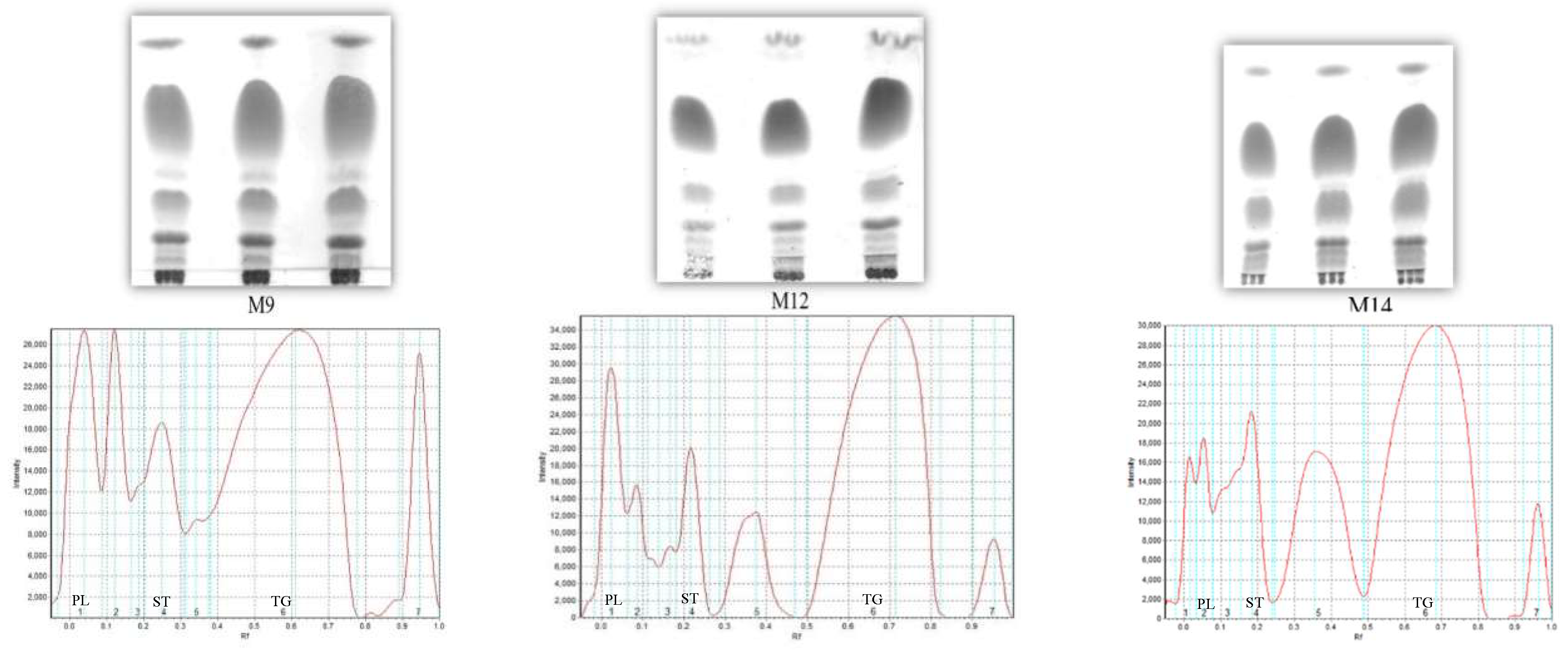
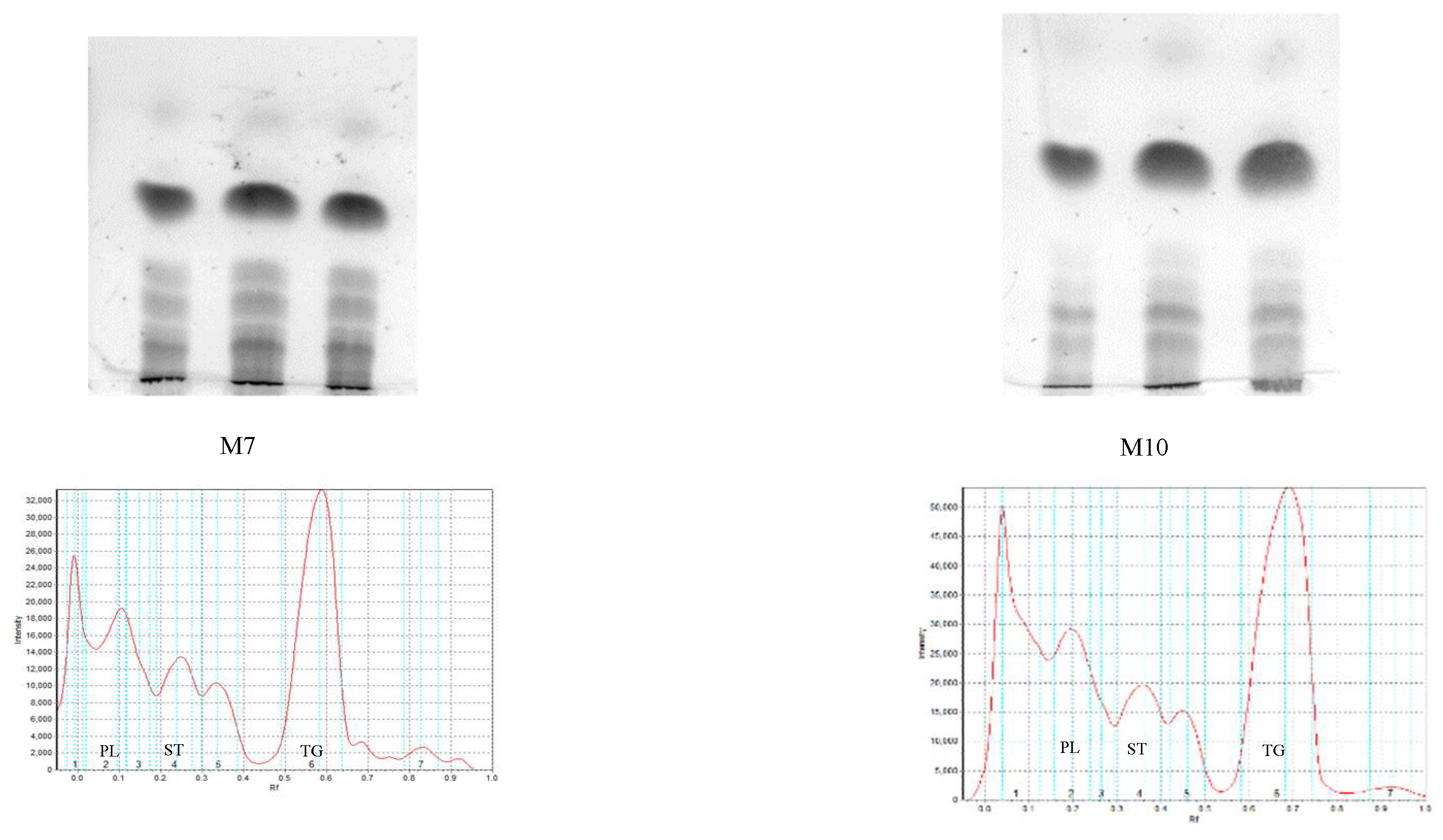
2.4. Composition and Content of Lipid Class Analysis

Total lipid was extracted from fresh seeds by the Bligh and Dyer method [22]. The seeds (10 g) were crushed, and the lipid was extracted at 4 °C for 24 h by stirring with a mixture of chloroform/methanol (30/60 mL, v/v). After that, the mixture was filtered and the biphasic system was obtained by adding chloroform (30 mL) and H2O (30 mL). After settling (24 h), the lower layer was separated, and the aqueous layer was subject to another extraction with chloroform (30 mL). The total lipid mixture, obtained by mixing and evaporating chloroform layers under low pressure, was stored at −20 °C for further investigation. The total lipid content was determined gravimetrically [23]. One-dimensional thin layer chromatography (TLC), with pre-coated silica gel plates (10 cm × 10 cm), Sorbfil PTLC-AF-V (Sorbfil, Krasnodar, Russia), was employed to determine total lipid compositions. First, development of the plates took place to full length with n-hexan/Et2O/AcOH (70/20/1, by vol.). Following that, the plates were developed to 25% length with CHCl3/MeOH/C6H6/NH4OH (65/30/5/10, by vol.). The plates were then air dried, sprayed with 10% H2SO4/MeOH, and heated at 240 °C for 10 min. To obtain chromatograms, an image scanner (Epson Perfection 2400 PHOTO), operating in a grayscale mode, was utilized. Lipid contents were quantified in percentages using an image analysis program (Sorbfil TLC Videodensitometer, Krasnodar, Russia) with band intensities as inputs. Peak areas and lipid class percentages were calculated according to the previously described method [24,25].

3. Results and Discussion

3.1. Lipid Content and Fatty Acid Composition

The total lipid contents of five collected seeds were in the range of 1.24 and 16.91% (Table 1). Only Glycine soja seed contained 16.91% oil whereas other seeds contained oil less than 3%. These results were in line with some previously reported results. To be specific, it was reported that the crude oil content varied from 2.8 to 18.0% with a mean of 10.2% for some Korean wild soybean (Glycine soja) lines [25] and from 1.3–2.3 g/100 g for some lima bean varieties [26]. The oils from five Fabaceae seeds were further investigated for fatty acid, tocopherol, and sterol compositions. Table 1 lists the fatty acid compositions of five samples, showing considerable compositional variation among seed samples. All seed oils shared several saturated fatty acids (SFA), but in different contents, including palmitic acid (16:0) or stearic acid (18:0), and some unsaturated fatty acids (USFA) such as oleic acid (18:1n-9), linoleic acid (18:2n-6), or α-linolenic acid (18:3n-3).
In five samples, USFA content was very high, ranging from 66.49% to 87.87% in which Phaseolus vulgarisa and Phaseolus coccineus species achieved the highest contents. This is a special case because α-linolenic acid (ALA) is the predominant USFA identified in P. vulgaris and P. coccineus (59.39% and 49.53%, respectively), making them valuable sources for this fatty acid. This ALA acid was also reported in considerable amounts in several plant samples including some green leafy vegetables, soybeans, linseed, rapeseed, flaxseed, and canola oils, as well as in phytoplankton, algae, and fish [27]. In some specific plant species with very high nutritional values, this ALA acid was found in excess amounts, such as in Salvia hispanica (64%), Perilla frutescens (58%), Linum usitatissimum (55%), and Phaseolus vulgaris (49%) [28]. The fatty acid composition patterns exhibited in the examined samples in this study are quite similar to those reported for seeds of Perilla frutescens, a species in the mint Lamiaceae family. The oil extracted from perilla seed oil exhibited strong anti-inflammatory activities and is one of the sources with the richest contents of omega-3 fatty acids, which are medicinally beneficial and important in preventing cardiovascular disorders, cancer, and inflammatory and rheumatoid arthritis. In addition, perilla leaves were also probed for anti-inflammatory properties [29].
Two other bean samples (P. lunatus and V. angularis) have a lower content of ALA acid, ranging from 17.77% to 26.10%. The content of oleic acid is low among the total fatty acids, ranging from 5.13% (P. coccineus) to 29.66% (G. soja). There are four bean samples containing oleic acid with content less than 7% of the total fatty acids and only one sample with a content of 29.66%, which is G. soja. Other momounsaturated fatty acids were also detected with three other acids including C16:1(n-7), C18:1(n-11), and C20:1(n-9), which have a very low content (<2.5% total fatty acid).
Several species were characterized by high amounts of saturated fatty acids including Phaseolus aureus (38.30%), P. lunatus (30.00%), and V. angularis (25.00%). Among the common saturated fatty acids, the content of palmitic acid ranged from 7.91% (P. vulgaris) to 23.42% (P. lunatus) while the content of stearic acid was marginal, ranging between 1.30% (P. coccineus) and 5.80% (P. lunatus). C22:0 behenic acid and C24:0 lignoceric acid were found in four of the seed oils in very low content.

3.2. Tocochromanol Composition

The seed oils were assessed with regard to two indicators, namely vitamin-E-active compounds and tocochromanols. The tocochromanol composition of the sample oils are presented in Table 2.
Similar to the previous result, the oils showed a wide range in the total tocochromanol content and composition. The content of tocochromanols varied from 14.85 mg/kg (G. soja) to 266.92 mg/kg (P. coccineus). With regard to the contents of tocochromanols, while G. soja, V. angularis, and P. lunatus contained tocochromanols at less than 25 mg/kg, the remaining species, including P. vulgaris and P. coccineus, showed very high amounts of tocochromanols of more than 100 mg/kg (164.55 and 266.92 mg/kg, respectively). Tocochromanol-rich seeds find a myriad of applications in the manufacture of food and functional products, medicine fields, and in stabilizing the oxidation of fats and oils. The health benefits of tocochromanols have been confirmed in various studies. Tocopherols in the examined species also possess significant antioxidant and anti-inflammatory activities, evidenced by high γ-tocopherol contents varying from 3.94 to 247.85 mg/kg. The highest amounts of γ-tocopherol were found in the seed oils of P. coccineus (247.85 mg/kg) and V. angularis (152.58 mg/kg). In addition, the amount of α-tocopherol in the examined seeds was relatively low (less than 20.90 mg/kg). Even though all principal tocopherols were found in the examined samples, the examined samples had lower tocopherol contents in comparison with those found in other legumes [30,31]. One of notable functions of tocopherols is to prevent the peroxidation of polyunsaturated fatty acids (PUFA) [32]. Evidently, in a previous analysis where the relationship between the level of tocopherol and unsaturation was explored in vegetable oils, it was found that γ-tocopherol was positively related with α-linolenic acid [33]. While α-tocopherol is considered to have significant nutritional value, recent evidence pointed out that γ-tocopherol may act as a superior risk-reducing factor for certain types of cancer and myocardial infarctions in comparison with α-tocopherol [33].

3.3. Sterol Composition

Several typical sterols were predominant in the samples, including campesterol, stigmasterol, ∆5,23-stigmastadienol, β-sitosterol, sitostanol, ∆5-avenasterol, and ∆7-stigmastenol (Table 3).
β-sitosterol was the major constituent, with the content ranging from 42.70 mg/g (P. lunatus) to 83.30 mg/g (V. angularis). β-sitosterol has been shown to exhibit multiple beneficial properties including anti-hypercholesterolemic, anti-inflammatory, antibacterial, antifungal, and anti-hyperlipoproteinemic activities. Other discovered functions of this component include the inhibition of carcinogenesis [34], the simultaneous reduction of glycated hemoglobin and serum glucose levels, and the promotion of seruminsulin levels [35]. With regard to stigmasterol, five examined samples possessed the constituent at varying content from 2.10 mg/g (V. angularis) to 39.00 mg/g (P. lunatus). Some typical sterols also appeared in large quantities in a few species such as campesterol in G. soja (15.70%).

3.4. Phenolic Acids Composition

Phenolic constituents in the samples were detected using a standard mixture consisting of thirteen phenolic acids. It was found that most standard compounds existed in the samples with contents ranging from 0.16 to 34.47 mg/g (Table 4). In general, ferulic acid and chlorogenic acid were the two components existing in most seed samples at considerable quantities. The content of ferulic acid varied from 2.08 mg/kg (P. lunatus) to 14.49 mg/kg (V. angularis). The content of chlorogenic acid varied from 1.89 mg/kg (G. soja) to 11.06 mg/kg (P. vulgaris). Caffeic acid only presents in four seed samples, and the percentages ranged from 0.32 mg/g (G. soja) to 8.00 mg/g (P. coccineus). There are three samples containing p-coumaric acid with content at less than 6 mg/g, including P. lunatus, at 11.41 mg/kg, and V. angularis, at 13.21 mg/kg. P. vulgaris seeds also had naringinin acid and taxifolin content at 6.03 and 3.99 mg/kg, respectively [34]. The values reported here are different from those reported for leguminous seeds (pea, bean, lentil, faba bean, broad bean, everlasting bean, and chickpea) elsewhere [36].

3.5. Composition and Content of Lipid Classes from Total Lipid

According to the result in Table 5 and Figure 1, the total lipid of bean seed samples is composed of six main layers with content fluctuating from 10% to 20%. Layers included polar lipid (PL), sterol (ST), triacylglycerol (TG), free fatty acid (FFA), diacylglycerol (DG), hydrocarbons, and wax (HC + W). Among them, triacylglycerol and polar lipid layers were found in the highest content in lima bean, bush bean, and red bean samples. In addition, free fatty acid content also occupied a relatively high proportion (average of 15% across the five samples). The content of the phospholipid layer was also high, suggesting the existence of a phospholipid class. This form of polar lipid has high biological activity and also presents abundantly in the soybean seed’s lipid.

3.6. Antioxidant Activity

The total extractable compounds (EC), total phenolic compounds (PC), and DPPH free radical scavenging activity of methanol extracts of five seed samples are provided in Table 6. The results demonstrated that all samples showed moderate antioxidant capacity, in which V. angularis appeared to have the most potential (Table 6). Methanol extracts of the five oil samples exhibited moderate antioxidant activity, as demonstrated by SC50 values ranging from 14.27 to 91.56 μg/mL. Ascorbic acid, which serves as the standard compound (Vitamin C), achieved an SC50 value of 26.48 μg/mL. The antioxidant potential of seeds showed that V. angularis varieties were characterized by the highest DPPH scavenging activity of 14.27 µg /mL, followed by the extracts of P. lunatus and G. soja seeds at 23.69 µg/mL–25.43 µg/mL. While the species with the strongest antioxidant potential, V. angularis, was abundantly composed of ferulic acid (14.49%) and β-Coumaric acid (13.21%), P. coccineus, the sample with the second highest antioxidant activities, was mainly composed of caffeic acid (8%).
As suggested by Oomah et al. [37], the antioxidant activity of bean phenolics could be best characterized by total phenolic content. To be specific, the antiradical activity could be largely described by total phenolics and the antioxidant activity is moderately related to flavonols with the correlation degree varying with bean type. Due to redox properties, phenolics may take the role of reducing agents, hydrogen donors, and singlet oxygen quenchers, in turn enhancing antioxidant activity. Among five seed extracts, the Vigna angularis seed extract showed the highest content of total phenolic content (18.46 mg/g), followed by the P. coccineus extract at 16.11 mg/g.

4. Conclusions

The present study compared the total lipid, the composition of fatty acids, tocopherols, sterols, phenolics, and the antioxidant potential of five common bean varieties with white coats in Vietnam. The most dominant fatty acid in P. vulgaris seed oil was polyunsaturated α-linolenic acid (ALA) with an abundance of 59.39% and 49.53% in P. vulgaris and P. coccineus seed oil, respectively. V. angularis seed oil was the richest source of linoleic acid content (49.01%), total phenolic content (18.46 mg/g), ferulic acid (14.49%), and β-Coumaric acid (13.21%) and had higher antioxidant activity than other seed beans. Our findings suggest that phenolic extracts from seed beans have high commercial potential as antioxidant sources for use in food or cosmetics.

Author Contributions

Investigation, T.T.N., M.Q.P., Q.T.T., Q.L.P., D.Q.T., and V.T.T.; writing—original draft, L.P.D.; writing—review and editing, L.G.B.

Funding

The authors are thankful to the Ministry of Science and Technology, Vietnam (44/2014/HĐ-NĐT) for financial support of the present research.

Conflicts of Interest

The authors declare no conflict of interest.

References

  1. Matthäus, B.; Vosmann, K.; Long, P.Q.; Aitzetmüller, K. Fatty acid and Tocopherol Composition of Vietnamese Oilseeds. J. Am. Oil Chem. Soc. 2003, 80, 1013–1020. [Google Scholar] [CrossRef]
  2. Lanza, E.; Hartman, T.J.; Albert, P.S.; Shields, R.; Slattery, M.; Caan, B.; Paskett, E.; Iber, F.; Kikendall, J.W.; Lance, P.; et al. High dry bean intake and reduced risk of advanced colorectal adenoma recurrence among participants in the polyp prevention trial. J. Nutr. 2006, 136, 1896–1903. [Google Scholar] [CrossRef] [PubMed]
  3. Saraçoðlu, H.T.; Zengin, G.; Akin, M.; Aktümsek, A. A comparative study on the fatty acid composition of the oils from five Bupleurum species collected from Turkey. Turk. J. Biol. 2012, 36, 527–532. [Google Scholar]
  4. Baum, S.J.; Kris-Etherton, P.M.; Willett, W.C.; Lichtenstein, A.H.; Rudel, L.L.; Maki, K.C.; Whelan, J.; Ramsden, T. Fatty acids in cardiovascular health and disease: A comprehensive update. J. Clin. Psychiatry 2012, 6, 216–234. [Google Scholar] [CrossRef] [PubMed]
  5. Nutrient Data Laboratory Home Page. USDA National Nutrient Database for Standard Reference, Release 20; U.S. Department of Agriculture, Agricultural Research Service: Beltsville, MD, USA, 2007.
  6. Campos, H.; Baylin, A.; Willett, W.C. Alpha-linolenic acid and risk of nonfatal acute myocardial infarction. Circulation 2008, 118, 339–345. [Google Scholar] [CrossRef] [PubMed]
  7. Nguemeni, C.; Delplanque, B.; Rovere, C.; Simon-Rousseau, N.; Gandin, C. Dietary supplementation of alpha-linolenic acid in an enriched rapeseed oil diet protects from stroke. Pharmacol. Res. 2010, 61, 226–233. [Google Scholar] [CrossRef]
  8. Sanderson, P.; Finnegan, Y.E.; Williams, C.M.; Calder, P.C.; Burdge, G.C. UK Food Standards Agency alpha-linolenic acid workshop report. Br. J. Nutr. 2002, 88, 573–579. [Google Scholar] [CrossRef]
  9. Gross, M.D. Lipids, Oxidation, and Cardiovascular Disease. In Atherosclerosis and Oxidant Stress; Springer: Berlin, Germany, 2008; pp. 79–95. [Google Scholar]
  10. Franzen-Castle, L.D.; Paula-Ritter, G. Omega-3 and Omega-6 Fatty Acids; NebGuide—University of Nebraska, Institute of Agriculture and Natural Resources: Lincoln, NE, USA, 2010. [Google Scholar]
  11. Simopoulos, A.P. Omega-3 fatty acids in wild plants, nuts and seeds. Asia J. Clin. Nutr. 2002, 11, 63–173. [Google Scholar] [CrossRef]
  12. Burdge, G.C.; Calder, P.C. Dietary alpha-linolenic acid and health-related outcomes: A metabolic perspective. Nutr. Res. 2006, 19, 26–52. [Google Scholar] [CrossRef]
  13. Stark, A.H.; Crawford, M.A.; Reifen, R. Update on alpha-linolenic acid. Nutr. Res. 2008, 66, 326–332. [Google Scholar] [CrossRef]
  14. International Organization for Standardization. Oil Seeds—Determination of Oil Content; Standard No. 659; ISO: Geneva, Switzerland, 1988. [Google Scholar]
  15. International Organization for Standardization. Animal and Vegetable Fats and Oils—Preparation of Methyl Esters of Fatty Acids; Standard No. 5509; ISO: Geneva, Switzerland, 1988. [Google Scholar]
  16. International Organization for Standardization. Animal and Vegetable Fats and Oils—Determination of Tocopherol and Tocotrienol Contents by High-performance Liquid Chromatography; Standard No. 9936; ISO: Geneva, Switzerland, 2006. [Google Scholar]
  17. International Olive Council. Determination of the Composition and Content of Sterols by Capillary Column Gas Chromatography; COI/T, 20/Doc. No. 10 Rev; Madrid, Spain, 2001. [Google Scholar]
  18. International Olive Council. Determination of the Composition and Content of Sterols and Triterpenedialcohols by Capillary Column Gas Chromatography; COI/T, 20/Doc. No. 30/Rev; Madrid, Spain, 2013. [Google Scholar]
  19. Saeed, N.; Khan, M.R.; Shabbir, M. Antioxidant activity, total phenolic and total flavonoid contents of whole plant extracts Torilis leptophylla L. BMC Complement. Altern. Med. 2012, 16, 221–233. [Google Scholar]
  20. Radwan, S.S.; Mangold, H.K. Lipids in plant tissue cultures V. Effect of environmental conditions on the lipids of Glycine soja and Brassica napus cultures. Chem. Phys. Lipids 1975, 14, 87–91. [Google Scholar] [CrossRef]
  21. Taga, M.S.; Miller, E.E.; Pratt, D.E. Chia seeds as a source of natural lipid antioxidants. J. Am. Oil Chem. Soc. 1984, 61, 928–931. [Google Scholar] [CrossRef]
  22. Bligh, E.G.; Dyer, W.J. A rapid method for total lipid extraction and purification. Can. J. Biochem. Physiol. 1959, 37, 911–917. [Google Scholar] [CrossRef] [PubMed]
  23. Hamoutene, D.; Puestow, T.; Miller-Banoub, J.; Wareham, V. Main lipid classes in some species of deep sea corals in the Newfoundland and Labrador region (Northwest Atlantic Ocean). Coral Reefs 2008, 27, 237–246. [Google Scholar] [CrossRef]
  24. Khotimchenko, S.V.; Kulikova, I.V. Lipids of different parts of the lamina of Laminaria japonica Aresch. Bot. Mar. 2000, 43, 87–141. [Google Scholar] [CrossRef]
  25. Mabaleha, M.B.; Yeboah, S.O. Characterization and compositional studies of the oils from some legume cultivars, Phaseolus vulgaris, grown in southern Africa. J. Am. Oil Chem. Soc. 2004, 81, 361–364. [Google Scholar] [CrossRef]
  26. Krupa, U. Main nutritional and antinutritional compounds of bean seeds Polish. J. Food Nutr. Sci. 2008, 58, 149–155. [Google Scholar]
  27. Seed Oil Fatty Acids Database. Available online: http://sofa.mri.bund.de (accessed on 1 July 2019).
  28. Michiyuki, K.; Masao, O.; Seisuke, I. Composition and molecular species of ceramide and cerebroside in scarlet runner beans (Phaseoluscoccineus, L.) and kidney beans (Phaseolus vulgaris L.). J. Agric. Food Chem. 1991, 39, 1709–1714. [Google Scholar]
  29. Asif, M. Health effects of omega-3, 6, 9 fatty acids: Perillafrutescens is a good example of plant oils. Orient. Pharm. Exp. Med. 2011, 11, 51–59. [Google Scholar] [CrossRef]
  30. Ramón, A.; Laura, R.; Miguel, A.A.; Francisca, G. Effect of Various Compounds on Virgin Olive Oil Stability Measured by Rancimat. J. Agric. Food Chem. 2011, 47, 4150–4155. [Google Scholar]
  31. Loizou, S.; Lekakis, I.; Chrousos, G.P.; Moutsatsou, P. Beta-sitosterol exhibits anti-inflammatory activity in human aortic endothelial cells. Mol. Nutr. Food Res. 2010, 54, 551–559. [Google Scholar] [CrossRef] [PubMed]
  32. Ju, J.; Hao, X.; Lee, M.J.; Lambert, J.D.; Lu, G.; Xiao, H.; Newmark, H.L.; Yang, C.S. A gamma-tocopherol-rich mixture of tocopherols inhibits colon inflammation and carcinogenesis in azoxymethane and dextran sulfate sodium-treated mice. Cancer Prev. Res. (Phila.) 2009, 2, 143–152. [Google Scholar] [CrossRef] [PubMed]
  33. Matthaus, B.; Ozcan, M.M. Chemical Evaluation of Flower Bud and Oils of Tumbleweed (Gundeliatourneforti L.) as a New Potential Nutrition Sources. J. Food Biochem. 2011, 35, 1257–1266. [Google Scholar] [CrossRef]
  34. Ling, W.H.; Jones, P.J.H. Dietary phytosterols: A review of metabolism, benefits and side effects. Life Sci. 1995, 57, 195–206. [Google Scholar] [CrossRef]
  35. Shubrook, J.H.; Shubrook, J. Risks and benefits of attaining HbA(1c) goals: Examining the evidence. J. Am. Osteopath. Assoc. 2010, 11, 7–12. [Google Scholar]
  36. Rocha-Guzmán, N.E.; Herzog, A.; González-Laredo, R.F.; Ibarra-Pérez, F.J.; Zambrano-Galván, G.; Gallegos-Infante, J.A. Antioxidant and antimutagenic activity of phenolic compounds in three different colour groups of common bean cultivars (P. vulgaris). Food Chem. 2007, 103, 521–527. [Google Scholar] [CrossRef]
  37. Oomah, B.D.; Cardador-Martinez, A.; Loarca-Pina, G. Phenolics and antioxidative activities in common beans (Phaseolus vulgaris L.). J. Sci. Food Agric. 2005, 85, 935–942. [Google Scholar] [CrossRef]
Figure 1. Thin layer chromatography (TLC) images and chromatograms of content of lipid layers of bean seeds. Polar Lipid (PL), Sterol (ST), Triacylglycerol (TG).
Processes 07 00456 g001aProcesses 07 00456 g001b
Table 1. List of compositions fatty acids from five Fabaceae seed samples.
SamplesM7M12M9M14M10
Fatty Acids
Total lipid (% weight of fresh seaweed)16.912.011.271.891.24
Fatty acids (% total fatty)
116:0Hexadecanoic acidPalmitic acid11.1312.6423.427.919.47
216:1(n-7)9-Hexadecenoic acidPalmitoleic acid0.090.620.170.260.15
317:0Heptadecanoic acidMargaric acid0.110.180.280.210.17
418:0Octadecanoic acidStearic acid2.763.255.801.621.30
518:1(n-9)cis-9-octadecenoic acidOleic acid29.666.565.615.575.13
618:1(n-11)cis-7-octadecenoic acidVaccenic acid0.080.461.142.162.11
718:2(n-6)cis-9,12-octadecadienoic acidLinoleic acid48.0949.0141.6420.3429.20
818:3(n-3)cis-9,12,15-octadecatrienoic acidLinolenic acid (ALA)6.7626.1017.7759.3949.53
920:0Eicosanoic acidArachidic acid (AA)0.380.481.010.390.38
1020:1(n-9)11-eicosenoic acidEicosenoic acid0.290.020.160.150.15
1122:0Docosanoic acidBehenic acid0.581.042.081.281.90
1224:0Tetracosanoic acidLignoceric acid0.070.180.920.720.51
Saturated15.0317.7733.5112.1313.73
Unsaturated84.9782.7766.4987.8786.27
Table 2. Tocopherol compositions (mg/Kg) of five seed oil samples.
Compositions (mg/Kg)α-Tα-T3β-Tγ-Tβ-T3P8γ-T3δ-Tδ-T3Sum
Samples
M72.17<LQ0.423.94<LQ<LQ<LQ8.31<LQ14.85
M121.412.70<LQ12.74<LQ<LQ<LQ1.06<LQ17.92
M9<LQ0.44<LQ19.960.15<LQ<LQ0.911.0022.47
M146.74<LQ0.35152.58<LQ2.02<LQ2.87<LQ164.55
M10<LQ5.15<LQ247.852.61<LQ<LQ10.700.62266.92
LQ: Limit of quantification.
Table 3. Sterol compositions (%) of five seed samples.
NameM7M9M14M10M12
Cholesterol0.600.100.100.100.10
Brassicasterol0.30<LQ0.10<LQ0.20
24-methylenecholesterol0.50<LQ0.10<LQ0.90
Campesterol15.702.505.204.708.50
Campestanol1.303.80<LQ<LQ<LQ
Stigmasterol15.4039.0028.6026.902.10
7-camersterol0.800.300.200.301.10
5,23-stigmastadienol0.70<LQ1.10<LQ<LQ
Chlerosterol0.300.90Nd1.100.70
β-sitosterol53.6042.7053.2057.8083.30
Sitostanol2.500.900.50NdNd
5-Avenasterol2.306.809.106.701.70
5,24-Stigmastadienol0.201.101.601.000.40
7-Stigmastenol4.500.700.200.900.80
7-Avenastenol1.301.300.100.700.10
Total100100100100100
LQ: Limit of quantification.
Table 4. Phenolic compositions (%) in the eight seed oil samples.
NameM7M9M14M10M12
Chlorogenic acid1.894.1311.066.545.15
Gallic acid0.61.853.062.621.70
Caffeic acid0.32<LOQ4.688.001.30
Vanillic acid<LOQ2.154.371.880.92
Isovanillic acid<LOQ<LOQ<LOQ1.741.66
Vanillin1.03<LOQ<LOQ<LOQ<LOQ
β-Coumaric acid5.1811.41<LOQ<LOQ13.21
Ferulic acid4.282.086.333.214.49
Taxifolin1.713.11<LOQ<LOQ3.99
Rosmarinic acid2.548.372.79<LOQ6.51
Daidzein<LOQ1.442.26<LOQ0.88
Cinnamic acid<LOQ1.670.64<LOQ1.17
Naringinin<LOQ0.716.03<LOQ3.25
Table 5. Lipid class composition (% of total lipid).
NoLipid LayersContent %
M7M9M14M10M12
1Polar Lipid (PL)18.5119.120.824.8822.5
2Sterol (ST)15.3719.310.79.598.3
3Free fatty acid17.5213.216.514.3115.3
4Triacylglycerol (TG)31.4229.637.735.5437.2
5Diacyl glyxerol7.815.58.28.159.5
6Wax Hydrocarbon9.3713.46.17.537.2
Total100100100100100
Table 6. Total extractable compounds (EC) [mg/g], total phenolic compounds (PC) [mg/g], and 2,2-diphenyl-1-picrylhydrazyl (DPPH) free radical scavenging activity of methanol extracts of five seed samples (SC50) [µg/mL].
NoCodeScientific NameEC (mg/g)PC (mg/g)DPPH Free Radical Scavenging Activity (SC50, µg/mL)
1M7Glycine soja1655.306425.43
2M9Phaseolus lunatus768.796523.69
3M14Phaseolus vulgaris67.412.921491.56
4M10Phaseolus coccineus8016.116168.60
5M12Vigna angularis80.518.469014.27
6Vitamin C 26.48

