Article

Research on Permeation Influencing Factors of Cosmetics UV Filters and Improve In Vitro Permeation Tests (IVPTs)

1
School of Perfume and Aroma Technology, Shanghai Institute of Technology, Shanghai 201418, China
2
The Oriental Beauty Valley Research Institute, Shanghai 201403, China
*
Author to whom correspondence should be addressed.
Processes 2023, 11(11), 3139; https://doi.org/10.3390/pr11113139
Submission received: 25 July 2023 / Revised: 11 October 2023 / Accepted: 30 October 2023 / Published: 2 November 2023

Abstract

:
The ideal UV absorber should be safe and should have excellent properties. Therefore, transdermal absorption is essential for the safety risk assessment of sunscreen cosmetics. The Franz diffusion cell method is the most common means of studying in vitro penetration, but there is a lack of standard methods for the in vitro permeation of UV absorbers. This paper used the Franz diffusion cell method to improve an in vitro permeation test (IVPT) for UV absorbers; three commonly used UV absorbers were tested: Octinoxate (EHM), Diethylaminohydroxybenzoyl hexyl benzoate (DHHB), and Ensulizole (PBSA). The final parameters were as follows: porcine ear skin was chosen for the membrane; the temperature of the receptor fluid was 37 °C; a PBS solution with 50% ethanol was chosen for the receptor fluid; and the dose of the test substance was 3 g. The improved IVPT method will help to accurately quantify the in vitro permeation of difficult-to-permeate components. In addition, the method can also be applied to evaluate the permeability of UV absorbers under different formulation conditions, which will help to address the difficulties related to the safety and application of sunscreen products.

1. Introduction

Solar UV radiation has recently increased due to climate change and air pollution. UV radiation can damage human skin [1]. In addition to physical sun protection, the safe and correct use of sunscreen products containing UV absorbers can also reduce the risk of UV radiation. The ideal UV absorber should be safe, bland, and have excellent broad-spectrum protection, photostability, chemical stability, and compatibility. However, in recent years, studies have found that some UV absorbers can penetrate the skin [2] and cause skin damage by causing photosensitivity, phototoxic reactions, and skin irritation [3]. In 2019, Matta et al. [2] evaluated whether the UV absorber components (avobenzone, oxybenzone, octocrylene, and ecamsule) of four commercial sunscreen products could be absorbed into the body’s circulation. The plasma concentrations of the four sunscreen ingredients exceeded thresholds under maximum use conditions, as revealed by their study. The transdermal absorption process describes the passage of a compound through the skin. The transdermal absorption of cosmetic products is mainly achieved through the stratum corneum. Its penetration depends largely on the properties of the skin [4], the physicochemical properties of the constituent ingredients of the cosmetic [5], and the nature of the cosmetic formulation [6]. Current permeation detection methods include in vitro methods (the Franz diffusion cell method, the 3D skin model permeation assay [7], and the confocal Raman spectroscopy method [8]) and in vivo methods [9] (the tape application method and the confocal Raman microscopy method). Compared to the in vivo methods, the in vitro permeation assays have the advantages of lower cost, ease of implementation, and excellent reproducibility.
The Franz diffusion cell method is the most used method in the study of in vitro permeation and is widely used in pharmaceutical and cosmetic applications [10]. Many factors influence the Franz diffusion cell method for assessing the in vitro permeation of cosmetic components, including the temperature [11] of the permeation experiment, the type of membrane [12], the composition of the receptor fluid, and the dose of the test substance [13]. Currently, human skin, porcine dorsal skin, porcine ear skin, rat skin, and artificial skin are used in the Franz diffusion cell as the cortices. The epidermal histological appearance of porcine skin is similar to that of human skin, and the follicular structure of porcine skin is identical to that of humans. Porcine ear skin typically has 20 hairs per cm on average. compared to 14–32 hairs per cm in humans [14], and the epidermal–dermal junction of the pig is similar to that of humans. A summary of 41 in vitro alternative studies of porcine skin by Barbero et al. [15] found 41 indices of permeability between porcine and human skin. The correlation coefficient (r) was 0.88 (p < 0.0001). Because of the high similarity between porcine and human skin characteristics, the pig is a suitable alternative animal model for in vitro skin permeation studies [16]. The WHO and OECD Guideline 428 [17] recommend a limited dose of 1–5 mg cm−2 or 10 µL cm−2 for permeation testing. The limited dose is the recommended dosage that closely corresponds with the consumer’s daily habits and actual usage. However, the limited dose can only be used to simulate the absorption of a substance and is not suitable for evaluating the safety of difficult-to-permeate substances due to the large deviations. Therefore, the infinite dose method should be used for assessment when comparing the permeation of various active ingredients [18]. An infinite dose is an applied dose that results in a negligible amount diffusing into the skin and concentration in the receiving pool. It helps to measure the substance’s steady-state flux, diffusion rate, and lag time [19]. Finite and infinite doses have different applications in transdermal drug delivery. It is difficult to find a good correlation between the in vitro penetration results of these two doses; consequently, the determination of the dose to be used is based on the primary properties of the chemicals [20]. As absorption by the skin is based on diffusion and obeys the diffusion principle [21] and Fick’s law [
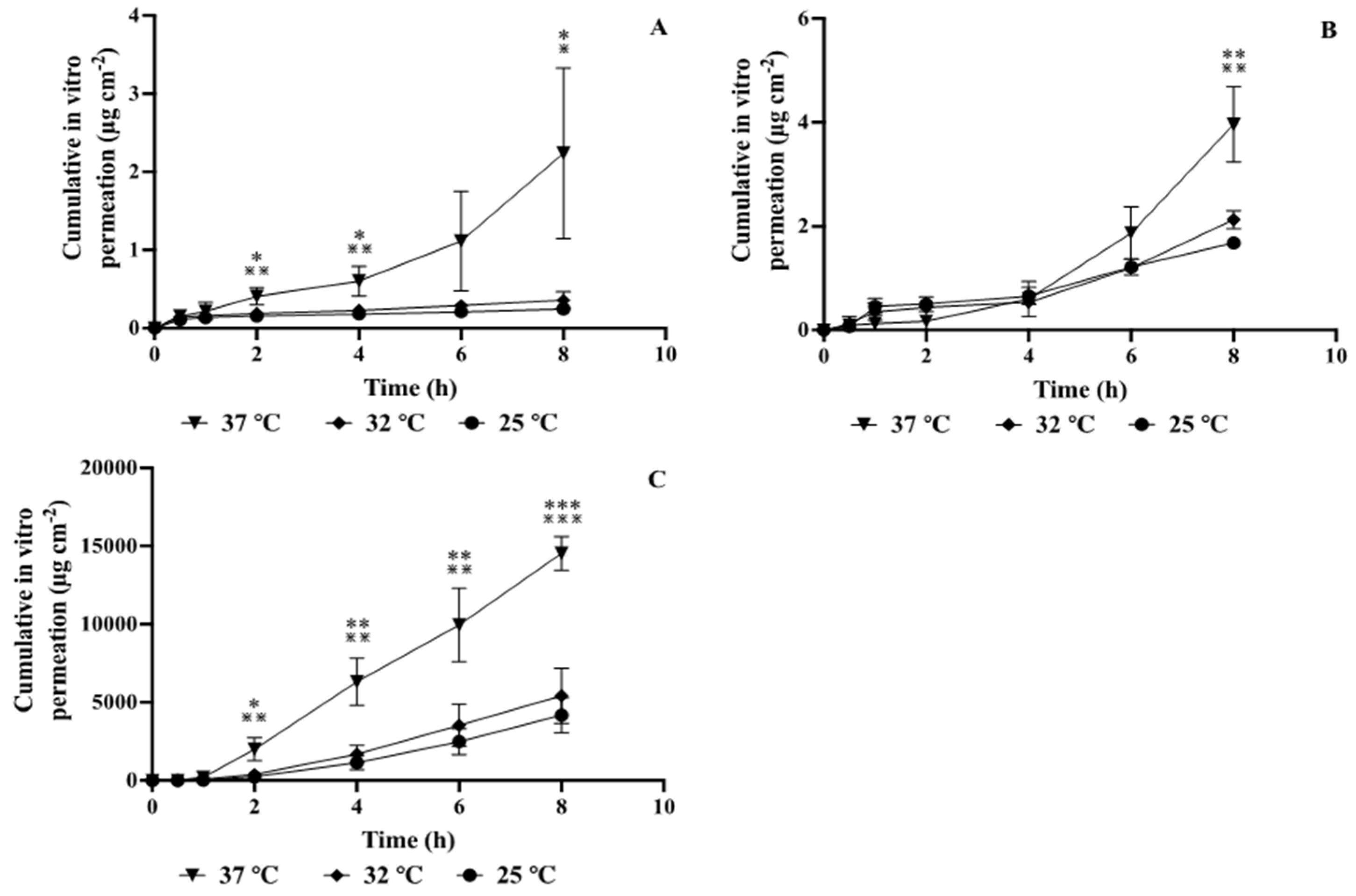
22], temperature affects the in vitro penetration of UV absorbers to a certain extent. In accordance with the recommendations of the OECD guidelines, a skin surface temperature of 32 °C is chosen for in vitro permeation experiments. As the human proximal skin and the core temperature are in the vicinity of 37 °C [23], a different commonly used temperature of 25 °C was chosen as the low temperature for the experiments. The composition of the receptor fluid significantly affects the permeation, and the receptor fluid must be both like the human environment and sufficiently soluble for the substance to be tested. Phosphate-buffered saline (pH 7.4) is close to the human environment in terms of pH and ionic concentration and is generally used for cosmetic studies of water-soluble substances. For substances that are difficult to dissolve in water, certain solubilizers, such as bovine albumin [5], polyethene glycol [13], and ethanol [6], are generally added to the phosphate-buffered saline (pH 7.4); according to OECD Guideline 428 (2004a, b) [24], a PBS-buffered solution containing 50% ethanol as a receptor fluid does not significantly affect the integrity of the skin. The sample’s formulation may affect the UV absorber’s penetration as the stratum corneum is lipophilic and the dermis is hydrophilic. The enhanced penetration of the compound may be due to an increase in diffusivity within the stratum corneum or an increase in the compound’s partitioning between the stratum corneum and the receiving fluid [
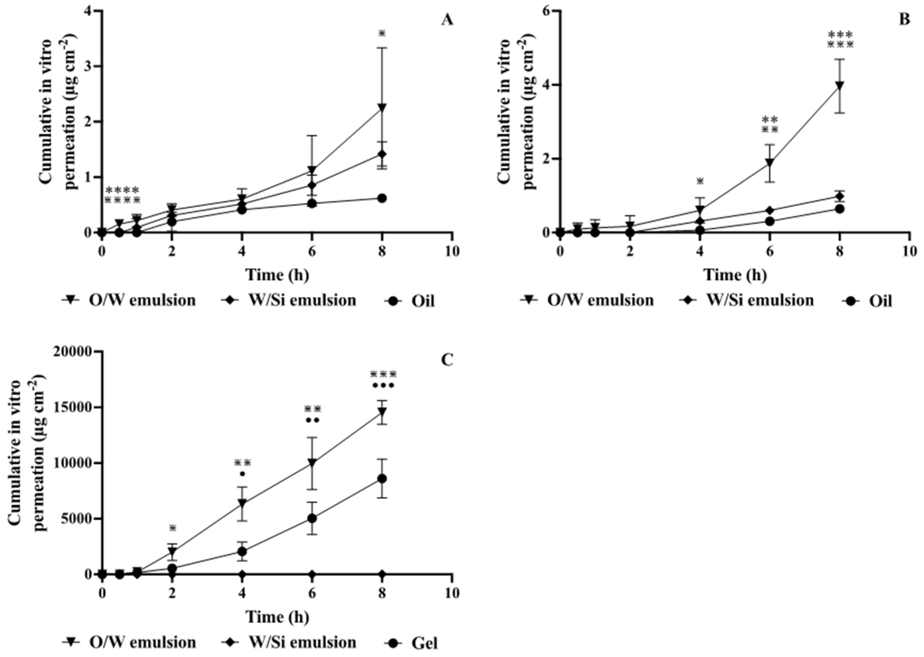
25]. For O/W emulsions, the amount of retention in the stratum corneum may be three times greater than that for W/O emulsions [26].
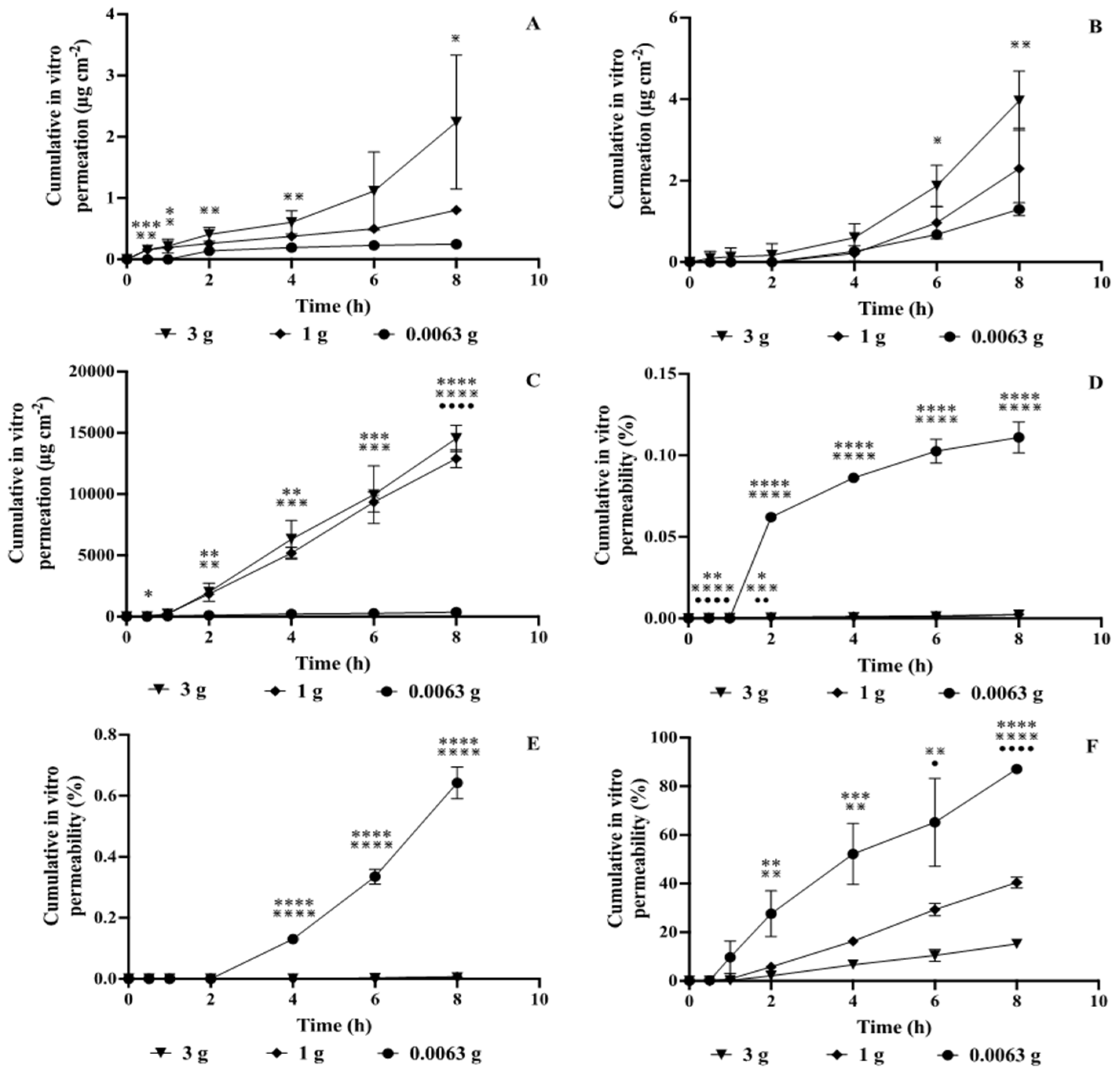
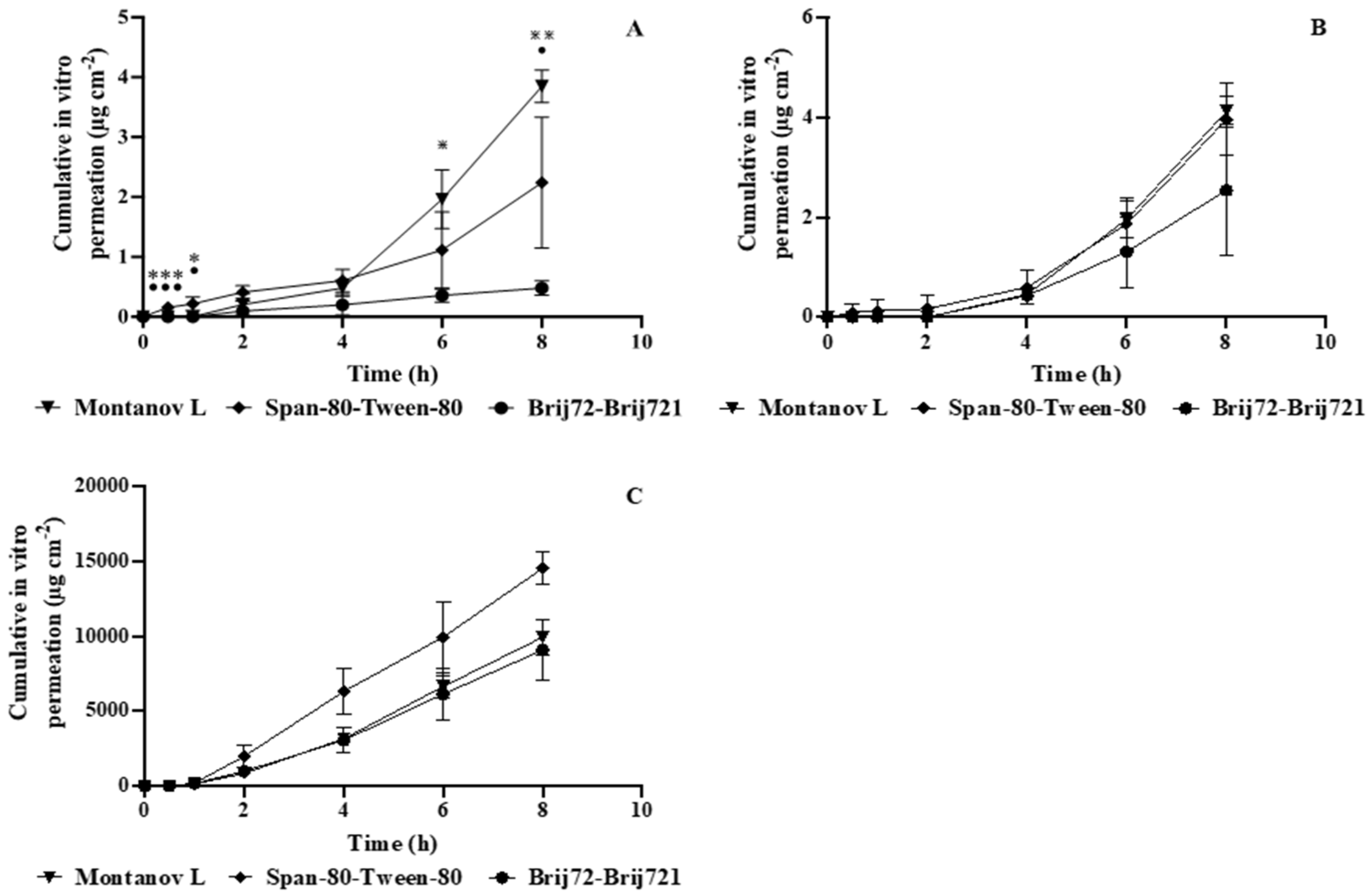
To date, there is no established IVPT for the UV absorbers for the human replacement of skin. Three commonly used UV absorbers (EHM, DHHB, and PBSA) were selected for this paper. (1) The IVPT parameters for the UV absorbers were evaluated and improved using a Franz diffusion cell to establish a theoretical basis for exploring clinical penetration test conditions. The parameters contained the temperature range (25, 32, 37 °C); the dose of the test substance (liquid supply 0.0063, 1, 3 g, i.e., 2, 318.47, 955.41 mg cm−2); the receptor fluid (PBS solution (pH = 7.4) and PBS solution with 50% ethanol); and the type of membrane (porcine ear skin, porcine dorsal skin, Strat-3M) for the diffusion cell’s receptor fluid. (2) To design safer sunscreen products, the evaluation employed the improved IVPT method to assess the penetration of UV absorbers in various formulation samples.

2. Materials and Methods

2.1. Materials

Ethylhexyl methoxycinnamate (Uvinul® MC 80, EHM) and diethylamino benzoyl hexyl benzoate (Uvinul® A plus, DHHB) was purchased from BASF (Ludwigshafen, Germany). TEA-Phenyl benzimidazole sulfonate (Parsol® HS, PBSA) was purchased from DSM (Heerlen, The Netherlands). Jojoba oil and caprylic/capric triglyceride were purchased from Evonik Industries AG (Essen, Germany). 1,2-pentanediol was purchased from Sigma-Aldrich (St. Louis, MO, USA). Sepimax Zen (polyacrylate cross polymer-6) and Montanov L (C14-22 Alcohol (and) C12-20 Alkyl Glucoside) were purchased from Seppic (Courbevoie, France). Stearyl polyether-2-21 was purchased from Croda (Cowick Hall, UK). Isopropyl myristate, ethanol, glycerol, Hansen gum Tween-80 (sorbitan monooleate) and Span-80 (polyoxyethylene (20) monooleate dehydrated sorbitol ester) were purchased from Greagent (Shanghai, China). PBS (phosphate-buffered saline) was purchased from Solarbio (Beijing, China). Methanol (HPLC grade) was purchased from Adamas (Shanghai, China). Formic acid (LC/MS grade) was purchased from Fisher (Hampton, NH, USA).

2.2. Preparation of Sunscreen Samples

The formulations of the O/W emulsions of EHM, DHHB, and PBSA are described in Table 1, Table 2, Table 3 and Table S1. The formulations of the W/Si emulsions of EHM, DHHB, and PBSA are described in Table 4, Table 5, Table 6 and Table S1.
The procedure for the preparation of the O/W and W/Si emulsions was as follows: Accurately weigh the components in phase A in beaker A; accurately weigh the deionized water in beaker B; then, disperse the remaining components in phase B in the aqueous phase one by one. Place beakers A and B in an HWS-12 electric thermostatic water bath (Shanghai Yiheng Scientific Instruments Co., Shanghai, China) at 85 °C. The solution in beaker B (RW 20 digital display overhead mechanical stirrer, IKA, Königswinter, Germany) was stirred for 5 min at 500 rpm and homogenized for 3 min at 13,200 rpm (T 18 high-speed disperser, IKA, Germany). After homogenization, stir at 300 rpm to reduce the temperature to 50 °C, add the C-phase components, and continue stirring until room temperature.
The formulation of the gel of PBSA is described in Table 7. The gel is prepared by accurately weighing the deionized water in a beaker; then dispersing the UV absorber, deionized water, glycerol, and Hansen gum one by one in the aqueous phase; heating the solution in beaker B to 80 °C while stirring at 500 rpm; then stirring at 300 rpm to lower the temperature; and then adding 1,2-pentanediol when the temperature drops to 50 °C and continuing to stir until room temperature.

2.3. Quantification of UV Filters

The quantification of the UV filters in the samples was analyzed with an Agilent 1220 series HPLC system equipped with an autosampler, a degasser, a quaternary pump, a diode array detector (DAD), and ChemStation software and separated on an Eclipse Plus C18 column (5 µm, 250 × 4.6 mm). The binary mobile phase consisted of 0.1% formic acid (v/v) (solvent A) and methanol-formic acid (99.9/0.1, v/v) (solvent B). The solvent gradient was as follows: 0–2.00 min, 32% B; 2.00–2.10 min, 70% B; 2.10–5.00 min, 70% B-80% B; 5.00–15.00 min, 85% B-90% B; 15.00–15.10 min, 90% B; 15.10–20.00 min, 100% B; 20.00–21.00 min, 100% B-32% B. The column temperature was 40 °C. The injection volume was 10 µL, and the flow rate was kept constant at 1.0 mL/min for a total run time of 21 min. The peaks were monitored at 307 nm (PBSA), 310 nm (EHM), and 361 nm (DHHB), respectively [27]. The peaks were tentatively identified by matching the retention time (tR) and UV absorption spectra with the standards.

2.4. Preparation of Skin Samples for Permeation Studies

The three types of membrane selected for this study were porcine ear skin, porcine dorsal skin, and Strat-3M. The porcine ear skin and porcine dorsal skin were prepared as described below.
The freshly excised porcine ears were carefully peeled out of the outer epidermis and cut into 3 × 3 cm2 pieces after the fat and fascia were removed from the skin. The pretreated porcine skin was stored at −20 °C and used within one month. Before the in vitro skin permeation experiments, the skin was thawed, and the integrity of the skin was checked according to Hewitt et al. [5]. The undamaged porcine ear skin with TEWL values of 25–35 g h-1 m-2 and thicknesses of 0.7–1.0 mm (porcine back skin thicknesses of 0.95–0.25 mm) was selected for further experiments was selected for further experiments.

2.5. In Vitro Skin Permeation Experiments

The method chosen for the in vitro skin permeation assay in this paper was that of Hewitt et al. [5]; the isolated skin (porcine dorsal skin, porcine ear skin, and Strat-3M) was mounted on a Franz vertical diffusion cell (TK-12D type transdermal diffusion tester, Shanghai Yuyan Scientific Instruments Co., Ltd., Shanghai, China) with an effective permeation area of 3.14 cm2 in the diffusion cell and the cuticle facing the supply cell. The receiving cell was filled with 8.0 mL of receptor fluid (PBS solution, PBS solution with 50% ethanol (v:v)), and the supply cell was filled with a specific dose (0.0063, 1, 3 g) of samples containing 10 wt% UV absorbers (O/W emulsion, oil, gel, and W/Si emulsion). The diffusion cell was placed in a constant-temperature water bath (25, 32, and 37 °C). The cell was stirred at a constant magnetic speed of 300 rpm, and 200 μL of receiver solution was aspirated at 0, 0.5, 1, 2, 4, 6, and 8 h, respectively, while 200 μL of blank receiver solution was added to the cell. The receiver solution was filtered through a 0.22 μm microporous membrane and then subjected to HPLC analysis. The cumulative permeate volume per unit area (Qn) was calculated using the following formula:
Q n = V C n + i = 1 n 1 C i V i A
where Qn denotes the total permeate per unit area (g cm−2); Cn is the UV absorber concentration (µg mL−1) measured at the nth sampling point; V is the total volume of the receptor fluid (mL); Ci is the drug concentration (µg mL−1) measured at the ith sampling point; Vi is the sampling volume (mL); and A represents the effective permeate area (cm2).
At the end of the permeation experiment, the UV absorber was recovered from the residual sample on the skin surface (Mw), the remaining skin sample (Ms), and the sample from the permeation experiment receptor fluid. The surface of the skin was washed three times with 1 mL of PBS solution containing 50% ethanol (v:v). Regarding the residual skin surface sample, the washing solution was collected and fixed to 10 mL with PBS solution containing 50% ethanol (v:v); the skin in the infiltrated area was cut off using medical surgical scissors and dried; this was followed by the addition of 10 mL of methanol and sonication at 50 Hz for 15 min (KQ-300DA CNC Ultrasonic Cleaner, Kunshan Ultrasonic Instruments Co., Ltd., Kunshan, China). The content of the UV absorber in each of the recovered samples was determined, and the permeation assay recovery (Vr) was calculated according to the following formula:
V r = M w + M s + A × Q n C u × M u × 100 %
Cu denotes the UV absorber concentration in the supply cell sample, and Mu denotes the total mass of the sample added to the supply cell.

2.6. Statistical Analysis

All the data were expressed as mean ± standard deviation (SD) and were statistically analyzed using Microsoft Excel 2023 (Microsoft) and GraphPad Prism 9.02 (GraphPad); differences between the groups for the three conditions were analyzed using one-way analysis of variance (ANOVA), followed by Tukey’s test (p < 0.05). The differences between the groups for the two conditions were analyzed using the Student’s t-test (p < 0.05).

3. Results and Discussion

3.1. Improvement of IVPT Conditions

3.1.1. Membrane Type

In this paper, we investigate the effect of three membrane types (Strat-3M, porcine ear skin, and porcine dorsal skin) on the in vitro permeation of UV absorbers. The penetration was measured at a temperature of 37 °C; the dose of the test substance was 1 g of O/W emulsion containing 10 wt% UV absorbers (EHM, DHHB, and PBSA); and a PBS solution with 50% ethanol was used as the receptor fluid (PBS solution for PBSA). The results are shown in Figure 1. After 8 h, the cumulative penetration of EHM and DHHB on Strat-3M was significantly higher than that on the porcine ear and dorsal skin (p < 0.0001). In comparison, the cumulative penetration of PBSA on the porcine ear skin was significantly higher than that on Strat-3M and porcine dorsal skin (p < 0.0001), while the cumulative penetration on Strat-3M was more than that on the porcine dorsal skin, but there was no significant difference.
In vitro, the transdermal absorption mainly occurred through the stratum corneum and the skin appendages (about 10% of the total skin absorption). The thicker epidermal layer of porcine dorsal skin compared to that of porcine ear skin (which can play a vital barrier role) may affect the amount of UV absorber penetration in porcine dorsal skin [28]. As shown in Figure 1, the penetration profiles of the UV absorber under porcine ear skin, porcine dorsal skin, and Strat-3M are comparable, with the pig ears receiving less penetration and a smaller coefficient of variation in the penetration. Depending on the location of the skin, porcine ear skin exhibits varying penetration level, its thickness, and the tissue properties of the skin, among other factors. In addition, the Strat-3M is composed of a multilayer structure with a total thickness of approximately 325 µm [29], mimicking the laminar structure and lipids of human skin, with the outermost layer consisting of two layers of porous polyolefin non-woven sulfone (PES), which simulates the stratum corneum barrier and prevents drug penetration into the skin. The two synthetic lipids form a polymeric membrane layer consisting of polyolefin non-woven fabric support (PNS), which contains various lipids, such as phospholipids and ceramides, which impart hydrophobicity to the skin. This gives Strat-3M its lipophilic properties [30,31]. This may lead to a higher cumulative penetration of the lipophilic UV absorbers EHM and DHHB at 8 h for Strat-3M than for the porcine ear and dorsal skin. For the hydrophilic UV absorber PBSA, the cumulative penetration at 8 h was higher in the porcine ear skin than in Strat-3M. In accordance with the principle of higher cumulative permeation and a lower coefficient of variation in the case of similar cumulative permeations, this paper selected the porcine ear skin with higher cumulative permeation and a lower coefficient of variation for the subsequent experimental study.

3.1.2. Dose of Test Substance

As UV absorbers do not readily penetrate the skin, we investigated the effect of two unlimited doses (1 and 3 g) and a limited dose (0.0063 g) on the in vitro penetration of UV absorbers. The penetration was measured at a temperature of 37 °C with porcine ear skin as the membrane and a PBS solution with 50% ethanol as the receptor fluid (PBS solution for PBSA); the results are shown in Figure 2. The cumulative permeation of EHM at a dose of 3 g was significantly higher than that of the sample with 0.0063 g added (p < 0.05), while the cumulative permeation at an additive amount of 3 g was greater than that of 1 g, and the cumulative permeation at an additive amount of 1 g was greater than that of 0.0063 g, but there was no significant difference. The cumulative permeation of DHHB at a dose of 3 g was significantly higher than that of the sample with 0.0063 g added (p < 0.005). The cumulative permeation of PBSA at 1 and 3 g was significantly higher than that at 0.0063 g (p < 0.0001), while the cumulative permeation at 3 g was more significant than that at 1 g. The cumulative permeation at 1 g was more significant than 0.0063 g but not significantly different. The cumulative infiltration at 3 g of the additive was more significant than that at 1 g, but there was no significant difference.
According to Fick’s law [22], the diffusive material flux of a substance per unit of time through a unit cross-sectional area perpendicular to the diffusion direction is proportional to the concentration gradient at that cross-section. All steps of skin absorption are based on diffusion, and a more significant concentration gradient leads to a greater diffusive flux and a greater in vitro penetration of the UV absorber. As shown in Figure 2, the cumulative penetration at dose additions of 1 and 3 g is higher than at dose additions of 0.0063 g. This may be because the total amount of UV absorber in the supply pool sample is not very high at the additive amount of 0.0063 g. As the penetration time increases, the concentration of the UV absorber on the supply pool sample decreases, which affects the in vitro penetration of the UV absorber. According to Figure 2, when the supply cell samples had a 0.0063 g dose, the penetration rate was higher compared to adding a 1.3 g dose as the amount of additive increased. This may be because the UV absorber’s cumulative penetration increases as the applied dose increases until the penetration becomes constant under infinite dose conditions [32]. The skin penetration is expected to decrease as the dose applied to the supply cell continues to increase. The method used in this study, with the highest cumulative penetration, was prioritized for subsequent experimental studies, following the principle of higher cumulative penetration. The sample with the highest cumulative penetration was chosen for subsequent experimental studies, following the principle of higher cumulative penetration in similar cases.

3.1.3. Receptor Media Temperature

In this paper, we investigated the effects of three receptor media temperatures (25, 32, and 37 °C) on the in vitro permeation of UV absorbers. The penetration was measured using porcine ear skin, with the dose of the test substance being 1 g of O/W emulsion containing 10 wt% UV absorbers (EHM, DHHB, and PBSA) and a PBS solution with 50% ethanol as the receptor fluid (PBS, solution for PBSA). The results are shown in Figure 3. After 8 h, the cumulative permeation of the three UV absorbers at a receptor fluid temperature of 37 °C was significantly higher than that at 25 and 32 °C (EHM: p < 0.05; DHHB: p < 0.005; PBSA: p < 0.0005). The cumulative permeation of the three UV absorbers at a receptor fluid temperature of 32 °C was higher than that at 25 °C, but there was no significant difference.
The stratum corneum is the main barrier to penetration, and changes in the highly ordered and densely arranged lipid matrix in the stratum corneum at elevated temperatures can affect the penetration of UV absorbers [11]. As shown in Figure 3, the cumulative penetration of the UV absorber increases with increasing temperature, and the cumulative penetration of the UV absorber is significantly higher at 37 °C than at 25 and 32 °C. Silva et al. [33] used high-speed differential scanning calorimetry to identify transitions in the state of the human stratum corneum and detected eight phase transitions, including those between 30 °C and 41 °C. At 30 °C, changes in the organization and mobility of the stratum corneum lipids occurred. At 41 °C, the lipids changed from an orthogonal structure to a hexagonal shape, and the entropy of the lipids increased. Apoorva Panda et al. [34] studied the effect of heat therapy on the drug release and skin permeability of nicotine transdermal patches and found in an in vitro permeation study that the transdermal permeation flux of nicotine from the patches was higher at 42 °C (100.1 ± 14.83 µg cm−2 h−1) than at 32 °C (33.3 ± 14.83 µg cm−2 h−1). Subsequent research revealed that, independent of the substance’s solubility at various temperatures, the main method of improving drug permeability under thermal treatment conditions was to increase skin permeability by reducing skin resistance and boosting skin TEWL. The higher cumulative permeation principle was used in this paper to select 37 °C with higher cumulative permeation for the next experimental study.

3.1.4. Receptor Media Composition

Due to the extremely poor water solubility of the oil-soluble UV absorbers EHM and DHHB, selecting a receptor fluid with appropriate settling conditions is crucial in permeation experiments [35]. In the previous study, we chose a PBS solution with 50% ethanol as the receptor fluid for oil-soluble UV absorber permeation experiments. In this paper, To study the permeation, we utilized pig ear skin with a liquid supply of 3 g 10 wt% UV absorber O/W emulsion at 37 °C. The different receptor fluid results are shown in Figure 4. After 8 h, the cumulative permeation of EHM in a PBS solution with 50% ethanol was higher than in a PBS solution, but there was no significant difference. The cumulative permeation of DHHB was significantly higher (p < 0.05) than that of the PBS solution with 50% ethanol in the receptor fluid. The cumulative permeation of PBSA was significantly higher (p < 0.0005) than that of the PBS solution with 50% ethanol in the receptor fluid. From the experimental results, the cumulative permeation of oil-soluble sunscreens was 0 at 8 h when phosphate-buffered saline (pH 7.4) was used as the receptor fluid, and it was even more significant at 8 h when the PBS solution with 50% ethanol was used as the receptor fluid. This paper chose the PBS solution with 50% ethanol as the higher cumulative permeate for the subsequent experimental study.

3.1.5. Comparative Analysis of Dosage Form under Improved In Vitro Conditions

In this paper, the penetrations of UV absorbers (EHM, DHHB, and PBSA) in different dosage forms were investigated according to the IVPT method improved in Section 3.1 and Section 3.2, and the results are shown in Figure 5. After 8 h, the cumulative permeation of EHM in the O/W emulsion was significantly higher than that in the oil agent (p < 0.05), where the cumulative permeation in the O/W emulsion was higher than that in the W/Si emulsion, and the cumulative permeation in the W/Si emulsion was higher than that in the oil agent, but not significantly. The cumulative permeation of DHHB in the O/W emulsion was significantly higher than that in the oil and W/Si emulsions (p < 0.0005), where the cumulative permeation in the W/Si emulsions was higher than that in the oil but not significantly. The cumulative permeation of PBSA in the O/W emulsion and gel was significantly higher than that in the W/Si emulsions (p < 0.0005), where the cumulative permeation in the O/W emulsions was higher than that in the gel, but not significantly. This paper’s permeation of the different dosage forms was ranked as follows: O/W emulsion ≈ gel > W/Si emulsion > pure oil agent. Lucia et al. [6] used the Franz diffusion cell method to evaluate the effects of the times of use and the different formulations (O/W, W/O cream, and mixed oil phase carrier) on the skin permeability of sunscreen. They found that compared with the O/W lotion, the amount of sunscreen in the W/O sunscreen was more. It was different in our case. This may be due to differences in other components in the formula.

3.2. Comparative Analysis of O/W Vehicle under Improved In Vitro Conditions

3.2.1. Emulsifier

The type of emulsifier may influence the emulsion’s penetration of UV absorbers. The penetration of UV absorbers (EHM, DHHB, and PBSA) in O/W emulsions with different emulsifiers (Montanov L, Span-80-Tween-80, and Brij-72-Brij-721) was investigated according to the IVPT improved in 3.1–3.2, and the results are shown in Figure 6. After 8 h, the cumulative permeation of EHM under the emulsions containing Montanov L emulsifier was significantly higher than that in Brij-72-Brij-721(p < 0.005)and Span-80-Tween-80 (p < 0.05). The cumulative permeation of DHHB and PBSA under the emulsions containing Montanov L emulsifier was higher than that in Span-80-Tween-80 and Brij-72-Brij-721 (p < 0.05). However, there was no significant difference. Regarding chemical structure, surfactants consist of lipophilic and hydrophilic head groups [36], and surfactants can interact with components on the skin or alter the saturation state of compounds within the formulation [37,38].
On the one hand, to modify the stratum corneum’s barrier properties, surfactants may interact with protein components and denature keratin or interact with lipids and increase their mobility. Surfactants may be inserted into the lipid bilayer of the stratum corneum, leading to bilayer interfacial defects or structural disruptions and facilitating the diffusion of substances [39]. On the other hand, surfactants form micelles, and the lubrication of drug molecules by surfactant micelles may reduce the thermodynamic activity of the drug, thereby reducing the diffusion of the substance [40]. The effect of surfactants on substance penetration combines these opposing effects.

3.2.2. Fats and Oils

The type of oil may influence UV absorber permeation in the emulsion. According to the IVPT method improved in Section 3.1 and Section 3.2, the permeation of UV absorbers (EHM, DHHB, PBSA) in O/W emulsions with different oils (jojoba oil, isopropyl myristate, caprylic/capric triglyceride) was investigated. In this study, we use Montanov L as the emulsifier; the results are shown in Figure 7. After 8 h, the cumulative permeation of EHM and PBSA under emulsions containing jojoba oil was higher than that under isopropyl myristate and caprylic/capric triglyceride, but there was no significant difference. The cumulative permeation of DHHB under the emulsion containing jojoba oil was significantly higher than that under isopropyl myristate and caprylic/capric triglyceride (p < 0.0001), and the cumulative permeation under the emulsion containing isopropyl myristate was significantly higher than that in the emulsions containing caprylic/capric triglyceride (p < 0.001).

3.2.3. Humectant

The humectant type may influence the emulsion’s penetration of UV absorbers, according to the IVPT method improved in Section 3.1 and Section 3.2. We investigated the permeation of UV absorbers (EHM, DHHB, and PBSA) in O/W emulsions with different humectants (glycerol, 1,3-butanediol, propylene glycol, and sodium hyaluronate). In this study, we used Montanov L as the emulsifier; the oil was jojoba oil. The results are shown in Figure 8.
The cumulative permeation of EHM in emulsions containing 1,3-butanediol and propylene glycol was significantly higher than that in sodium hyaluronate after 8 h (p < 0.05). The cumulative permeation of DHHB in emulsions containing 1,3-butanediol and propylene glycol was significantly higher than that in sodium hyaluronate (p < 0.005), and the cumulative permeation of DHHB in emulsions containing glycerol was significantly higher than that in sodium hyaluronate (p < 0.05). The cumulative permeation of PBSA in emulsions containing glycerol, 1,3-butanediol, and propylene glycol was higher than that in sodium hyaluronate (p < 0.0005), but there was no significant difference.

4. Conclusions

Assessing the in vitro penetration of cosmetic products using the Franz diffusion cell requires the determination of many factors (membrane type, temperature and composition of the receptor fluid, and dose of test substance). To facilitate accurate quantification of the in vitro permeation of UV absorbers in cosmetics, this paper improves the IVPT method for UV absorbers. The following test parameters were considered suitable for UV absorber permeation experiments: porcine ear skin was chosen for the membrane; the temperature of receptor fluid was 37 °C; a PBS solution with 50% ethanol was chosen for the receptor fluid; and the dose of the test substance was 3 g. The improved IVPT method facilitates accurate quantification of the in vitro permeation of difficult-to-permeate components, including UV absorbers.
In addition, this IVPT method was used to compare the permeation of UV absorbers in samples of different emulsion types and the effect of the three main components of the O/W emulsions (emulsifiers, oils, and humectants) on the permeation of the UV absorbers. On the one hand, it is realistic to apply the improved IVPT method. The permeation of the different dosage forms is ranked as follows: O/W emulsion ≈ gel > W/Si emulsion > oil agent. In O/W emulsions, the cumulative permeation of UV absorbers in the emulsions with jojoba oil was higher than that in isopropyl myristate and the caprylic/capric triglycerides, but there was no significant difference. On the other hand, To avoid the penetration of UV absorbers and create safer sunscreen products, it is necessary to opt for components with low or no penetration promotion.

Supplementary Materials

The following supporting information can be downloaded at https://www.mdpi.com/article/10.3390/pr11113139/s1, Table S1: The table of the permeation assay recovery.

Author Contributions

Conceptualization, W.Z.; methodology, L.G.; software, H.W. and S.Z.; formal analysis, C.C.; investigation, C.C.; writing—original draft preparation, C.C.; writing—review and editing, C.C. All authors have read and agreed to the published version of the manuscript.

Funding

This research received no external funding.

Data Availability Statement

The data presented in this study are available on request from the corresponding author.

Conflicts of Interest

The authors declare no conflict of interest.

References

  1. Krutmann, J. Sun-damaged skin (photoaging): What is new? Zeitschrift fuer Dermatologie, Venerologie, und verwandte Gebiete. Der Hautarzt 2021, 72, 2–5. [Google Scholar] [CrossRef] [PubMed]
  2. Matta, M.K.; Zusterzeel, R.; Pilli, N.R.; Patel, V.; Strauss, D.G. Effect of Sunscreen Application Under Maximal Use Conditions on Plasma Concentration of Sunscreen Active Ingredients: A Randomized Clinical Trial. JAMA J. Am. Med. Assoc. 2019, 321, 2082–2091. [Google Scholar] [CrossRef] [PubMed]
  3. Yang, K.; Song, S.; Zhang, Y.; Shen, S.; Xu, X.; Yue, Z. Programmed gene expression change in mouse skin after ultraviolet radiation damage. Exp. Dermatol. 2022, 31, 862–868. [Google Scholar] [CrossRef] [PubMed]
  4. Law, R.M.; Mai, A.N.; Maibach, H.I. Twenty Clinically Pertinent Factors/Observations for Percutaneous Absorption in Humans. Am. J. Clin. Dermatol. 2019, 21, 85–95. [Google Scholar] [CrossRef] [PubMed]
  5. Hewitt, N.J.; Grégoire, S.; Cubberley, R.; Duplan, H.; Eilstein, J.; Ellison, C.; Lester, C.; Fabian, E.; Fernandez, J.; Géniès, C. Measurement of the penetration of 56 cosmetic relevant chemicals into and through human skin using a standardized protocol. Wiley-Blackwell Online Open 2020, 40, 403–415. [Google Scholar] [CrossRef] [PubMed]
  6. Lucia, M.; Rita, T.; Carmela, P.; Lorella, P. In Vitro Evaluation of Sunscreen Safety: Effects of the Vehicle and Repeated Applications on Skin Permeation from Topical Formulations. Pharmaceutics 2018, 10, 27. [Google Scholar]
  7. Xiao, M.; Wang, F.; Wang, B. Application of an in vitro reconstructed human skin on cosmetics in skin irritation tests. J. Cosmet. Dermatol. 2020, 20, 1933–1941. [Google Scholar]
  8. Meng, R. Study on Skin Penetration of Chemical Sunscreens Based on Confocal Raman Imaging Technology. Master’s Thesis, Shanghai Normal University, Shanghai, China, 2021. [Google Scholar]
  9. Adlhart, C.; Baschong, W. Surface distribution and depths profiling of particulate organic UV absorbers by Raman imaging and tape stripping. Int. J. Cosmet. Sci. 2011, 33, 527–534. [Google Scholar] [CrossRef]
  10. Ravenzwaay, B.V.; Leibold, E. A comparison between in vitro rat and human and in vivo rat skin absorption studies. Hum. Exp. Toxicol. 2004, 23, 421–430. [Google Scholar] [CrossRef]
  11. Lacount, T.D.; Zhang, Q.; Hao, J.; Ghosh, P.; Li, S.K. Modeling Temperature-Dependent Dermal Absorption and Clearance for Transdermal and Topical Drug Applications. AAPS J. 2020, 22, 70. [Google Scholar] [CrossRef]
  12. Cubberley, R.; Duplan, H.; Hewitt, N.; Jacques-Jamin, R.; Eilstein, C. Comparison of protocols for measuring cosmetic ingredient distribution in human and pig skin. Toxicol. Vitr. Int. J. Publ. Assoc. Bibra 2016, 34, 153–160. [Google Scholar]
  13. Kim, K.B. Effect of Application Amounts on In Vitro Dermal Absorption Test Using Caffeine and Testosterone. Pharmaceutics 2021, 13, 641. [Google Scholar]
  14. Capt, A.; Luzy, A.P.; Esdaile, D.; Blanck, O. Comparison of the human skin grafted onto nude mouse model with in vivo and in vitro models in the prediction of percutaneous penetration of three lipophilic pesticides. Regul. Toxicol. Pharmacol. RTP 2007, 47, 274–287. [Google Scholar] [CrossRef] [PubMed]
  15. Barbero, A.M.; Frasch, H.F. Pig and guinea pig skin as surrogates for human in vitro penetration studies: A quantitative review. Toxicol. Vitr. 2009, 23, 1–13. [Google Scholar] [CrossRef] [PubMed]
  16. Jung, E.C.; Maibach, H.I. Animal models for percutaneous absorption. J. Appl. Toxicol. 2015, 35, 1–10. [Google Scholar] [CrossRef] [PubMed]
  17. OECD. Test No. 428: Skin Absorption: In Vitro Method. OECD Guidel. Test. Chem. 2006, 1, 1–8. [Google Scholar]
  18. Brain, K.R.; Walters, K.A.; Watkinson, A.C. Methods for Studying Percutaneous Absorption. In Dermatological and Transdermal Formulations; CRC Press: Boca Raton, FL, USA, 2002; pp. 197–270. [Google Scholar]
  19. Selzer, D.; Abdel-Mottaleb, M.; Hahn, T.; Schaefer, U.F.; Neumann, D. Finite and infinite dosing: Difficulties in measurements, evaluations and predictions. Adv. Drug Deliv. Rev. 2013, 65, 278–294. [Google Scholar] [CrossRef] [PubMed]
  20. Coderch, L.; Collini, I.; Carrer, V.; Barba, C.; Alonso, C. Assessment of Finite and Infinite Dose In Vitro Experiments in Transdermal Drug Delivery. Pharmaceutics 2021, 13, 364. [Google Scholar] [CrossRef]
  21. Brodin, B.; Steffansen, B.; Nielsen, C.U. Passive diffusion of drug substances: The concepts of flux and permeability. In Molecular Biopharmaceutics: Aspects of Drug Characterisation, Drug Delivery and Dosage form Evaluation; Pharmaceutical Press: London, UK, 2010; pp. 135–152. [Google Scholar]
  22. Fick, A. Ueber Diffusion. Ann. Der Phys. 1855, 170, 59–86. [Google Scholar] [CrossRef]
  23. Te Lindert, B.H.; Van Someren, E.J. Skin temperature, sleep, and vigilance. Handb. Clin. Neurol. 2018, 156, 353–365. [Google Scholar]
  24. Heylings, J.R. OECD 428: A new in vitro test guideline for human risk assessment following dermal exposure to industrial chemicals. In Proceedings of the BTS 2004 Annual Congress, Glasgow, UK, 4–5 September 2004. [Google Scholar]
  25. Ghafourian, T.; Nokhodchi, A.; Kaialy, W. Surfactants as penetration enhancers for dermal and transdermal drug delivery. In Percutaneous Penetration Enhancers Chemical Methods in Penetration Enhancement; Springer: Berlin/Heidelberg, Germany, 2015; pp. 207–230. [Google Scholar]
  26. Chatelain, E.; Gabard, B.; Surber, C. Skin Penetration and Sun Protection Factor of Five UV Filters: Effect of the Vehicle. Skin Pharmacol. Appl. Ski. Physiol. 2003, 16, 28–35. [Google Scholar] [CrossRef] [PubMed]
  27. Xiqin, M.; Haitao, B.; Baocheng, Q. Determination of 17 ultraviolet-filters in cosmetics by high performance liquid chromatography. Chin. J. Chromatogr. 2013, 31, 775–780. [Google Scholar]
  28. Todo, H. Transdermal Permeation of Drugs in Various Animal Species. Pharmaceutics 2017, 9, 33. [Google Scholar] [CrossRef]
  29. Haq, A.; Goodyear, B.; Ameen, D.; Joshi, V.; Michniak-Kohn, B. Strat-M Synthetic Membrane: Permeability Comparison to Human Cadaver Skin. Int. J. Pharm. 2018, 547, 432–437. [Google Scholar] [CrossRef] [PubMed]
  30. Uchida, T.; Kadhum, W.R.; Kanai, S.; Todo, H.; Oshizaka, T.; Sugibayashi, K. Prediction of skin permeation by chemical compounds using the artificial membrane, Strat-M™. Eur. J. Pharm. Sci. Off. J. Eur. Fed. Pharm. Sci. 2015, 67, 113–118. [Google Scholar] [CrossRef] [PubMed]
  31. Clément, P.; Laugel, C.; Marty, J.P. Influence of three synthetic membranes on the release of caffeine from concentrated W/O emulsions. J. Control. Release 2000, 66, 243–254. [Google Scholar] [CrossRef] [PubMed]
  32. Frasch, H.F.; Dotson, G.S.; Bunge, A.L.; Chen, C.P.; Cherrie, J.W.; Kasting, G.B.; Kissel, J.C.; Sahmel, J.; Semple, S.; Wilkinson, S. Analysis of finite dose dermal absorption data: Implications for dermal exposure assessment. J. Expo. Sci. Environ. Epidemiol. 2014, 24, 65–73. [Google Scholar] [CrossRef] [PubMed]
  33. Silva, C.L.; Nunes, S.; Eusébio, M.; Pais, A.; Sousa, J. Thermal behaviour of human stratum corneum. A differential scanning calorimetry study at high scanning rates. Ski. Pharmacol. Physiol. 2006, 19, 132. [Google Scholar] [CrossRef]
  34. Panda, A.; Sharma, P.K.; Narasimha Murthy, S. Effect of Mild Hyperthermia on Transdermal Absorption of Nicotine from Patches. AAPS PharmSciTech 2019, 20, 77. [Google Scholar] [CrossRef]
  35. Marto, J.; Baltazar, D.; Duarte, A.; Fernandes, A.; Gouveia, L.; Militão, M.; Salgado, A.; Simões, S.; Oliveira, E.; Ribeiro, H.M. Topical gels of etofenamate: In vitro and in vivo evaluation. Pharm. Dev. Technol. 2015, 20, 710–715. [Google Scholar] [CrossRef]
  36. Malmsten, M. Surfactants and polymers in drug delivery. Eur. J. Pharm. Biopharm. 2004, 57, 421–422. [Google Scholar]
  37. Quatresooz, P.; Pierard, G.; Rald, E. Handbook of Cosmetic Science and Technology; CRC Press: Boca Raton, FL, USA, 2009; Volume 138, pp. 1262–1263. [Google Scholar]
  38. Martínez, V.; Sánchez, L.; Busquets, M.A.; Infante, M.R.; Vinardell, M.P.; Mitjans, M. Disturbance of erythrocyte lipid bilayer by amino acid-based surfactants. Amino Acids 2007, 33, 459–462. [Google Scholar] [CrossRef]
  39. Almeida, J.O.A.S.; Marques, E.F.; Jurado, A.S.; Pais, A. The effect of cationic gemini surfactants upon lipid membranes. An experimental and molecular dynamics simulation study. Phys. Chem. Chem. Phys. 2010, 12, 14462–14476. [Google Scholar] [CrossRef]
  40. Nokhodchi, A.; Shokri, J.; Dashbolaghi, A.; Hassan-Zadeh, D.; Ghafourian, T.; Barzegar-Jalali, M. The enhancement effect of surfactants on the penetration of lorazepam through rat skin. Int. J. Pharm. 2003, 250, 359–369. [Google Scholar] [CrossRef]
Figure 1. Cumulative permeation and coefficient of variation of UV absorbers under different cortices (n = 3): cumulative permeation: (A) EHM; (B) DHHB; (C) PBSA; coefficient of variation: (D) EHM; (E) DHHB; (F) PBSA, where * refers to Strat-M with porcine ear skin (* means p < 0.05, *** means p < 0.0005, **** means p < 0.0001), ⋇ refers to Strat-M with porcine dorsal skin (⋇ means p < 0.05, ⋇⋇⋇ means p < 0.0005, ⋇⋇⋇⋇ means p < 0.0001).
Processes 11 03139 g001
Figure 2. Cumulative permeation and permeability of UV absorbers at different doses of test substance (n = 3): cumulative permeation: (A) EHM; (B) DHHB; (C) PBSA; cumulative permeability: (D) EHM; (E) DHHB; (F) PBSA, where * refers to 0.0063 with 1 g (* means p < 0.05, ** means p < 0.005, *** means p < 0.0005, **** means p < 0.0001), ⋇ refers to 0.0063 with 3 g (⋇ means p < 0.05, ⋇⋇ means p < 0.005, ⋇⋇⋇ means p < 0.0005, ⋇⋇⋇⋇ means p < 0.0001), • refers to 1 with 3 g (• means p < 0.05,•• means p < 0.005, •••• means p < 0.0001).
Processes 11 03139 g002
Figure 3. Cumulative permeation of UV absorbers at different receiver fluid temperatures (n = 3): (A) EHM; (B) DHHB; (C) PBSA, where * refers to 37 with 32 °C (* means p < 0.05, ** means p < 0.005, *** means p < 0.0005), ⋇ refers to 37 with 25 °C (⋇ means p < 0.05, ⋇⋇ means p < 0.005, ⋇⋇⋇ means p < 0.0005).
Processes 11 03139 g003
Figure 4. Cumulative permeation of UV absorbers at different receiving fluids (n = 3): (A) EHM; (B) DHHB; (C) PBSA, where * refers to 50% ethanol + 50% PBS (v:v) with PBS (* means p < 0.05, *** means p < 0.0005).
Processes 11 03139 g004
Figure 5. Cumulative permeation of UV absorbers for different sample emulsification types (n = 3): (A) EHM; (B) DHHB; (C) PBSA, where * refers to O/W emulsion with W/Si emulsion (** means p < 0.005, *** means p < 0.0005, **** means p < 0.0001), ⋇ refers to O/W emulsion with oil (⋇ means p < 0.05, ⋇⋇ means p < 0.005, ⋇⋇⋇ means p < 0.0005, ⋇⋇⋇⋇ means p < 0.0001), • refers to W/Si emulsion with oil (• means p < 0.05, •• means p < 0.005, ••• means p < 0.0005).
Processes 11 03139 g005
Figure 6. Cumulative permeation of UV absorbers with different emulsifiers (n = 3): (A) EHM; (B) DHHB; (C) PBSA, where * refers to Montanov L with Span-80-Tween-80 (* means p < 0.05, *** means p < 0.0005), ⋇ refers to Montanov L with Brij72-Brij721 (⋇ means p < 0.05, ⋇⋇ means p < 0.005), • refers to Span-80-Tween-80 with Brij72-Brij721 (• means p < 0.05, ••• means p < 0.0005).
Processes 11 03139 g006
Figure 7. Cumulative permeation of UV absorbers under different oils and fats (n = 3): (A) EHM; (B) DHHB; (C) PBSA, where * refers to jojoba oil–isopropyl myristate (* means p < 0.05, ** means p < 0.005, **** means p < 0.0001), ⋇ refers to jojoba oil with caprylic/capric triglyceride (⋇⋇⋇⋇ means p < 0.0001), • refers to isopropyl myristate with caprylic/capric triglyceride (•• means p < 0.005, •••• means p < 0.0001).
Processes 11 03139 g007
Figure 8. Cumulative permeation of UV absorbers with different humectants (n = 3): (A) EHM; (B) DHHB; (C) PBSA, where * refers to propylene glycol with sodium hyaluronate (* means p < 0.05, ** means p < 0.005, *** means p < 0.0005), ⋇ refers to 1,3-butanediol with glycerin (⋇ means p < 0.05), • refers to 1,3-butanediol with sodium hyaluronate (• means p < 0.05, •• means p < 0.005), ∴ refers to glycerin with sodium hyaluronate (∴ means p < 0.05, ∴∴∴ p < 0.0005, ∴∴∴∴ means p < 0.0001).
Processes 11 03139 g008
Table 1. Compositions of EHM O/W emulsions.
PhaseIngredientConcentration (% w/w)
3 wt% 5 wt% 10 wt%
AEHM3.005.0010.00
caprylic/capric triglyceride25.0025.0025.00
sorbitan monooleate1.201.201.20
polyoxyethylene (20) monooleate dehydrated sorbitol ester4.804.804.80
BWaterq.s.100q.s.100q.s.100
polyacrylate cross-polymer-60.150.150.15
glycerol10.0010.0010.00
C1.2-pentyl glycolproper amountproper amountproper amount
Table 2. Composition of DHHB O/W emulsions.
PhaseIngredientConcentration (% w/w)
3 wt% 5 wt% 10 wt%
ADHHB3.005.0010.00
caprylic/capric triglyceride25.0025.0025.00
sorbitan monooleate1.201.201.20
polyoxyethylene (20) monooleate dehydrated sorbitol ester4.804.804.80
BWaterq.s.100q.s.100q.s.100
polyacrylate cross-polymer-60.150.150.15
glycerol10.0010.0010.00
C1.2-pentyl glycolproper amountproper amountproper amount
Table 3. Composition of PBSA O/W emulsions.
PhaseIngredientConcentration (% w/w)
3 wt% 5 wt% 10 wt%
Acaprylic/capric triglyceride25.0025.0025.00
sorbitan monooleate1.201.201.20
polyoxyethylene (20) monooleate dehydrated sorbitol ester4.804.804.80
BWaterq.s.100q.s.100q.s.100
PBSA3.005.0010.00
Triethanolamine1.682.805.60
polyacrylate cross-polymer-60.150.150.15
glycerol10.0010.0010.00
C1.2-pentyl glycolproper amountproper amountproper amount
Table 4. Composition of EHM W/Si emulsions.
PhaseIngredientConcentration (% w/w)
10 wt%
AEHM25.00
Cyclopentasiloxane and Cyclohexasiloxane1.20
Cetearyl polyethene glycol/polypropylene glycol-10/1 dimethicone4.80
BWaterq.s.100
glycerol10.00
C1.2-pentyl glycolproper amount
Table 5. Composition of DHHB W/Si emulsions.
PhaseIngredientConcentration (% w/w)
10 wt%
ADHHB25.00
Cyclopentasiloxane and Cyclohexasiloxane1.20
Cetearyl polyethene glycol/polypropylene glycol-10/1 dimethicone4.80
BWaterq.s.100
glycerol10.00
C1.2-pentyl glycolproper amount
Table 6. Composition of PBSA W/Si emulsions.
PhaseIngredientConcentration (% w/w)
10 wt%
ACyclopentasiloxane and Cyclohexasiloxane1.20
Cetearyl polyethene glycol/polypropylene glycol-10/1 dimethicone4.80
BWaterq.s.100
PBSA10.00
Triethanolamine5.60
glycerol10.00
C1.2-pentyl glycolproper amount
Table 7. Composition of PBSA gel.
PhaseIngredientConcentration (% w/w)
10 wt%
AWaterq.s.100
PBSA10.00
Triethanolamine5.60
Hansen gum0.10
glycerol10.00
B 1.2-pentyl glycolproper amount
Disclaimer/Publisher’s Note: The statements, opinions and data contained in all publications are solely those of the individual author(s) and contributor(s) and not of MDPI and/or the editor(s). MDPI and/or the editor(s) disclaim responsibility for any injury to people or property resulting from any ideas, methods, instructions or products referred to in the content.

