Article

Overview of Different Modes and Applications of Liquid Phase-Based Microextraction Techniques

1
CQM-Centro de Química da Madeira, NPRG, Universidade da Madeira, Campus Universitário da Penteada, 9020-105 Funchal, Portugal
2
Departamento de Química, Faculdade de Ciências Exatas e Engenharia, Universidade da Madeira, Campus da Penteada, 9020-105 Funchal, Portugal
3
Department of Chemistry and Biochemistry, Florida International University, Miami, FL 33199, USA
4
Department of Pharmacy, Faculty of Allied Health Science, Daffodil International University, Dhaka 1207, Bangladesh
*
Author to whom correspondence should be addressed.
Processes 2022, 10(7), 1347; https://doi.org/10.3390/pr10071347
Submission received: 7 June 2022 / Revised: 6 July 2022 / Accepted: 8 July 2022 / Published: 11 July 2022

Abstract

:
Liquid phase-based microextraction techniques (LPµETs) have attracted great attention from the scientific community since their invention and implementation mainly due to their high efficiency, low solvent and sample amount, enhanced selectivity and precision, and good reproducibility for a wide range of analytes. This review explores the different possibilities and applications of LPμETs including dispersive liquid–liquid microextraction (DLLME) and single-drop microextraction (SDME), highlighting its two main approaches, direct immersion-SDME and headspace-SDME, hollow-fiber liquid-phase microextraction (HF-LPME) in its two- and three-phase device modes using the donor–acceptor interactions, and electro membrane extraction (EME). Currently, these LPμETs are used in very different areas of interest, from the environment to food and beverages, pharmaceutical, clinical, and forensic analysis. Several important potential applications of each technique will be reported, highlighting its advantages and drawbacks. Moreover, the use of alternative and efficient “green” extraction solvents including nanostructured supramolecular solvents (SUPRASs, deep eutectic solvents (DES), and ionic liquids (ILs)) will be discussed.

1. Introduction

Always considered as the bottleneck of the analytical process, sample preparation is a crucial step in the whole analytical procedure, being necessary to eliminate endogenous components and/or other interfering compounds. In turn, this allows for improvements in the precision and accuracy, in addition to the detectability and selectivity of the analytical method, parameters that are highly dependent on adequate sample treatment. In recent decades, phenomenal developments in analytical systems and applications have been made. Nevertheless, the analysis of trace compounds in complex samples requires a sample preparation step before using analytical systems. In this sense, classical treatments including solid-phase extraction (SPE) and liquid–liquid extraction (LLE) have been proposed to extract trace analytes from complex matrices. These extraction techniques present several disadvantages such as the consumption of large volumes of harmful organic solvents in noncompliance with green analytical chemistry, require large sample volumes, are generally time-consuming, and are prone to emulsion formation, depending on the sample nature and complexity. These shortcomings have sparked research into more eco-friendly miniaturized versions of LLE, which produces a great reduction in the ratio of extraction solvent volume to sample amounts. This includes some extraction techniques based on the liquid phase known as liquid-phase microextractions (LPμEs) that combine the greener versions of LLE with the miniaturization nature of solid-phase microextraction, namely single-drop microextraction (SDME) [1], dispersive liquid–liquid microextraction (DLLME) [2], ultrasound-assisted dispersive liquid-liquid microextraction (UA-DLLME), hollow-fiber LPME (HF-LPME) [3], solidification of floating organic drop microextraction (SFOME) [4], ionic liquids (ILs) [5], and ultrasound-assisted back extraction (UABE). The new LPME techniques have been accomplished by extraction into small water-immiscible drops of organic solvents (microdrop) or small volumes of acceptor solution contained in the inner lumen of porous hollow fibers [6]. LPME requires low solvent volumes (a few 25–50 µL), ease of automation, minimized steps of operation at a relatively moderate cost, and the use of a small sample volume, allowing for a more selective isolation of analytes, in addition to a preconcentration and clean-up of the sample [7]. The most popular modes of LPME and its main application fields are summarized in Figure 1.
In 1996, Jeannot and Cantwell developed a liquid phase extraction method that entailed the sequential immersion and stirring of 8 μL of an organic solvent placed at the end of a Teflon rod in a stirred aqueous sample. The withdrawal of the probe from the sample preceded the removal of the organic layer using a micro-syringe, and finally, the last step was direct injection into the inlet of the gas chromatograph (GC) [8]. As a result of the efforts to minimize the analytical errors caused by the intermittent human intervention as well as to improve the effectiveness of the extraction, studies on assigning the self-operation of the LPE techniques are reported. The solvent solution was blended with the aqueous sample and the elution of the complex analytes was performed using 300 μL isobutylmethylketone before injection into the nebulizer of the flame atomic absorption spectrometer (FAAS) [9]. A related study for the quantification of cadmium and lead in natural water samples involving a modification was reported by Anthemidis and Ioannou (2010) [10]. Mechanization has also been settled with SDME combined with electrothermal atomic absorption spectrometry for the determination of Cr(VI) in water samples [11].
The following sections report on LPμETs, emphasizing the physic-chemical principles that determine the separations, the most important advantages, and shortcomings, in addition to selected applications in differentiated samples such as food samples, environmental samples, and biological samples, highlighting the performance of each studied LPμET. Moreover, a brief explanation of the analytical technique used is provided, in addition to a discussion on green extraction solvents such as deep eutectic solvents (DES), ionic liquids (ILs), and nanostructured supramolecular solvents (SUPRASs).

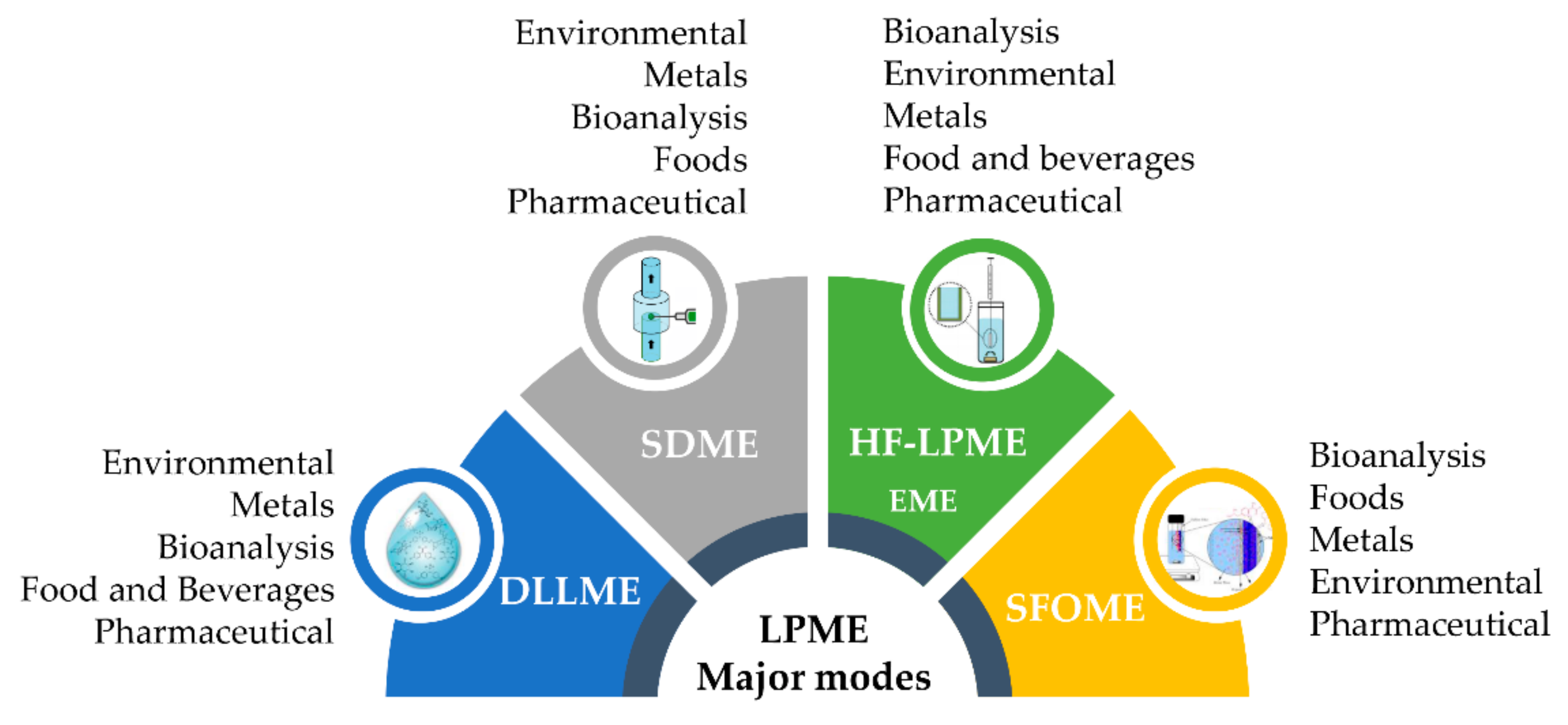
2. Different Modes of LPME

LPμETs can be categorized in different modes, where the most commonly used and widely applied is (i) DLLME, where an acceptor phase, a high-density organic extracting solvent (hydrophobic phase), and an amphiphilic solvent, a dispersing solvent miscible in the hydrophobic and hydrophilic (aqueous sample) phase, are quickly added to the sample, forming small emulsified droplets of the organic solvent within the sample; (ii) SDME, where a drop of the acceptor phase (extraction solvent) is suspended into the sample or the headspace above the sample, from the tip of a polytetrafluoroethylene (PTFE) rod or a syringe needle, and extraction occurs under equilibrium or non-equilibrium conditions; and (iii) HP-LPME, where the acceptor phase (extracting solvent) and the donor phase (sample) are separated by a porous hydrophobic polymeric membrane filled with the extracting solvent and typically assembled using a hollow fiber (HF). The extractions can be either dynamic or static and can be operated using a two- or three-phase system. These different LPμETs modes are discussed in the following sections.

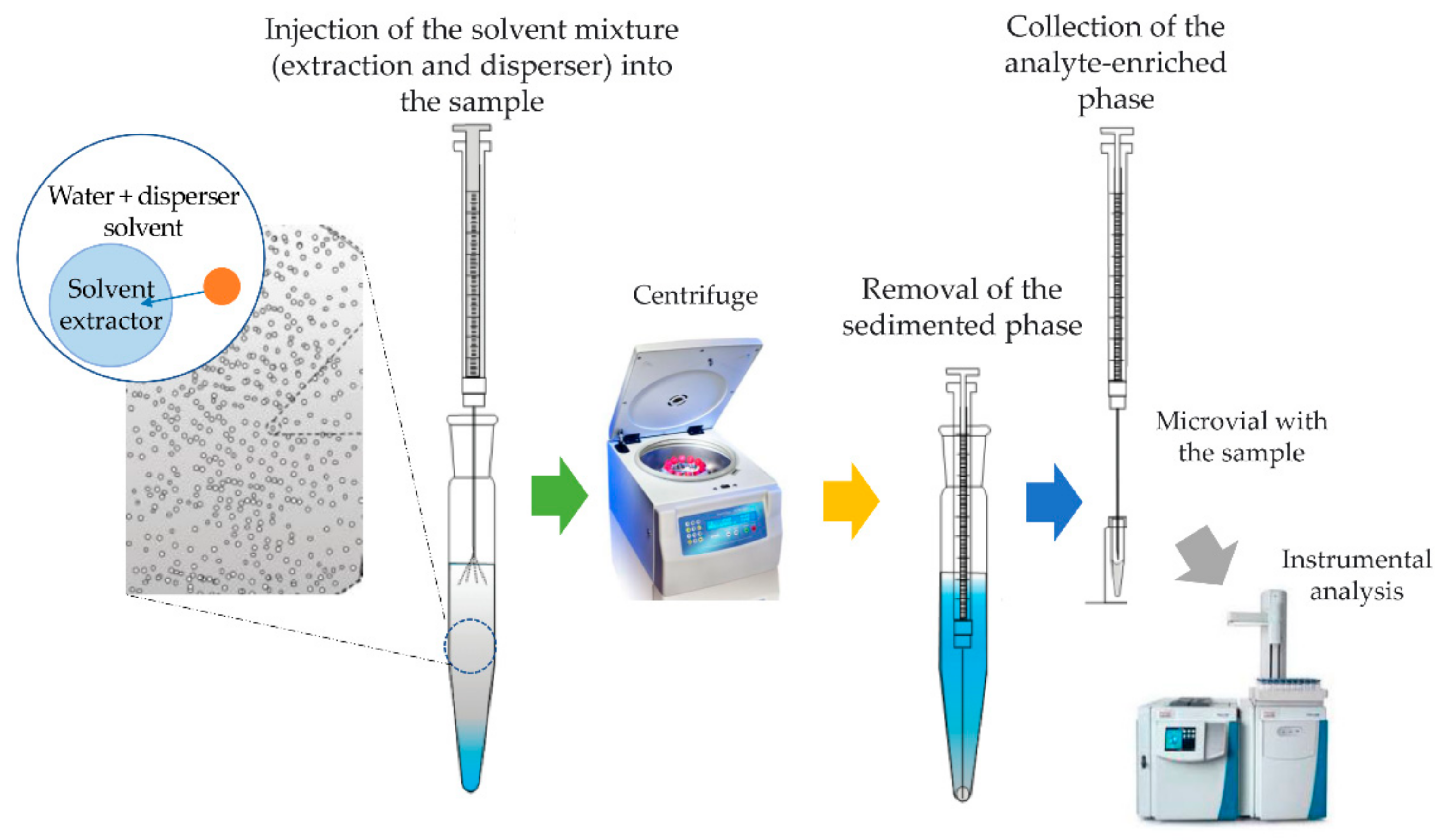
2.1. Dispersive Liquid-Liquid Microextraction (DLLME) Technique

DLLME was first implemented by Razaee et al. [2] for the extraction of organic pollutants such as polycyclic aromatic hydrocarbons (PAHs) before their instrumental analysis using GC-FID [2]. It is a three-solvent-based technique using a microliter of the organic solvent to achieve the extraction and preconcentration of organic pollutants. As a miniaturized form of LLE, the amount of organic solvent (acceptor phase) used in DLLME is significantly reduced. This is a three-phase-based technique: the extraction solvent (acceptor phase), dispersive solvent, and aqueous phase (donor phase, where the analyte is located) (Figure 2). In the aqueous phase, a mixture of a few milliliters of an organic solvent and diffuse solvent with miscibility in the extractant and aqueous phases is quickly added to the sample, leading to the formation of a high degree of turbulence and small droplets originating from a cloudy solution. The equilibrium state is achieved quickly due to the very large area between the acceptor phase (extractor solvent) and the donor phase (aqueous sample), making the time of isolation very short.
The isolation effectiveness is affected by (i) the extraction solvent, which must present very low hydrophilicity, a high extraction capability of the target analytes, and higher density than water (CCl4, CHCl3); (ii) the spreading solvent, which should be mixable with the extractant solvent and aqueous phase, with the most widely used being acetone, acetonitrile, and methanol; (iii) the extraction time; (iv) the effect of the ionic strength adjusted by electrolyte addition, (NaCl); and (v) the sample pH.
The simplicity of the technique, its low cost, high enrichment and recovery factors, and rapidity are the main primacies of DLLME, while the use of three solvents (hydrophobic, hydrophilic, and amphiphilic), the limited choice of solvents, and the fact that it is considered as a non-selective extraction method constitute their main disadvantages. The merits of DLLME that have gained so much attention include simplicity, speed, ease of operation, cost-effectiveness, a reduced volume of generated toxic organic waste as well as its capability to combine ultra-concentration and clean-up [2,12]. Since its invention, it has been complemented by other methods such as ultrasonication [13] and solidification of the floating organic drop method [14,15] for the analysis of a myriad of analytes in different matrices to boost the extraction efficiency of the whole procedure. The involvement of ultrasonication in a DLLME procedure also helps to circumvent the challenges that accompany conventional DLLME including the requirement of a high volume of disperser solvent and drawbacks in terms of the selection of the appropriate disperser solvent.
Figure 2. A simplified schematic diagram demonstrating the DLLME principle (adapted from Yazdi and Amiri [16].
Processes 10 01347 g002

DLLME Applications

Since its implementation, DLLME has found a wide range of applications (Table 1), being an effective method of sample preparation used for the extraction and preconcentration of organic compounds (pesticides [17,18], triazole fungicides [19], parabens [20], alkylbenzenes and bisphenol A [21]) and UV filters [22] from samples of different compositions and complexity, mainly environmental samples [23], foodstuffs [24], pharmaceuticals and cosmetics, and biological samples [25,26].
Altunay [13] developed a UA-DLLME-based method for the isolation of niacinamide in vitamin supplements, syrups, and cosmetics using a UV–Vis spectrophotometer at 265 nm. The preparation of a deep eutectic solvent (DES) entails the mixing of a hydrogen donor sugar and a hydrogen acceptor sugar on a magnetic stirrer in a glass tube while maintaining a temperature of 50 °C–80 °C, after which the mixture is allowed to cool. A total of 335 µL of the DES solution (glycerol and sorbitol, molar ratio of 1:2) was placed into a centrifuge tube containing 10 mL of the sample solutions with pre-added 75 ng mL−1 of the target compound (niacinamide). The tube containing the solution was sonicated for 1.5 min at 30 °C to establish the dispersion of the extraction solvent before the DES phase was separated from the aqueous phase by centrifugation at 4000 rpm. The extracted solution was diluted using methanol before analyzing at 265 nm on a UV–Vis spectrophotometer. This method provided a linear range within the studied range of concentration (1–400 ng mL−1), a limit of detection of 0.33 ng mL−1, and a satisfactory recovery from 97.3% to 98.9%.
Non-steroidal anti-inflammatory drugs (NSAIDS) have been used for use in animals due to their analgesic and inflammatory properties. However, as a result of health threats such as cardiovascular complications, cerebrovascular risks, and gastrointestinal ulcers posed by their presence in the environment, they have been tagged as ‘emerging pollutants’ [27,28]. The development of a method for the isolation of four NSAIDs was investigated by Qiao et al. [29]. The extraction process involved the transfer of 10 mL of the sample spiked with 1 μg mL−1 each of diclofenac and oxaprozin, 2 μg mL−1 of ibuprofen, and 1.5 μg mL−1 of salicylic acid before the addition of 200 μL of the synthesized HDES. The mixture was vortexed and sonicated while the resulting cloudy solution was centrifuged to obtain the HDES phase, which was later diluted with H2O:MeOH (25:75, v/v). Finally, 5 μL of the diluted solution was injected into HPLC-UV and the resulting chromatogram is depicted in Figure S1 (Supplementary Materials). The validation of the method using different figures of merit such as LOD (0.5–1 μg L−1), LOQ (1–5 μg L−1), and recovery (79.4% to 107.5%) at a linear range of 5–2000 μg L−1 suggested that the method is suitable for the everyday analysis of NSAIDs in different water and milk samples. In another study, a solution of extraction solvent (chloroform, 500 μL) and dispersive solvent (acetonitrile, 500 μL) was added to 8 mL of a sample solution containing five fluoroquinolones (enrofloxacin, sarafloxacin, ciprofloxacin, norfloxacin, and levofloxacin) with the pH adjusted to 8, after which the samples were vortexed (2 min) and sonicated (2 min). The solution was centrifuged for 5 min at 4000 rpm followed by the dissolution of the sedimented phase in 50 μL of methanol in a 1.5 mL vial before injection into HPLC-FLD for analysis. A limit of detection of 0.11 µg L−1, an enrichment factor (EF) of 54.7, and recoveries in the range of 72 ± 6.1% were obtained for river water and 27 ± 8.3% for seawater, suggesting that the method is viable for the routine analysis of the targeted fluoroquinolones in low salinity water samples [30].
Another application that reported using UA-DLLME was in combination with HPLC-DAD, primarily for the quantitative analysis of empagliflozin (EMPA), dapagliflozin (DAPA), and canagliflozin (CANA) in human plasma in which the protein precipitating agent/disperser and extracting solvent were methanol and 1-dodecanol. The LODs were 0.67, 1.66, and 0.37 ng mL−1 for EMPA, DAPA, and CANA, respectively, whereas LOQs were 2.00, 3.5, and 1.10 ng mL−1, respectively, with recoveries in the range of 92.9 to 113% [31].
Phenol and phenolic compounds constitute a class of harmful substances whose anthropogenic sources can be traced back to industries such as pharmaceuticals, textiles, plastic, and dyes, to mention a few. As a result of the adverse health impact, the long-term oral exposure exerted on the living organisms including the severe damage to vital organs of the body (i.e., genitourinary tract, liver, kidney and lungs [32]) means that the need for the development of a cheap, in situ deployable method has been on the rise. Moslemzadeh et al. [33] proposed a method for the quantification of phenol in environmental water and wastewater samples. In this method, the colorimetric reaction between phenol and 4-amino antipyrine (4-AAP) was set up in a conical tube followed by the UA-DLLME, while the quantitative analysis of RGB values was accomplished with an Android app Color Grab. Recovery, LOD, and LOQ were 93.7–103.6%, 1.7 μg L−1, and 5.7 μg L−1, respectively, in the linear range of 5.0 to 300 μg L−1.
Table 1. The selected DLLME application examples.
SampleAnalytesExtraction Solvent
(Used Volume)
Analytical InstrumentLinear RangeLODLOQRecoveryRef.
Vitamin supplements, syrups and cosmetic samples Niacinamide DES (sugar alcohol-based, 335 μL DES) UV–VIS >1–400 ng mL−1 0.33 ng mL−1- 97–99 [13]
Environmental water and milk samples NSAIDs >DES (hydrophobic based, 200 μL HDES) HPLC-UV- 0.5–1 μg L−1- 79–107 [29]
Water, beverages and vegetables Cr SUPRAS-5 (1-decanol: Bu4NOH, 1:1 v/v, 250 μL) >FAAS 0.1–350 μg L−1 >0.03 μg L−1 0.1 μg L−1 95–104 [34]
Surface water Fluoroquinolones 1000 μL of CH3Cl (extracting solvent), and dispersive solvent (ACN) in 1:1 ratio HPLC-FLD- 0.11 µg L−1- 27–72 [30]
Environmental water and wastewater samples Phenol >Dichloromethane
(300 μL)
Android app Color Grab(smartphone)5.0 – 300 μg L−1 1.7 μg L−1 5.7 μg L−1 94–104 [33]
>Human plasma Empagliflozin, dapagliflozin, canagliflozin Methanol, 1-dodecanol
(300 μL CH2Cl2)
HPLC-DAD 1.1–2500 ng mL−1 0.37–1.66 ng mL−1 1.10–3.5 ng mL−1 93–113 [31]
Wastewater Siloxanes Chlorobenzene
(13 μL)
GC-MS 2–25 μg L−1 0.002–1.4 µg L−1 >0.00–4.7 µg L−1 71–116 [35]
Legend: DES—deep eutectic solvent; HDES—hydrophobic deep eutectic solvents; CAN—acetonitrile; FAAS—flame atomic absorption spectrometry; GC-MS—gas chromatography-mass spectrometry; HPLC-DAD—high-performance liquid chromatography with a diode-array detector; HPLC-FLD—high-performance liquid chromatography with fluorescence detector; HPLC-UV—high-performance liquid chromatography-ultraviolet; LOD—limit of detection; LOQ—limit of quantification; NSAIDs—non-steroidal anti-inflammatory drugs; UV–Vis—ultraviolet–visible spectroscopy.
The existence of chromium in the environment has been traced to different anthropogenic sources including industries such as petroleum refineries, leathering, electroplating, leathering, textiles, and many more [36,37]. Analytical results characterized by the recovery, LOD, LOQ, and RSD of 95–104%, 0.03 μg L−1, 0.09 μg L−1, and 2.5%, respectively, operated in the linear concentration range from 0.1 to 350 μg L−1, was obtained for the ultrasound-assisted supramolecular solvent dispersive liquid–liquid microextraction (AA-SUPRAS-DLLME) combined with FAAS employed for the quantitative analysis of chromium (Cr) in water, vegetable, and beverage samples. In this method, 200 μL of 10−4 mol L−1 of the complexing agent (4-hydroxy-2-[(E)-(4-sulfonate-1-naphthyl) diazenyl] naphthalene-1-sulfonate (azorubine) and 250 μL of SUPRAS-5 (solution of Bu4NOH and 1-decanol, 1:1 M ratio, extraction solvent) were added to 10 mL of the sample with 100 μg L−1 of Cr(VI). The aqueous phase was discarded after subjecting the mixture to centrifugation for 5 min at 4000 rpm while the remaining component was diluted to 1 mL with 1 M of HNO3 in ethanol before injection into the inlet of the FAAS [34].

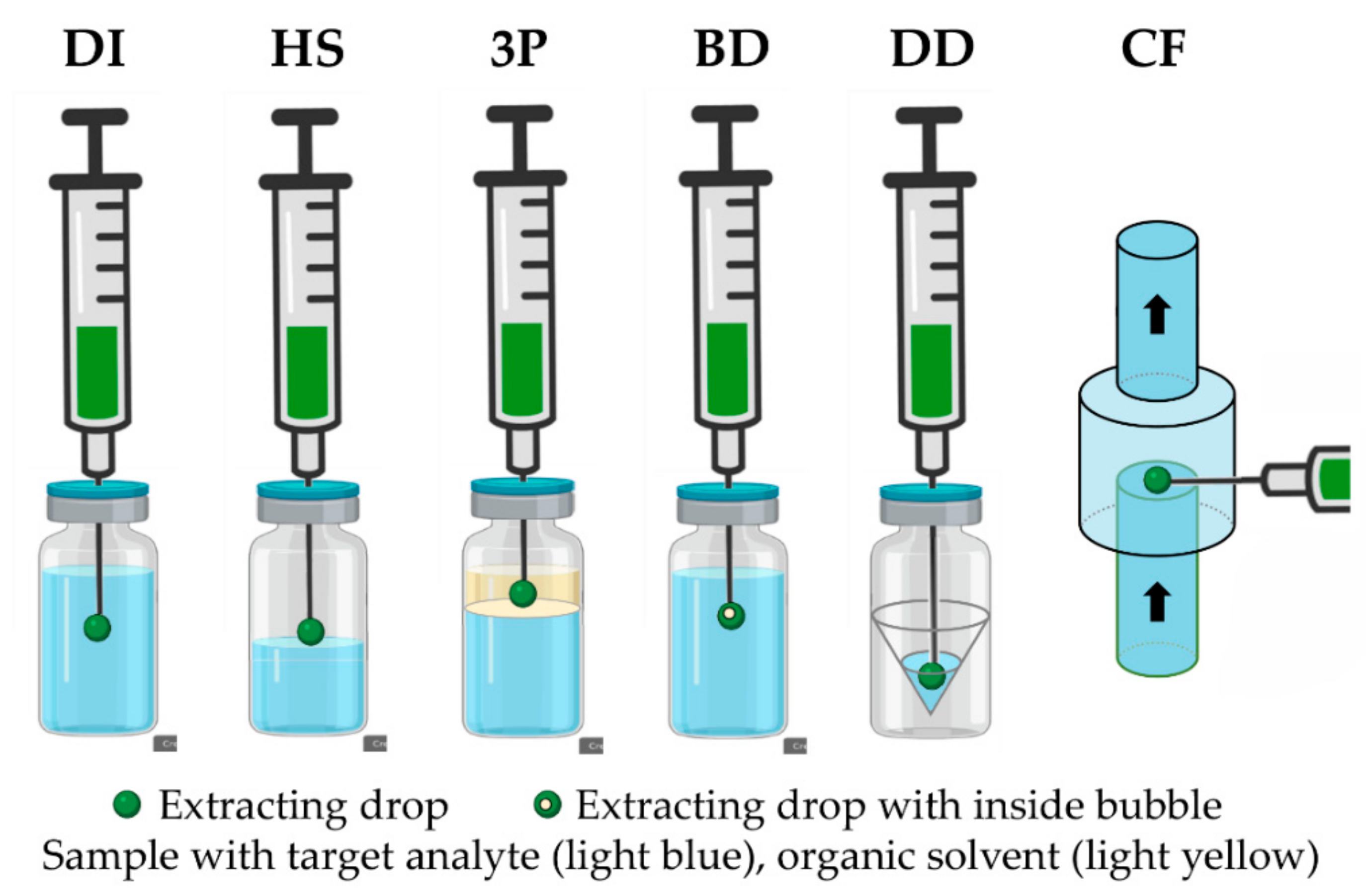
2.2. Single Drop Microextraction

SDME emerged in the mid-90s by Liu and Dasgupta as the first approach to LPME, in which ammonia and sulfur dioxide were extracted by a drop of water before the spectrophotometric analysis [1]. SDME involves mass transfer into a little droplet (few μL) of an organic solvent (acceptor phase) located at the tip of a micro-syringe needle. SDME can operate in different modes (Figure 3) such as (i) direct immersion (DI), mostly used for non-volatile analytes in which the extraction solvent is submerged in the sample solution, allowing for the diffusion of the analytes, followed by the withdrawal of the drop before analysis; and (ii) headspace (HS-SDME), which is suitable for the extraction of volatile and semi-volatile compounds [38], with other variants such as bubble-in-drop (BD-SDME) launched by Williams et al. [39] to increase the droplet surface area, and continuous-flow microextraction (CF) introduced by Liu and Lee to improve the contact region among the extraction solvent and the analyte [40]. The drop-to-drop (DD) liquid–liquid microextraction, developed by Wijethunga et al. [41], using a reduced sample volume, constitutes another option.
This LPμET has been perceived as an effective approach for the extraction and preconcentration of a wide range of analytes from very complex matrices including environmental, food and beverages, pharmaceutical, and clinical samples. Thus, SDME approaches are cost-effective and less nocive to the environment, supporting green analytical chemistry principles. Nevertheless, SDME approaches also present some disadvantages, the most obvious being the instability of the extraction drop [5,42,43], particularly at high stirring and high temperature [44]. Furthermore, it is an equilibrium-based technique, therefore limiting the enrichment factor that it is possible to attain [45].

2.2.1. SDME Applications

HS-SDME has been rapidly employed for the isolation of volatile compounds in conjunction with sophisticated analytical instruments such as GC and LC (Table 2). The disadvantages of such methods lie in their inability to be deployed for the in situ analysis, high-level expertise of the analyst, expensive cost of maintenance, and the size of the space to accommodate them. In recent times, the nanocolorimetric detection of volatile analytes using a smartphone color app has been employed in tandem with HS-SDME [46,47]. Qi et al. [47] developed a smartphone nanocolorimetric method for the determination of formaldehyde in octopus and chicken flesh. The analytical procedure entails the preconcentration of the analyte in a 3 μL gold nanoprism/Tollen’s reagent (Au-np/TR) complex at the underside of the polypropylene centrifuge tube cap. The heating of the sample inside the tube led to the gradual release of the formaldehyde, which was captured by the extraction solvent on the tube cap. The image of the analyte complex was captured by a smartphone camera and analyzed for the RGB value via EKColorPicker software A limit of detection of 30 nM was observed and recoveries were in the range of 94–98.34% for octopus and 93.20–100.92% for chicken in the 0.1 to 100 μM.
The unique features of deep eutectic solvents (DES) such as high thermal stability, moderate cost, reduced level of toxicity, excellent solubility, and negligible vapor pressure have attracted their deployment in separation science as green solvents considered to possess competitive advantages over conventional solvents [48,49,50]. In a method described by Mehravar et al. [51], DES was synthesized using a different molar ratio of hydrogen bond donor (choline chloride) and hydrogen bond acceptors (oxalic acid, urea, and glycerol). The best DES solvent was selected and hung on the edge of a micro-syringe to extract the polycyclic aromatic hydrocarbons in a 15 mL vial before injection in the GC-MS for instrumental quantitation. The method’s figures of merit revealed that LODs and LOQs were 0.003–0.012 μg L−1 and 0.009–0.049 μg L−1, respectively, with recoveries in the range of 94.40–105.98%.
A method of homocysteine thiolactone (HTL) assessment in human urine involving an automated coupling of SDME to capillary electrophoresis (CE) was reported by Purgat et al. [52]. The LOQ, LOD, and recoveries for HTL were 50 nM, 25 nM, and 92.7–115.5%, respectively, at a linear range of 50–200 nM. The five-step analysis proceeded from the centrifugation of the urine sample, dilution with phosphate buffer and methanol, the addition of chloroform to the donor phase, isolation of the analyte in automated SDME-CE, and finally, separation and detection at 240 nm in the CZE-UV.
The ionic liquid 1-hexyl-3-methylimidazolium hexafluorophosphate ([C6MIM][PF6]) was employed for the direct immersion-SDME isolation and preconcentration of vanadium before the analysis of the RGB value of the captured image. The extraction was conducted under stirring for 9 min in a 5 mL vial and the microdrop, which was suspended at the tip of the chromatographic syringe, was submerged in the aqueous donor phase to isolate the analyte. The extraction solvent showing a bluish color was fixed on a wooden box and the image was captured for digital image colorimetry (DIC) analysis. The method was reported to yield a LOD of 0.6 μg L−1 for a 3.5 mL sample volume, an LOQ (50.0 μg L−1) was obtained of 1.8 μg L−1, and a relative standard deviation of 4.8% [53].
Table 2. A summary of the selected SDME examples.
SampleAnalytesExtraction Solvent
(Used Volume)
Analytical InstrumentLinear RangeLODLOQRecovery
(%)
Ref.
Octopus and chicken fleshFormaldehydeGold nanoprism and TR (Au-np/TR) complexUV-Vis, Smartphone, naked eye for qualitative analysis0.1–100 μM30 nM 93–101[47]
Environmental waterPAHs15 μL DESGC-MS---0.003–0.012 μg L−10.009–0.049 μg L−194-106[51]
Urinehomocysteine thiolactonePhosphoric acid (the acceptor phase)
Organic phase (chloroform) (40 μL)
CE-UV50–200 nM25 nM50 nM92.7–115.5%[52]
WaterVanadium1-hexyl-3-methylimidazolium hexafluorophosphate ([C6MIM][PF6], 7 μL)Smartphone---0.6 μg L−11.8 μg L−191 and 103%[53]
Spices (cinnamon, cumin, fennel, clove, thyme, and nutmeg)TerpenesDES (N4444Br and dodecanol, 1:2, 1.5 μL drop at the tip of the needle)GC-MS1 to 500 μg/g 0.47–86.40 μg g−1---[54]
UrinePsilocin and MuscimolOrganic phase (a drop of octanol layer)CE0.05–50 mg L−10.004–0.016 mg L−10.014–0.045 mg L−1---[55]
Legend: [C6MIM][PF6]—1-hexyl-3-methylimidazolium hexafluorophosphate; CE—capillary electrophoresis; CE-UV—capillary electrophoresis with ultra-violet detection; LOD—limit of detection; LOQ—limit of quantification; PAHs—polycyclic aromatic hydrocarbon; UV–Vis—ultraviolet-visible spectroscopy; GC-MS—gas chromatography-mass spectrometry.

2.2.2. Solidification of Floating Organic Drop Microextraction

The solidification of floating organic drop microextraction (SFOME) method was developed by Khalili Zanjani et al. [56] for the analysis of polycyclic aromatic hydrocarbons (PAHs). In this technique, the assembly of the compounds in a microdrop of the acceptor phase (organic extraction solvent) under stirring is succeeded by the solidification of the suspended microdrop organic layer in ice. Before injection into the instrument for quantitative assessment, the solidified microdrop is allowed to melt. The low melting point of the extraction solvent, usually in the range of 10–30 °C, constitutes its main notable characteristic. The use of a small quantity of acceptor phase indicates the conformity of this method with the demand for green analytical chemistry (GAC). Individually or in combination with other extraction methods, it has been widely used for the analysis of different contaminants in environmental samples [57,58], food samples [59,60,61], and biological samples [15]. Similarly to SDME, SFOME is an equilibrium-based extraction approach and requires extensive optimization [62]. SFOME is not restricted to the extraction of inorganic metal ions such as lead [63] and cadmium [64] as they have been established for the extraction of organic compounds. Table 3 presents the recent literature with the application of SFOME as an extraction technique for the isolation of the target compounds in a diversity of samples.

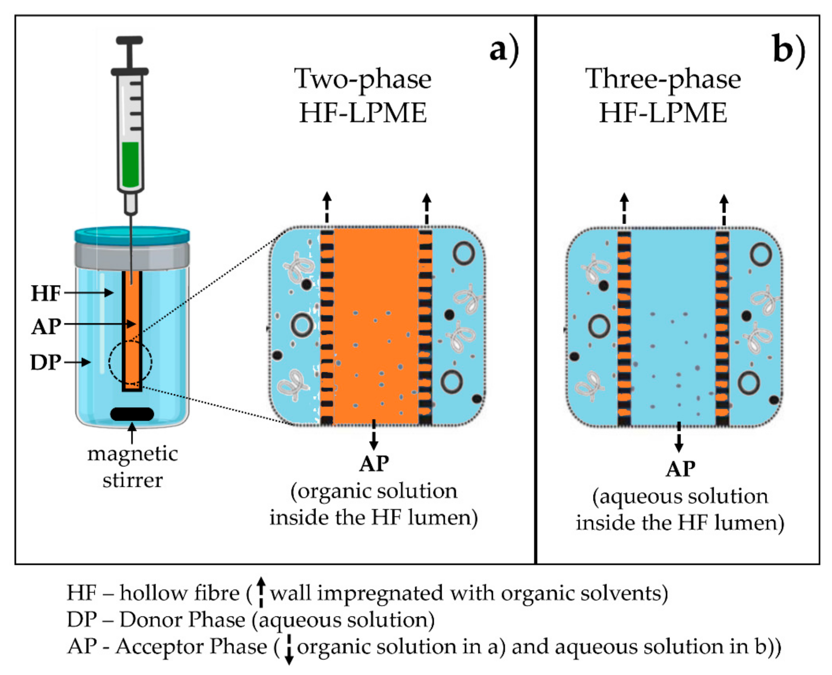
2.3. Hollow-Fiber LPME (HF-LPME) and Electro Membrane Extraction (EME)

The drop instability led to the development of HF-LPME. This was implemented by Pedersen-Bjergaard and Rasmussen [3] for the determination of methamphetamine in urine and plasma. In this analytical approach, the drop, a small volume (few mL) of the acceptor phase (hydrophobic organic solvent) is accommodated within the lumen of a porous hydrophobic hollow fiber to protect it (Figure 4).
HF-LPME can be performed in two- and three-phase systems (Figure 4). Even though both versions allocate a coincidence in that they involve the partitioning of the analytes from the sample solution (donor phase) to other phases (acceptor phase), a few lines of distinction can be perceived. In the two-phase system, the analytes are diffused from the aqueous phase to the acceptor phase based on their affinity. In contrast, the three-phase system involves the partitioning from the aqueous donor across the organic SLM into the aqueous acceptor phase in the lumen of the hollow fiber [71]. As a result of the benefits that attend automation, we can refer to the reduction in the number of employees and the reduced use of chemicals and time of analysis, just to indicate a few. In contrast, HF-LPME approaches are often time-consuming, with extraction times up to 45–90 min [45].

2.3.1. HF-LPME and EME Applications

HF-LPME has been applied to several fields and analytes including the analysis of hormonal drugs [72]. Recoveries in the range of 86.2–102.3% were obtained for megestrol acetate and levonorgestrel in water and urinary samples. In another study, HF-LPME was coupled with UHPLC-QTOF-MS for the analysis of 2,4-, 2,5-, and 2,6-dinitrophenol in urine and saliva samples [73]. LODs of 0.18 µg L−1, 0.38 µg L−1, and 0.14 µg L−1 in urine samples and 0.32 µg L−1, 0.67 µg L−1 and 0.24 µg L−1 in saliva samples, respectively, were obtained. The extraction entailed the dilution of 400 µL of saliva or 700 µL of human urine in 50 mL of DI water before the insertion of the hexyl ether pre-treated hollow fibers containing 30 µL of the aqueous acceptor phase. The withdrawal of the hollow fiber was preceded by stirring of the sample solution at 300 rpm with a magnetic stirrer after which the acceptor phase was collected by cutting on the end of the fiber and injected into the UHPLC system [73].
An electrochemical-based differential pulse voltammetry analysis preceded the HF-LPME extraction of vanillylmandelic acid in human urine as proposed by [74]. In this method, the support liquid membrane was butyl benzoate. To begin with, the polypropylene hollow fiber was pre-rinsed with acetone, soaked in an organic solvent and then sealed with custom-made glass plugs. The pre-treated hollow fiber was dipped into the donor solution and stirred on a magnetic stirrer at 1500 rpm. The withdrawal of the fiber from the donor solution was succeeded by transferring the acceptor phase into the surface of the working electrode for voltammetric analysis and led to a limit of detection of 0.5 μmol L−1 and 1.7 μmol L−1 at a linear range of 0.5 to 100 μmol L−1.
The environmental application of effervescence-assisted spiral-HF-LPME has been applied for the quantitative assessment of disinfection by-product (DBP) levels in water samples. The proposed method was accompanied by the desired advantages of a reduction in analysis time, an increase in the number of processed samples, and the lack of need for mechanical stirring due to the effervescence by CO2 bubbles, which aided in the extraction of the analytes with detection limits (ng L−1) ranging from 17 to 79 for halo acetonitriles, from 10 to 35 for trihalomethanes, from 12 to 220 for halo nitromethanes, and 10 to 16 for halo ketones. The enrichment factors ranged from 13.1 to 140.1 and recoveries from 80 to 113% [75]. Table 4 describes some powerful applications of HF-LPME in different matrices.

3. New Solvents in LPME as Promising Environmentally Friendly Extraction Phases

Recently, there has been an increasing interest in the application of new reagents in LPME due to the environmental sustainability and toxicity of classical organic solvents, along with the search for solvents for specific sample matrices and extraction methods.

3.1. Ionic Liquids (ILs)

ILs are low melting organic salts constituted by an organic cations such as tetra-alkyl ammonium or imidazolium ions, and a smaller organic or inorganic anions such as hexafluorophosphate or bromide, with melting points below 100 °C. ILs are major designer solvents due to the large potential combinations of cations and anions through the inclusion of some functional groups in their structures, which in turn improves the specificity and selectivity of the target analytes [5,45,83]. ILs are more ecological than common solvents due to their negligible vapor pressure, thermal and chemical stabilities, adequate solubility in inorganic and organic solvents, and low flammability [83,84,85]. ILs also offer multiple interactions and range from hydrophilic water-soluble compounds to hydrophobic water-insoluble compounds, with properties varying with the polarity or non-polarity of the functional groups [45].
Some properties of ILs such as viscosity, melting point, water miscibility, and density can be challenging to handle [84,85]. For instance, the extent of molecular interactions between ionic constituents determines the melting point and viscosity of ILs [86]. Moreover, many ILs have a relatively high viscosity, which, due to the slow diffusion, is not beneficial in terms of mass transfer. For this reason, ILs are mostly used in DLLME procedures as the microdroplets of the dispersed extraction phase allow for short diffusion paths and thus fast extractions. Bamorowat et al. [87] assessed the level of benzoylurea insecticides (chlorfluazuron, diflubenzuron, hexaflumuron, and triflumuron) in vegetable and fruit juice samples through an HPLC-DAD method preceded by ultrasonic-assisted DLLME. An IL of 1-butyl-3-methylimidazolium hexafluorophosphate was used as an extraction solvent while a mixture of sodium bicarbonate, tartaric acid, and potassium bromide was used as a dispersant agent to transfer the target compounds from the aqueous phase. The enrichment factors, extraction recoveries, detection limit, and limit of quantification obtained were 370 to 465, 74% to 93%, 0.04–0.19 ng mL–1, and 0.13–0.64 ng mL–1, respectively. Other DLLME applications of ILs include the use of 1-butyl-3-methylimidazolium hexafluorophosphate for the extraction of cortisone and cortisol from saliva samples [88] and 1-butyl-1-methylpyrrolidinium bis(trifluoromethylsulfonyl)imide for the extraction of bilirubin from urine and serum [89].
Their high viscosity and surface tension make ILs particularly useful for larger and stable droplets for DI-SDME. On the other hand, the low thermal stabilities and volatilities make them appropriate for HS-SDME [45,86], but incompatible with GC, which means that the analyte extracts require the use of HPLC instrumentation. However, ILs present some challenges for MS instrumentation due to contamination problems, so HPLC methods with UV and DAD have to be used [45]. An et al. [90] developed two tetrachloromanganate ([MnCl42-])-based magnetic ILs as extraction solvents for the analysis of 11 aromatic compounds in lake water through HS-SDME. The results were assessed by reversed-phase HPLC and the obtained LODs varied from 0.04 to 1.0 μg L−1. Nevertheless, ILs show environmental threat, as they are slow to degrade and might be bioaccumulative. The toxicity of ILs seems to be influenced by the structure of the cation, especially by the alkyl chain length [83]. ILs containing the imidazolium group have shown toxic effects [45,86]. Moreover, hydrophobic ILs become persistent contaminants in the environment since they are strongly adsorbed onto the sediments, while hydrophilic ILs are expected to enter aquatic ecosystems [83,91].

3.2. Deep Eutectic Solvents (DES)

DESs are constituted by two or more solid organic compounds that form hydrogen bonds, in which one organic compound is a hydrogen bond donor (HBD) and the other is a hydrogen bond acceptor (HBA) [86]. DESs have similar characteristics to ILs including low volatility and low melting temperatures, high tunability (designer solvents), chemical, non-flammability, thermal stability, and the probability to dissolve organic and inorganic compounds. In comparison to ILs, DESs are superior in cost, ease of preparation, biodegradability, produce less viscous solutions, and toxicity, as they can be obtained by a cheap and simple synthesis procedure, based on heating a solution of their constituents [45,83]. Another green advantage is the chance to obtain natural DESs (NADESs) derived from naturally occurring compounds and metabolites such as sugars, organic acids, choline derivatives, amino acids, and terpenes. NADESs have shown a growing interest due to their low toxicity, low environmental impact, and low cost [86].
The physical-chemical properties of DESs and NADESs are dependent on the choice of HBD and HBA components. Furthermore, the ratio of HBD to HBA determines the melting temperature and influences the solvent properties [45,86]. NADESs are stable at room temperature and compatible with GC analysis since many decompose to the individual molecules in the GC injection port at temperatures higher than 200 °C. Some NADESs such as DESs commonly have moderate to high viscosities, and for this reason, they have been used in some DLLME and SDME procedures. For instance, Altunay [13] used a sugar alcohol-based DES consisting of a mixture of sorbitol and glycerol (2:1) for the extraction of niacinamide in syrups, cosmetic samples, and vitamin supplements by ultrasound-assisted DLLME (Table 2). The niacinamide content was then determined by UV–Vis at 265 nm. The linear range, detection limit, and quantitative recovery obtained were 1–400 ng mL−1, 0.33 ng mL−1, and 97.3–98.9%, respectively, with a precision of 1.4–2.8%. Hydrophobic DESs and NADESs are liquids at room temperature and have been increasingly used in all modes of LPME as green solvents [45]. Qiao et al. [29] extracted NSAIDs in environmental water and milk samples through an ultrasound-assisted DLLME method (Table 1). The hydrophobic DES was composed of guanidinium chloride and thymol, and the results showed the potential of the synthesized DES with low limits of detection of 0.5–1 μg L−1 and acceptable recoveries in the range of 79.42%–107.52%. Yang et al. [92] developed a DES composed of a range of fatty acids C8:C9:C12 (3:2:1) for the determination of phenolic compounds in tap water, lake water, and river water through gas-assisted LLME, in which LODs were obtained in the range of 0.22–0.53 μg L−1. In another study, Khataei et al. [93] proposed a phosphonium-based DES for HF-LPME of steroidal hormones in urine and plasma samples. In this study, the LODs obtained were in the range of 0.5–2 µg L−1.

3.3. Nanostructured Supramolecular Solvents (SUPRASs)

Nanostructured SUPRASs are constituted of three-dimensional amphiphilic surfactants that immediately form nanostructured micelles or vesicles through self-assembly and coacervation induced by the modification of pH, temperature, organic solvent, and/or salt addition. SUPRASs are water-immiscible and offer a wide variety of molecular interactions due to their distinct physiochemical properties including good solvation, non-volatility, and non-flammability. Moreover, these environmentally friendly solvents are available at a low cost [83,86,94]. SUPRASs are designer solvents, and their conform properties depend on the hydrophobicity or non-polarity of the surfactant, as the supramolecular aggregates have regions of different polarity, providing excellent solvation properties for a wide range of inorganic and organic compounds [83,86,94].
SUPRASs are constant in DLLME-based methods with coacervation induced in situ, in which the extraction processes quickly. The extraction phase is typically collected by centrifugation or supported by the SFO principle [86,95]. For instance, hexanol-based SUPRASs were used in the extraction of phenethylamines in oral fluids [96] and amphetamine-type stimulants in breast milk, serum, urine, oral fluids, sweat, and digested hair or fingernails [97] through LC-MS/MS analysis. Other applications of SUPRASs include the extraction and preconcentration of amphiphilic compounds such as surfactants, bisphenols, drugs, pesticides, mycotoxins, and bioactive compounds from both solid and liquid environmental samples [94]. Tuzen et al. [34] developed an ultrasound-assisted SUPRAS-DLLME for the preconcentration and analysis of Cr ions in waters, beverages, and vegetables (Table 1). The SUPRASs prepared consisted of combinations of THF and Bu4NOH with 1-undecanol, 1-decanol, and 1-dodecanol. The enhancement factor and the LODs and LOQs were found to be 134, 0.03 μg L−1, and 0.1 μg L−1, respectively. Ezoddin et al. [98] developed a SUPRAS containing reverse micelles of 1-dodecanol in THF for the extraction of methadone from plasma and saliva samples through ultrasonic-assisted SFOME. In this study, the limits of detection and quantitation were 0.5–1.2 µg L−1 and 1.2–2.5 µg L−1 in water and biological samples, respectively. Another application of these solvents was in the vortex assisted-LLME of mercury in hair samples using quinalizarin as a chelating agent, forming a metal–quinalizarin complex extracted in 1-decanol/THF as a SUPRAS solvent, with a LOD of 0.30 µg L−1 [99]. Nevertheless, surfactants are petroleum-based and are only moderately biodegradable. Moreover, SUPRAS synthesis is laborious and time-consuming, and its stability is limited to a narrow pH range [5]. The negative impact of SUPRASs can be overcome by using bio SUPRASs developed by the coacervation of biosurfactants with fatty alcohols and synthetic alkyl-carboxylic acids in solutions of water and ethanol [83,100].

4. Final Remarks

The focus of this review was on the discussion of major modes of LPμETs and their applications in a wide range of fields. Commonly, the determination of analytes present in complex samples such as in environmental, food and beverages, pharmaceutical and clinical analysis, at very low concentrations, requires analytical procedures that include extraction, clean-up, and enrichment. To overwhelm the major disadvantages of the classical extraction techniques including the use of a large volume of organic solvents and samples, the risk of analyte loss and contamination, the lack of automation, the harm to the operator of the use of large amounts of organic solvents, and the fact that they are time-consuming and laborious techniques, we need to move the paradigm of extraction techniques toward the facilitation and miniaturization of sample preparation as well as reduce the volume of solvents and samples in addition to the employment of safe and non-toxic solvents such as ILs, DES, and SUPRASs. This will allow us to decrease the environmental impact as well as the harmfulness to the operator while improving the main figures of the merit of the methods. Due to its simplicity, high recovery, short extraction time, and high extraction efficiency, accompanied by the good analytical results achievable, LPμETs have received much attention in the last few years. HF-LPME and EME are universally used modes based on the complexity of samples, DLLME owes its popularity to its efficient and quick extractions, whereas SDME in HS and DI is easily adaptable to automated procedures.

Supplementary Materials

The following supporting information can be downloaded at: https://www.mdpi.com/article/10.3390/pr10071347/s1, Figure S1. Schematic illustrations of the experimental procedure of UA-HDES-DLLME (adapted from Qiao, Sun, Yu, Tao and Yan [29]).

Author Contributions

J.S.C. planned and managed the manuscript; R.P. was responsible for Section 1; J.S.C. and J.A.M.P. were responsible for Section 2.1 and Section 2.2 and Section 3; R.P.; A.K. and B.O. collaborated on Section 2.3 and Section 2.3.1.; R.P., C.V.B. and J.S.C. were responsible for Section 3.1, Section 3.2 and Section 3.3; J.S.C. and J.A.M.P. designed all of the figures and edited the manuscript; J.S.C. submitted the paper. All authors have read and agreed to the published version of the manuscript.

Funding

This research was funded by FCT-Fundação para a Ciência e a Tecnologia through the CQM Base Fund, UIDB/00674/2020, Programmatic Fund, UIDP/00674/2020, and by ARDITI-Agência Regional para o Desenvolvimento da Investigação Tecnologia e Inovação, through the project M1420-01-0145-FEDER-000005—Centro de Química da Madeira—CQM + (Madeira 14-20 Program). The authors also acknowledge FCT and the Madeira 14-2020 program to the Portuguese Mass Spectrometry Network (RNEM) through the PROEQUIPRAM program, M14-20 M1420-01-0145-FEDER-000008). ARDITI is also acknowledged for the postdoctoral fellowship granted to J.A.M.P. (Project M1420-09-5369-FSE-000001).

Institutional Review Board Statement

Not applicable.

Informed Consent Statement

Not applicable.

Data Availability Statement

Not applicable.

Acknowledgments

The authors acknowledge Ofélia Anjos from the Polytechnic Institute of Castelo Branco for the kind invitation to participate in this Special Issue.

Conflicts of Interest

The authors declare no conflict of interest.

References

  1. Liu, S.; Dasgupta, P.K. Liquid Droplet. A Renewable Gas Sampling Interface. Anal. Chem. 1995, 67, 2042–2049. [Google Scholar] [CrossRef]
  2. Vinas, P.; Bravo-Bravo, M.; Lopez-Garcia, I.; Pastor-Belda, M.; Hernandez-Cordoba, M. Pressurized liquid extraction and dispersive liquid-liquid microextraction for determination of tocopherols and tocotrienols in plant foods by liquid chromatography with fluorescence and atmospheric pressure chemical ionization-mass spectrometry detection. Talanta 2014, 119, 98–104. [Google Scholar] [CrossRef]
  3. Pedersen-Bjergaard, S.; Rasmussen, K.E. Liquid-liquid-liquid microextraction for sample preparation of biological fluids prior to capillary electrophoresis. Anal. Chem. 1999, 71, 2650–2656. [Google Scholar] [CrossRef]
  4. Nemati, M.; Farajzadeh, M.A.; Mohebbi, A.; Khodadadeian, F.; Afshar Mogaddam, M.R. Development of a stir bar sorptive extraction method coupled to solidification of floating droplets dispersive liquid–liquid microextraction based on deep eutectic solvents for the extraction of acidic pesticides from tomato samples. J. Sep. Sci. 2020, 43, 1119–1127. [Google Scholar] [CrossRef]
  5. da Silva Burato, J.S.; Medina, D.A.V.; de Toffoli, A.L.; Maciel, E.V.S.; Lancas, F.M. Recent advances and trends in miniaturized sample preparation techniques. J. Sep. Sci. 2020, 43, 202–225. [Google Scholar] [CrossRef] [PubMed]
  6. Ho, T.S.; Pedersen-Bjergaard, S.; Rasmussen, K.E. Recovery, enrichment and selectivity in liquid-phase microextraction. J. Chromatogr. A 2002, 963, 3–17. [Google Scholar] [CrossRef]
  7. de la Calle, I.; Pena-Pereira, F.; Lavilla, I.; Bendicho, C. Liquid-phase microextraction combined with graphite furnace atomic absorption spectrometry: A review. Anal. Chim. Acta 2016, 936, 12–39. [Google Scholar] [CrossRef] [PubMed]
  8. Tankiewicz, M.; Fenik, J.; Biziuk, M. Solventless and solvent-minimized sample preparation techniques for determining currently used pesticides in water samples: A review. Talanta 2011, 86, 8–22. [Google Scholar] [CrossRef] [PubMed]
  9. Anthemidis, A.N.; Ioannou, K.-I.G. On-line sequential injection dispersive liquid–liquid microextraction system for flame atomic absorption spectrometric determination of copper and lead in water samples. Talanta 2009, 79, 86–91. [Google Scholar] [CrossRef]
  10. Anthemidis, A.N.; Ioannou, K.-I.G. Development of a sequential injection dispersive liquid–liquid microextraction system for electrothermal atomic absorption spectrometry by using a hydrophobic sorbent material: Determination of lead and cadmium in natural waters. Anal. Chim. Acta 2010, 668, 35–40. [Google Scholar] [CrossRef]
  11. Pena, F.; Lavilla, I.; Bendicho, C. Immersed single-drop microextraction interfaced with sequential injection analysis for determination of Cr(VI) in natural waters by electrothermal-atomic absorption spectrometry. Spectrochim. Acta Part B At. Spectrosc. 2008, 63, 498–503. [Google Scholar] [CrossRef]
  12. Alves, A.; Vanermen, G.; Covaci, A.; Voorspoels, S. Ultrasound assisted extraction combined with dispersive liquid–liquid microextraction (US-DLLME)—a fast new approach to measure phthalate metabolites in nails. Anal. Bioanal. Chem. 2016, 408, 6169–6180. [Google Scholar] [CrossRef] [PubMed]
  13. Altunay, N. Optimization of ultrasound-assisted dispersive liquid–liquid microextraction of niacinamide in pharmaceutical and cosmetic samples using experimental design. Microchem. J. 2021, 170, 106659. [Google Scholar] [CrossRef]
  14. Sun, X.; Xing, X.; Du, Z. Application of dispersive liquid–liquid microextraction based on solidification of floating organic drop for the determination of extractables from pharmaceutical packaging materials. Talanta 2020, 209, 120540. [Google Scholar] [CrossRef] [PubMed]
  15. Golpayegani, M.R.; Akramipour, R.; Fattahi, N. Sensitive determination of deferasirox in blood of patients with thalassemia using dispersive liquid-liquid microextraction based on solidification of floating organic drop followed by HPLC–UV. J. Pharm. Biomed. Anal. 2021, 193, 113735. [Google Scholar] [CrossRef]
  16. Sarafraz-Yazdi, A.; Amiri, A. Liquid-phase microextraction. TrAC Trends Anal. Chem. 2010, 29, 1–14. [Google Scholar] [CrossRef]
  17. Ahmad, W.; Al-Sibaai, A.A.; Bashammakh, A.S.; Alwael, H.; El-Shahawi, M.S. Recent advances in dispersive liquid-liquid microextraction for pesticide analysis. TrAC Trends Anal. Chem. 2015, 72, 181–192. [Google Scholar] [CrossRef]
  18. Primel, E.G.; Caldas, S.S.; Marube, L.C.; Escarrone, A.L.V. An overview of advances in dispersive liquid–liquid microextraction for the extraction of pesticides and emerging contaminants from environmental samples. Trends Environ. Anal. Chem. 2017, 14, 1–18. [Google Scholar] [CrossRef]
  19. Bordagaray, A.; Millán, E.; Garcia-Arrona, R. A Review on Microextraction Techniques for Selected Triazole Fungicides Determination in Water and Food Samples. J. Food Chem. Nanotechnol. 2016, 2, 128–137. [Google Scholar] [CrossRef]
  20. Ocaña-González, J.A.; Villar-Navarro, M.; Ramos-Payán, M.; Fernández-Torres, R.; Bello-López, M.A. New developments in the extraction and determination of parabens in cosmetics and environmental samples. A review. Anal. Chim. Acta 2015, 858, 1–15. [Google Scholar] [CrossRef]
  21. Salgueiro-Gonzalez, N.; Muniategui-Lorenzo, S.; Lopez-Mahia, P.; Prada-Rodriguez, D. Trends in analytical methodologies for the determination of alkylphenols and bisphenol A in water samples. Anal. Chim. Acta 2017, 962, 1–14. [Google Scholar] [CrossRef] [PubMed]
  22. Yücel, C.; Ertaş, H.; Ertaş, F.N.; Karapinar, İ. Determination of UV Filters in Surface Water by VA-DLLME-SFOD Technique Coupled with GC-MS/MS. Clean–Soil Air Water 2022, 2100246. [Google Scholar] [CrossRef]
  23. Hashemi, B.; Zohrabi, P.; Kim, K.-H.; Shamsipur, M.; Deep, A.; Hong, J. Recent advances in liquid-phase microextraction techniques for the analysis of environmental pollutants. TrAC Trends Anal. Chem. 2017, 97, 83–95. [Google Scholar] [CrossRef]
  24. Quigley, A.; Cummins, W.; Connolly, D. Dispersive Liquid-Liquid Microextraction in the Analysis of Milk and Dairy Products: A Review. J. Chem. 2016, 2016, 4040165. [Google Scholar] [CrossRef] [Green Version]
  25. Jain, R.; Singh, R. Applications of dispersive liquid–liquid micro-extraction in forensic toxicology. TrAC Trends Anal. Chem. 2016, 75, 227–237. [Google Scholar] [CrossRef]
  26. Mansour, F.R.; Khairy, M.A. Pharmaceutical and biomedical applications of dispersive liquid-liquid microextraction. J. Chromatogr. B 2017, 1061–1062, 382–391. [Google Scholar] [CrossRef] [PubMed]
  27. Duan, L.; Wu, A.H.; Sullivan-Halley, J.; Bernstein, L. Nonsteroidal Anti-inflammatory Drugs and Risk of Esophageal and Gastric Adenocarcinomas in Los Angeles County. Cancer Epidemiol. Biomark. Prev. 2008, 17, 126–134. [Google Scholar] [CrossRef] [Green Version]
  28. Fanelli, A.; Ghisi, D.; Aprile, P.L.; Lapi, F. Cardiovascular and cerebrovascular risk with nonsteroidal anti-inflammatory drugs and cyclooxygenase 2 inhibitors: Latest evidence and clinical implications. Ther. Adv. Drug Saf. 2017, 8, 173–182. [Google Scholar] [CrossRef] [PubMed] [Green Version]
  29. Qiao, L.; Sun, R.; Yu, C.; Tao, Y.; Yan, Y. Novel hydrophobic deep eutectic solvents for ultrasound-assisted dispersive liquid-liquid microextraction of trace non-steroidal anti-inflammatory drugs in water and milk samples. Microchem. J. 2021, 170, 106686. [Google Scholar] [CrossRef]
  30. Dias, R.A.S.; Sousa, E.R.; Silva, G.S.; Silva, L.K.; Freitas, A.S.; Lima, D.L.D.; Sousa, E.M.L. Ultrasound-assisted dispersive liquid-liquid microextraction for determination of enrofloxacin in surface waters. Microchem. J. 2021, 160, 105633. [Google Scholar] [CrossRef]
  31. Mabrouk, M.M.; Soliman, S.M.; El-Agizy, H.M.; Mansour, F.R. Ultrasound-assisted dispersive liquid–liquid microextraction for determination of three gliflozins in human plasma by HPLC/DAD. J. Chromatogr. B 2020, 1136, 121932. [Google Scholar] [CrossRef] [PubMed]
  32. Manan, F.A.A.; Hong, W.W.; Abdullah, J.; Yusof, N.A.; Ahmad, I. Nanocrystalline cellulose decorated quantum dots based tyrosinase biosensor for phenol determination. Mater. Sci. Eng. C 2019, 99, 37–46. [Google Scholar] [CrossRef] [PubMed]
  33. Moslemzadeh, M.; Larki, A.; Ghanemi, K. A combination of dispersive liquid–liquid microextraction and smartphone-based colorimetric system for the phenol measurement. Microchem. J. 2020, 159, 105583. [Google Scholar] [CrossRef]
  34. Tuzen, M.; Elik, A.; Altunay, N. Ultrasound-assisted supramolecular solvent dispersive liquid-liquid microextraction for preconcentration and determination of Cr(VI) in waters and total chromium in beverages and vegetables. J. Mol. Liq. 2021, 329, 115556. [Google Scholar] [CrossRef]
  35. Cortada, C.; dos Reis, L.C.; Vidal, L.; Llorca, J.; Canals, A. Determination of cyclic and linear siloxanes in wastewater samples by ultrasound-assisted dispersive liquid–liquid microextraction followed by gas chromatography–mass spectrometry. Talanta 2014, 120, 191–197. [Google Scholar] [CrossRef] [Green Version]
  36. Islam, A.; Ahmad, H.; Zaidi, N.; Kumar, S. A graphene oxide decorated with triethylenetetramine-modified magnetite for separation of chromium species prior to their sequential speciation and determination via FAAS. Microchim. Acta 2016, 183, 289–296. [Google Scholar] [CrossRef]
  37. Filik, H.; Avan, A.A. Magnetic nanostructures for preconcentration, speciation and determination of chromium ions: A review. Talanta 2019, 203, 168–177. [Google Scholar] [CrossRef]
  38. Przyjazny, A.; Kokosa, J.M. Analytical characteristics of the determination of benzene, toluene, ethylbenzene and xylenes in water by headspace solvent microextraction. J. Chromatogr. A 2002, 977, 143–153. [Google Scholar] [CrossRef]
  39. Williams, D.B.G.; George, M.J.; Meyer, R.; Marjanovic, L. Bubbles in Solvent Microextraction: The Influence of Intentionally Introduced Bubbles on Extraction Efficiency. Anal. Chem. 2011, 83, 6713–6716. [Google Scholar] [CrossRef]
  40. Liu, W.; Lee, H.K. Continuous-Flow Microextraction Exceeding1000-Fold Concentration of Dilute Analytes. Anal. Chem. 2000, 72, 4462–4467. [Google Scholar] [CrossRef]
  41. Wijethunga, P.A.L.; Nanayakkara, Y.S.; Kunchala, P.; Armstrong, D.W.; Moon, H. On-Chip Drop-to-Drop Liquid Microextraction Coupled with Real-Time Concentration Monitoring Technique. Anal. Chem. 2011, 83, 1658–1664. [Google Scholar] [CrossRef] [PubMed]
  42. Tang, S.; Qi, T.; Ansah, P.D.; Fouemina, J.C.N.; Shen, W.; Basheer, C.; Lee, H.K. Single-drop microextraction. TrAC Trends Anal. Chem. 2018, 108, 306–313. [Google Scholar] [CrossRef]
  43. Dugheri, S.; Mucci, N.; Bonari, A.; Marrubini, G.; Cappelli, G.; Ubiali, D.; Campagna, M.; Montalti, M.; Arcangeli, G. Liquid phase microextraction techniques combined with chromatography analysis: A review. Acta Chromatogr. 2020, 32, 69–79. [Google Scholar] [CrossRef]
  44. Havlikova, M.; Cabala, R.; Pacakova, V.; Bursova, M.; Bosakova, Z. Critical evaluation of microextraction pretreatment techniques-Part 1: Single drop and sorbent-based techniques. J. Sep. Sci. 2019, 42, 273–284. [Google Scholar] [CrossRef] [Green Version]
  45. Kokosa, J.M. A guide to recent trends in green applications of liquid phase microextraction for bioanalytical sample preparations. Sustain. Chem. Pharm. 2021, 22, 100478. [Google Scholar] [CrossRef]
  46. Tang, S.; Qi, T.; Xia, D.; Xu, M.; Xu, M.; Zhu, A.; Shen, W.; Lee, H.K. Smartphone Nanocolorimetric Determination of Hydrogen Sulfide in Biosamples after Silver-Gold Core-Shell Nanoprism-Based Headspace Single-Drop Microextraction. Anal. Chem. 2019, 91, 5888–5895. [Google Scholar] [CrossRef]
  47. Qi, T.; Xu, M.; Yao, Y.; Chen, W.; Xu, M.; Tang, S.; Shen, W.; Kong, D.; Cai, X.; Shi, H.; et al. Gold nanoprism/Tollens’ reagent complex as plasmonic sensor in headspace single-drop microextraction for colorimetric detection of formaldehyde in food samples using smartphone readout. Talanta 2020, 220, 121388. [Google Scholar] [CrossRef] [PubMed]
  48. Plastiras, O.E.; Andreasidou, E.; Samanidou, V. Microextraction Techniques with Deep Eutectic Solvents. Molecules 2020, 25, 6026. [Google Scholar] [CrossRef]
  49. Toniolo, R.; Dossi, N.; Bortolomeazzi, R.; Bonazza, G.; Daniele, S. Volatile aldehydes sensing in headspace using a room temperature ionic liquid-modified electrochemical microprobe. Talanta 2019, 197, 522–529. [Google Scholar] [CrossRef]
  50. Godwin, P.M.; Pan, Y.; Xiao, H.; Afzal, M.T. Progress in Preparation and Application of Modified Biochar for Improving Heavy Metal Ion Removal From Wastewater. J. Bioresour. Bioprod. 2019, 4, 31–42. [Google Scholar] [CrossRef]
  51. Mehravar, A.; Feizbakhsh, A.; Sarafi, A.H.M.; Konoz, E.; Faraji, H. Deep eutectic solvent-based headspace single-drop microextraction of polycyclic aromatic hydrocarbons in aqueous samples. J. Chromatogr. A 2020, 1632, 461618. [Google Scholar] [CrossRef] [PubMed]
  52. Purgat, K.; Olejarz, P.; Kośka, I.; Głowacki, R.; Kubalczyk, P. Determination of homocysteine thiolactone in human urine by capillary zone electrophoresis and single drop microextraction. Anal. Biochem. 2020, 596, 113640. [Google Scholar] [CrossRef] [PubMed]
  53. Nunes, L.S.; Korn, M.G.A.; Lemos, V.A. A novel direct-immersion single-drop microextraction combined with digital colorimetry applied to the determination of vanadium in water. Talanta 2021, 224, 121893. [Google Scholar] [CrossRef] [PubMed]
  54. Triaux, Z.; Petitjean, H.; Marchioni, E.; Boltoeva, M.; Marcic, C. Deep eutectic solvent–based headspace single-drop microextraction for the quantification of terpenes in spices. Anal. Bioanal. Chem. 2020, 412, 933–948. [Google Scholar] [CrossRef] [PubMed]
  55. Poliwoda, A.; Zielińska, K.; Wieczorek, P.P. Direct Analysis of Psilocin and Muscimol in Urine Samples Using Single Drop Microextraction Technique in-Line with Capillary Electrophoresis. Molecules 2020, 25, 1566. [Google Scholar] [CrossRef] [Green Version]
  56. Zanjani, M.R.K.; Yamini, Y.; Shariati, S.; Jönsson, J.Å. A new liquid-phase microextraction method based on solidification of floating organic drop. Anal. Chim. Acta 2007, 585, 286–293. [Google Scholar] [CrossRef]
  57. Zhang, K.; Li, S.; Wang, Y.; Fan, J.; Zhu, G. Air-assisted liquid-liquid microextraction based on solidification of floating deep eutectic solvent for the analysis of ultraviolet filters in water samples by high performance liquid chromatography with the aid of response surface methodology. J. Chromatogr. A 2020, 1618, 460876. [Google Scholar] [CrossRef]
  58. Silva, L.K.; Rangel, J.H.G.; Brito, N.M.; Sousa, E.R.; Sousa, É.M.L.; Lima, D.L.D.; Esteves, V.I.; Freitas, A.S.; Silva, G.S. Solidified floating organic drop microextraction (SFODME) for the simultaneous analysis of three non-steroidal anti-inflammatory drugs in aqueous samples by HPLC. Anal. Bioanal. Chem. 2021, 413, 1851–1859. [Google Scholar] [CrossRef]
  59. Mogaddam, M.R.A.; Farajzadeh, M.A.; Damirchi, S.A.; Nemati, M. Dispersive solid phase extraction combined with solidification of floating organic drop–liquid–liquid microextraction using in situ formation of deep eutectic solvent for extraction of phytosterols from edible oil samples. J. Chromatogr. A 2020, 1630, 461523. [Google Scholar] [CrossRef]
  60. Fazaieli, F.; Mogaddam, M.R.A.; Farajzadeh, M.A.; Feriduni, B.; Mohebbi, A. Development of organic solvents-free mode of solidification of floating organic droplet–based dispersive liquid–liquid microextraction for the extraction of polycyclic aromatic hydrocarbons from honey samples before their determination by gas chromatograp. J. Sep. Sci. 2020, 43, 2393–2400. [Google Scholar] [CrossRef]
  61. Mao, X.; Wan, Y.; Li, Z.; Chen, L.; Lew, H.; Yang, H. Analysis of organophosphorus and pyrethroid pesticides in organic and conventional vegetables using QuEChERS combined with dispersive liquid-liquid microextraction based on the solidification of floating organic droplet. Food Chem. 2020, 309, 125755. [Google Scholar] [CrossRef]
  62. Câmara, J.S.; Perestrelo, R.; Berenguer, C.V.; Andrade, C.F.P.; Gomes, T.M.; Olayanju, B.; Kabir, A.; Rocha, C.M.R.; Teixeira, J.A.; Pereira, J.A.M. Green Extraction Techniques as Advanced Sample Preparation Approaches in Biological, Food, and Environmental Matrices: A Review. Molecules 2022, 27, 2953. [Google Scholar] [CrossRef] [PubMed]
  63. Dadfarnia, S.; Salmanzadeh, A.M.; Shabani, A.M.H. A novel separation/preconcentration system based on solidification of floating organic drop microextraction for determination of lead by graphite furnace atomic absorption spectrometry. Anal. Chim. Acta 2008, 623, 163–167. [Google Scholar] [CrossRef]
  64. Dadfarnia, S.; Shabani, A.M.; Kamranzadeh, E. Separation/preconcentration and determination of cadmium ions by solidification of floating organic drop microextraction and FI-AAS. Talanta 2009, 79, 1061–1065. [Google Scholar] [CrossRef] [PubMed]
  65. Chen, S.; Yan, J.; Liu, Y.; Wang, C.; Lu, D. Determination of Mn(II) and Mn(VII) in beverage samples using magnetic dispersive micro-solid phase extraction coupled with solidified floating organic drop microextraction followed by graphite furnace atomic absorption spectrometry. Food Chem. 2021, 359, 129958. [Google Scholar] [CrossRef]
  66. Nemati, M.; Mogaddam, M.R.A.; Farazajdeh, M.A.; Tuzen, M.; Khandaghi, J. In-situ formation/decomposition of deep eutectic solvent during solidification of floating organic droplet-liquid-liquid microextraction method for the extraction of some antibiotics from honey prior to high performance liquid chromatography-tandem mass spectrometry. J. Chromatogr. A 2021, 1660, 462653. [Google Scholar] [CrossRef]
  67. Jouyban, A.; Farajzadeh, M.A.; Mogaddam, M.R.A. In matrix formation of deep eutectic solvent used in liquid phase extraction coupled with solidification of organic droplets dispersive liquid-liquid microextraction; application in determination of some pesticides in milk samples. Talanta 2020, 206, 120169. [Google Scholar] [CrossRef]
  68. Zahiri, E.; Khandaghi, J.; Farajzadeh, M.A.; Mogaddam, M.R.A. Combination of dispersive solid phase extraction with solidification organic drop–dispersive liquid–liquid microextraction based on deep eutectic solvent for extraction of organophosphorous pesticides from edible oil samples. J. Chromatogr. A 2020, 1627, 461390. [Google Scholar] [CrossRef]
  69. Caleb, J.; Alshana, U.; Ertaş, N. Smartphone digital image colorimetry combined with solidification of floating organic drop-dispersive liquid-liquid microextraction for the determination of iodate in table salt. Food Chem. 2021, 336, 127708. [Google Scholar] [CrossRef] [PubMed]
  70. Khan, W.A.; Arain, M.B.; Yamini, Y.; Shah, N.; Kazi, T.G.; Pedersen-Bjergaard, S.; Tajik, M. Hollow fiber-based liquid phase microextraction followed by analytical instrumental techniques for quantitative analysis of heavy metal ions and pharmaceuticals. J. Pharm. Anal. 2020, 10, 109–122. [Google Scholar] [CrossRef] [PubMed]
  71. Madikizela, L.M.; Pakade, V.E.; Ncube, S.; Tutu, H.; Chimuka, L. Application of Hollow Fibre-Liquid Phase Microextraction Technique for Isolation and Pre-Concentration of Pharmaceuticals in Water. Membranes 2020, 10, 311. [Google Scholar] [CrossRef] [PubMed]
  72. Tajik, M.; Yamini, Y.; Esrafili, A.; Ebrahimpour, B. Automated hollow fiber microextraction based on two immiscible organic solvents for the extraction of two hormonal drugs. J. Pharm. Biomed. Anal. 2015, 107, 24–31. [Google Scholar] [CrossRef] [PubMed]
  73. Kazakova, J.; Villar-Navarro, M.; Pérez-Bernal, J.L.; Ramos-Payán, M.; Bello-López, M.Á.; Fernández-Torres, R. Urine and saliva biomonitoring by HF-LPME-LC/MS to assess dinitrophenols exposure. Microchem. J. 2021, 166, 106193. [Google Scholar] [CrossRef]
  74. Hrdlička, V.; Navrátil, T.; Barek, J. Application of hollow fibre based microextraction for voltammetric determination of vanillylmandelic acid in human urine. J. Electroanal. Chem. 2019, 835, 130–136. [Google Scholar] [CrossRef]
  75. Dominguez-Tello, A.; Dominguez-Alfaro, A.; Gómez-Ariza, J.L.; Arias-Borrego, A.; García-Barrera, T. Effervescence-assisted spiral hollow-fibre liquid-phase microextraction of trihalomethanes, halonitromethanes, haloacetonitriles, and haloketones in drinking water. J. Hazard. Mater. 2020, 397, 122790. [Google Scholar] [CrossRef]
  76. Kraševec, I.; Prosen, H. Determination of polar benzotriazoles in aqueous environmental samples by hollow-fibre microextraction method with LC-MS/MS and its comparison to a conventional solid-phase extraction method. Microchem. J. 2021, 166, 106191. [Google Scholar] [CrossRef]
  77. Bombana, H.S.; dos Santos, M.F.; Muñoz, D.R.; Leyton, V. Hollow-fibre liquid-phase microextraction and gas chromatography-mass spectrometric determination of amphetamines in whole blood. J. Chromatogr. B 2020, 1139, 121973. [Google Scholar] [CrossRef]
  78. Mlunguza, N.Y.; Ncube, S.; Mahlambi, P.N.; Chimuka, L.; Madikizela, L.M. Determination of selected antiretroviral drugs in wastewater, surface water and aquatic plants using hollow fibre liquid phase microextraction and liquid chromatography-tandem mass spectrometry. J. Hazard. Mater. 2020, 382, 121067. [Google Scholar] [CrossRef]
  79. Li, F.; Song, Y.; Wu, J.; Chen, X.; Hu, S.; Zhao, H.; Bai, X. Hollow fibre cell fishing and hollow fibre liquid phase microextraction research on the anticancer coumarins of Radix Angelicae dahuricae in vitro and in vivo. J. Liq. Chromatogr. Relat. Technol. 2019, 42, 79–88. [Google Scholar] [CrossRef]
  80. Rahimi, A.; Nojavan, S.; Maghsoudi, M. Analysis of basic drugs in biological samples using dynamic single-interface hollow fiber liquid-phase microextraction combined with fast electromembrane extraction. Microchem. J. 2020, 157, 105001. [Google Scholar] [CrossRef]
  81. Mlunguza, N.Y.; Ncube, S.; Mahlambi, P.N.; Chimuka, L.; Madikizela, L.M. Optimization and application of hollow fiber liquid-phase microextraction and microwave-assisted extraction for the analysis of non-steroidal anti-inflammatory drugs in aqueous and plant samples. Environ. Monit. Assess. 2020, 192, 557. [Google Scholar] [CrossRef]
  82. Piao, H.; Jiang, Y.; Li, X.; Ma, P.; Wang, X.; Song, D.; Sun, Y. Matrix solid-phase dispersion coupled with hollow fiber liquid phase microextraction for determination of triazine herbicides in peanuts. J. Sep. Sci. 2019, 42, 2123–2130. [Google Scholar] [CrossRef] [PubMed]
  83. Armenta, S.; Esteve-Turrillas, F.A.; Garrigues, S.; de la Guardia, M. Alternative green solvents in sample preparation. Green Anal. Chem. 2022, 1, 100007. [Google Scholar] [CrossRef]
  84. Kogelnig, D.; Stojanovic, A.; Galanski, M.; Groessl, M.; Jirsa, F.; Krachler, R.; Keppler, B.K. Greener synthesis of new ammonium ionic liquids and their potential as extracting agents. Tetrahedron Lett. 2008, 49, 2782–2785. [Google Scholar] [CrossRef]
  85. Trujillo-Rodriguez, M.J.; Nan, H.; Varona, M.; Emaus, M.N.; Souza, I.D.; Anderson, J.L. Advances of Ionic Liquids in Analytical Chemistry. Anal. Chem. 2019, 91, 505–531. [Google Scholar] [CrossRef] [PubMed]
  86. Hansen, F.; Øiestad, E.L.; Pedersen-Bjergaard, S. Bioanalysis of pharmaceuticals using liquid-phase microextraction combined with liquid chromatography-mass spectrometry. J. Pharm. Biomed. Anal. 2020, 189, 113446. [Google Scholar] [CrossRef] [PubMed]
  87. Bamorowat, M.; Mogaddam, M.R.A.; Farajzadeh, M.A. Development of an ultrasonic-assisted and effervescent tablet-assisted dispersive liquid–liquid microextraction based on ionic liquids for analysis of benzoylurea insecticides. Int. J. Environ. Anal. Chem. 2020, 102, 1834–1848. [Google Scholar] [CrossRef]
  88. Abujaber, F.; Ricardo, A.I.C.; Ríos, Á.; Bernardo, F.J.G.; Martín-Doimeadios, R.C.R. Ionic liquid dispersive liquid-liquid microextraction combined with LC-UV-Vis for the fast and simultaneous determination of cortisone and cortisol in human saliva samples. J. Pharm. Biomed. Anal. 2019, 165, 141–146. [Google Scholar] [CrossRef]
  89. Avan, A.A.; Filik, H. Dispersive Liquid-Liquid Microextraction Based on Ionic Liquid and Spectrophotometric Determination of Bilirubin in Biological Samples. Curr. Anal. Chem. 2020, 16, 652–659. [Google Scholar] [CrossRef]
  90. An, J.; Rahn, K.L.; Anderson, J.L. Headspace single drop microextraction versus dispersive liquid-liquid microextraction using magnetic ionic liquid extraction solvents. Talanta 2017, 167, 268–278. [Google Scholar] [CrossRef] [Green Version]
  91. Pirkwieser, P.; López-López, J.A.; Schagerl, M.; Kandioller, W.; Keppler, B.K.; Moreno, C.; Jirsa, F. Heavy Metal Extraction under Environmentally Relevant Conditions Using 3-Hydroxy-2-Naphthoate- Based Ionic Liquids: Extraction Capabilities vs. Acute Algal Toxicity. Appl. Sci. 2020, 10, 3157. [Google Scholar] [CrossRef]
  92. Yang, D.; Wang, Y.; Peng, J.; Xun, C.; Yang, Y. A green deep eutectic solvents microextraction coupled with acid-base induction for extraction of trace phenolic compounds in large volume water samples. Ecotoxicol. Environ. Saf. 2019, 178, 130–136. [Google Scholar] [CrossRef] [PubMed]
  93. Khataei, M.M.; Yamini, Y.; Nazaripour, A.; Karimi, M. Novel generation of deep eutectic solvent as an acceptor phase in three-phase hollow fiber liquid phase microextraction for extraction and preconcentration of steroidal hormones from biological fluids. Talanta 2018, 178, 473–480. [Google Scholar] [CrossRef] [PubMed]
  94. Carasek, E.; Merib, J.; Mafra, G.; Spudeit, D. A recent overview of the application of liquid-phase microextraction to the determination of organic micro-pollutants. TrAC Trends Anal. Chem. 2018, 108, 203–209. [Google Scholar] [CrossRef]
  95. Romera-García, E.; Caballero-Casero, N.; Rubio, S. Saliva-induced coacervation of inverted aggregates of hexanol for simplifying human biomonitoring: Application to the determination of free bisphenols. Talanta 2019, 204, 465–474. [Google Scholar] [CrossRef] [PubMed]
  96. Accioni, F.; Nieddu, M.; Corona, P.; Boatto, G. Hexanol-Based Supramolecular Solvents Tool for the Determination of 11 Illicit Phenethylamines in Oral Fluid by LC-MS/MS. J. Anal. Toxicol. 2020, 44, 15–21. [Google Scholar] [CrossRef] [PubMed]
  97. Accioni, F.; García-Gómez, D.; Girela, E.; Rubio, S. SUPRAS extraction approach for matrix-independent determination of amphetamine-type stimulants by LC-MS/MS. Talanta 2018, 182, 574–582. [Google Scholar] [CrossRef]
  98. Ezoddin, M.; Adlnasab, L.; Kaveh, A.A.; Karimi, M.A. Ultrasonically formation of supramolecular based ultrasound energy assisted solidification of floating organic drop microextraction for preconcentration of methadone in human plasma and saliva samples prior to gas chromatography-mass spectrometry. Ultrason. Sonochem. 2019, 50, 182–187. [Google Scholar] [CrossRef]
  99. Gouda, A.A.; Alshehri, A.M.; El Sheikh, R.; Hassan, W.S.; Ibrahim, S.H. Development of green vortex-assisted supramolecular solvent-based liquid–liquid microextraction for preconcentration of mercury in environmental and biological samples prior to spectrophotometric determination. Microchem. J. 2020, 157, 105108. [Google Scholar] [CrossRef]
  100. Torres-Valenzuela, L.S.; Ballesteros-Gómez, A.; Sanin, A.; Rubio, S. Valorization of spent coffee grounds by supramolecular solvent extraction. Sep. Purif. Technol. 2019, 228, 115759. [Google Scholar] [CrossRef]
Figure 1. The most popular modes of LPME techniques and a thematic distribution of their main applications (decreasing order as reported in the literature). LPME: liquid phase membrane extraction; DLLME: dispersive liquid-liquid phase microextraction-, SDME: -single drop micriextraction, HF-LPME: hallow fibre liquid phase microextraction; -SFOME: solidification of floating organic drop.
Processes 10 01347 g001
Figure 3. Different modes of SDME: DI—direct immersion SDME, HS—headspace SDME, 3 P—three-phase SDME, BD—bubble-in-drop SDME, DD—drop-to-drop microextraction, CF—continuous-flow microextraction (adapted from Tang et al. [42]).
Processes 10 01347 g003
Figure 4. The representation scheme of the experimental HF-LPME system, with (a) two-phase and (b) three-phase HF-LPME systems (adapted from [70]).
Processes 10 01347 g004
Table 3. Some applications of SFOME.
SampleAnalytesExtraction Solvent
(Used Volume)
Analytical InstrumentLinear RangeLODLOQRecoveryRef.
Drinking water and beveragesMn(II)1-undecanol (60 µL)GFAAS0.05–50 ng mL−10.005–0.007 ng mL−1---93–106[65]
Edible oilPhytosterolsn-butyric acid (80 µL), ChCl (0.065 g)GC-MS 0.5 2–1.6 ng mL–11.7–5.6 ng mL–175–90[59]
HoneyPAHsDES (menthol: decanoic acid, 1:2, 65 µL)GC-MS47–50,000 ng kg−114–52 ng kg−147–173 ng kg−176–93[60]
Environmental water(NSAIDs) naproxen (NPX), diclofenac (DCF), and mefenamic acid (MFN)1-dodecanol (30 µL), 150 μL of ACNHPLC-UV/Vis0.6–5 μg L−10.09–0.25 μg L−10.29–0.82 μg L−190–116[58]
Water samplesBenzophenone and salicylate ultraviolet filtersDES (C12 with C8, C9 or C10, 65 µL)HPLC-UV/Vis0.15–800 μg L−10.045–0.54 µg L−10.15–2.0 µg L−187–106[57]
Organic and conventional vegetableOrganophosphorus and pyrethroid pesticidesn-hexadecane (20 µL)GC-MS5–500 ng g−10.3–1.5 μg kg−10.9–4.7 μg kg−162–119[61]
HoneyAntibiotics (penicillin G, dihydrostreptomycin, enrofloxacin, and ciprofloxacin)DES ([CH3(CH2)3]4NCl: p-cresol, 0.27 g: 0.21 g)HPLC-MS/MS1.9–500 ng g−10.55–0.79 ng g−11.9–2.6 ng g−170–92[66]
TomatoAcidic pesticides Dalapon, Fenoxaprop, Haloxyfob, 2,4–dichlorophenoxyacetic acid, 2–methyl–4–chlorophenoxyacetic acid)DES (C5H14NO•Cl: ethylene glycol, 58 µL)GC-MS23–2 × 10⁶ ng L−17–14 ng L−123–47 ng L−176–90[4]
MilkPesticides (carbaryl, hexythiazox, pretilachlor, iprodione, famoxadone, sethoxydim, and fenazaquin)ChCl: ethylene glycol (1.04 g: 0.94 g)GC-FID13–5000 ng mL−10.90–3.9 ng mL−13.1–13 ng mL−164–89[67]
Edible oilOrganophosphorus pesticides
(etrimfos, fenthion, diazinon, and chloropyrifos)
DES (Acetone, C5H14NO•Cl: 3,3–dimethyl butyric acid) (15 µL)GC-NPD0.20–2000 ng mL–10.06–0.24 ng mL–10.20–0.56 ng mL–168–77[68]
BloodDeferasiroxDouble solvent system (1-undecanol, 1-decanol, 2:5, 40 µL) HPLC-UV0.2–200 μg L−10.06 μg L−10.2 μg L−188[15]
Pharmaceutical packaging materials Dodecanol (100 µL), methanol (300 µL)UHPLC-QTOFMS1–50 μg L−10.3 μg L−11.0 μg L−181–118[14]
Table salt 1-Undecanol (500 µL)Smartphone 0.1 µg mL−10.3 µg mL−189–109[69]
Legend: [CH3(CH2)3]4NCl—tetrabutylammonium chloride; C5H14NO•Cl—choline chloride; DES—deep eutectic solvent; GC-FID—gas chromatography flame ionization detector; GC-MS—gas chromatography-mass spectrometry; GC-NPD—Gas chromatography-nitrogen phosphorous detector; GFAAS—graphite furnace atomic absorption spectrometry; HPLC-MS/MS—high-performance liquid chromatography-tandem mass spectrometry; HPLC-UV/vis—high-performance liquid chromatography-ultraviolet-visible detector; LOD—limit of detection; LOQ—limit of quantification; NSAIDs—non-steroidal anti-inflammatory drugs; PAHs—polycyclic aromatic hydrocarbons; UHPLC-QTOFMS—ultra-high performance liquid chromatography-quadrupole time-of-flight mass spectrometry.
Table 4. A summary of the selected application examples of HF-LPME.
SampleAnalytesExtraction Solvent
(Used Volume)
Analytical InstrumentLinear RangeLODLOQRecovery
(%)
Ref.
Human urine and saliva2,4-, 2,5- and 2,6-dinitrophenolsAqueous acceptor phase (30 µL)UHPLC-QTOF-MS 0.14–0.67 µg L−1-75–80[73]
UrineVanillylmandelic acidButyl benzoate (SLM, 10 µL)DPV0.5–100 μmol L−10.5 μmol L−11.7 μmol L−1 [74]
Drinking waterTrihalomethanes, halonitromethanes, haloacetonitriles, and haloketonesAcceptor phase (1-octanol, 50 µL)GC-ECD-10–220 ng L−1--[75]
Environmental waterBenzotriazoleAcceptor phase (0.10 M NaOH, 50 µL)LC-MS/MS-0.0020–0.16 μg L−1 [76]
BloodAmphetaminesAcceptor phase (0.10 M HCl, 50 µL)GC–MS-1–3 ng mL−12–5 ng mL−1 [77]
Water and plant samplesAntiretroviral drugs (emtricitabine, tenofovir disoproxil and efavirenz)Acceptor phase (0.10 M NaOH, 50 µL)UHPLC-HRMS-0.002–0.16 μg L−10.033–0.53 μg L−191–108[78]
Abdominal aortal blood and the rats’ liver and kidney tissuesCoumarins of psoralen, bergapten, oxypeucedanin, imperatorin, and isoimperatorin---HPLC-UV-0.7–10.5 ng mL−11.3–21.0  ng mL−180–109[79]
Urine and plasmaBasic drugs (propranolol, diltiazem, and lidocaine)Aqueous acceptor phase (30 µL 100 mM HCl)HPLC-UV5–1000 ng mL−10.32–1.32 ng mL−1 36–46[80]
Wastewater and aquatic plant, Eichhornia crassiNSAIDs (naproxen, fenoprofen, diclofenac, and ibuprofen)Acceptor phase (NaOH, pH = 10, 50 µL)HPLC-HRMS-0.1–0.41 μg L−10.09–0.59 μg L−186–116[81]
PeanutsTriazine herbicides standards: desmetryn, secbumeton,
prometon, prometryn, and terbutryn
Triazine herbicides standards: desmetryn, secbumeton,
prometon, prometryn, and terbutryn
Triazine herbicides standards: desmetryn, secbumeton,
prometon, prometryn, and terbutryn
Triazine desmetryn, secbumeton, prometon, prometryn, terbutryn
ACN (7 mL)HPLC-UV/Vis2–800 μg/kg
2–800 μg/kg
2–800 μg kg−1
0.05–1.71 μg kg−11.68–5.71 μg kg−180–120[82]
Legend: ACN—acetonitrile; DPV—differential pulse voltammetry; GC-ECD—gas chromatography with electron capture detector; GC-MS—gas chromatography-mass spectrometry; HPLC-HRMS—high-performance liquid chromatography-high resolution mass spectrometry; LC-MS/MS—liquid chromatography-tandem mass spectrometry; LOD—limit of detection; LOQ—limit of quantification; NSAIDs—non-steroidal anti-inflammatory drugs; HPLC-UV—high-performance liquid chromatography-ultraviolet; UHPLC-HRMS—ultra-performance liquid chromatography-high-resolution; UHPLC-QTOF-MS—ultra-high performance liquid chromatography-quadrupole time-of-flight mass spectrometry.
Publisher’s Note: MDPI stays neutral with regard to jurisdictional claims in published maps and institutional affiliations.

