Article

Reappraisal of VEGF in the Pathogenesis of Kawasaki Disease

1
Congenital Structural Heart Disease Center, Department of Pediatrics, Kaohsiung Veterans General Hospital, Kaohsiung 813414, Taiwan
2
Department of Pediatrics, Chi Mei Medical Center, Tainan 71004, Taiwan
3
Department of Nursing, Fooyin University, Kaohsiung 83102, Taiwan
4
Division of Clinical Laboratory, Department of Pathology and Laboratory Medicine, Kaohsiung Veterans General Hospital, Kaohsiung 813414, Taiwan
5
Department of Food Safety/Hygiene and Risk Management, Medical College, National Cheng Kung University, Tainan 701401, Taiwan
6
Research Center of Environmental Trace Toxic Substances, National Cheng Kung University, Tainan 701401, Taiwan
7
Department of Pediatrics, China Medical University Children’s Hospital, Taichung 40447, Taiwan
8
School of Medicine, National Yang Ming Chiao Tung University, Taipei 11221, Taiwan
*
Author to whom correspondence should be addressed.
Children 2022, 9(9), 1343; https://doi.org/10.3390/children9091343
Submission received: 1 August 2022 / Revised: 15 August 2022 / Accepted: 31 August 2022 / Published: 1 September 2022
(This article belongs to the Special Issue Kawasaki Disease, MIS-C and COVID-19)

Abstract

:
Vascular endothelial growth factor (VEGF) is an important factor in mediating the inflammation of Kawasaki disease (KD). The literature regarding the relationship between VEGF and KD is sparse. The purpose of this study was to investigate the correlation of VEGF and KD. In a prospective study of 42 Taiwanese KD patients (18.9 ± 12.2 months, M/F 22/20) treated with intravenous immunoglobulin (IVIG), a series of VEGF levels was measured from the acute to convalescent phases. KD patients were classified into two subgroups with (n =20) and without (n = 22) acute coronary artery lesions (CALs). Control samples were obtained from 30 febrile controls (19.1 ± 13.7 months, M/F 13/17). In KD patients, VEGF levels in the pre-IVIG acute phase were significantly higher than those in the subacute and convalescent phases (both p < 0.001). In KD patients with CALs, VEGF levels significantly increased immediately in the post-IVIG phase (p = 0.039), and then significantly decreased in the subacute phase (p = 0.002). KD patients with acute CALs had higher median VEGF levels than those without acute CALs from acute to convalescent phases. In the subacute phase, KD patients with acute CALs had significantly higher VEGF levels (p = 0.022) than those without acute CALs. Our data show that VEGF did not decrease after IVIG treatment, and increased significantly after IVIG treatment in KD patients with acute CALs in acute phase. VEGF might be related to the complications of CALs in KD patients.

1. Introduction

Kawasaki disease (KD) is a systemic vasculitis, predominantly affecting children less than 5 years of age [1,2]. KD patients are susceptible to developing complications through formation of coronary artery lesions (CALs) if diagnosis and treatment are delayed. Treatment with intravenous immunoglobulin (IVIG) can decrease the incidence of CALs from 15–20% to <5%, but 8–38% of IVIG-resistant KD patients are at high risk for CALs [3,4]. It is an important issue to identify high-risk factors for CALs in the early stage of KD and start additional anti-inflammatory therapy.
Strong immune reactivation and secretion of cytokines have represented the pathogenesis of KD [4,5]. Vascular leakage and tissue edema are key features during the initial phase of KD [4,6]. The degree of edema serves as an important predictor of CALs in KD [7,8]. Vascular endothelial growth factor (VEGF) is related to angiogenesis and lymphangiogenesis [9]. Elevated VEGF levels have been reported to be associated with the risk of developing CALs in KD patients [10,11,12]. Maeno et al. reported that serum VEGF levels in KD patients with CALs increased from a low level in the acute phase to a very high level in the subacute phase, but patients without CALs had high VEGF levels only in the acute phase of KD [10]. Ohno et al.’ study showed that VEGF level was a major risk factor for the development of CALs in KD patients [11]. The findings in the Takeshita et al. study demonstrated that KD patients with a high VEGF/endostatin ratio were at high risk of CALs [12]. The murine model in the Lin et al. study showed that local VEGF-A and its signaling pathway are associated with the occurrence of CALs [13]. Despite the evidence for supporting the correlation of VEGF with CALs in KD, further research is needed to elucidate the effect of VEGF on the outcome of KD patients.
The purpose of this study is to evaluate the correlations of VEGF with clinical features in different phases of KD, especially formation of CALs in KD children.

2. Materials and Methods

2.1. Subject Enrollment

This prospective study consisted of 42 KD children, who received IVIG (2 g/kg) therapy in the acute phase at the Department of Pediatrics, Kaohsiung Veterans General Hospital (KVGH), Taiwan. Medical chart review was conducted for the following data: age, sex, symptoms, times of IVIG treatment, complications, and laboratory data as follows: white blood cell (WBC), platelet count, hemoglobin level, alanine aminotransferase (ALT) level, aspartate aminotransferase (AST) level, lipid profile, and C-reactive protein (CRP) level. IVIG resistance is defined as persistent fever for 3 days after initial IVIG treatment. KD patients received two-dimensional echocardiography at the time of diagnosis and again at weeks 2, 4, 8, and 12 after treatment. Acute CALs based on Z score are defined as Z score ≥ 2 within 2 weeks of illness. KD patients were divided into two subgroups with/without acute CALs [4,14]. The control group comprised 30 age- and sex-matched febrile controls with a clinically viral syndrome and without individual history of KD, autoimmune, allergic, or inflammation-associated diseases. This research was performed after the approval of the Institutional Review Board of KVGH (IRB number: VGHKS10-CT9-04). All guardians signed the informed consent form.

2.2. Blood Sample Collection

Blood (about 3 mL) was obtained from KD patients in the pre-IVIG acute phase (5.2 ± 1.6 days from initial fever), post-IVIG acute phase (7.9 ± 2.3 days from initial fever), subacute phase (39.2 ± 18.6 days from initial fever), and convalescent phase (182.0 ± 92.8 days from initial fever). Blood (about 3 mL) was obtained once in the acute phase (4.8 ± 1.9 days from initial fever) from febrile controls with a clinically viral syndrome. Blood samples were processed in aliquots of 1 mL of serum. Serum samples were then frozen at −80 °C until analysis of VEGF.

2.3. Determination of Serum VEGF

Serum VEGF concentration was measured by the sandwich enzyme-linked immunosorbent assay kit for human VEGF (Quantikine human VEGF; R&D System, Minneapolis, MN, USA). The assay was performed according to the manufacturer’s instructions. The reaction mixture was quantified using the Bio-Plex protein array reader. The VEGF levels were automatically calculated by Bio-Plex Manager software using the appropriate standard curve. The detection limit of VEGF was 31.2 pg/mL.

2.4. Statistical Analysis
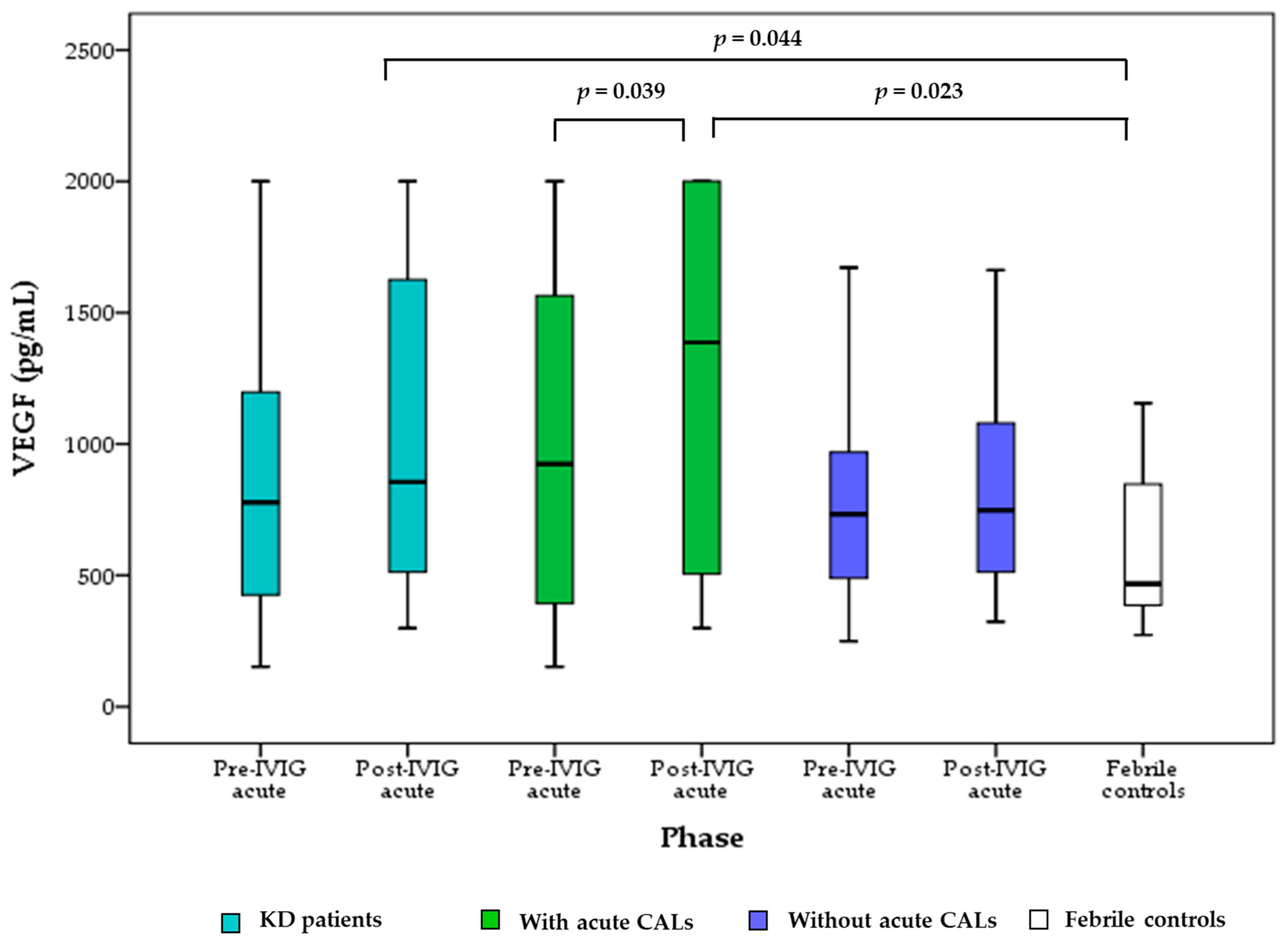
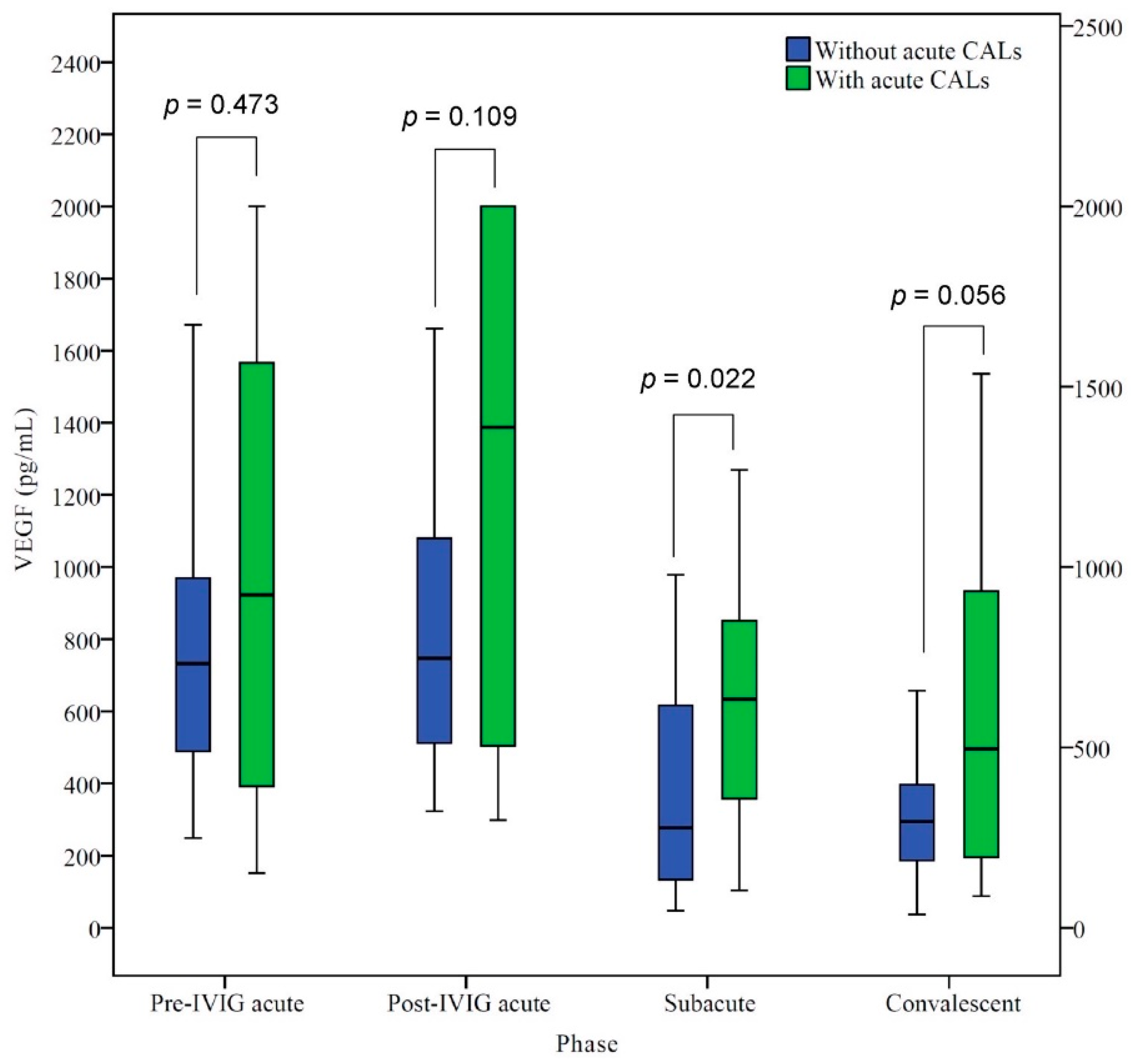
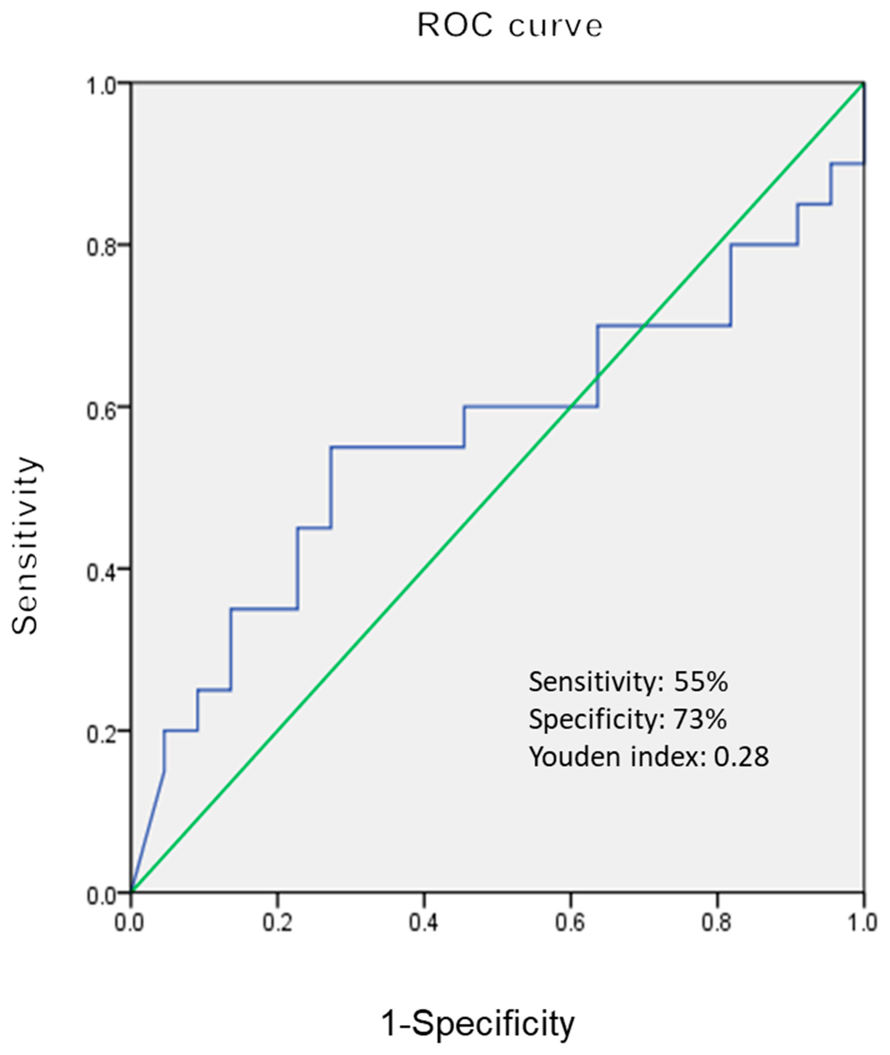
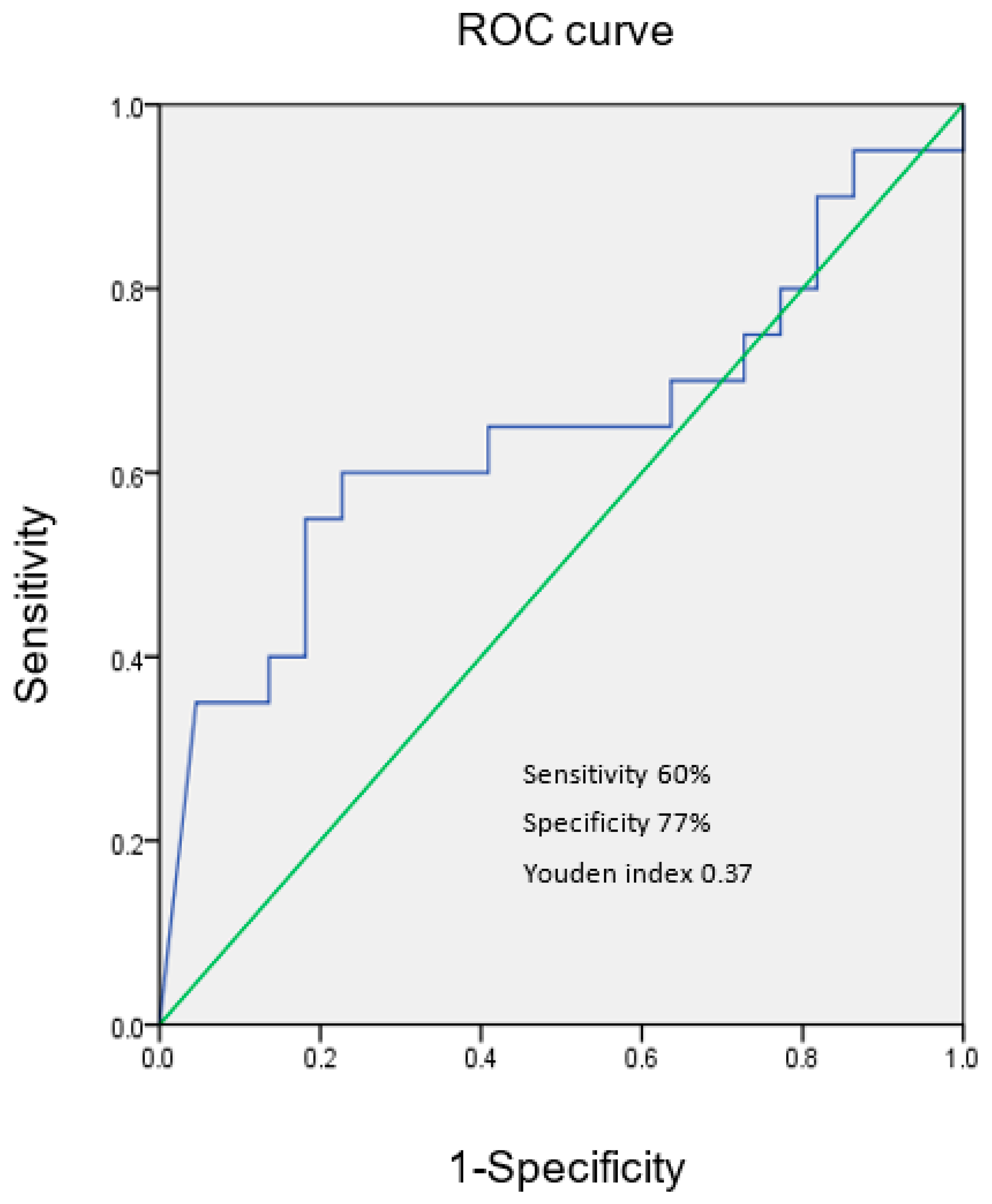
Data were expressed as mean ± standard deviation or median (quartiles). Student t test, Mann–Whitney U test, chi-square test, and Wilcoxon signed-rank test were used to determine differences in age, gender, and laboratory data between KD patients with/without acute CALs, and between KD patients and febrile controls. The correlation between VEGF and other laboratory data in KD patients was analyzed by Spearman’s rank correlation test. The receiver operating characteristic (ROC) curve was used to calculate the cutoff values of VEGF to predict KD with acute CALs. The best possible cutoff value was indicated by the highest Youden’s index. A p value of less than 0.05 was considered statistically significant. Statistical analyses were performed using SPSS version 22.0 (SPSS Inc., Chicago, IL, USA).

3. Results

Forty-two KD patients (M/F 22/20; mean age, 18.9 ± 12.2 months) and thirty febrile controls (M/F 13/17; mean age 19.1 ± 13.7 months) were enrolled in this study. The numbers of KD patients with/without acute CALs were 20 and 22, respectively. No KD patients had IVIG resistance (Table 1). Z scores of the coronary artery were 2.7 ± 0.5 in KD patients with acute CALs and 1.3 ± 0.6 in those without acute CALs. There were no KD patients with myocarditis with systolic dysfunction, arrhythmias, or pericarditis with pericardial effusion.
As shown in Figure 1, KD patients in the post-IVIG acute phase had significantly higher VEGF levels than febrile controls (p = 0.044). In KD patients with acute CALs, the VEGF levels significantly increased (p = 0.039) from the pre-IVIG acute phase to the post-IVIG acute phase. VEGF levels in KD patients with acute CALs were also significantly higher (p = 0.023) in the post-IVIG acute phase compared to those in the febrile controls. In KD patients without acute CALs, VEGF levels did not significantly increase from the pre-IVIG acute phase to the post-IVIG acute phase. KD patients without acute CALs did not have significantly higher VEGF levels in the acute phase than the febrile controls.
In KD patients, VEGF levels in the pre-IVIG acute phase were significantly higher than those in the subacute and convalescent phases (899.5 ± 549.1 vs. 508.9 ± 389.4 and 449.5 ± 362.8 pg/mL, both p < 0.001). In KD patients with acute CALs, VEGF levels significantly decreased (1241.8 ± 689.3 vs. 654.7 ± 443.3 pg/mL, p = 0.002) from the post-IVIG acute phase to the subacute phase. In KD patients without acute CALs, VEGF levels in the post-IVIG acute stage were also significantly higher (861.0 ± 483.8 vs. 376.4 ± 282.1 pg/mL, p < 0.001) than those in the subacute phase.
The difference in VEGF levels at various phases between KD patients with/without acute CALs is shown in Figure 2. KD patients with acute CALs had higher median VEGF levels than those without acute CALs in the various phases. VEGF ratios (with acute CALs/without acute CALs) from the acute to the convalescent phase were as follows: 1.2, 1.4, 1.7., and 1.8, respectively. However, there was no significant difference in VEGF levels in the various phases except the subacute phase. In the subacute phase, KD patients with acute CALs had significantly higher VEGF levels (p = 0.022) than those without CALs.
Table 2 shows Spearman correlation coefficients between VEGF levels and the laboratory data at the acute pre-IVIG and post-IVIG phases in all KD patients. In the acute pre-IVIG stage, VEGF was significantly negatively related to albumin (p = 0.039), and significantly positively related to platelet (p = 0.021) as well as C-reactive protein (CRP) (p = 0.021). In the acute post-IVIG phase, VEGF was still significantly positively related to platelet (p = 0.036) as well as CRP (p = 0.003).
Figure 3 shows the best cutoff value of VEGF 902.4 pg/mL to differentiate KD patients with/without acute CALs in the pre-IVIG acute phase.
Figure 4 shows the best cutoff value of VEGF 1106.7 pg/mL to differentiate KD patients with/without acute CALs in the post-IVIG acute phase.

4. Discussion

This study demonstrated that VEGF level in KD patients with acute CALs increased significantly after IVIG treatment in the acute stage. The significant positive correlation of VEGF with CRP as well as platelet in KD patients was found in both pre-IVIG and post-IVIG acute phases. In contrast, there was a negative correlation of VEGF with albumin only in the acute pre-IVIG phase. Compatible with previous studies [6,7,8,9,10,11,12,13], our results confirmed the involvement of VEGF in the systemic vasculitis of KD. The results in this series further imply that VEGF might play an important role in the pathogenesis of KD patients with acute CALs.
In this series, there was a negative correlation of VEGF with albumin in the acute pre-IVIG phase in KD patients. The literature regarding the effect of VEGF on the clinical features of KD is inconsistent. Maeno et al.’s results showed that serum VEGF levels did not correlate with the inflammatory markers and clinical symptoms in KD patients (n = 20) [10]. The increased VEGF levels in acute KD patients (n = 30) were independent of IVIG therapy and albumin as well as CRP levels in Terai et al.’s study [15]. In contrast, elevation of serum VEGF levels was negatively significantly correlated with low serum albumin in acute KD patients (n = 220, r = −0.53, p < 0.001) in Yasukawa et al.’s study [16]. Hung et al. also reported that VEGF levels were inversely correlated with albumin levels in acute KD patients (n = 25) [9]. Zhou et al. reported that VEGF and platelet are the important risk factors for KD patients with CALs complications (n = 60) [17]. All Yasukawa et al.’s [16], Hung et al.’s [9], and our findings might partially support the role of vascular leakage as a key feature of KD pathophysiology. We further found the significant positive correlation of VEGF with CRP as well as platelet in KD patients in both pre-IVIG and post-IVIG acute phases. CRP is a powerful inflammatory biomarker of acute KD, and has been used for predicting IVIG resistance in some scoring systems [18,19,20,21]. In this series, VEGF did not decrease after IVIG treatment, and increased significantly after IVIG treatment in KD patients with acute CALs. Our findings might suggest that high VEGF level is another risk factor for CALs other than IVIG resistance. Platelets are the major source of VEGF in the blood, and platelet VEGF levels in KD patients (n = 80) were reported to be significantly correlated with CALs in Ueno et al.’s study [22]. The previous findings [17,22] may partially explain the significant positive correlation of VEGF and platelet in our KD patients in the acute stage. Further studies are required to elucidate the interaction of VEGF, CRP, and platelet in the pathogenesis of KD.
In this series, KD patients with acute CALs had higher median VEGF levels than those without acute CALs in the various phases, but there is a significant difference in VEGF levels between the two groups only in the subacute phase. The ROC curve analysis further showed that the cutoff values of VEGF (902.4 and 1106.7 pg/mL, respectively) in the acute phase could differentiate KD with/without acute CALs with modest sensitivity and specificity. We speculate that the persistent high VEGF levels would be a prognostic marker for CALs complications and related outcomes in KD patients. Our results are generally in agreement with the previous studies [10,11,12]. The effect of VEGF on the CALs in KD patients has been demonstrated in both the immunohistochemical and animal studies. Suzuki et al.’s immunohistochemical study showed that extensive expression of VEGF was observed in the smooth muscle cells of the thickened intima at stenotic sites and at recanalized vessels in KD patients [23]. The immunohistochemical examination in Suzuki el al.’s research showed that VEGF was expressed in the intimal smooth muscle cells of the KD, but not the normal coronary artery without a history of KD [24]. Breunis et al. also reported that immunohistochemistry demonstrated VEGF expression in the coronary artery wall in autopsy tissue [25]. The murine model in Lin et al.’s study showed that local VEGF-A and its signaling pathway are associated with the development of CALs [13]. The rabbit model in An et al.’s study indicated that the phosphatase and tensin homo-log/phosphoinositide 3-kinase/VEGF pathway is important in the vascular injury in KD [26]. It is still a controversial issue to treat IVIG-resistant KD with an additional agent. Miura et al.’s study showed that both additional IVIG therapy and MTP pulse in IVIG-resistant KD patients (n = 15) suppressed TNF-α, but not VEGF [27]. Hirono et al.’s research showed that serum levels of proinflammatory cytokines decreased dramatically after infliximab treatment, but VEGF level was not suppressed in IVIG-resistant KD patients (n = 11) [28]. Recently, Su et al. reported that IL-35 levels in KD patients (n = 90) were negatively associated with VEGF levels, and suggested IL-35 may have the potential effect of preventing KD patients from developing CALs [29]. The potential agent against VEGF merits further investigation in terms of decreasing the risk of CALs in IVIG-resistant KD patients.
There are some limitations in this study. First, this is an observational study with a small number of patients in a single center. However, serial measurement of VEGF levels in four different phases increases the statistical power in this series. Second, the control group consisted of febrile subjects without definite viral diagnosis that had VEGF measurement only in the acute stage. VEGF is an important biomarker in various viral infections, including COVID-19 infection [30,31]. The role of VEGF and associated biomarkers will require in vivo and in vitro studies for clinical utility in the future. Third, the relationship between VEGF and IVIG resistance cannot be analyzed because of the lack of IVIG-resistant KD patients in this series. Fourth, the definition of CALs based on Z score may not reflect the real situation of coronary arteritis and myocarditis in KD patients. All KD patients in this series were IVIG-sensitive and had no echocardiographic findings of myocarditis with systolic dysfunction, arrhythmias, or pericarditis with pericardial effusion. Therefore, the statistical deviation can be decreased when comparing KD patients with/without acute CALs. Further multiple-center studies with large numbers of subjects are suggested.

5. Conclusions

Our data show that VEGF did not decrease after IVIG treatment, and increased significantly after IVIG treatment in KD patients with acute CALs in the acute phase. VEGF might be related to the complications of CALs in KD patients.

Author Contributions

Conceptualization, C.-Y.C., S.-H.H., K.-J.C. and K.-P.W.; methodology, C.-Y.C., S.-H.H., K.-J.C. and K.-P.W.; software, S.-H.H.; validation, C.-Y.C., K.-J.C., W.-H.C. and K.-P.W.; formal analysis, T.-J.L., W.-H.C. and S.-H.H.; investigation, C.-Y.C., W.-H.C. and K.-P.W.; resources, K.-S.H.; data curation, C.-Y.C., S.-H.H. and K.-S.H.; writing—original draft preparation, C.-Y.C., S.-H.H., K.-J.C., T.-J.L., W.-H.C., K.-S.H. and K.-P.W.; writing—review and editing, C.-Y.C., S.-H.H., K.-J.C., W.-H.C., K.-S.H. and K.-P.W.; visualization, T.-J.L.; supervision, K.-P.W.; project administration, K.-P.W.; funding acquisition, C.-Y.C. and K.-J.C. All authors have read and agreed to the published version of the manuscript.

Funding

This research was funded partly by the Kaohsiung Veterans General Hospital (KSVGH111-101) and VTY Joint Research Program (VGHUST110-G3-3-3).

Institutional Review Board Statement

The study was conducted according to the guidelines of the Declaration of Helsinki, and approved by the Institutional Review Board (or Ethics Committee) of KVGH (IRB number: VGHKS10-CT9-04).

Informed Consent Statement

After providing a detailed explanation of the study, informed consent was obtained from all guardians.

Conflicts of Interest

The authors declare no conflict of interest.

References

  1. Chang, L.S.; Weng, K.P.; Yan, J.H.; Lo, W.S.; Guo, M.M.; Huang, Y.H.; Kuo, H.C. Desquamation in Kawasaki Disease. Children 2021, 8, 317. [Google Scholar] [CrossRef]
  2. Weng, K.P.; Li, S.C.; Chien, K.J.; Tsai, K.W.; Kuo, H.C.; Hsieh, K.S.; Huang, S.H. Prediction Model for Diagnosis of Kawasaki Disease Using iTRAQ-Based Analysis. Children 2021, 8, 576. [Google Scholar] [CrossRef]
  3. Weng, K.P.; Hsieh, K.S.; Ho, T.Y.; Huang, S.H.; Lai, C.R.; Chiu, Y.T.; Huang, S.C.; Lin, C.C.; Hwang, Y.T.; Ger, L.P. IL-1B polymorphism in association with initial intravenous immunoglobulin treatment failure in Taiwanese children with Kawasaki disease. Circ. J. 2010, 74, 544–551. [Google Scholar] [CrossRef]
  4. McCrindle, B.W.; Rowley, A.H.; Newburger, J.W.; Burns, J.C.; Bolger, A.F.; Gewitz, M.; Baker, A.L.; Jackson, M.A.; Takahashi, M.; Shah, P.B.; et al. Diagnosis, Treatment, and Long-Term Management of Kawasaki Disease: A Scientific Statement for Health Professionals From the American Heart Association. Circulation 2017, 135, e927–e999. [Google Scholar] [CrossRef]
  5. Weng, K.P.; Ho, T.Y.; Chiao, Y.H.; Cheng, J.T.; Hsieh, K.S.; Huang, S.H.; Ou, S.F.; Liu, K.H.; Hsu, C.J.; Lu, P.J.; et al. Cytokine genetic polymorphisms and susceptibility to Kawasaki disease in Taiwanese children. Circ. J. 2010, 74, 2726–2733. [Google Scholar] [CrossRef]
  6. Park-Windhol, C.; D’Amore, P.A. Disorders of vascular permeability. Annu. Rev. Pathol. 2016, 11, 251–281. [Google Scholar] [CrossRef]
  7. Ohno, T.; Yuge, T.; Kariyazono, H.; Igarashi, H.; Joh-o, K.; Kinugawa, N.; Kusuhara, K.; Hara, T. Serum hepatocyte growth factor combined with vascular endothelial growth factor as a predictive indicator for the occurrence of coronary artery lesions in Kawasaki disease. Eur. J. Pediatr. 2002, 161, 105–111. [Google Scholar] [CrossRef] [PubMed]
  8. Terai, M.; Honda, T.; Yasukawa, K.; Higashi, K.; Hamada, H.; Kohno, Y. Prognostic impact of vascular leakage in acute Kawasaki disease. Circulation 2003, 108, 325–330. [Google Scholar] [CrossRef]
  9. Huang, J.; Zhang, S. Overexpressed Neuropilin-1 in Endothelial Cells Promotes Endothelial Permeability through Interaction with ANGPTL4 and VEGF in Kawasaki Disease. Mediat. Inflamm. 2021, 2021, 9914071. [Google Scholar] [CrossRef]
  10. Maeno, N.; Takei, S.; Masuda, K.; Akaike, H.; Matsuo, K.; Kitajima, I.; Maruyama, I.; Miyata, K. Increased serum levels of vascular endothelial growth factor in Kawasaki disease. Pediatr. Res. 1998, 44, 596–599. [Google Scholar] [CrossRef] [Green Version]
  11. Ohno, T.; Igarashi, H.; Inoue, K.; Akazawa, K.; Joho, K.; Hara, T. Serum vascular endothelial growth factor: A new predictive indicator for the occurrence of coronary artery lesions in Kawasaki disease. Eur. J. Pediatr. 2000, 159, 424–429. [Google Scholar] [CrossRef] [PubMed]
  12. Takeshita, S.; Kawamura, Y.; Takabayashi, H.; Yoshida, N.; Nonoyama, S. Imbalance in the production between vascular endothelial growth factor and endostatin in Kawasaki disease. Clin. Exp. Immunol. 2005, 139, 575–579. [Google Scholar] [CrossRef] [PubMed]
  13. Lin, I.C.; Sheen, J.M.; Tain, Y.L.; Chou, M.H.; Huang, L.T.; Yang, K.D. Vascular endothelial growth factor-a in lactobacillus casei cell wall extract-induced coronary arteritis of a murine model. Circ. J. 2014, 78, 752–762. [Google Scholar] [CrossRef] [PubMed]
  14. Lin, M.T.; Chang, C.H.; Hsieh, W.C.; Chang, C.E.; Chang, Y.M.; Chen, Y.C.; Hsu, J.Y.; Huang, Y.L.; Ma, J.Y.; Sun, L.C.; et al. Coronary Diameters in Taiwanese Children Younger than 6 Years Old: Z-Score Regression Equations Derived from Body Surface Area. Acta Cardiol. Sin. 2014, 30, 266–273. [Google Scholar] [PubMed]
  15. Terai, M.; Yasukawa, K.; Narumoto, S.; Tateno, S.; Oana, S.; Kohno, Y. Vascular endothelial growth factor in acute Kawasaki disease. Am. J. Cardiol. 1999, 83, 337–339. [Google Scholar] [CrossRef]
  16. Yasukawa, K.; Terai, M.; Shulman, S.T.; Toyozaki, T.; Yajima, S.; Kohno, Y.; Rowley, A.H. Systemic production of vascular endothelial growth factor and fms-like tyrosine kinase-1 receptor in acute Kawasaki disease. Circulation 2002, 105, 766–769. [Google Scholar] [CrossRef]
  17. Zhou, Y.; Wang, S.; Zhao, J.; Fang, P. Correlations of complication with coronary arterial lesion with VEGF, PLT, D-dimer and inflammatory factor in child patients with Kawasaki disease. Eur. Rev. Med. Pharmacol. Sci. 2018, 22, 5121–5126. [Google Scholar]
  18. Fukunishi, M.; Kikkawa, M.; Hamana, K.; Onodera, T.; Matsuzaki, K.; Matsumoto, Y.; Hara, J. Prediction of non-responsiveness to intravenous highdose gamma-globulin therapy in patients with Kawasaki disease at onset. J. Pediatr. 2000, 137, 172–176. [Google Scholar] [CrossRef]
  19. Kobayashi, T.; Inoue, Y.; Takeuchi, K.; Okada, Y.; Tamura, K.; Tomomasa, T.; Kobayashi, T.; Morikawa, A. Prediction of intravenous immunoglobulin unresponsiveness in patients with Kawasaki disease. Circulation 2006, 113, 2606–2612. [Google Scholar] [CrossRef]
  20. Sano, T.; Kurotobi, S.; Matsuzaki, K.; Yamamoto, T.; Maki, I.; Miki, K.; Kogaki, S.; Hara, J. Prediction of non-responsiveness to standard high-dose gamma-globulin therapy in patients with acute Kawasaki disease before starting initial treatment. Eur. J. Pediatr. 2007, 166, 131–137. [Google Scholar] [CrossRef]
  21. Egami, K.; Muta, H.; Ishii, M.; Suda, K.; Sugahara, Y.; Iemura, M.; Matsuishi, T. Prediction of resistance to intravenous immunoglobulin treatment in patients with Kawasaki disease. J. Pediatr. 2006, 149, 237–240. [Google Scholar] [CrossRef] [PubMed]
  22. Ueno, K.; Nomura, Y.; Hashiguchi, T.; Masuda, K.; Morita, Y.; Hazeki, D.; Eguchi, T.; Maruyama, I.; Kawano, Y. Platelet vascular endothelial growth factor is a useful predictor for prognosis in Kawasaki syndrome. Br. J. Haematol. 2010, 148, 285–292. [Google Scholar] [CrossRef] [PubMed]
  23. Suzuki, A.; Miyagawa-Tomita, S.; Komatsu, K.; Nishikawa, T.; Sakomura, Y.; Horie, T.; Nakazawa, M. Active remodeling of the coronary arterial lesions in the late phase of Kawasaki disease: Immunohistochemical study. Circulation 2000, 101, 2935–2941. [Google Scholar] [CrossRef] [PubMed]
  24. Suzuki, A.; Miyagawa-Tomita, S.; Komatsu, K.; Nakazawa, M.; Fukaya, T.; Baba, K.; Yutani, C. Immunohistochemical study of apparently intact coronary artery in a child after Kawasaki disease. Pediatr. Int. 2004, 46, 590–596. [Google Scholar] [CrossRef]
  25. Breunis, W.B.; Davila, S.; Shimizu, C.; Oharaseki, T.; Takahashi, K.; van Houdt, M.; Khor, C.C.; Wright, V.J.; Levin, M.; Burns, J.C.; et al. International Kawasaki Disease Genetics Consortium. Disruption of vascular homeostasis in patients with Kawasaki disease: Involvement of vascular endothelial growth factor and angiopoietins. Arthritis. Rheum. 2012, 64, 306–315. [Google Scholar] [CrossRef]
  26. An, X.; Lv, H.; Tian, J.; He, X.; Ling, N. Role of the PTEN/PI3K/VEGF pathway in the development of Kawasaki disease. Exp. Ther. Med. 2016, 11, 1318–1322. [Google Scholar] [CrossRef]
  27. Miura, M.; Kohno, K.; Ohki, H.; Yoshiba, S.; Sugaya, A.; Satoh, M. Effects of methylprednisolone pulse on cytokine levels in Kawasaki disease patients unresponsive to intravenous immunoglobulin. Eur. J. Pediatr. 2008, 167, 1119–1123. [Google Scholar] [CrossRef]
  28. Hirono, K.; Kemmotsu, Y.; Wittkowski, H.; Foell, D.; Saito, K.; Ibuki, K.; Watanabe, K.; Watanabe, S.; Uese, K.; Kanegane, H.; et al. Infliximab reduces the cytokine-mediated inflammation but does not suppress cellular infiltration of the vessel wall in refractory Kawasaki disease. Pediatr. Res. 2009, 65, 696–701. [Google Scholar] [CrossRef]
  29. Su, Y.; Feng, S.; Luo, L.; Liu, R.; Yi, Q. Association between IL-35 and coronary arterial lesions in children with Kawasaki disease. Clin. Exp. Med. 2019, 19, 87–92. [Google Scholar] [CrossRef]
  30. Page, A.V.; Liles, W.C. Biomarkers of endothelial activation/dysfunction in infectious diseases. Virulence 2013, 4, 507–516. [Google Scholar] [CrossRef]
  31. Norooznezhad, A.H.; Mansouri, K. Endothelial cell dysfunction, coagulation, and angiogenesis in coronavirus disease 2019 (COVID-19). Microvasc. Res. 2021, 137, 104188. [Google Scholar] [CrossRef] [PubMed]
Figure 1. Comparisons of vascular endothelial growth factor (VEGF) levels between KD patients with/without acute CALs in acute phase and febrile controls. Central box, values from the lower to upper quartile (25th–75th percentile). In the box plots, the middle line represents the median. The p-value was estimated by the Wilcoxon sign-rank test in KD patients in different phases. The p-value was estimated by the Mann–Whitney U test between KD patients and febrile controls.
Children 09 01343 g001
Figure 2. Comparisons of vascular endothelial growth factor (VEGF) levels between KD patients with/without acute CALs. Central box, values from the lower to upper quartile (25th–75th percentile). In the box plots, the middle line represents the median. The p-value was estimated by the Mann–Whitney U test.
Children 09 01343 g002
Figure 3. ROC curve for differentiating KD patients with/without acute CALs in the pre-IVIG acute phase at the cutoff value of VEGF 902.4 pg/mL.
Children 09 01343 g003
Figure 4. ROC curve for differentiating KD patients with/without acute CALs in the post-IVIG acute phase at the cut-off value of VEGF 1106.7 pg/mL.
Children 09 01343 g004
Table 1. Demographic data of subjects.
Factors\CategoryKD Patients
(n = 42)
Febrile Controls
(n = 30)
p Value
n (%)n (%)
Sex
Male22 (52.4)13 (43.3)0.537
Female20 (47.6)17 (56.7)
Age (months)18.9 ± 12.219.1 ± 13.70.835
Acute CALs
Yes20 (47.6)
No22 (52.4)
IVIG resistance
Yes0 (0%)
No42 (100%)
KD: Kawasaki disease; CALs: coronary artery lesions; IVIG: intravenous immunoglobulin.
Table 2. The correlation between VEGF and laboratory data of KD patients in the acute pre-IVIG and post-IVIG phases.
VariablesPre-IVIG Acute Phase
(n = 42)
Post-IVIG Acute Phase
(n = 42)
Correlation
Coefficient
p ValueCorrelation
Coefficient
p Value
WBC0.0240.8810.0080.960
PMN0.1880.2340.2010.203
Lymphocyte−0.0630.692−0.2000.204
Monocyte0.0300.849−0.0260.869
Eosinophil−0.0080.9600.2010.201
Albumin−0.3200.039−0.0410.804
Hemoglobin−0.0500.7540.0920.561
Platelet0.3560.0210.3250.036
AST0.0280.861−0.0950.549
ALT−0.0630.691−0.0690.663
CRP0.3110.0450.4430.003
TG−0.0730.6470.2800.072
TC−0.0550.7300.0050.976
HDL−0.2070.189−0.1100.490
TC/HDL ratio0.1420.3680.0860.588
LDL−0.0310.8440.0190.906
D-dimer0.0360.8230.1390.381
KD: Kawasaki disease; VEGF: vascular endothelial growth factor; WBC: white blood cell; PMN: polymorphonuclear; AST: aspartate aminotransferase; ALT: alanine aminotransferase; CRP: C-reactive protein; TG: triglyceride; TC: total cholesterol; HDL: high-density lipoprotein; LDL: low-density lipoprotein.
Publisher’s Note: MDPI stays neutral with regard to jurisdictional claims in published maps and institutional affiliations.

