
Management of Severely Aberrant Permanent First Molars in Molar Root–Incisor Malformation Patients: Case Series and a Guideline


Abstract
:1. Introduction
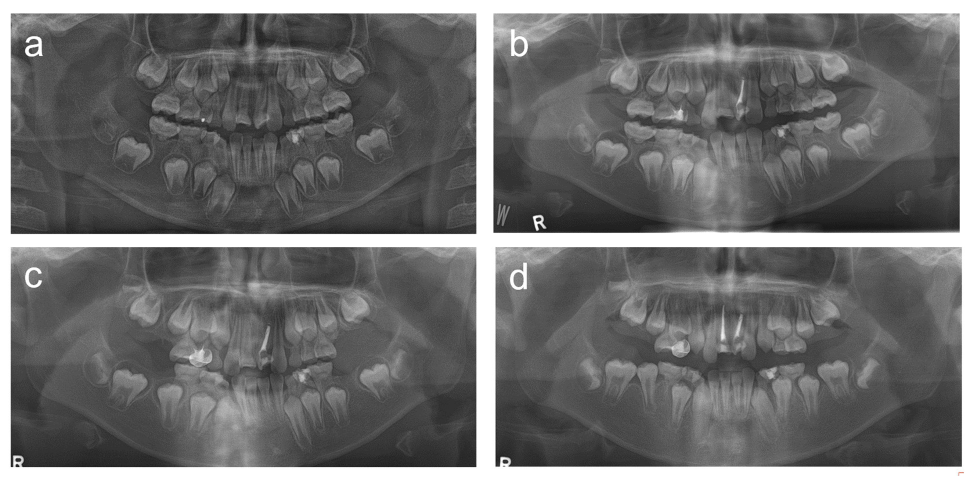
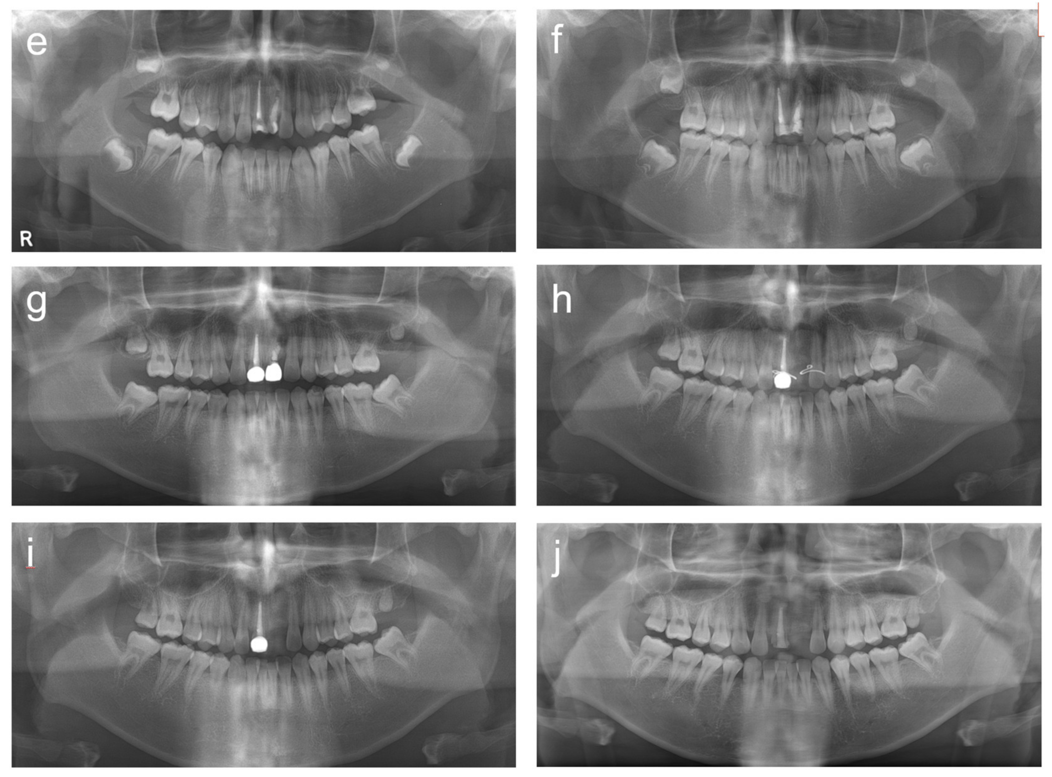
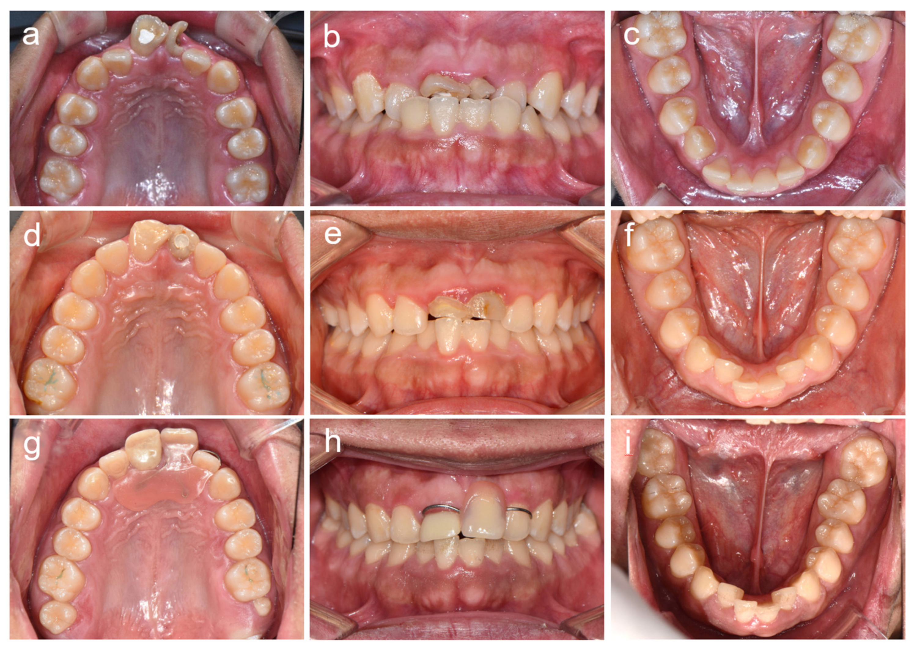
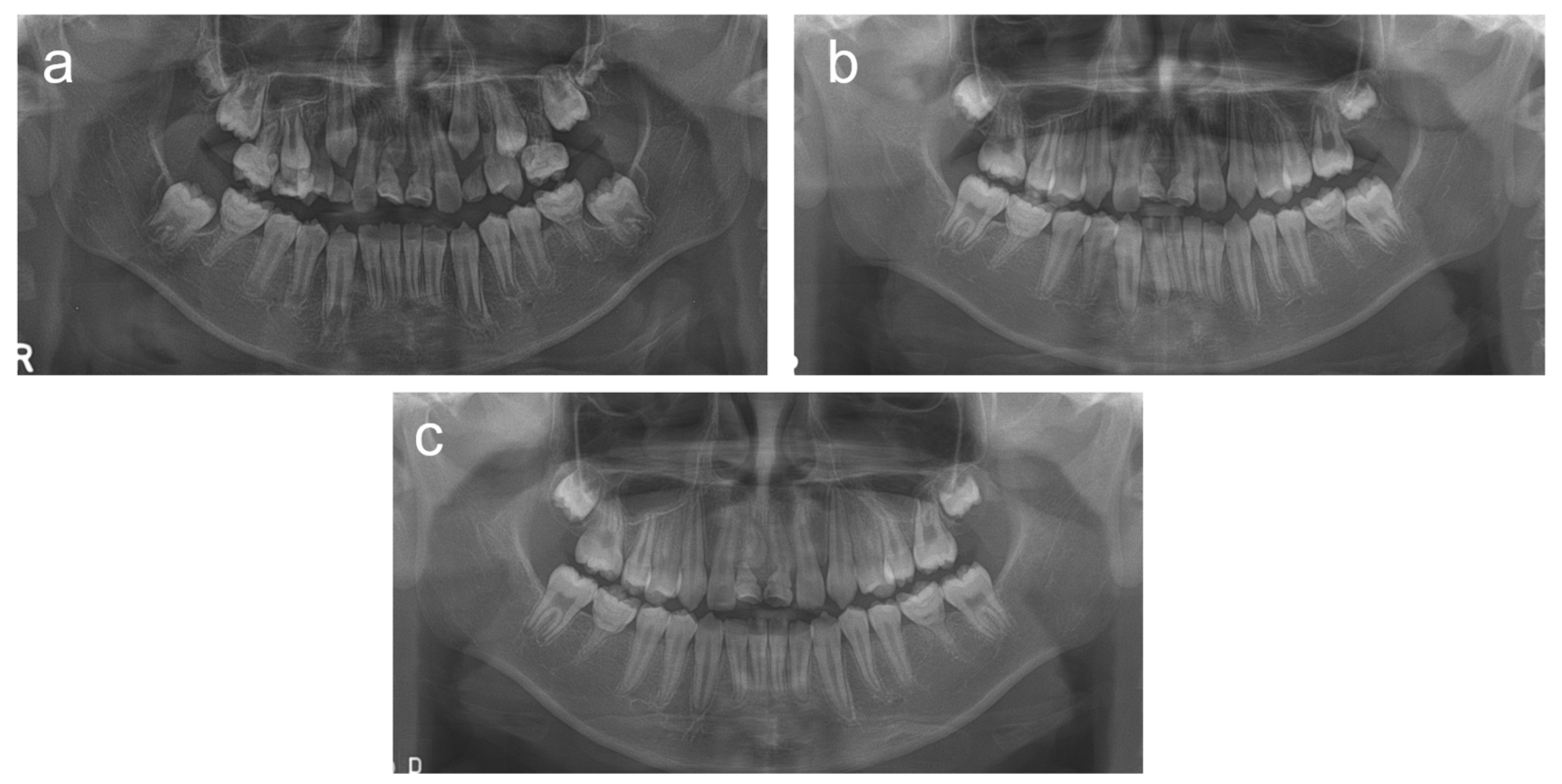
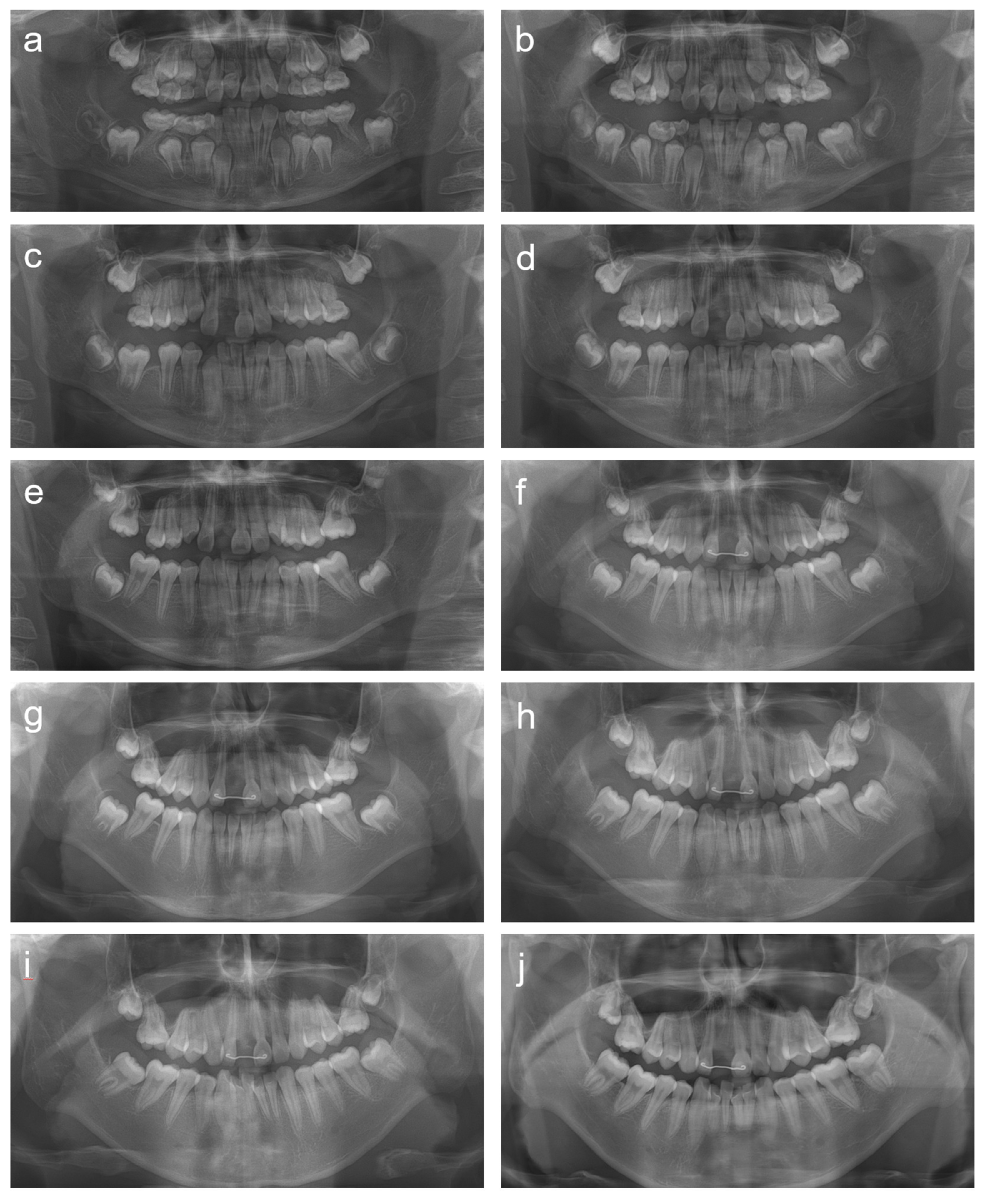
2. Cases
2.1. Case 1
2.2. Case 2
2.3. Case 3
2.4. Case 4
2.5. Case 5
3. Discussion
4. Conclusions
Author Contributions
Funding
Institutional Review Board Statement
Informed Consent Statement
Acknowledgments
Conflicts of Interest
References
- Witt, C.V.; Hirt, T.; Rutz, G.; Luder, H.U. Root malformation associated with a cervical mineralized diaphragm--a distinct form of tooth abnormality? Oral Surg. Oral Med. Oral Pathol. Oral Radiol. 2014, 117, e311–e319. [Google Scholar] [CrossRef] [PubMed] [Green Version]
- Lee, H.S.; Kim, S.H.; Kim, S.O.; Lee, J.H.; Choi, H.J.; Jung, H.S.; Song, J.S. A new type of dental anomaly: Molar-incisor malformation (MIM). Oral Surg. Oral Med. Oral Pathol. Oral Radiol. 2014, 118, 101–109.e3. [Google Scholar] [CrossRef] [PubMed]
- Brusevold, I.J.; Bie, T.M.G.; Baumgartner, C.S.; Das, R.; Espelid, I. Molar incisor malformation in six cases: Description and diagnostic protocol. Oral Surg. Oral Med. Oral Pathol. Oral Radiol. 2017, 124, 52–61. [Google Scholar] [CrossRef] [PubMed] [Green Version]
- McCreedy, C.; Robbins, H.; Newell, A.; Mallya, S.M. Molar-incisor Malformation: Two Cases of a Newly Described Dental Anomaly. J. Dent. Child. 2016, 83, 33–37. [Google Scholar]
- Neo, H.L.; Watt, E.N.; Acharya, P. Molar-incisor malformation: A case report and clinical considerations. J. Orthod. 2019, 46, 343–348. [Google Scholar] [CrossRef] [PubMed]
- Vargo, R.J.; Reddy, R.; Da Costa, W.B.; Mugayar, L.R.F.; Islam, M.N.; Potluri, A. Molar-incisor malformation: Eight new cases and a review of the literature. Int. J. Paediatr. Dent. 2020, 30, 216–224. [Google Scholar] [CrossRef] [PubMed]
- Wright, J.T.; Curran, A.; Kim, K.J.; Yang, Y.M.; Nam, S.H.; Shin, T.J.; Hyun, H.K.; Kim, Y.J.; Lee, S.H.; Kim, J.W. Molar root-incisor malformation: Considerations of diverse developmental and etiologic factors. Oral Surg. Oral Med. Oral Pathol. Oral Radiol. 2016, 121, 164–172. [Google Scholar] [CrossRef] [PubMed]
- Qari, H.; Kessler, H.; Narayana, N.; Premaraj, S. Symmetric multiquadrant isolated dentin dysplasia (SMIDD), a unique presentation mimicking dentin dysplasia type 1b. Oral Surg. Oral Med. Oral Pathol. Oral Radiol. 2017, 123, e164–e169. [Google Scholar] [CrossRef] [PubMed]
- Luder, H.U. Malformations of the tooth root in humans. Front. Physiol. 2015, 6, 307. [Google Scholar] [CrossRef] [PubMed] [Green Version]
- Lee, H.S.; Kim, S.H.; Kim, S.O.; Choi, B.J.; Cho, S.W.; Park, W.; Song, J.S. Microscopic analysis of molar-incisor malformation. Oral Surg. Oral Med. Oral Pathol. Oral Radiol. 2015, 119, 544–552. [Google Scholar] [CrossRef] [PubMed]
- Zschocke, J.; Schossig, A.; Bosshardt, D.D.; Karall, D.; Glueckert, R.; Kapferer-Seebacher, I. Variable expressivity of TCTEX1D2 mutations and a possible pathogenic link of molar-incisor malformation to ciliary dysfunction. Arch. Oral Biol. 2017, 80, 222–228. [Google Scholar] [CrossRef] [PubMed]
- Pavlič, A.; Vrecl, M.; Jan, J.; Bizjak, M.; Nemec, A. Case report of a molar-root incisor malformation in a patient with an autoimmune lymphoproliferative syndrome. BMC Oral Health 2019, 19, 49. [Google Scholar] [CrossRef] [PubMed]
- Park, S.; Byun, S.; Kim, J.; Yang, B.; Oh, S. Treatment of Molar Incisor Malformation and the short term follow-up: Case reports. Eur. J. Paediatr. Dent. 2020, 21, 238–242. [Google Scholar] [CrossRef] [PubMed]
- Yue, W.; Kim, E. Nonsurgical Endodontic Management of a Molar-Incisor Malformation-affected Mandibular First Molar: A Case Report. J. Endod. 2016, 42, 664–668. [Google Scholar] [CrossRef] [PubMed]
- Kim, J.E.; Hong, J.K.; Yi, W.J.; Heo, M.S.; Lee, S.S.; Choi, S.C.; Huh, K.H. Clinico-radiologic features of molar-incisor malformation in a case series of 38 patients: A retrospective observational study. Medicine 2019, 98, e17356. [Google Scholar] [CrossRef] [PubMed]
- Lee, H.-S.; Kim, H.J.; Lee, K.; Kim, M.S.; Nam, O.H.; Choi, S.-C. Complications of Teeth Affected by Molar-Incisor Malformation and Pathogenesis According to Microbiome Analysis. Appl. Sci. 2021, 11, 4. [Google Scholar]


Publisher’s Note: MDPI stays neutral with regard to jurisdictional claims in published maps and institutional affiliations. |
© 2021 by the authors. Licensee MDPI, Basel, Switzerland. This article is an open access article distributed under the terms and conditions of the Creative Commons Attribution (CC BY) license (https://creativecommons.org/licenses/by/4.0/).
Share and Cite
Song, J.-S.; Yang, Y.-M.; Kim, Y.-J.; Kim, J.-W. Management of Severely Aberrant Permanent First Molars in Molar Root–Incisor Malformation Patients: Case Series and a Guideline. Children 2021, 8, 904. https://doi.org/10.3390/children8100904
Song J-S, Yang Y-M, Kim Y-J, Kim J-W. Management of Severely Aberrant Permanent First Molars in Molar Root–Incisor Malformation Patients: Case Series and a Guideline. Children. 2021; 8(10):904. https://doi.org/10.3390/children8100904
Chicago/Turabian StyleSong, Ji-Soo, Yeon-Mi Yang, Young-Jae Kim, and Jung-Wook Kim. 2021. "Management of Severely Aberrant Permanent First Molars in Molar Root–Incisor Malformation Patients: Case Series and a Guideline" Children 8, no. 10: 904. https://doi.org/10.3390/children8100904