1. Introduction
It is well-known that the surgical objective in gastroschisis and omphalocele is to integrate the viscera in the abdominal cavity and then to restore the abdominal wall [
1]. But we must take into account that visceral–abdominal disproportion impairs repositioning viscera in the abdominal cavity and, in this regard, intra-abdominal pressure (IAP) represents the keystone for postoperative evolution of patients with congenital abdominal wall defects.
Increased IAP has an impact on several organs and systems. The respiratory function is influenced by the elevation and compression of the diaphragm, causing decreased tidal volume, atelectasis, hypoxemia, and hypercarbia [
2,
3]. Due to compression on the mesenteric vessels, portal vein, and inferior vena cava, increased IAP leads to decreased venous return to the right heart and consequently decreased cardiac output. Compression of the heart reduces its compliance and decreases the stroke volume, leading to diminished cardiac output [
4]. Owing to decreased cardiac output and compression on renal parenchyma and renal vessels, renal perfusion declines, leading to a reduced glomerular filtration rate [
5,
6]. Reduced mesenteric circulation, as a consequence of increased IAP, causes poor intestinal perfusion and ischemia and also diminishes lymphatic drainage with intestinal edema and bacterial translocation [
7]. Cerebral venous return is impaired by increased central venous pressure due to increased intrathoracic pressure, causing hypoxia and edema manifesting as lethargy, irritability, somnolence, or unresponsiveness [
2,
8].
The World Society of the Abdominal Compartment Syndrome (WSACS) established different thresholds when defining intraabdominal hypertension (IAH) for adults and for children. According to the 2006 consensus, IAP ≥ 2 mmHg defines IAH [
9]. Subsequently, 2013 WSACS consensus changes IAH definition to IAP > 10 mmHg, only for pediatric patients [
10]. Furthermore, abdominal compartment syndrome (ACS) is defined differently for adults and for children. ACS is defined, for adults, as IAP > 20 mmHg associated with new organ dysfunction and, for children, as IAP > 10 mmHg associated with worsening or new organ dysfunction [
9,
10].
Due to the fluid-like behavior of the abdomen, we can apply Pascal’s law, which states that the force is uniformly transmitted in a confined fluid and we can measure the pressure in any point [
11,
12]. Therefore, there are various sites for the indirect measuring of IAP described in the literature, namely, bladder, stomach, rectum, uterus, or inferior vena cava [
13]. An experimental animal study compared the direct measurement of IAP with piezoresistive probes placed in the upper and lower abdomen, and the indirect measurement of intravesical pressure (IVP) and intragastral pressure. The mean baseline pressure values were similar for the caudal peritoneal probe and IVP measurement [
14]. Other studies comparing pressures measured via an indwelling urinary catheter and via an intraperitoneal dialysis catheter demonstrated that IVP represents an accurate delineation of IAP [
15,
16]. Another study, performed on patients undergoing laparoscopic cholecystectomy, revealed that urinary bladder pressure showed parallel increments to elevated IAP during intraperitoneal carbon dioxide insufflation and also a similar decrease of IVP during gradual deflation of the intraperitoneal cavity [
17].
IVP represents a valid reflection of IAP, and WSACS consensus from 2013 recommends the indirect trans-bladder technique for the measurement of IAP [
10].
The aim of the study was to highlight the occurrence of postoperative abdominal compartment syndrome in patients treated for gastroschisis and omphalocele, in the Neonatal Intensive Care Unit of “Marie Sklodowska Curie” Emergency Clinical Hospital for Children, over the last decade. Identifying the pitfall of postoperative abdominal compartment syndrome when performing abdominal closure without quantifying the IAP, we decided to start monitoring IVP in order to have an objective evaluation of the IAP. We intend to improve our outcomes by adjusting the surgical procedure based on IVP measurement in an attempt to avoid IAH.
2. Materials and Methods
We performed a retrospective study on 47 patients born with gastroschisis and omphalocele that were admitted from January 2011 to June 2021 to the Neonatal Intensive Care Department of “Marie Sklodowska Curie” Emergency Clinical Hospital for Children.
Data were extracted retrospectively from our hospital medical records. We developed a database that included the following parameters: gender, gestational age, birth weight, Apgar score, type of abdominal wall defect, associated congenital anomalies, type of surgical procedure performed for abdominal wall closure, additional surgical procedures, time from birth until surgical procedure, and presence of abdominal compartment syndrome. The identification data were not included in our database. Our main aim was to emphasize the presence of postoperative abdominal compartment syndrome for patients with gastroschisis and omphalocele.
Starting from July 2021, we introduced IVP measurement as a means to assess patients with congenital abdominal wall defects, and we began a prospective study with the purpose of reducing the incidence of postoperative abdominal compartment syndrome. We included in the study all the patients with congenital abdominal wall defects that were admitted to our Neonatal Intensive Care Unit. There were no exclusion criteria for this study. Up to this point, we included in our survey nine patients with gastroschisis and omphalocele.
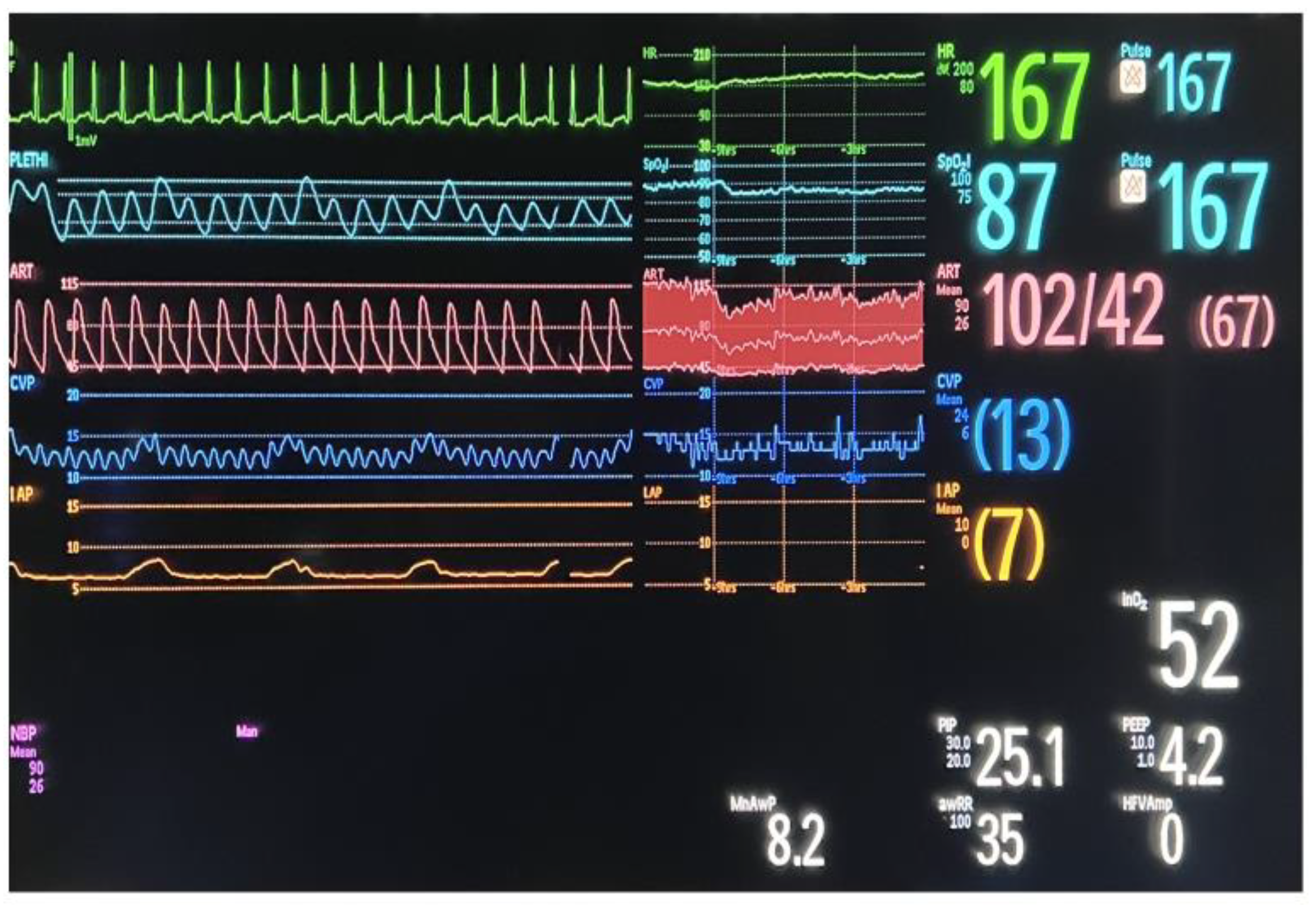
We inserted a Foley catheter in the bladder and connected it to a pressure transducer that is usually used in our Neonatal Intensive Care Unit for invasive blood pressure monitoring. The patient was placed in the supine position, with no elevation of the head of the bed. Then, we introduced in the bladder, through the Foley catheter, 1 mL/kg of room-temperature sterile 0.9% NaCl solution. After 1 min, we read the value of IVP shown on the monitor as a substitute of central venous pressure in mmHg (
Figure 1).
We created a database similar to the one for the retrospective study and, additionally, we included daily measurement of arterial pressure, heart rate, diuresis, and oxygen saturation and noted the value of Peak Inspiratory Pressure (PIP) for mechanically ventilated patients, the presence of intestinal transit, and the presence of edema.
Data were recorded using Microsoft Excel (Microsoft Corporation (2018). Microsoft Excel) and were analyzed using statistical software JASP (JASP Team (2022). JASP (Version 0.16.3) [Computer software])
Informed consent for surgery was obtained from the parents of all the subjects involved in the study. Operative technique was not standardized and was chosen by the lead surgeon based on their experience.
Since 2018, our informed consent has included a section dedicated to permission to participate in clinical teaching and to use their medical data for research studies, considering our institution is a teaching hospital. For the cases prior to 2018, the medical data collection was approved by the ethics committee of our hospital.
The approval of the ethics committee was obtained both in terms of data collection and analysis and in terms of publishing the results (no. 11231/31 March 202).
3. Results
3.1. Retrospective Study Results
Between January 2011 and June 2021, forty-seven patients with gastroschisis and omphalocele were admitted to the Neonatal Intensive Care Department of our hospital, of whom 30 were diagnosed with gastroschisis and 17 with omphalocele.
Demographics for each group are described in
Table 1. We can observe that there are no significant differences between the analyzed parameters, with a mean birth weight of 2404 g (±507 g) for the gastroschisis group and 2681 g (±591 g) for the omphalocele group, a mean gestational age of 36 weeks for both groups, and a mean Apgar score of 7 (±1) for the gastroschisis group and 8 (±1) for the omphalocele group.
In the gastroschisis group, the number of male and female patients was equal: fifteen males and fifteen females, while in the omphalocele group it was a female preponderance: eleven female patients and six male patients.
Additional congenital anomalies were identified in twenty-three of the gastroschisis patients, of whom seven had multiple associated anomalies (more than two anomalies in different organ systems) and sixteen had a single additional anomaly. Additional anomalies were found in fifteen omphalocele patients, of whom five had multiple associated anomalies and ten had a single additional anomaly.
Figure 2 summarizes the distribution of associated anomalies related to the affected organ system.
There were no standardized criteria for electing the type of abdominal wall closure, the decision of the surgical procedure being based on the experience of the surgeon. Primary closure was chosen for 27 of the 30 patients with gastroschisis, in five cases using additional abdominal wall stretching and in one case using dissection to create skin flaps for closure of the abdomen. In the other six cases, an umbilical cord patch was used for primary closure. Two patients needed intestinal resection and anastomosis due to associated intestinal atresia. Staged closure was chosen in three cases using three different methods: using a polyvinyl chloride tailored bag, a silo bag, or a polyvinyl chloride patch (
Table 2).
For omphalocele, primary closure was chosen in 16 of the 17 cases. Delayed closure was performed in one case, after daily wound dressing for 28 days. Regarding additional procedures, abdominal wall stretching was used in one case of primary closure. For one patient with ileal atresia and Ladd band, intestinal resection with ileostomy and lysis of the Ladd band was performed, and one patient with patent omphaloenteric duct required resection of the omphaloenteric duct (
Table 3).
On the grounds that gastroschisis requires emergency surgical intervention, we performed surgery shortly after the patients were admitted to the Neonatal Intensive Care Unit. Because patients are transferred to our hospital from different institutions, the time from birth to surgical procedure varied according to transport time. For gastroschisis, he median time from birth to surgical procedure was 6 h, with a standard deviation of 5.85 h. For omphalocele, the surgical procedure can be postponed since this congenital anomaly does not represent a surgical emergency if the omphalocele membrane is not ruptured. For omphalocele there was a median time of 48 h, with a minimum time from the delivery until surgical procedure of 7 h and a maximum time of 672 h for the patient that was managed 4 weeks with daily wound dressings before the abdominal wall closure was performed (
Table 4).
Studying the medical files, we observed that 12.77% of the patients developed clinical signs of abdominal compartment syndrome. From the total of six patients with abdominal compartment syndrome, five patients with gastroschisis underwent primary closure of the abdominal wall and the sixth underwent staged closure using a polyvinyl chloride patch (
Table 5).
For the patients included in the retrospective study, abdominal compartment syndrome was diagnosed based on clinical signs. Based on their medical charts, all six patients had tense abdomen upon palpation. Decreased urinary output and an increase in serum creatinine levels were used to diagnose renal failure. According to the Neonatal Acute Kidney Injury (AKI) classification of Kidney Disease Improving Global Outcomes (KDIGO) [
18], there were three patients with stage 1 AKI, two patients with stage 2 AKI, and one patient with stage 3 AKI. Moreover, one of the patients developed tachycardia and hypotension and was diagnosed with acute heart failure.
Regarding medical management of the abdominal compartment syndrome, medical treatment with diuretics (Furosemide) was prescribed for all the patients. Four patients required inotropic support with Dopamine, two of them needed Dopamine and Adrenaline administration and, for the patient with acute heart failure, Dopamine, Adrenaline, and Dobutamine were administered.
Four of the six patients described with ACS needed subsequent surgical procedures. Due to abdominal distension and the absence of stools, one patient required surgical intervention, and a 4 cm segment of ileal stenosis was identified with intestinal distension proximal to this segment. The stenotic segment was resected and Mikulicz ileostomy was performed. Closure of the abdomen was not possible due to abdominal–visceral disproportion, and prosthetic material (Gore-Tex Dual Mesh) was used for abdominal wall closure. Two weeks later, intestinal anastomosis was performed to restore intestinal continuity and the abdominal wall was restored. One patient had jejunal stenosis, and segmental resection with end-to-end jejunal anastomosis was performed one month after the first procedure. Two patients required lysis of bowel adhesions and one of them had a covered ileal perforation; therefore, an ileostomy was performed. Gentle maneuvers for intestinal content evacuation were performed for the last three patients described. In that manner, the previously distended bowel loops were easily reintegrated in the abdominal cavity and closure of the abdominal wall without tension was possible.
3.2. Prospective Study Preliminary Results
Since we implemented the IVP measurement, nine patients with congenital abdominal wall defects were admitted in our hospital—five patients with gastroschisis (three males and two females) and four with omphalocele (two males and two females).
The mean birth weight was 2835 g, with a minimum weight of 2000 g and a maximum weight of 3900 g. The gestational age ranged between 37 weeks and 40 weeks, and the Apgar score ranged between 7 and 9 (
Table 6).
Regarding associated congenital anomalies, four patients with gastroschisis and three patients with omphalocele had cardiac malformations: all seven were diagnosed by sonography with atrial septal defect and patent ductus arteriosus and one of them with aortic arch stenosis. At the time of surgery, two of the patients with omphalocele and all of the gastroschisis patients were diagnosed with intestinal malrotation; two of the patients with gastroschisis also had associated microcolon. Additionally, one patient with gastroschisis and one with omphalocele had associated renal ectopia.
Two patients with gastroschisis underwent primary closure and three underwent staged closure. For staged closure, a modified Schuster procedure was performed using a suspended tailored polyvinyl chloride (PVC) bag with serial reduction while measuring IVP and later reintegration. In the omphalocele group, two patients had primary closure, one patient underwent partial reduction and ligation of the omphalocele sac with abdominal wall closure the next day, and another underwent staged closure with the Schuster procedure using a suspended tailored polyvinyl chloride bag and serial reduction (
Table 7).
Additionally, for the patients that underwent the Schuster procedure, we measured the IVP at the time of the serial reductions and one or two hours after the procedure. In the cases where we performed staged closure, there were no complications related to the type of prosthetic material we used to create the bag for gradual reduction of the viscera.
We evaluated daily respiratory and cardiac function, measured diuresis, and observed intestinal transit. There were no clinical signs of developing abdominal compartment syndrome and the IVP values monitored were all below 10 mmHg.
Normal values of diuresis were noted, with a minimal value of 1 mL/kg/h in one case for a day, and all the other recorded values of diuresis were above 2 mL/kg/h (
Table 8).
Regarding cardiac function,
Table 9 and
Table 10 depict the value ranges of heart rate and mean arterial pressure (MAP). We reported values of heart rate and MAP between the normal ranges according to the reference values for the neonatal period.
We monitored IVP in the Operating Room at the time of abdominal wall closure, then one or two hours after the procedure in the Neonatal Intensive Care Unit, and then daily until we reached a plateau. A descriptive analysis of the IVP values measured displays no value above 10 mmHg (
Table 11).
Figure 3 is a graphical representation of the evolution of IVP values recorded postoperatively. With the red line, we marked the 10 mmHg threshold, as this is the value above which WSACS consensus defines IAH. Variations of pressure values were observed, especially for patients with staged closure—pressure increased after ligation of the PVC tailored bag and after definitive abdominal wall repair. For instance, Patient 6 underwent staged closure with the modified Schuster procedure with a PVC tailored bag, for a giant omphalocele (liver, spleen, small bowel, and colon protruding through the abdominal wall defect). When serial reduction of the viscera in the abdomen with ligation of the bag was performed, the IVP increased as shown: day 2—from 8 mmHg to 10 mmHg, day 6—from 5 mmHg to 6 mmHg, day 12—from 6 mmHg to 7 mmHg, day 16—from 5 mmHg to 7 mmHg, day 19—from 2 mmHg to 5 mmHg and, at day 21, at the time of definitive wall closure, the pressure increased from 2 mmHg to 7 mmHg.
4. Discussion
Despite the surgical goal of reducing viscera in the abdomen in congenital abdominal wall defects, it is important to be cautious when selecting the type of abdominal wall closure. Overenthusiastic primary closure could lead, as we observed in our retrospective study, to abdominal compartment syndrome.
A 30-year retrospective study from a different Romanian hospital, regarding risk factors of the unfavorable evolution of their patients with gastroschisis, mentioned the presence of clinical signs of abdominal compartment syndrome in 19 out of 159 patients, reflecting percentages similar to ours [
19]. These results reveal the fact that surgical experience is not enough to achieve the best results; we also need an objective quantification of abdominal pressure when performing abdominal closure.
Based on our results regarding the presence of postoperative abdominal compartment syndrome, we inferred that we should estimate the IAP at the time of abdominal wall closure as a way to improve outcomes for patients with abdominal wall defects. In this regard, we decided to implement, starting from June 2021, the protocol for intraabdominal pressure monitoring at the time of surgical procedure and performed primary closure only if the IVP was below 10 mmHg after integrating the viscera into the abdominal cavity.
Studying published articles regarding IVP measurement, we decided to measure the introduction of 1 mL/kg of sterile saline solution in the bladder. Defontaine et al. studied the required volume of saline solution instillation for IVP measurement and concluded that 1 mL/kg is the optimal volume for newborns weighing less than 4.5 kg [
20]. Another study recorded IAP via an intraperitoneal dialysis catheter and an indwelling urinary catheter with different bladder filling volumes and determined that IAP is best estimated with an intravesical volume of 1 mL/kg [
16].
Regarding the temperature of saline solution used for IVP measurement, a comparative study theorized that 35 °C saline solution, being close to body temperature, would not stimulate the bladder muscle wall. They compared the results of IVP using saline solution at 15 °C, 25 °C, and 35 °C. There were significant differences between IVP using 35 °C and 15 °C saline solution, probably because of bladder wall muscle stimulation due to lower temperatures. When comparing IVP values using 35 °C and 25 °C saline solution, the results were similar. They concluded that it is not necessary to heat the saline solution and that room-temperature solution could be directly used to measure IVP [
21]. In consideration of this study, we agreed to use room-temperature solution for our measurements.
Nevertheless, the pressure should be measured with the patient in supine position with no elevation of the head of the table, because studies show increased values of IVP with bed elevation, probably due to gravitational visceral compression of the bladder [
22,
23,
24].
For the patients included in the prospective study, clinical signs of abdominal compartment syndrome were absent and the IVP values monitored were all below 10 mmHg. In the cases where we performed staged closure, there were no complications related to the type of prosthetic material we used to create the bag for gradual reduction of the viscera.
Following our few cases after we implemented measurement of IVP, we are optimistic about the future regarding postoperative management of patients with congenital abdominal wall defects. Undoubtedly, we need a substantial number of cases in order to make a valid conclusion.
Unfortunately, as shown by a few studies, pediatric guidelines for IAH and ACS are not very widely acknowledged. In 2010, 127 heads of pediatric intensive care units from Germany responded to a questionnaire that revealed that the vast majority (99 respondents) use exclusively clinical signs for the diagnosis of IAH and ACS. Also, only 3.9% chose the correct IAH/ACS criteria and 16.5% chose definitions in accordance with WSACS consensus [
25]. After the 2013 WSACS updated guidelines, 156 questionnaires were returned from heads of pediatric/neonatal intensive care units from Germany, Austria, and Switzerland. Only 6% of the physicians chose the correct definition for IAH and 58% chose the correct definition of ACS in conformity with WSACS updated guidelines. Furthermore, there were still a high number of centers that relied exclusively on clinical signs to diagnose IAH (50%) and ACS (40%) [
26]. Other recent surveys from Saudi Arabia also revealed that many physicians are not aware of the actual definitions of IAH [
27,
28].
Presenting our experience aims to emphasize the importance of staying current in our field in order to improve our patients’ outcomes. Better results are linked to understanding our past results, observing what we could improve and, consequently, searching for solutions. Also, by enhanced cooperation between the surgical team and the neonatal intensive care team, we could find inventive solutions for our problems—for instance, in this case, we used for IVP measurement the pressure transducers that were already available in the Neonatal Intensive Care Unit for invasive blood pressure monitoring.
As a future perspective, we trust that, if we continue to perform abdominal closure only at pressures lower than 10 mmHg and monitor the IVP until we reach a plateau, there will be no postoperative compartment syndrome attributable to abdominal closure. In this manner, intravesical pressure measurement is of great value in preventing complications related to increased abdominal pressure, as it is a non-invasive method that could be easily applied either in the Operating Room or in the Neonatal Intensive Care Unit.
5. Conclusions
Developing abdominal compartment syndrome after the closure of congenital abdominal wall defects represents a risk even in the most experienced hands. For this reason, we believe that we should use a quantifiable method with which to assess intraabdominal pressure. As stated by the WSASC consensus, indirect measurement of intraabdominal pressure via a bladder catheter connected to a pressure transducer is a reliable method. In this manner, measuring the abdominal pressure while closing the abdominal wall and not allowing pressures above 10 mmHg would help us prevent postoperative abdominal compartment syndrome. This approach should aid us to achieve better postoperative evolution for patients with gastroschisis and omphalocele.
Author Contributions
Conceptualization, R.-A.M. and N.S.I.; methodology, N.S.I. and C.C.; software, R.-A.M.; validation, N.S.I., C.C., M.B. and I.F.S.; formal analysis R.-A.M.; investigation, R.-A.M.; resources, C.C., M.B. and I.F.S.; data curation, R.-A.M.; writing—original draft preparation, R.-A.M.; writing—review and editing, R.-A.M., C.C., M.B., I.F.S. and N.S.I.; visualization, R.-A.M.; supervision, N.S.I.; project administration, C.C. and N.S.I. All authors have read and agreed to the published version of the manuscript.
Funding
This research received no external funding.
Institutional Review Board Statement
The study was conducted in accordance with the Declaration of Helsinki, and was approved by the Institutional Review Board (or Ethics Committee) of ‘Marie S. Curie’ Emergency Clinical Hospital for Children, Bucharest, Romania (11231/approval date 31 March 2023).
Informed Consent Statement
Informed consent was obtained from the parents of all subjects involved in the study.
Data Availability Statement
Data sharing not applicable.
Conflicts of Interest
The authors declare no conflict of interest.
References
- Bhat, V.; Moront, M.; Bhandari, V. Gastroschisis: A state-of-the-art review. Children 2020, 7, 302. [Google Scholar] [CrossRef] [PubMed]
- Reitsma, J.; Schumacher, B. Nursing Assessment of Intra-abdominal Hypertension and Abdominal Compartment Syndrome in the Neonate. Adv. Neonatal Care 2018, 18, 7–13. [Google Scholar] [CrossRef] [PubMed]
- Stassen, N.A.; Lukan, J.K.; Dixon, M.S.; Carrillo, E.H.; Carrillo, E.H. Abdominal Compartment Syndrome. Scand. J. Surg. 2002, 91, 104–108. [Google Scholar] [CrossRef] [PubMed]
- Newcombe, J.; Mathur, M.; Ejike, J.C.; Williams, L.-M.; Hubbard, K.E.; Daye, O.; Barden, C. Abdominal compartment syndrome in children. Crit. Care Nurse 2012, 32, 51–61. [Google Scholar] [CrossRef] [PubMed] [Green Version]
- Hunter, J.D.; Damani, Z. Intra-abdominal hypertension and the abdominal compartment syndrome. Anaesthesia 2004, 59, 899–907. [Google Scholar] [CrossRef] [PubMed] [Green Version]
- Sugrue, M. Abdominal compartment syndrome. Curr. Opin. Crit. Care 2005, 11, 333–338. [Google Scholar] [CrossRef]
- Lewis, M.; Benjamin, E.R.; Demetriades, D. Intra-abdominal hypertension and abdominal compartment syndrome. Curr. Probl. Surg. 2021, 58, 159–169. [Google Scholar] [CrossRef]
- Thabet, F.C.; Ejike, J.C. Intra-abdominal hypertension and abdominal compartment syndrome in pediatrics. A review. J. Crit. Care 2017, 41, 275–282. [Google Scholar] [CrossRef]
- Malbrain, M.L.N.G.; Cheatham, M.L.; Kirkpatrick, A.; Sugrue, M.; Parr, M.; De Waele, J.; Balogh, Z.; Leppäniemi, A.; Olvera, C.; Ivatury, R.; et al. Results from the International Conference of Experts on Intra-abdominal Hypertension and Abdominal Compartment Syndrome. I. Definitions. Intensiv. Care Med. 2006, 32, 1722–1732. [Google Scholar] [CrossRef]
- Kirkpatrick, A.W.; The Pediatric Guidelines Sub-Committee for the World Society of the Abdominal Compartment Syndrome; Roberts, D.J.; De Waele, J.; Jaeschke, R.; Malbrain, M.L.N.G.; De Keulenaer, B.; Duchesne, J.; Bjorck, M.; Leppaniemi, A.; et al. Intra-abdominal hypertension and the abdominal compartment syndrome: Updated consensus definitions and clinical practice guidelines from the World Society of the Abdominal Compartment Syndrome. Intensiv. Care Med. 2013, 39, 1190–1206. [Google Scholar] [CrossRef] [Green Version]
- Muturi, A.; Ojuka, D.; Ndaguatha, P.; Kibet, A. Abdominal Compartment Syndrome in Surgical Patients. Ann. Afr. Surg. 2018, 14, 48–52. [Google Scholar] [CrossRef] [Green Version]
- Malbrain, M.L.N.G.; Peeters, Y.; Wise, R. The neglected role of abdominal compliance in organ-organ interactions. Crit. Care 2016, 20, 67. [Google Scholar] [CrossRef] [PubMed] [Green Version]
- Malbrain, M.L.N.G. Different techniques to measure intra-abdominal pressura (IAP): Time for a critical re-appraisal. Intensiv. Care Med. 2004, 30, 357–371. [Google Scholar] [CrossRef] [PubMed]
- Chopra, S.S.; Wolf, S.; Rohde, V.; Freimann, F.B. Pressure measurement techniques for abdominal hypertension: Conclusions from an experimental model. Crit. Care Res. Pract. 2015, 2015, 278139. [Google Scholar] [CrossRef] [PubMed] [Green Version]
- Davis, P.J.; Koottayi, S.; Taylor, A.; Butt, W.W. Comparison of indirect methods of measuring intra-abdominal pressure in children. Intensiv. Care Med. 2005, 31, 471–475. [Google Scholar] [CrossRef]
- Suominen, P.K.; Pakarinen, M.P.; Rautiainen, P.; Mattila, I.; Sairanen, H. Comparison of direct and intravesical measurement of intraabdominal pressure in children. J. Pediatr. Surg. 2006, 41, 1381–1385. [Google Scholar] [CrossRef]
- Al-Abassi, A.A.; Al Saadi, A.S.; Ahmed, F. Is intra-bladder pressure measurement a reliable indicator for raised intra-abdominal pressure? A prospective comparative study. BMC Anesthesiol. 2018, 18, 69. [Google Scholar] [CrossRef] [Green Version]
- Selewski, D.T.; Charlton, J.R.; Jetton, J.G.; Guillet, R.; Mhanna, M.J.; Askenazi, D.J.; Kent, A.L. Neonatal acute kidney injury. Pediatrics 2015, 136, e463–e473. [Google Scholar] [CrossRef] [Green Version]
- Ţarcă, E.; Roșu, S.T.; Cojocaru, E.; Trandafir, L.; Luca, A.C.; Lupu, V.V.; Moisă, Ș.M.; Munteanu, V.; Butnariu, L.I.; Ţarcă, V. Statistical analysis of the main risk factors of an unfavorable evolution in gastroschisis. J. Pers. Med. 2021, 11, 1168. [Google Scholar] [CrossRef]
- Defontaine, A.; Tirel, O.; Costet, N.; Beuchée, A.; Ozanne, B.; Gaillot, T.; Arnaud, A.P.; Wodey, E. Transvesical Intra-Abdominal Pressure Measurement in Newborn: What Is the Optimal Saline Volume Instillation? Pediatr. Crit. Care Med. 2016, 17, 144–149. [Google Scholar] [CrossRef]
- Zou, J.; Zheng, L.; Shuai, W.; Li, Q.; Wang, Q.; Zhang, Z.; Li, D. Comparison of Intra-Abdominal Pressure Measurements in Critically Ill Patients Using Intravesical Normal Saline at 15 °C, 25 °C, and 35 °C; Medical Science Monitor; International Scientific Information, Inc.: New York, NY, USA, 2021. [Google Scholar]
- Cheatham, M.L.; De Waele, J.J.; De Laet, I.; De Keulenaer, B.; Widder, S.; Kirkpatrick, A.W.; Cresswell, A.B.; Malbrain, M.; Bodnar, Z.; Mejia-Mantilla, J.H.; et al. The impact of body position on intra-abdominal pressure measurement: A multicenter analysis. Crit. Care Med. 2009, 37, 2187–2190. [Google Scholar] [CrossRef]
- McBeth, P.B.; Zygun, D.A.; Widder, S.; Cheatham, M.; Zengerink, I.; Glowa, J.; Kirkpatrick, A.W. Effect of patient positioning on intra-abdominal pressure monitoring. Am. J. Surg. 2007, 193, 644–647. [Google Scholar] [CrossRef] [PubMed]
- Samimian, S.; Ashrafi, S.; Mohammadi, T.K.; Yeganeh, M.R.; Ashraf, A.; Hakimi, H.; Dehghani, M. The Correlation between Head of Bed Angle and IntraAbdominal Pressure of Intubated Patients; a Pre-Post Clinical Trial. Arch. Acad. Emerg. Med. 2021, 9, e23. [Google Scholar] [PubMed]
- Kaussen, T.; Steinau, G.; Srinivasan, P.K.; Otto, J.; Sasse, M.; Staudt, F.; Schachtrupp, A. Recognition and management of abdominal compartment syndrome among German pediatric intensivists: Results of a national survey. Ann. Intensiv. Care 2012, 2012, S8. [Google Scholar] [CrossRef] [PubMed] [Green Version]
- Wiegandt, P.; Jack, T.; von Gise, A.; Seidemann, K.; Boehne, M.; Koeditz, H.; Beerbaum, P.; Sasse, M.; Kaussen, T. Awareness and diagnosis for intra-abdominal hypertension (IAH) and abdominal compartment syndrome (ACS) in neonatal (NICU) and pediatric intensive care units (PICU)—A follow-up multicenter survey. BMC Pediatr. 2023, 23, 82. [Google Scholar] [CrossRef]
- Qutob, R.; A Alkhannani, A.H.; Alassaf, T.Y.; Alhokail, S.O.; Bagazi, G.A.; Alsaleh, A.A.; Alqarni, M.K.; Alammari, Y.; Al Harbi, K.; Elhazmi, A.; et al. Physicians’ Knowledge of Abdominal Compartment Syndrome and Intra-Abdominal Hypertension in Saudi Arabia: An Online Cross-Sectional Survey Study. Int. J. Gen. Med. 2022, 15, 8509–8526. [Google Scholar] [CrossRef]
- Rezeni, N.; Thabet, F. Awareness and management of intra-abdominal hypertension and abdominal compartment syndrome by paediatric intensive care physicians: A national survey. Anaesthesiol. Intensiv. Ther. 2022, 54, 315–319. [Google Scholar] [CrossRef]
| Disclaimer/Publisher’s Note: The statements, opinions and data contained in all publications are solely those of the individual author(s) and contributor(s) and not of MDPI and/or the editor(s). MDPI and/or the editor(s) disclaim responsibility for any injury to people or property resulting from any ideas, methods, instructions or products referred to in the content. |
© 2023 by the authors. Licensee MDPI, Basel, Switzerland. This article is an open access article distributed under the terms and conditions of the Creative Commons Attribution (CC BY) license (https://creativecommons.org/licenses/by/4.0/).

 ,
, 


