
Pediatric Interventional Neuroradiology: Opportunities and Challenges
Abstract
:1. Introduction
2. Neurointerventional Procedures in the Pediatric Population
2.1. Vascular Malformations
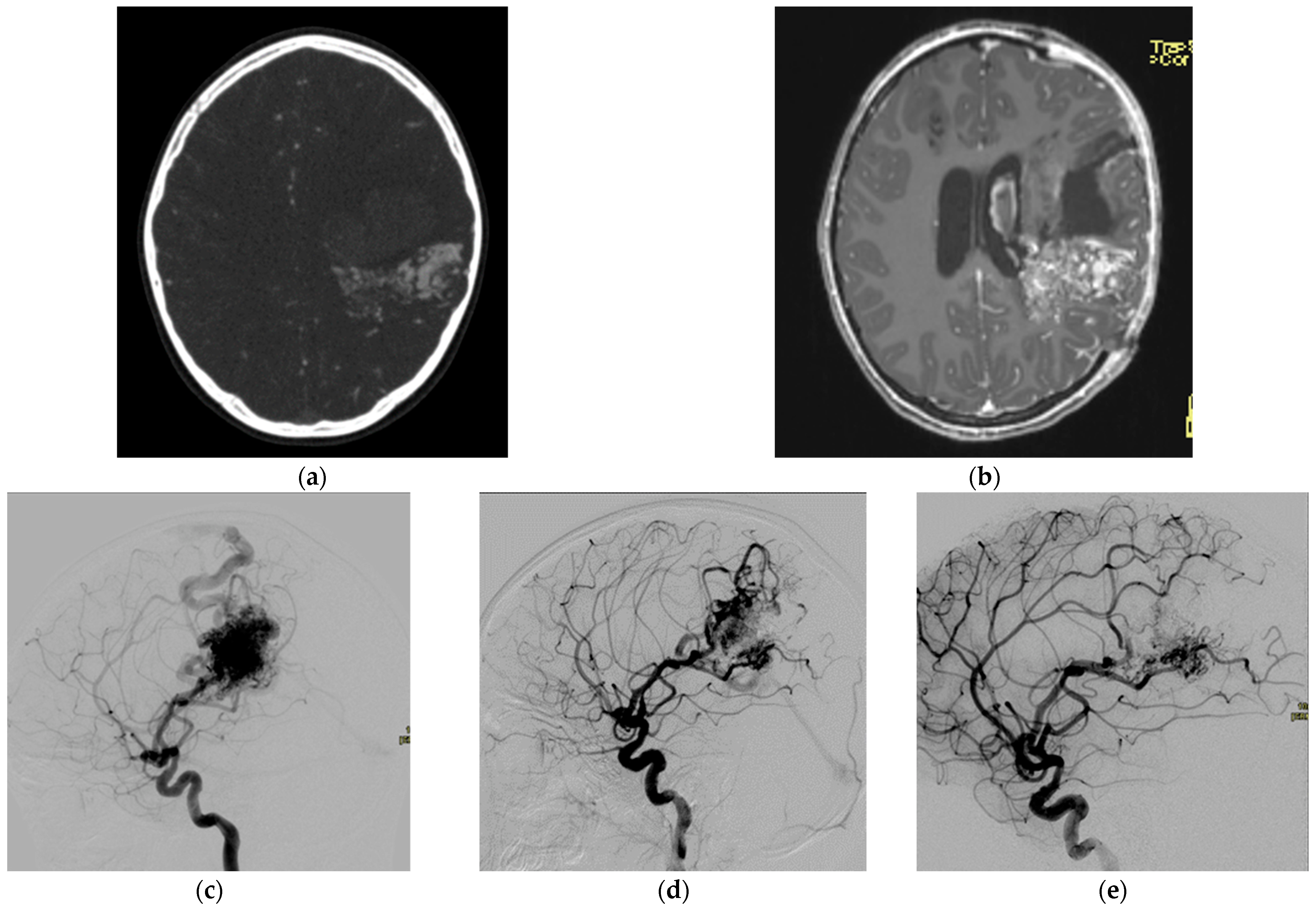
2.1.1. Arteriovenous Malformations
2.1.2. Vein of Galen Malformation
2.1.3. Venous Malformations
2.2. Ischemic Disease
2.3. Aneurysms
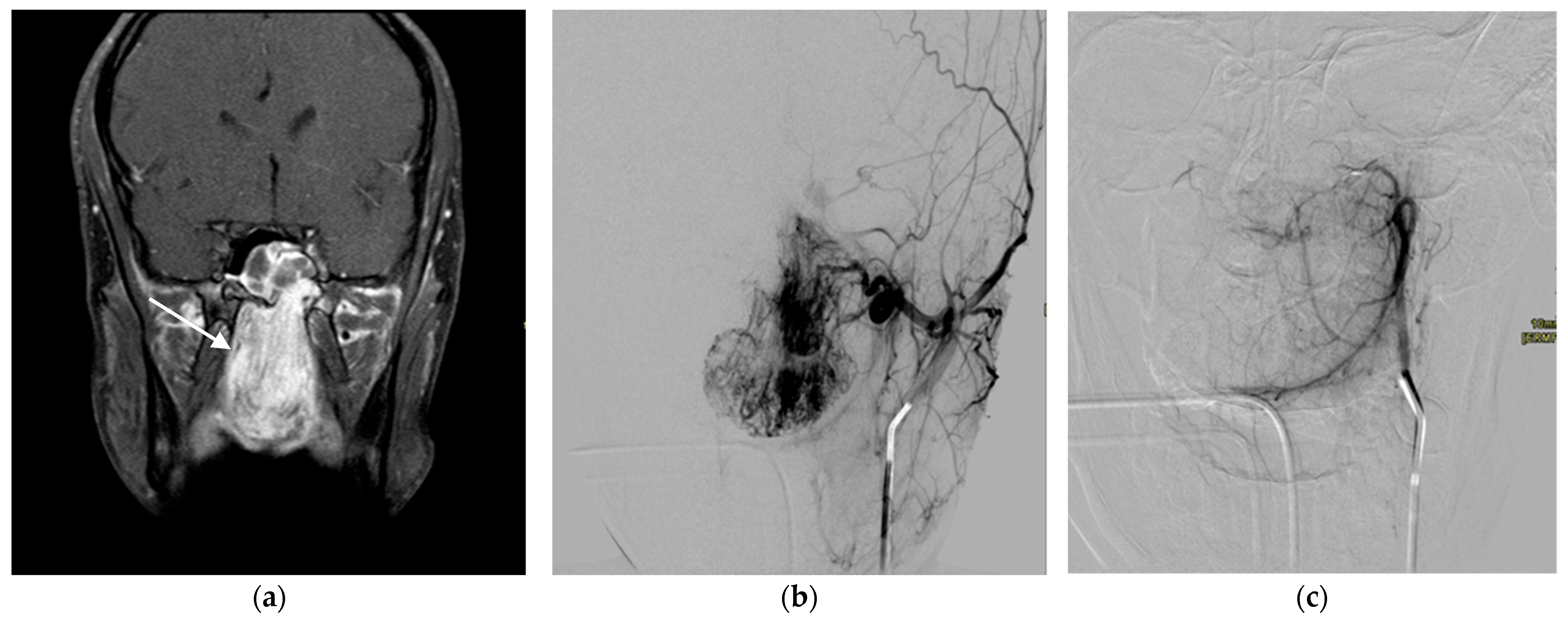
2.4. Tumors
2.5. Intraarterial Chemotherapy for Retinoblastoma
2.6. Petrosal Sinus Sampling
3. Special Considerations for Pediatric Patients
3.1. Sedation/Anesthesia
3.2. Radiation
3.3. Contrast Agent
3.4. Equipment
3.5. Vascular Access
4. Conclusions
Author Contributions
Funding
Institutional Review Board Statement
Informed Consent Statement
Data Availability Statement
Conflicts of Interest
References
- Towbin, R.B.; Ball, W.S., Jr. Pediatric interventional radiology. Radiol. Clin. N. Am. 1988, 26, 419–440. Available online: https://pubmed.ncbi.nlm.nih.gov/3277233/ (accessed on 7 February 2023). [CrossRef] [PubMed]
- Roebuck, D.J.; McLaren, C.A. Pediatric interventional radiology—does it add value? Pediatr. Radiol. 2021, 51, 570–573. [Google Scholar] [CrossRef] [PubMed]
- Kunimoto, K.; Yamamoto, Y.; Jinnin, M. ISSVA Classification of Vascular Anomalies and Molecular. Biology. Int. J. Mol. Sci. 2022, 23, 2358. [Google Scholar] [CrossRef] [PubMed]
- Alomari, A.; Dubois, J. Interventional Management of Vascular Malformations. Tech. Vasc. Interv. Radiol. 2011, 14, 22–31. [Google Scholar] [CrossRef]
- Mulligan, P.R.; Prajapati, H.J.S.; Martin, L.G.; Patel, T.H. Vascular anomalies: Classification, imaging characteristics and implications for interventional radiology treatment approaches. Br. J. Radiol. 2014, 87, 1035. [Google Scholar] [CrossRef] [Green Version]
- Lv, X.; Jiang, C.; Wang, J. Pediatric intracranial arteriovenous shunts: Advances in diagnosis and treatment. Eur. J. Paediatr. Neurol. 2020, 25, 29–39. [Google Scholar] [CrossRef]
- Zúñiga-Castillo, M.; Teng, C.L.; Teng, J.M.C. Genetics of vascular malformation and therapeutic implications. Curr. Opin. Pediatr. 2019, 31, 498–508. [Google Scholar] [CrossRef]
- Umansky, D.; Corn, B.W.; Strauss, I.; Shtraus, N.; Constantini, S.; Frolov, V.; Maimon, S.; Kanner, A.A. Combined treatment approach to cerebral arteriovenous malformation in pediatric patients: Stereotactic radiosurgery to partially Onyx-embolized AVM. Child’s Nerv. Syst. 2018, 34, 2269–2274. [Google Scholar] [CrossRef]
- Rodriguez-Calienes, A.; Bustamante-Paytan, D.; Camacho, K.; Mayoria-Vargas, A.; Saal-Zapata, G.; Rodriguez-Varela, R. Early Outcomes and Complications of Endovascular Treatment of Cerebral Arteriovenous Malformations in Pediatric Patients. Pediatr. Neurosurg. 2021, 56, 116–124. [Google Scholar] [CrossRef]
- Heran, M.K.S.; Burrill, J. Vascular pediatric interventional radiology. Can. Assoc. Radiol. J. 2012, 63, S59–S73. [Google Scholar] [CrossRef] [Green Version]
- Bhattacharya, J.J. Vein of galen malformations. J. Neurol. Neurosurg. Psychiatry 2003, 74, 42i–44i. [Google Scholar] [CrossRef] [Green Version]
- Recinos, P.F.; Rahmathulla, G.; Pearl, M.; Recinos, V.R.; Jallo, G.I.; Gailloud, P.; Ahn, E.S. Vein of Galen Malformations: Epidemiology, Clinical Presentations, Management, Neurosurg. Clin. N. Am. 2012, 23, 165–177. [Google Scholar] [CrossRef]
- Nelson, N.; Dickinson, D.; Wilson, N. Transtorcular coil embolisation of In, malformations of the vein of Galen: Rapid resolution of heart failure. Int. J. Cardiol. 1988, 18, 437e41. [Google Scholar] [CrossRef]
- Duran, D.; Karschnia, P.; Gaillard, J.R.; Karimy, J.K.; Youngblood, M.W.; DiLuna, M.L.; Matouk, C.C.; Aagaard-Kienitz, B.; Smith, E.R.; Orbach, D.B.; et al. Human genetics and molecular mechanisms of vein of Galen malformation. J. Neurosurg. Pediatr. 2018, 21, 367–374. [Google Scholar] [CrossRef]
- Heuer, G.G.; Gabel, B.; Beslow, L.A.; Stiefel, M.F.; Schwartz, E.S.; Storm, P.B.; Ichord, R.N.; Hurst, R.W. Diagnosis and treatment of vein of Galen aneurysmal malformations. Child’s Nerv. Syst. 2010, 26, 879–887. [Google Scholar] [CrossRef]
- Karadeniz, L.; Coban, A.; Sencer, S.; Has, R.; Ince, Z.; Can, G. Vein of Galen aneurysmal malformation: Prenatal diagnosis and early endovascular management. J. Chin. Med. Assoc. 2011, 74, 134–137. [Google Scholar] [CrossRef] [Green Version]
- Wagner, M.W.; Vaught, A.J.; Poretti, A.; Blakemore, K.J.; Huisman, T.A.G.M. Vein of Galen aneurysmal malformation: Prognostic markers depicted on fetal MRI. Neuroradiol. J. 2015, 28, 72–75. [Google Scholar] [CrossRef]
- Lasjaunias, P.L.; Chng, S.M.; Sachet, M.; Alvarez, H.; Rodesch, G.; Garcia-Monaco, R. The management of vein of Galen aneurysmal malformations. Neurosurgery 2006, 59, S3–S184. [Google Scholar] [CrossRef] [Green Version]
- Li, A.H.; Armstrong, D.; TerBrugge, K.G. Endovascular treatment of vein of Galen aneurysmal malformation: Management strategy and 21-year experience in Toronto—Clinical article. J. Neurosurg. Pediatr. 2011, 7, 3–10. [Google Scholar] [CrossRef] [Green Version]
- Johnston, I.H.; Whittle, I.R.; Besser, M.; Morgan, M.K. Vein of galen malformation: Diagnosis and management. Neurosurgery 1987, 20, 747–758. [Google Scholar] [CrossRef]
- Berenstein, A.; Masters, L.T.; Nelson, P.K.; Setton, A.; Verma, R. Transumbilical catheterization of cerebral arteries. Neurosurgery 1997, 41, 846–850. [Google Scholar] [CrossRef] [PubMed]
- Legiehn, G.M.; Heran, M.K.S. Venous Malformations: Classification, Development, Diagnosis, and Interventional Radiologic Management. Radiol. Clin. N. Am. 2008, 46, 545–597. [Google Scholar] [CrossRef]
- Vogel, S.A.; Hess, C.P.; Dowd, C.F.; Hoffman, W.Y.; Kane, A.J.; Rajaii, R.; Frieden, I.J. Early Versus Later Presentations of Venous Malformations: Where and Why? Pediatr. Dermatol. 2013, 30, 534–540. [Google Scholar] [CrossRef] [PubMed]
- Cooke-Barber, J.; Kreimer, S.; Patel, M.; Dasgupta, R.; Jeng, M. Venous malformations. Semin. Pediatr. Surg. 2020, 29, 150976. [Google Scholar] [CrossRef] [PubMed]
- Mattila, K.A.; Aronniemi, J.; Salminen, P.; Rintala, R.J.; Kyrklund, K. Intra-articular venous malformation of the knee in children: Magnetic resonance imaging findings and significance of synovial involvement. Pediatr. Radiol. 2020, 50, 509–515. [Google Scholar] [CrossRef] [Green Version]
- Schmidt, V.F.; Olivieri, M.; Häberle, B.; Masthoff, M.; Deniz, S.; Sporns, P.B.; Wohlgemuth, W.A.; Wildgruber, M. Interventional Treatment Options in Children with Extracranial Vascular Malformations. Hamostaseologie 2022, 42, 131–141. [Google Scholar] [CrossRef]
- Hage, A.N.; Chick, J.F.B.; Srinivasa, R.N.; Bundy, J.J.; Chauhan, N.R.; Acord, M.; Gemmete, J.J. Treatment of Venous Malformations: The Data, Where We Are, and How It Is Done. Tech. Vasc. Interv. Radiol. 2018, 21, 45–54. [Google Scholar] [CrossRef]
- Leung, M.; Leung, L.; Fung, D.; Poon, W.L.; Liu, C.; Chung, K.; Tang, P.; Tse, S.; Fan, T.W.; Chao, N.; et al. Management of the low-flow head and neck vascular malformations in children: The sclerotherapy protocol. Eur. J. Pediatr. Surg. 2014, 24, 97–101. [Google Scholar] [CrossRef] [Green Version]
- Markovic, J.N.; Nag, U.; Shortell, C.K. Safety and efficacy of foam sclerotherapy for treatment of low-flow vascular malformations in children. In Journal of Vascular Surgery: Venous and Lymphatic Disorders; Elsevier Inc.: Amsterdam, The Netherlands, 2020; pp. 1074–1082. [Google Scholar] [CrossRef]
- Sporns, B.; Fullerton, H.J.; Lee, S.; Kim, H.; Lo, W.D.; Mackay, M.T.; Wildgruber, M. Childhood stroke. Nat. Rev. Dis. Prim. 2022, 8, 12. [Google Scholar] [CrossRef]
- Chiang, K.L.; Cheng, C.Y. Epidemiology, risk factors and characteristics of pediatric stroke: A nationwide population-based study. QJM 2018, 111, 445–454. [Google Scholar] [CrossRef] [Green Version]
- Sporns, P.B.; Fullerton, H.J.; Lee, S.; Kirton, A.; Wildgruber, M. Current treatment for childhood arterial ischaemic stroke. Lancet Child Adolesc. Health 2021, 5, 825–836. [Google Scholar] [CrossRef]
- Benjamin, E.J.; Muntner, P.; Alonso, A.; Bittencourt, M.S.; Callaway, C.W.; Carson, A.P.; Chamberlain, A.M.; Chang, A.R.; Cheng, S.; Das, S.R.; et al. Heart Disease and Stroke Statistics-2019 Update: A Report From the American Heart Association. Circulation 2019, 139, e56–e528. [Google Scholar] [CrossRef]
- Riela, A.R.; Roach, E.S. Topical Review Article: Etiology of Stroke in Children. J. Child Neurol. 1993, 8, 201–220. [Google Scholar] [CrossRef]
- Williamson, C.; Morgan, L.; Klein, J.P. Imaging in Neurocritical Care Practice. Semin. Respir. Crit. Care Med. 2017, 38, 840–852. [Google Scholar] [CrossRef] [Green Version]
- Sinha, R.; Ramji, S. Neurovascular disorders in children: An updated practical guide. Transl. Pediatr. 2021, 10, 1100–1116. [Google Scholar] [CrossRef]
- Sporns, P.B.; Kemmling, A.; Hanning, U.; Minnerup, J.; Sträter, R.; Niederstadt, T.; Heindel, W.; Wildgruber, M. Thrombectomy in Childhood Stroke. J. Am. Heart Assoc. 2019, 8, e011335. [Google Scholar] [CrossRef] [Green Version]
- Bhatia, K.; Kortman, H.; Blair, C.; Parker, G.; Brunacci, D.; Ang, T.; Worthington, J.; Muthusami, P.; Shoirah, H.; Mocco, J.; et al. Mechanical thrombectomy in pediatric stroke: Systematic review, individual patient data meta-analysis, and case series. J. Neurosurg. Pediatr. 2019, 24, 558–571. [Google Scholar] [CrossRef]
- Fragata, I.; Morais, T.; Silva, R.; Nunes, A.P.; Loureiro, P.; Martins, J.D.; Pamplona, J.; Carvalho, R.; Baptista, M.; Reis, J. Endovascular treatment of pediatric ischemic stroke: A single center experience and review of the literature. Interv. Neuroradiol. 2021, 27, 16–24. [Google Scholar] [CrossRef]
- Satti, S.; Chen, J.; Sivapatham, T.; Jayaraman, M.; Orbach, D. Mechanical thrombectomy for pediatric acute ischemic stroke: Review of the literature. J. Neurointerv. Surg. 2017, 9, 732–737. [Google Scholar] [CrossRef]
- Shoirah, H.; Shallwani, H.; Siddiqui, A.H.; Levy, E.I.; Kenmuir, C.L.; Jovin, T.G.; Levitt, M.R.; Kim, L.J.; Griauzde, J.; Pandey, A.S.; et al. Endovascular thrombectomy in pediatric patients with large vessel occlusion. J. Neurointerv. Surg. 2019, 11, 729–732. [Google Scholar] [CrossRef]
- Ashour, R.; Orbach, D.B. Interventional neuroradiology in children: Diagnostics and therapeutics. Curr. Opin. Pediatr. 2015, 27, 700–705. [Google Scholar] [CrossRef] [PubMed]
- Montaser, A.; Smith, E.R. Intracranial Vascular Abnormalities in Children. Pediatr. Clin. N. Am. 2021, 68, 825–843. [Google Scholar] [CrossRef] [PubMed]
- Gemmete, J.J.; Toma, A.K.; Davagnanam, I.; Robertson, F.; Brew, S. Pediatric cerebral aneurysms. Neuroimaging Clin. N. Am. 2013, 23, 771–779. [Google Scholar] [CrossRef] [PubMed]
- Heredia-Gutiérrez, A.; Carbarín-Carbarín, M.E. Cerebral aneurysms in pediatrics: A case report and review of the literature. Bol. Med. Hosp. Infant. Mex. 2021, 78, 636–641. [Google Scholar] [CrossRef]
- Garg, K.; Singh, P.K.; Sharma, B.S.; Chandra, P.S.; Suri, A.; Singh, M.; Kumar, R.; Kale, S.S.; Mishra, N.K.; Gaikwad, S.K.; et al. Pediatric intracranial aneurysms—Our experience and review of literature. Child’s Nerv. Syst. 2014, 30, 873–883. [Google Scholar] [CrossRef]
- Ghali, M.G.Z.; Srinivasan, V.M.; Cherian, J.; Kim, L.; Siddiqui, A.; Aziz-Sultan, M.A.; Froehler, M.; Wakhloo, A.; Sauvageau, E.; Rai, A.; et al. Pediatric Intracranial Aneurysms: Considerations and Recommendations for Follow-Up Imaging. World Neurosurg. 2018, 109, 418–431. [Google Scholar] [CrossRef]
- Takemoto, K.; Tateshima, S.; Golshan, A.; Gonzalez, N.; Jahan, R.; Duckwiler, G.; Vinuela, F. Endovascular treatment of pediatric intracranial aneurysms: A retrospective study of 35 aneurysms. J. Neurointerv. Surg. 2014, 6, 432–438. [Google Scholar] [CrossRef]
- Ashour, R.; Aziz-Sultan, A. Preoperative tumor embolization. Neurosurg. Clin. N. Am. 2014, 25, 607–617. [Google Scholar] [CrossRef]
- Amuluru, K.; Al-Mufti, F.; Singh, I.P.; Prestigiacomo, C.J.; Gandhi, C.D. Embolization of pediatric intracranial and skull base vascular tumors. In Pediatric Vascular Neurosurgery: Principles and Practice of Neurovascular Disorders (Part 1); Springer International Publishing: Berlin/Heidelberg, Germany, 2017; pp. 273–284. [Google Scholar] [CrossRef]
- Wang, H.H.; Luo, C.B.; Guo, W.Y.; Wu, H.M.; Lirng, J.F.; Wong, T.T.; Lu, Y.H.; Chang, F.C. Preoperative embolization of hypervascular pediatric brain tumors: Evaluation of technical safety and outcome. Child’s Nerv. Syst. 2013, 29, 2043–2049. [Google Scholar] [CrossRef]
- Ampie, L.; Choy, W.; Lamano, J.B.; Kesavabhotla, K.; Kaur, R.; Parsa, A.T.; Bloch, O. Safety and outcomes of preoperative embolization of intracranial hemangioblastomas: A systematic review. Clin. Neurol. Neurosurg. 2016, 150, 143–151. [Google Scholar] [CrossRef]
- Rodriguez-Galindo, C.; Orbach, D.B.; VanderVeen, D. Retinoblastoma. Pediatr. Clin. N. Am. 2015, 62, 201–223. [Google Scholar] [CrossRef]
- Dimaras, H.; Corson, T.W. Retinoblastoma, the visible CNS tumor: A review. J. Neurosci. Res. 2019, 97, 29–44. [Google Scholar] [CrossRef] [Green Version]
- Fabian, I.D.; Onadim, Z.; Karaa, E.; Duncan, C.; Chowdhury, T.; Scheimberg, I.; Ohnuma, S.I.; Reddy, M.A.; Sagoo, M.S. The management of retinoblastoma. Oncogene 2018, 37, 1551–1560. [Google Scholar] [CrossRef]
- Farhat, W.; Yeung, V.; Ross, A.; Kahale, F.; Boychev, N.; Kuang, L.; Chen, L.; Ciolino, J.B. Advances in biomaterials for the treatment of retinoblastoma. Biomater. Sci. 2022, 10, 5391–5429. [Google Scholar] [CrossRef]
- Monroy, J.E.G.; Orbach, D.B.; Vanderveen, D. Complications of intra-Arterial chemotherapy for retinoblastoma. Semin. Ophthalmol. 2014, 29, 429–433. [Google Scholar] [CrossRef]
- Manjandavida, F.; Stathopoulos, C.; Zhang, J.; Honavar, S.; Shields, C. Intra-arterial chemotherapy in retinoblastoma—A paradigm change. Indian J. Ophthalmol. 2019, 67, 740. [Google Scholar] [CrossRef]
- Zampetti, B.; Grossrubatscher, E.; Ciaramella, P.D.; Boccardi, E.; Loli, P. Bilateral inferior petrosal sinus sampling. Endocr. Connect. 2016, 5, R12–R25. [Google Scholar] [CrossRef] [Green Version]
- Raff, H. Cushing Syndrome. Update on Testing. Endocrinol. Metab. Clin. N. Am. 2015, 44, 43–50. [Google Scholar] [CrossRef]
- Aguado, A. Role of Interventional Radiology in Pediatric Cancer Patients. Curr. Oncol. Rep. 2022, 24, 1731–1740. [Google Scholar] [CrossRef]
- Nelson, O.; Bailey, P.D. Pediatric Anesthesia Considerations for Interventional Radiology. Anesthesiol. Clin. 2017, 35, 701–714. [Google Scholar] [CrossRef]
- Desai, S.B.; Kukreja, K.U.; Recognize, H.T. Avoid, or Get Out of Trouble in Pediatric Interventional Radiology. Tech. Vasc. Interv. Radiol. 2018, 21, 242–248. [Google Scholar] [CrossRef] [PubMed]
- U.S. Food and Drug Administration (FDA). FDA Drug Safety Communication: FDA Approves Label Changes for Use of General Anesthetic and Sedation Drugs in Young Children. 2017. Available online: https://www.fda.gov/drugs/drug-safety-and-availability/fda-drug-safety-communication-fda-approves-label-changes-use-general-anesthetic-and-sedation-drugs (accessed on 9 January 2023).
- Barton, K.; Nickerson, J.P.; Higgins, T.; Williams, R.K. Pediatric anesthesia and neurotoxicity: What the radiologist needs to know. Pediatr. Radiol. 2018, 48, 31–36. [Google Scholar] [CrossRef] [PubMed]
- Koenig, T.R.; Wolff, D.; Mettler, F.A.; Wagner, L.K. Skin injuries from fluoroscopically guided procedures: Part I, characteristics of radiation injury. Am. J. Roentgenol. 2001, 177, 3–11. [Google Scholar] [CrossRef] [PubMed] [Green Version]
- Vano, E.; Goicolea, J.; Galvan, C.; Gonzalez, L.; Meiggs, L.; Ten, J.I.; Macaya, C. Skin radiation injuries in patients following repeated coronary angioplasty procedures. Br. J. Radiol. 2001, 74, 1023–1031. [Google Scholar] [CrossRef]
- Koenig, T.R.; Mettler, F.A.; Wagner, L.K. Skin injuries from fluoroscopically guided procedures: Part 2, review of 73 cases and recommendations for minimizing dose delivered to patient. Am. J. Roentgenol. 2001, 177, 13–20. [Google Scholar] [CrossRef] [Green Version]
- Brenner, D.J.; Hall, E.J. Computed Tomography—An Increasing Source of Radiation Exposure. N. Engl. J. Med. 2007, 357, 2277–2284. [Google Scholar] [CrossRef] [Green Version]
- Wakeford, R. The risk of childhood leukaemia following exposure to ionising radiation—A review. J. Radiol. Prot. 2013, 33, 1–25. [Google Scholar] [CrossRef]
- Kodama, K.; Ozasa, K.; Okubo, T. Radiation and cancer risk in atomic-bomb survivors. J. Radiol. Prot. 2012, 32, N51. [Google Scholar] [CrossRef]
- Nagayama, Y.; Oda, S.; Nakaura, T.; Tsuji, A.; Urata, J.; Furusawa, M.; Utsunomiya, D.; Funama, Y.; Kidoh, M.; Yamashita, Y. Radiation Dose Reduction at Pedi-atric CT: Use of Low Tube Voltage and Iterative Reconstruction. Radiographics 2018, 38, 1421–1440. [Google Scholar] [CrossRef] [Green Version]
- Thierry-Chef, I.; Simon, S.L.; Land, C.E.; Miller, D.L. Radiation dose to the brain and subsequent risk of developing brain tumors in pediatric patients undergoing interventional neuroradiology procedures. Radiat. Res. 2008, 170, 553–565. [Google Scholar] [CrossRef] [Green Version]
- Rossi, A.; Argyropoulou, M.; Zlatareva, D.; Boulouis, G.; Pizzini, F.B.; van den Hauwe, L.; Raissaki, M.; Pruvo, J.P.; Rosendahl, K.; Hoffmann, C.; et al. European recommendations on practices in pediatric neuroradiology: Consensus document from the European Society of Neuroradiology (ESNR), European Society of Paediatric Radiology (ESPR) and European Union of Medical Specialists Division of Neuroradiology (UEMS). Pediatr. Radiol. 2022, 53, 159. [Google Scholar] [CrossRef]
- Riche, M.; Monfraix, S.; Balduyck, S.; Voglimacci-Stephanopoli, Q.; Rollin, A.; Mondoly, P.; Mandel, F.; Beneyto, M.; Delasnerie, H.; Derval, N.; et al. Radiation dose during catheter ablation in children using a low fluoroscopy frame rate. Arch. Cardiovasc. Dis. 2022, 115, 151–159. [Google Scholar] [CrossRef]
- Heran, M.K.S.; Marshalleck, F.; Temple, M.; Grassi, C.J.; Connolly, B.; Towbin, R.B.; Baskin, K.M.; Dubois, J.; Hogan, M.J.; Kundu, S.; et al. Joint Quality Improvement Guidelines for Pediatric Arterial Access and Arteriography: From the Societies of Interventional Radiology and Pediatric Radiology. J. Vasc. Interv. Radiol. 2010, 21, 32–43. [Google Scholar] [CrossRef]
- Harned, R.K.; Heran, M.K.S.; Patel, M.; Barnacle, A.; Cahill, A.M.; Braswell, L.; Feola, G.P.; Sierre, S.; Marshalleck, F. Challenges and opportunities for continued success and growth of pediatric interventional radiology: A communiqué from the society for pediatric interventional radiology. Am. J. Roentgenol. 2018, 211, 740–743. [Google Scholar] [CrossRef]
- Chait, P. Future directions in interventional pediatric radiology. Pediatr. Clin. N. Am. 1997, 44, 763–782. [Google Scholar] [CrossRef]
- Vranicar, M.; Hirsch, R.; Canter, C.E.; Balzer, D.T. Selective coronary angiography in pediatric patients. Pediatr. Cardiol. 2000, 21, 285–288. [Google Scholar] [CrossRef]
- Lock, J.E.; Keane, J.F.; Fellows, K.E. Diagnostic and Interventional Catheterization in Congenital Heart Disease, 2nd ed.; Kluwer Academic Publishers: Boston, MA, USA, 2000. [Google Scholar]
- Scognamiglio, R. The Science and Practice of Pediatric Cardiology, 2nd ed.; Garson, A., Jr., Bricker, J.T., Fisher, D.J., Neish, S.R., Eds.; Williams & Wilkins: Baltimore, MD, USA, 1998; Volumes I–II, p. 54. [Google Scholar] [CrossRef]
- Burrows, P.E.; Robertson, R.L.; Barnes, P.D. Angiography and the evaluation of cerebrovascular disease in childhood. Neuroimaging Clin. N. Am. 1996, 6, 561–588. [Google Scholar]


Disclaimer/Publisher’s Note: The statements, opinions and data contained in all publications are solely those of the individual author(s) and contributor(s) and not of MDPI and/or the editor(s). MDPI and/or the editor(s) disclaim responsibility for any injury to people or property resulting from any ideas, methods, instructions or products referred to in the content. |
© 2023 by the authors. Licensee MDPI, Basel, Switzerland. This article is an open access article distributed under the terms and conditions of the Creative Commons Attribution (CC BY) license (https://creativecommons.org/licenses/by/4.0/).
Share and Cite
Jerele, C.; Lovrič, D.; Kuhelj, D. Pediatric Interventional Neuroradiology: Opportunities and Challenges. Children 2023, 10, 715. https://doi.org/10.3390/children10040715
Jerele C, Lovrič D, Kuhelj D. Pediatric Interventional Neuroradiology: Opportunities and Challenges. Children. 2023; 10(4):715. https://doi.org/10.3390/children10040715
Chicago/Turabian StyleJerele, Cene, Dimitrij Lovrič, and Dimitrij Kuhelj. 2023. "Pediatric Interventional Neuroradiology: Opportunities and Challenges" Children 10, no. 4: 715. https://doi.org/10.3390/children10040715