Article

Randomized Trial of 21% versus 100% Oxygen during Chest Compressions Followed by Gradual versus Abrupt Oxygen Titration after Return of Spontaneous Circulation in Neonatal Lambs

1
Department of Pediatrics, University of California, Davis, Sacramento, CA 95817, USA
2
Department of Stem Cell Research, University of California, Davis, Sacramento, CA 95817, USA
*
Author to whom correspondence should be addressed.
Children 2023, 10(3), 575; https://doi.org/10.3390/children10030575
Submission received: 27 February 2023 / Revised: 15 March 2023 / Accepted: 16 March 2023 / Published: 17 March 2023
(This article belongs to the Special Issue Stabilization and Resuscitation of Newborns: 2nd Edition)

Abstract

:
The combination of perinatal acidemia with postnatal hyperoxia is associated with a higher incidence of hypoxic-ischemic encephalopathy (HIE) in newborn infants. In neonatal cardiac arrest, current International Liaison Committee on Resuscitation (ILCOR) and Neonatal Resuscitation Program (NRP) guidelines recommend increasing inspired O2 to 100% during chest compressions (CC). Following the return of spontaneous circulation (ROSC), gradual weaning from 100% O2 based on pulse oximetry (SpO2) can be associated with hyperoxia and risk for cerebral tissue injury owing to oxidative stress. We hypothesize that compared to gradual weaning from 100% O2 with titration based on preductal SpO2, abrupt or rapid weaning of inspired O2 to 21% after ROSC or use of 21% O2 during CC followed by upward titration of inspired O2 to achieve target SpO2 after ROSC will limit hyperoxia after ROSC. Nineteen lambs were randomized before delivery and asphyxial arrest was induced by umbilical cord occlusion. There was no difference in oxygenation during chest compressions between the three groups. Gradual weaning of inspired O2 from 100% O2 after ROSC resulted in supraphysiological PaO2 and higher cerebral oxygen delivery compared to 21% O2 during CC or 100% O2 during CC followed by abrupt weaning to 21% O2 after ROSC. The use of 21% O2 during CC was associated with very low PaO2 after ROSC and higher brain tissue lactic acid compared to other groups. Our findings support the current recommendations to use 100% O2 during CC and additionally suggest the benefit of abrupt decrease in inspired oxygen to 21% O2 after ROSC. Clinical studies are warranted to investigate optimal oxygen titration after chest compressions and ROSC during neonatal resuscitation.

1. Introduction

Many newly born infants impacted by perinatal asphyxia need resuscitation immediately after birth. Previous studies have shown that even brief exposure to 100% inspired oxygen augments systemic oxidative stress and lung oxidative injury in ventilated term newborn lambs [1]. The American Academy of Pediatrics—Neonatal Resuscitation Program (AAP-NRP) has provided guidelines for preductal pulse oximetry oxygen saturation (SpO2) targets during resuscitation [2]. The 2020 International Liaison Committee on Resuscitation (ILCOR) guidelines advocate the use of 100% oxygen when chest compressions are indicated [3]. Owing to poor circulation during cardiac arrest in spite of chest compressions, it is challenging to accurately monitor oxygenation status using a pulse oximeter. Once return of spontaneous circulation (ROSC) is achieved, SpO2 can be measured and can be utilized to guide the administration of supplemental oxygen. However, there are no clear guidelines on how quickly and by how much the supplemental O2 needs to be titrated. Hypercapnia following asphyxia is accompanied by a rise in cerebral blood flow and reperfusion injury. On the contrary, hypocarbia following ROSC can reduce cerebral blood flow [4,5]. Use of 100% oxygen during neonatal resuscitation with chest compressions and gradual weaning of inspired oxygen based on SpO2 can be associated with ischemia-reperfusion compounded by hyperoxia [6] and a risk for free-radical-mediated cerebral oxidative stress and tissue injury [7].
We have previously shown the benefit of abrupt weaning of inspired oxygen to 21% oxygen from 100% oxygen following ROSC after the initial use of 100% oxygen during chest compressions in limiting cerebral hyperoxia [8]. However, we did not randomize lambs to receive 21% oxygen during chest compressions in this study. Moreover, we did not evaluate markers of oxidative stress or lactic acid in the brain tissue in the previous study.
We hypothesized that gradual weaning of inspired O2 from 100% as currently recommended by NRP (solid red line in Figure 1) following ROSC will increase cerebral oxygen delivery to the newborn brain during chest compressions and exacerbate reperfusion injury after ROSC compared to use of 21% oxygen during chest compressions (dashed purple line) and 100% oxygen during chest compressions followed by abrupt weaning to 21% (dotted blue line). Use of 21% oxygen during chest compressions will decrease cerebral oxygen delivery (hyphenated purple line). We also hypothesized that gradual weaning of inspired oxygen from 100% following ROSC as currently recommended by the NRP will increase oxidative stress to the newborn brain and exacerbate ischemia-reperfusion injury compared to abrupt weaning to 21% or use of 21% oxygen during chest compressions.

2. Methods

The experimental protocol was approved by the Institutional Animal Care and Use Committee (IACUC, protocol #22544) at the University of California Davis, Davis, CA, USA. This protocol, which involves a perinatal model of asphyxial arrest in term newborn lambs, has been described previously [9]. Experiments for this protocol were performed per animal ethical guidelines (ARRIVE) [10]. Time-dated near-term (139–142 days) gestation pregnant ewes were procured from Van Laningham Farm (Arbuckle, CA, USA). After an overnight fast, the ewes underwent a cesarean section under general anesthesia with intravenous (IV) diazepam, ketamine, and inhaled 2% isoflurane [9].

2.1. Instrumentation in Utero

The fetal lamb was partially exteriorized and the airway was intubated (4.5 mm cuffed endotracheal tube/ETT). Lung fluid was passively drained by gravity, and the ETT was occluded to prevent the entry of air during gasping. The lamb instrumentation was performed under maternal anesthesia. Additional local anesthesia was provided using subcutaneous bupivacaine infiltration. A catheter was inserted into the right carotid artery for preductal arterial blood draws and invasive blood pressure and heart rate monitoring. Another catheter was inserted into the right jugular vein for intravenous access for fluid and medication administration. A left carotid artery flow probe (Transonics, Ithaca, NY, USA) was secured to measure and record blood flow to the brain. The hemodynamics were recorded at fetal baseline. Arterial blood gases and plasma samples were also obtained.

2.2. Asphyxial Cardiac Arrest and Resuscitation

Fetal lambs were asphyxiated by umbilical cord occlusion [9]. After a flat line was achieved on carotid arterial pressure tracing with a heart rate of zero, the umbilical cord was clamped, the lamb was delivered and placed under a radiant warmer. A low umbilical venous catheter was placed to a depth of 2–4 cm below the skin during the period of asystole. Resuscitation was commenced after 5 min of asystole. The ETT occluder was removed and PPV was initiated using 21% oxygen with a T-piece resuscitator with peak inflation pressures of 30–35 cm H2O, PEEP of 5 cm H2O, and rate of 40 breaths per minute. An end-tidal CO2 detector and exhaled tidal volume monitor were connected to the ETT. The research team was not blinded to the intervention due to the nature of the study.
Thirty seconds after the onset of PPV, chest compressions were initiated and further resuscitation was determined based on the randomization. Lambs were randomized with the help of opaque envelopes prior to starting the fetal instrumentation to the following 3 groups:
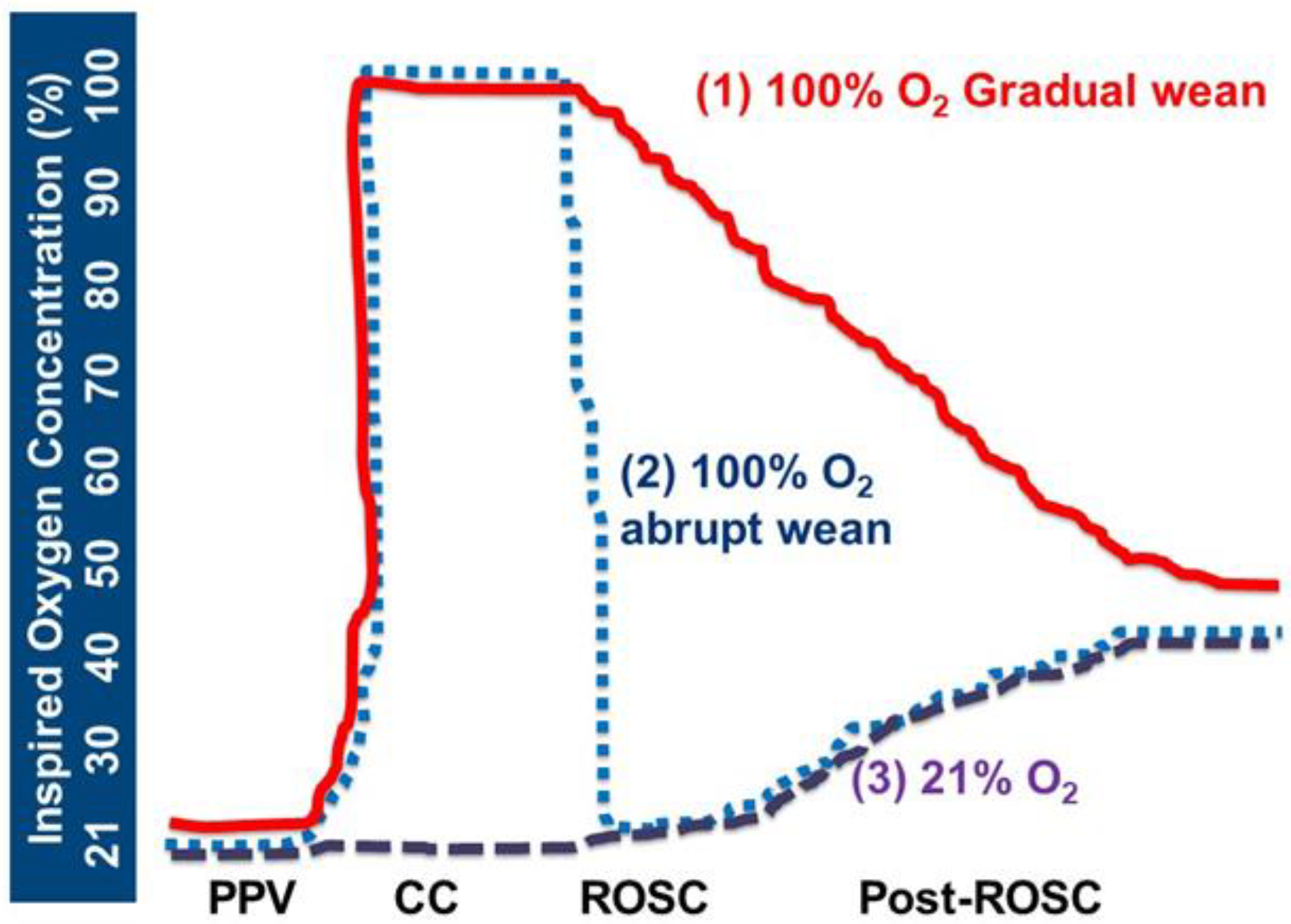
  • Group 1: 100% O2 CC—Gradual wean: Inspired oxygen was increased to 100% during CC and resuscitation was continued per AAP-NRP guidelines. Following ROSC, attempts were made to adjust inspired oxygen down from 100% O2 by 5–10% every 30 sec to achieve preductal SpO2 per NRP guidelines. After the first 10 min, we targeted 85–95% SpO2 in lambs that achieved ROSC during the first 30 min after ROSC, and 90–95% beyond 30 min after ROSC until 60 min for all the study lambs.
  • Group 2: 100% O2 CC—Abrupt wean: Inspired oxygen was increased to 100% during CC, and resuscitation was continued per NRP. After ROSC, the inspired oxygen was rapidly decreased to 21% and attempts were made to adjust inspired oxygen up by 5–10% every 30 sec to maintain preductal SpO2 as described above.
  • Group 3: 21% O2 CC: Inspired oxygen of 21% was administered during chest compressions. Following ROSC, inspired oxygen was titrated up by 5–10% every 30 sec to achieve preductal SpO2 as described above.
After initial resuscitation, the ETT was connected to a conventional mechanical ventilator. Target PaCO2 was 40–60 mm Hg to allow permissive hypercapnia and to avoid hypocapnia in the setting of hypoxic ischemic encephalopathy. Hemodynamics (carotid artery blood flow, heart rate, and carotid artery blood pressure) were continuously monitored, and arterial blood samples were obtained at fetal baseline, during resuscitation, and at serial intervals after ROSC. Lambs were monitored for up to 60 min after successful ROSC and were subsequently euthanized using IV pentobarbital (Fatal-Plus, Vortech Pharmaceuticals, Dearborn, MI, USA).
Plasma samples were frozen and analyzed at the Western Metabolomics center, UC Davis for markers of oxidative stress including hypoxanthine/xanthine ratios, methionine sulfoxide/methionine ratio, and lactic acid. Similarly, oxidative stress markers were assessed from brain tissue that was collected after euthanasia. After euthanasia, frozen brain tissue was sent to Western Metabolomics center for analysis to detect the same markers of oxidative stress and lactic acid concentrations.

2.3. Primary and Secondary Outcomes

Primary outcome measures were partial pressure of oxygen and cerebral oxygen delivery during chest compressions and after ROSC.
Secondary outcome measures were changes in left carotid artery blood flow, PaCO2, SaO2, and oxidative stress markers in plasma and brain tissue as described above.
Cerebral oxygen delivery (mL/kg/min) was calculated by multiplying carotid artery oxygen content (CaO2 = (1.36 × Hemoglobin in g/dL × SaO2%/100) + (partial pressure of O2 in mm Hg× 0.0031)) and left carotid artery blood flow. This is a representative measurement as it does not take into account the right carotid and vertebral arterial flow [11].

2.4. Sample Size Calculation

Sample size was calculated for the parameter cerebral oxygen delivery and based on prior data evaluating oxygen-weaning strategies in asphyxiated lambs [8]. We planned a study of a continuous response variable from independent control and experimental subjects with 1 control per experimental subject. If the true difference in the experimental and control means is 4 mL/kg/min (standard deviation 3 mL/kg/min) with contrast coefficients of (1,1,1) to factor the differences between groups, we need a total of 18 lambs with 6 lambs in each group to be able to reject the null hypothesis that the population means of cerebral oxygen delivery in the experimental groups (100% O2 CC—Abrupt wean and 21% O2 CC) and control (100% O2 CC—Gradual wean) group are equal to probability (power) of 0.97. The Type I error is 0.05.

2.5. Data Collection and Analysis

Hemodynamic data were continuously monitored and recorded using BIOPAC systems data acquisition software (Goleta, CA, USA). Blood gases were analyzed using a blood gas analyzer (Radiometer ABL90 FLEX, Denmark). Categorical data were analyzed using chi-squared test or Fisher’s exact test as appropriate, and changes in parametric continuous data over time were compared using repeated measures ANOVA and post hoc analysis using Statview 5.0.1 (SAS Institute Inc., New York, NY, USA). Statistical significance was defined as p < 0.05.

3. Results

Among nineteen near-term lambs that were asphyxiated, seven were randomized to 100% O2 CC—Gradual wean, six to 100% O2 CC2014Abrupt wean, and six to 21% O2 CC. Hemodynamic and arterial blood gas results were similar between the three groups at feta baseline (Table 1). Inspired oxygen concentration was different between the three groups based on the study design (Figure 2). All lambs achieved ROSC. However, one lamb in the 21% O2 CC group had a cardiac arrest after initial ROSC after the first dose of intravenous epinephrine, and chest compressions were restarted.

3.1. Hemodynamic and Arterial Blood Gas Parameters during Chest Compressions and after ROSC

There was no significant difference in PaO2, SaO2, and left carotid artery blood flow during chest compressions between the three study groups (Table 2). Cerebral oxygen delivery was low compared to fetal values but also not different among groups during chest compressions (Table 2).
Following ROSC, PaO2 was significantly higher in 100% O2 CC—Gradual wean compared to the other two groups, reaching supraphysiological levels (Figure 3a).
Preductal arterial oxygen saturation (SaO2) was significantly higher after ROSC with 100% O2 CC—Gradual wean compared to 100% O2—Abrupt wean and 21% O2 CC (p = 0.03, Figure 3b). SaO2 remained very low in the 21% O2 CC group up to 3 min after ROSC (Figure 3b). PaCO2 was not different during chest compressions and after ROSC (Figure 3c).
Left carotid artery blood flow was lower and closer to fetal baseline levels with 100% O2 CC—Abrupt wean (Figure 4a) at 3 and 4 min after ROSC but was not different between the groups by 5 and 10 min after ROSC. Mean blood pressures were not different between the groups at fetal baseline (Figure 4b). Following ROSC, mean blood pressures increased significantly compared to fetal baseline in all three groups, but were not different between 100% O2 CC—Gradual wean, 100% O2 CC—Abrupt wean, and 21% O2 CC groups.
Cerebral oxygen delivery was low during chest compressions compared to fetal values. However, after ROSC, it was higher in 100% O2 CC—Gradual wean compared to the abrupt wean and 21% O2 CC groups (Figure 5), reaching supraphysiological levels within 5 min after ROSC.

3.2. Comparison of Markers of Oxidative Stress in Plasma and Brain Tissue

Plasma markers of oxidative stress, including hypoxanthine/xanthine ratio, methionine sulfoxide ratio, and lactic acid, were not different between the three study groups at fetal baseline (Table 3). Although the plasma hypoxanthine/xanthine ratio was higher at 5 min after ROSC in the 100% O2 CC—Gradual wean group compared to the other two groups, it was not different at 10 min after ROSC. Plasma lactic acid and methionine sulfoxide/methionine ratios were not different after ROSC between the three study groups (Table 3). When oxidative stress markers were compared in brain tissue, lactic acid was higher in the 21% O2 CC group compared to the 100% O2 CC—Gradual wean and 100% O2 CC—Abrupt wean groups.

4. Discussion

The current study demonstrates the advantage of abrupt/rapid weaning of inspired oxygen to 21% immediately after ROSC following the use of 100% oxygen during chest compressions in limiting cerebral hyperoxia without resulting in systemic oxidative stress. There was no difference in the incidence of ROSC between the three study groups (100% O2 CC—Gradual wean, 100% O2 CC—Abrupt wean, and 21% O2 CC). Although our previous study compared weaning strategies after ROSC, we did not have a group of lambs randomized to receive 21% oxygen during chest compressions in that study [8]. Additionally, in the current study, we report lower cerebral oxygenation after ROSC with the use of 21% oxygen during chest compressions and elevated brain lactic acid compared to 100% oxygen supporting NRP recommendation to use 100% oxygen during CC. However, the current standard of gradual weaning from 100% oxygen following ROSC after chest compressions is associated with hyperoxemia in term lambs (Figure 6).
At the cellular level, a reduction in blood flow and oxygen delivery to the brain during perinatal asphyxia initiates biochemical events that ultimately lead to anerobic metabolism and generation of lactate. This further results in energy failure and neuronal death within hours to days after asphyxia and resuscitation. The neuronal injury in the acute phase (minutes to hours) is mediated by free radicals (including oxygen free radicals) and mitochondrial damage. Generation of oxygen free radicals and reactive nitrogen species at a rate that exceeds the capacity of endogenous antioxidant systems results in oxidative stress. Due to the inherent high lipid content, high metabolic rate, and reduced activity of antioxidant enzymes, the newborn brain is vulnerable to damage incited by free radicals. After successful resuscitation and return of spontaneous circulation, newborns are exposed to a deleterious combination of hyperoxia and hyperemia (cerebral tissue reperfusion secondary to hypercarbia and possibly high blood pressure). The cerebral vasoparalysis following asphyxia leads to a loss of autoregulation and an increase in cerebral blood flow in response to hypercarbia and hypertension [5]. Epinephrine administered by the ET route is dissolved in the lung liquid and the lungs may act as a reservoir leading to an epinephrine surge and systemic hypertension following ROSC (Figure 4b and Figure 6) [12,13]. Intravenous and intraosseous epinephrine can also contribute to post-resuscitation systemic hypertension and cerebral hyperperfusion (Figure 6). Thus, a newborn surviving with hypoxic-ischemic injury is further compromised by the combination of oxidative stress and cerebral hyperemia, and reperfusion. Increased superior vena cava flow has been associated with poor outcomes in infants with HIE undergoing therapeutic hypothermia possibly due to cerebral hyperperfusion and oxidative stress [14]. This can be potentially mitigated by minimizing oxygen exposure in the post-resuscitation phase after ROSC.
In one-day-old postnatal piglets with apnea-induced cardiac arrest [15,16], Linner et al. observed high brain tissue PaO2 with the use of 100% O2 during chest compressions and similar time to ROSC with 21% O2. In another study by Solevag et al., higher SpO2 and left ventricular oxidized glutathione ratios were reported after chest compressions with 100% O2 [17]. In a study comparing newly born lambs ventilated with 21% vs. 100% oxygen during chest compressions by Rawat et al., use of 100% O2 during chest compressions led to an increase in PaO2 and pulmonary blood flow immediately following ROSC compared to use of 21% O2 during chest compressions [6]. Cerebral oxygen delivery was significantly lower during chest compressions with both room air and 100% O2 comparable to fetal levels. In contrast, cerebral O2 delivery was significantly higher with 100% O2 immediately after ROSC. Our findings corroborate the results of Rawat et al. We have previously shown that gradual weaning of inspired oxygen following ROSC is associated with high PaO2 levels compared to abrupt weaning [8]. In this study, we randomized and compared three arms and included a third arm with 21% oxygen during chest compressions and up-titration of inspired oxygen after ROSC. In a recent meta-analysis, there was no difference in mortality or time to ROSC between 21% and 100% O2 during chest compressions [18]. A systemic review of 10 clinical studies comparing 21% and 100% O2 during resuscitation of asphyxiated term newborns (including many infants that did not need chest compressions) showed lowered mortality with 21% O2 [19]. There are no randomized clinical trials of 21% vs. 100% oxygen during chest compressions. Our study, for the first time, has demonstrated low cerebral oxygen delivery and increased brain tissue lactic acid (suggesting more anaerobic metabolism) with 21% oxygen use during chest compressions.
In asphyxiated term lambs, prolonged ventilation using 100% oxygen resulted in elevated brain tissue oxygen tension when compared to ventilation with 21% oxygen [20]. Koch et al. reported that brief exposure to hyperoxemia after hypoxic-ischemic insult increases secondary neuronal injury, interferes with myelination, and impairs functional recovery in rats [21]. Kapadia et al. reported that hyperoxia during the post-resuscitation phase after perinatal acidemia is associated with a higher incidence of moderate to severe hypoxic-ischemic encephalopathy [15]. Thus, it is crucial to decrease inspired O2 promptly to avoid hyperoxemia.
The AAP-NRP Textbook of Neonatal Resuscitation recommends initiating resuscitation with 21% O2 and increasing inspired oxygen to 100% if chest compressions are required [2]. Additionally, achieving a SpO2 target of 85–95% by 10 min after birth is recommended, without clear guidelines on how often and how much to titrate the inspired supplemental oxygen. We show the benefits of a rapid decrease in inspired oxygen to 21% immediately after ROSC in limiting cerebral hyperoxia when compared to the use of 21% oxygen during chest compressions and 100% oxygen during chest compressions followed by gradual weaning of inspired oxygen. Furthermore, higher brain lactic acid and a trend towards higher PaCO2 in the 21% O2 CC group are concerning for poor ventilation (likely secondary to a sustained elevation in pulmonary vascular resistance) and oxidative stress in the brain.
Analysis of oxidative stress markers in plasma and brain tissue revealed minimal differences between the three groups. A transient increase in plasma hypoxanthine/xanthine ratios was observed at 5 min after ROSC but not at 10 min after ROSC. We speculate that active weaning and titrating of inspired oxygen after ROSC in the study groups negated the initially observed differences (Table 3).
Our study has several limitations. Lambs were randomized before their delivery, thus resulting in an extra lamb in the 100% O2 CC—Gradual wean group due to a triplet delivery. There are species differences between lambs and humans that we have not accounted for in our study. We performed this study in a term ovine model of perinatal asphyxial cardiac arrest induced by umbilical cord compression. The response to our intervention may be different in models of asphyxial bradycardia, preterm lambs, and human newborns. However, the ovine model of asphyxial arrest is a well-established model used for investigating various interventions during neonatal resuscitation which are otherwise difficult to investigate in clinical trials due to ethical challenges in obtaining consent and recruiting newborns for randomization. We did not measure pulmonary blood flow as performing a thoracotomy can alter thoracic cage physics and mechanics during chest compressions. The perinatal lamb model closely mimics human cardiopulmonary physiology. Prior randomized trials in animal models investigating oxygen use have predominantly been performed in the postnatal piglet model after the cardiopulmonary transition has occurred [16].

5. Conclusions

In a perinatal term ovine model of asphyxial cardiac arrest, the use of 100% oxygen during chest compressions followed by abrupt weaning of inspired oxygen resulted in more physiological cerebral oxygen delivery and PaO2 compared to gradual weaning of inspired oxygen. The use of 21% oxygen during chest compressions resulted in suboptimal cerebral oxygen delivery and high lactic acid levels in brain tissue. These findings support the current NRP recommendations of using 100% oxygen during chest compressions, as the use of 21% oxygen is associated with hypoxemia and hypercarbia during chest compressions and after ROSC, respectively. Weaning abruptly to 21–30% oxygen from 100% soon after ROSC following chest compressions might potentially minimize reperfusion injury. Clinical trials investigating oxygenation, hemodynamics, and neurodevelopmental outcomes in term neonates comparing different oxygenation strategies during neonatal chest compressions and after ROSC are warranted.

Author Contributions

D.S.: procuring funds, conducting studies, data extraction and analysis, analysis of data, writing, critiquing, and revising the manuscript. E.M.G., E.C.A.L., A.L.L., M.E.H., K.C.T., V.L.H., H.M.J. and P.V.: conducting experiments, data extraction, critiquing and revising the manuscript. S.L.: concept, procuring funds, conducting experiments, writing, critiquing, and revising the manuscript. All authors have read and agreed to the published version of the manuscript.

Funding

This research was funded by NICHD 5R01 HD072929 (S.L.), Children’s Miracle Network at the University of California, Davis, First Tech Federal Credit Union and UC Davis Pediatrics, and Canadian Pediatric Society NRP research grant (D.S.).

Institutional Review Board Statement

The study was conducted according to the guidelines of the Declaration of Helsinki and approved by the Institutional Animal Care and Use Committee (IACUC) at the University of California Davis, Davis, CA, USA (protocol #22544).

Informed Consent Statement

Not applicable.

Data Availability Statement

The data presented in this study are available in this article.

Conflicts of Interest

The authors declare no conflict of interest. S.L. is a member of the AAP NRP steering committee. The views expressed in this article are his own and do not represent the official position of AAP or NRP. The funders had no role in the design of the study; in the collection, analysis, or interpretation of data; in the writing of the manuscript, or in the decision to publish the results.

References

  1. Kumar, V.H.; Patel, A.; Swartz, D.D.; Wang, H.; Wynn, K.; Nielsen, L.C.; Ryan, R.M. Exposure to Supplemental Oxygen and Its Effects on Oxidative Stress and Antioxidant Enzyme Activity in Term Newborn Lambs. Pediatr. Res. 2010, 67, 66–71. [Google Scholar] [CrossRef] [PubMed] [Green Version]
  2. Weiner, G.M.; Zaichkin, J. American Academy of Pediatrics. American Heart Association; In Textbook of Neonatal Resuscitation, 8th ed.; American Heart Association: Chicago, IL, USA, 2021. [Google Scholar]
  3. Aziz, K.; Lee, H.C.; Escobedo, M.B.; Hoover, A.V.; Kamath-Rayne, B.D.; Kapadia, V.S.; Magid, D.J.; Niermeyer, S.; Schmölzer, G.M.; Szyld, E.; et al. Part 5: Neonatal Resuscitation: 2020 American Heart Association Guidelines for Cardiopulmonary Resuscitation and Emergency Cardiovascular Care. Circulation 2020, 142, S524–S550. [Google Scholar] [CrossRef] [PubMed]
  4. Rosenberg, A.A. Response of the cerebral circulation to hypocarbia in postasphyxia newborn lambs. Pediatr. Res. 1992, 32, 537–541. [Google Scholar] [CrossRef] [PubMed] [Green Version]
  5. Pryds, O.; Greisen, G.; Lou, H.; Friis-Hansen, B. Vasoparalysis associated with brain damage in asphyxiated term infants. J. Pediatr. 1990, 117, 119–125. [Google Scholar] [CrossRef] [PubMed]
  6. Rawat, M.; Chandrasekharan, P.; Gugino, S.; Koenigsknecht, C.; Helman, J.; Alsaleem, M.; Mathew, B.; Nair, J.; Berkelhamer, S.; Vali, P.; et al. Oxygenation and Hemodynamics during Chest Compressions in a Lamb Model of Perinatal Asphyxia Induced Cardiac Arrest. Children 2019, 6, 52. [Google Scholar] [CrossRef] [PubMed] [Green Version]
  7. Perez, M.; Robbins, M.E.; Revhaug, C.; Saugstad, O.D. Oxygen radical disease in the newborn, revisited: Oxidative stress and disease in the newborn period. Free. Radic. Biol. Med. 2019, 142, 61–72. [Google Scholar] [CrossRef] [PubMed]
  8. Sankaran, D.; Vali, P.; Chen, P.; Lesneski, A.L.; Hardie, M.E.; Alhassen, Z.; Wedgwood, S.; Wyckoff, M.H.; Lakshminrusimha, S. Randomized trial of oxygen weaning strategies following chest compressions during neonatal resuscitation. Pediatr. Res. 2021, 90, 540–548. [Google Scholar] [CrossRef] [PubMed]
  9. Sankaran, D.; Chandrasekharan, P.K.; Gugino, S.F.; Koenigsknecht, C.; Helman, J.; Nair, J.; Mathew, B.; Rawat, M.; Vali, P.; Nielsen, L.; et al. Randomised trial of epinephrine dose and flush volume in term newborn lambs. Arch. Dis. Child. Fetal Neonatal Ed. 2021, 106, 578–583. [Google Scholar] [CrossRef] [PubMed]
  10. du Sert, N.P.; Hurst, V.; Ahluwalia, A.; Alam, S.; Avey, M.T.; Baker, M.; Browne, W.J.; Clark, A.; Cuthill, I.C.; Dirnagl, U.; et al. The ARRIVE guidelines 2.0: Updated guidelines for reporting animal research. PLoS Biol. 2020, 18, e3000410. [Google Scholar]
  11. Hooper, S.B.; Crossley, K.J.; Zahra, V.; van Vonderen, J.; Moxham, A.; Gill, A.W.; Kluckow, M.; Pas, A.B.T.; Wallace, E.M.; Polglase, G.R. Effect of body position and ventilation on umbilical artery and venous blood flows during delayed umbilical cord clamping in preterm lambs. Arch. Dis. Child. Fetal Neonatal Ed. 2017, 102, F312–F319. [Google Scholar] [CrossRef] [PubMed] [Green Version]
  12. Nair, J.; Vali, P.; Gugino, S.F.; Koenigsknecht, C.; Helman, J.; Nielsen, L.C.; Chandrasekharan, P.; Rawat, M.; Berkelhamer, S.; Mathew, B.; et al. Bioavailability of endotracheal epinephrine in an ovine model of neonatal resuscitation. Early Hum. Dev. 2019, 130, 27–32. [Google Scholar] [CrossRef] [PubMed]
  13. Vali, P.; Sankaran, D.; Rawat, M.; Berkelhamer, S.; Lakshminrusimha, S. Epinephrine in Neonatal Resuscitation. Children 2019, 6, 51. [Google Scholar] [CrossRef] [PubMed] [Green Version]
  14. Kumagai, T.; Higuchi, R.; Higa, A.; Tsuno, Y.; Hiramatsu, C.; Sugimoto, T.; Booka, M.; Okutani, T.; Yoshikawa, N. Correlation between echocardiographic superior vena cava flow and short-term outcome in infants with asphyxia. Early Hum. Dev. 2013, 89, 307–310. [Google Scholar] [CrossRef] [PubMed]
  15. Linner, R.; Werner, O.; Perez-De-Sa, V.; Cunha-Goncalves, D. Circulatory Recovery Is as Fast with Air Ventilation as with 100% Oxygen After Asphyxia-Induced Cardiac Arrest in Piglets. Pediatr. Res. 2009, 66, 391–394. [Google Scholar] [CrossRef] [PubMed] [Green Version]
  16. Sankaran, D.; Rawat, M.; Lakshminrusimha, S. Optimal oxygen use in neonatal advanced cardiopulmonary resuscitation—A literature review. Pediatr. Med. 2022, 1–17. [Google Scholar] [CrossRef]
  17. Solevåg, A.L.; Schmölzer, G.M.; O’Reilly, M.; Lu, M.; Lee, T.-F.; Hornberger, L.K.; Nakstad, B.; Cheung, P.-Y. Myocardial perfusion and oxidative stress after 21% vs. 100% oxygen ventilation and uninterrupted chest compressions in severely asphyxiated piglets. Resuscitation 2016, 106, 7–13. [Google Scholar] [CrossRef] [PubMed]
  18. Garcia-Hidalgo, C.; Cheung, P.-Y.; Solevåg, A.L.; Vento, M.; O’Reilly, M.; Saugstad, O.; Schmölzer, G.M. A Review of Oxygen Use During Chest Compressions in Newborns—A Meta-Analysis of Animal Data. Front. Pediatr. 2018, 6, 400. [Google Scholar] [CrossRef] [PubMed] [Green Version]
  19. Saugstad, O.D.; Ramji, S.; Soll, R.F.; Vento, M. Resuscitation of Newborn Infants with 21% or 100% Oxygen: An Updated Systematic Review and Meta-Analysis. Neonatology 2008, 94, 176–182. [Google Scholar] [CrossRef] [PubMed]
  20. Perez-De-Sa, V.; Cunha-Goncalves, D.; Nordh, A.; Hansson, S.; Larsson, A.; Ley, D.; Fellman, V.; Werner, O. High brain tissue oxygen tension during ventilation with 100% oxygen after fetal asphyxia in newborn sheep. Pediatr. Res. 2009, 65, 57–61. [Google Scholar] [CrossRef] [PubMed] [Green Version]
  21. Koch, J.D.; Miles, D.K.; Gilley, J.; Yang, C.-P.; Kernie, S.G. brief exposure to hyperoxia depletes the glial progenitor pool and impairs functional recovery after hypoxic-ischemic brain injury. J. Cereb. Blood Flow Metab. 2008, 28, 1294–1306. [Google Scholar] [CrossRef] [PubMed]
Figure 1. Study protocols: red line: 100% CC, gradual wean; blue dotted line: 100% CC, abrupt wean; and purple dashed line: 21% CC O2. PPV, positive pressure ventilation. CC, chest compressions. ROSC, return of spontaneous circulation. Post-ROSC, after return of spontaneous circulation.
Children 10 00575 g001
Figure 2. Change in inspired oxygen concentration during resuscitation and after ROSC. Per study design, the inspired oxygen concentration was significantly different between the study groups. * p < 0.01 ANOVA repeated measures among the 3 groups. Data presented as mean ± SEM.
Children 10 00575 g002
Figure 3. Comparison of PaO2 (a), SaO2 (b), and PaCO2 (c) between the study groups. PaO2 was significantly higher after ROSC, reaching supraphysiological levels with the use of 100% oxygen during chest compressions followed by gradual weaning of inspired oxygen after ROSC compared to the use of 100% O2 CC abrupt weaning and 21% O2 during chest compressions (p < 0.0001 100% O2 CC—Gradual wean vs. 21% O2 CC. p = 0.0005 100% O2 CC—Gradual wean vs. 100% O2 CC—Abrupt wean. SaO2 was significantly lower with the use of 21% oxygen during chest compressions compared to the other two groups (p = 0.03, 21% O2 CC vs. 100% O2 CC—Abrupt wean; p= 0.002, 21% O2 CC vs. 100% O2 CC—Gradual wean), and higher in 100% O2 CC—Gradual wean compared to 100% O2 CC—Abrupt wean (p = 0.03). PaCO2 was not significantly different between the three groups 100% O2 CC—Gradual wean, 100% O2 CC—Gradual wean, and 21% O2 CC by repeated measures ANOVA. * p< 0.05 compared to the other 2 groups. Data presented as mean ± SEM.
Children 10 00575 g003
Figure 4. Comparison of left carotid artery blood flow (a) and mean blood pressure (b). Left carotid artery blood flow was lower and closer to fetal baseline with 100% O2 CC—Abrupt wean compared to 100% O2 CC—Gradual wean and 21% O2 CC but was not different between the groups by repeated measures ANOVA by 10 min after ROSC (p = 0.8, 0.26 and 0.38 when comparing the 3 groups). Mean blood pressures were not different between the groups by repeated measures ANOVA (p > 0.8). Mean blood pressures were high in all three groups when compared to fetal baseline following ROSC after chest compressions. Data presented as mean ± SEM. * p = 0.04 left carotid artery blood flow between 21% O2 CC vs. other 2 groups at 3 and 4 min after ROSC. 3.3. Comparison of cerebral oxygen delivery during chest compressions and after ROSC.
Children 10 00575 g004
Figure 5. Cerebral oxygen delivery during resuscitation and after return of spontaneous circulation (ROSC). 100% O2—Gradual wean resulted in higher cerebral oxygen delivery reaching supraphysiological levels after ROSC compared to the 100% O2—Abrupt wean (p = 0.0001) and 21% O2 CC (p = 0.0002) groups. Data presented as mean ± SEM.
Children 10 00575 g005
Figure 6. Speculative mechanisms of reperfusion injury associated with gradual weaning from 100% oxygen during ROSC following chest compressions during neonatal resuscitation. Hypercarbia associated with asphyxial injury and hypertension secondary to epinephrine use can lead to cerebral hyperperfusion. Vasoparalysis induced by asphyxia renders cerebral vasculature pressure-passive with loss of autoregulation. Hyperoxia from gradual weaning of inspired oxygen along with cerebral hyperemia may contribute to reperfusion neuronal injury. Copyright Satyan Lakshminrusimha.
Children 10 00575 g006
Table 1. Fetal baseline hemodynamic and arterial blood gas parameters, and incidence of return of spontaneous circulation.
Parameter100% O2 CC—Gradual Wean (n = 7)100% O2 CC—Abrupt Wean (n = 6)21% O2 CC (n = 6)
Weight (kg)3.5 ± 0.63.6 ± 0.63.7 ± 0.6
Gestational Age (days)140 ± 1.5139.7 ± 0.5139.7 ± 1.4
Sex (F:M)4:35:15:1
Hemoglobin, g/dL12.0 ± 1.411.4 ± 1.013.4 ± 2
pH7.24 ± 0.107.18 ± 0.087.19 ± 0.10
PaCO2 (mm Hg)66 ± 1471 ± 1176 ± 16
PaO2 (mm Hg)22 ± 625 ± 320 ± 8
Cerebral oxygen delivery (mL/kg/min)3.0 ± 1.32.9 ± 0.72.7 ± 1.1
Lactate (mmol/L)3.5 ± 2.22.5 ± 1.73.1 ± 2.6
Mean Carotid Artery Blood Flow (mL/kg/min)31.3 ± 8.030.1 ± 6.4 29.7 ± 9.0
Duration of Asphyxia until Asystole (min)12.5 ± 2.915.1 ± 2.316.5 ± 4.4
Incidence of ROSC n (%)7 (100)6 (100)5 (83.3)
Number of epinephrine doses n (ROSC or not)1 dose in 7 lambs (ROSC)1 dose in 6 lambs (ROSC)1 dose in 5 lambs ROSC)
4 doses in 1 lamb (ROSC followed by rearrest)
Data presented as mean ± standard deviation. Categorical variables were not different by chi-square test or Fisher’s exact test as appropriate between the three groups. Continuous variables were not different by repeated measures ANOVA between the three groups. PaCO2 = arterial carbon dioxide pressure; PaO2 = arterial oxygen pressure; SpO2 = oxygen saturation; ROSC, return of spontaneous circulation.
Table 2. Comparison of oxygenation during chest compressions.
Parameter100% O2 CC—Gradual Wean (n = 7)100% O2 CC—Abrupt Wean (n = 6)21% O2 CC
(n = 6)
PaO2 (mm Hg)18.5 ± 5.513.7 ± 11.010.7 ± 4.3
Mean Carotid Artery Blood Flow (QCA, mL/kg/min)3.1 ± 0.93.6 ± 1.82.8 ± 0.8
Cerebral oxygen delivery (mL/kg/min)0.07 ± 0.070.08 ± 0.07 0.06 ± 0.02
Data presented as mean and standard deviation. There were no significant differences between the three groups by ANOVA.
Table 3. Comparison of markers of oxidative stress and lactic acid from plasma and brain tissue samples.
Parameter100% O2 CC—Gradual Wean (n = 7)100% O2 CC—Abrupt Wean (n = 6)21% O2 CC (n = 6)
Plasma markers
Hypoxanthine/xanthine ratio
Fetal Baseline
26 ± 1626 ± 1417 ± 12
5 min after ROSC40 * ± 825 ± 624 ± 10
10 min after ROSC31 ± 1628 ± 1524 ± 8
Methionine sulfoxide/methionine ratio
Fetal baseline5.5 ± 1.55.2 ± 0.15.4 ± 1.2
5 min after ROSC6.7 ± 1.25.5 ± 1.15.8 ± 1.6
10 min after ROSC6.6 ± 2.08.7 ± 1.07.6 ± 2.5
Lactic acid (×105)
Fetal baseline8.7 ± 0.98.5 ± 2.213.9 ± 11.4
5 min after ROSC39.4 ± 5.229.6 ± 9.432.4 ± 8.4
10 min after ROSC37.5 ± 7.533.2 ± 2.630.6 ± 12.3
Brain markers
Brain hypoxanthine/xanthine ratio24.2 ± 35.29.2 ± 1.910.6 ± 2.1
Brain methionine sulfoxide/methionine ratio2.2 ± 1.11.3 ± 0.11.2 ± 0.3
Brain lactic acid (×105)7.3 ± 1.76.3 ± 0.79.3 * ± 2
Data presented as mean and standard deviation. * p < 0.05 compared to the other two groups.
Disclaimer/Publisher’s Note: The statements, opinions and data contained in all publications are solely those of the individual author(s) and contributor(s) and not of MDPI and/or the editor(s). MDPI and/or the editor(s) disclaim responsibility for any injury to people or property resulting from any ideas, methods, instructions or products referred to in the content.

