Article

Biological Activity Evaluation and In Silico Studies of Polyprenylated Benzophenones from Garcinia celebica

1
Department of Chemistry, Faculty of Science and Data Analytics, Institut Teknologi Sepuluh Nopember (ITS), Kampus ITS Sukolilo, Surabaya 60111, Indonesia
2
Department of Chemistry Education, Faculty of Teacher Training and Education, Musamus University, Merauke 99600, Indonesia
*
Author to whom correspondence should be addressed.
Biomedicines 2021, 9(11), 1654; https://doi.org/10.3390/biomedicines9111654
Submission received: 6 October 2021 / Revised: 3 November 2021 / Accepted: 8 November 2021 / Published: 10 November 2021
(This article belongs to the Topic Compounds with Medicinal Value)

Abstract

:
This study aimed to isolate polyprenylated benzophenones from the rootbark of Garcinia celebica and assess their activities in vitro and in silico. The antioxidant activity was evaluated by the DPPH, ABTS, and FRAP methods. The cytotoxicity was evaluated against HeLa, MCF-7, A549, and B16 cancer cell lines. The antiplasmodial activity was performed against the chloroquine-sensitive Plasmodium falciparum strain 3D7. Molecular docking was analyzed on alpha-estrogen receptor (3ERT) and P. falciparum lactate dehydrogenase enzyme (1CET). The prediction of ADMET for the compounds was also studied. For the first time, (-)-cycloxanthochymol, isoxanthochymol, and xanthochymol were isolated from the root bark of Garcinia celebica. The antioxidant and cytotoxicity evaluation showed that all benzophenones exhibited antioxidant activity compared to gallic acid and quercetin as positive controls and also exhibited strong activity against HeLa, MCF-7, A549, and B16 cell lines compared to cisplatin as the positive control. The antiplasmodial evaluation showed that isoxanthochymol exhibited activity against the chloroquine-sensitive P. falciparum strain 3D7. In addition, the in silico molecular docking study supported in vitro activities. The ADMET analysis also indicated the isolated benzophenones are potential oral drug candidates.

1. Introduction

Cancer (or neoplasm and malignant tumor) is defined as an abnormal growth of cells beyond normal boundaries, to breach adjacent parts of the body and spread out to other organs, causing metastasis. This large group of disease is one of the leading causes of death in the world, with about 9.6 million deaths recorded in 2018 alone [1]. In Indonesia, breast cancer has the highest incidence rate of 42.1 per 100,000 population with an average death rate of 17 from the same population [2]. Meanwhile, malaria is considered a major global health problem and affects a large population of the world, especially in low-income countries, with about 229 million cases and 409 deaths recorded in 2019 alone. Malaria is caused by Plasmodium parasites transferred to humans through the bites of infected female Anopheles mosquitoes. According to The Ministry of Health of The Republic of Indonesia, P. falciparum is the prevalent parasite causing the endemic malaria in Papua, East Indonesia [3]. Furthermore, medicinal plants have been used worldwide as complementary or alternative medicines; therefore, studies on these medicinal plants, including biological and pharmacological evaluations, are important for drug discovery. Numerous medicinal compounds have been discovered from medicinal plants, for instance, the cancer drugs vincristine and vinblastine were isolated from Catharanthus roseus, paclitaxel was isolated from Taxus baccata, the malaria drug quinine was obtained from Cinchona ledgeriana, and artemisinin was isolated for the first time from Artemisia annua L. [4,5].
The Garcinia genera (family Clusiaceae) contains numerous plants rich in compounds with interesting biological properties, including antioxidant, anticancer, and antimalaria properties, for instance, xanthones, and polyprenylated benzophenones. Xanthones vieillardixanthone, isocudraniaxanthone A, 1,3,7-trihydroxyxanthone, cheffouxanthone, smeathxanthone, and benzophenones 2,4,6,3′,4′,6′-hexahydroxybenzophenone and garcinol exhibited impressive antioxidant activity against DPPH [6,7,8,9,10,11]. In addition, xanthones 9-hydroxycalabaxanthone, macluraxanthone, garcinoxanthocins A and B, 14-deoxygarcinol garcicowin C, isogarcinol, gaudichaudione H, cantleyanone A, oliganthin I, garcibractatin A, forbesione, isoforbesione, gambogic acid [12,13,14,15,16,17,18], and polyprenylated benzophenones guttiferone E, guttiferone H, garcinol, and picrorhizone H [9,19,20] were shown to exhibit cytotoxicity against cancer cell lines. Mckeanianones A–C, bannaxanthones E and I, pancixanthones A and B, and assiguxanthone A have also been reported to be active against P. falciparum strain TM4 and K1; α-mangostin and β-mangostin have been reported active against P. falciparum strain D6 and W2; while subelliptenone and 12b-hydroxy-des-D-garcigerrin A have also been reported to be active against P. falciparum strain 3D7 [21,22,23]. Meanwhile, several polyprenylated benzophenones, including guttiferone E and H, were discovered to be active against P. falciparum strain D6 and W2 [22].
Garcinia celebica L. is a medium-to-large-sized tree, which grows up to 30 m in height, with yellow latex and sour but edible fruit. G. celebica L. has been empirically used as a folk medicine to treat endemic malaria and breast tumor medication by local people in Papua. However, only a few phytochemical and biological studies on G. celebica have been published. Previous phytochemical studies on this species have isolated triterpenoids friedelin, garcihombronane D, methyl-3α,23-dihydroxy-17,14-friedolanstan-8,14,24-trien-26-oat, 3,23-dioxo-9,16-lanostadien-26-oic acid, (E)-3β,9α-dihydroxylanosta-24-en-26-oic acid, (22Z,24E)-3-oxo-17,14-friedolanosta-8,14,22,24-tetraen-26-oic acid, (24E)-3-oxo-17,14-friedolanosta-8,14,24-trien-26-oic acid, (22Z,24E)-9α-hydroxy-3-oxo-17,13-friedolanosta-12,22,24-trien-26-oic acid, mangiferolic acid, (22Z,24E)-9α-hydroxy-3-oxo-17,14-friedolanosta-14,22,24-trien-26-oic acid, (22Z,24E)-9α-hydroxy-3-oxo-13α,30-cyclo-17,13-friedolanosta-22,24-dien-26-oic acid, (22Z,24E)-3β-acetoxy-9α,hydroxy-17,14-friedolanosta-14,22,24-trien-26-oic acid, and (24E)-3b-acetoxy-9α-hydroxy-17,14-friedolanosta-14,24-dien-26-oic acid, xanthones macluraxanthone, toxyloxanthone B, nigrolineaxanthone A, nigrolineaxanthone E, 6-deoxyjacareubin, 6-deoxyisojacareubin, morusignin A, isocudraniaxanthone B, and depsidone garcinisidone H. Several of these compounds exhibited antiplasmodial and anticancer activity [13,24,25]. However, there are no known reports on polyprenylated benzophenones present in G. celebica. This study, therefore, aimed to perform isolation and structural elucidation of the isolated compounds, as well as an evaluation of the antioxidant activity, in vitro cytotoxicity against four cancer cell lines (HeLa, MCF-7, A549, and B16), and in vitro antiplasmodial activity against chloroquine-sensitive P. falciparum 3D7 strain. In addition, this study provides an explanation of the in silico studies of molecular docking, as well as the ADMET analysis of the compounds. The aims of this study were also in line with the policy of the government of Republic of Indonesia in the health sector, which craves independency in drugs and medicine-related areas. This program encourages researchers to explore bioactive compounds from native medicinal plants of Indonesia to overcome endemic and deadly diseases, such as malaria and cancer.

2. Materials and Methods

2.1. General Experimental Procedures

Figure 1 shows the general experimental procedures of this study.

2.2. Plant Material

Garcinia celebica root barks were collected from Wasur National Park, Merauke Papua, Indonesia (8°31′16.32″ S, 140°33′26.47″ E). Subsequently, the plant was identified by Mr. Deni Sahroni, and a voucher specimen of this plant was deposited at the Herbarium Bogoriense, Bogor Botanical Garden, Indonesia (specimen number Ch. Koster BW 15509).

2.3. Preparation of Root Bark Extracts

The root bark of G. celebica (20 kg) was collected, cut into small pieces, sun-dried, and ground into powder using a grinder. Subsequently, 10 g of the powder were extracted by maceration using n-hexane, CH2Cl2, EtOAc, and MeOH (1:10, w/v) [26], separately, for 24 h at room temperature. The n-hexane, CH2Cl2, EtOAc, and MeOH organic solvents were chosen for the extraction procedure because of their ability to extract the active compounds, affordability, and quick separation from the extract [27]. The extracts were then filtered and concentrated using a rotary evaporator to obtain n-hexane, CH2Cl2, EtOAc, and MeOH crude extracts.

2.4. Extraction and Isolation

The compounds were isolated through vacuum liquid chromatography (VLC) (silica gel 60 G), Sephadex LH-20 (25–100 μm; GE Chemical Corporation), and TLC analysis (silica gel 60 GF254, 0.25 mm Merck). The powdered root bark of G. celebica (7.5 kg) was extracted with EtOAc (3 days × 10 L) through maceration, and the extract was concentrated in vacuo to yield 966.0 g of extract. The maceration process is a prevalent technique for the extraction of active compounds from plant materials [5]. Generally, the isolation of natural products needs a large amount of sample and maceration is suitable for this condition. Additionally, this method is convenient and very suitable for thermolabile plant materials. Subsequently, 90 g of the extract were fractionated through VLC on silica gel (250.0 g) using 100% v/v n-hexane, 100% v/v methylene chloride (CH2Cl2), 100% v/v ethyl acetate (EtOAc), and 100% v/v methanol (MeOH) to obtain seven fractions (A to F). Fraction E (17 g) was further subjected to VLC on silica gel (150 g) using an n-hexane:EtOAc gradient (100:0, 90:10, 80:20, 70:30, 0:100 v/v, each 300 mL) to obtain subfractions E1 to E3. Subsequently, compound 1 (327.4 mg) was obtained by recrystallization of subfraction E3 from n-hexane. Meanwhile, subfraction E2 (4.08 g) was chromatographed into silica (50 g) using a gradient of n-hexane: EtOAc (100:0, 90:10, 80:20, 70:30, 0:100 v/v, each 200 mL) to produce 5 subfractions (E2.1 to E2.5). In addition, subfraction E2.3 (2.47 g) was separated on a Sephadex column using CH2Cl2:MeOH (1:1 v/v) to yield 6 subfractions (E2.3.1 to E2.3.6). Subfraction E2.3.3 was then recrystallized from petroleum benzene to obtain compound 2 (56 mg). Additionally, fraction D (37 g) was subjected to VLC on silica gel (250 g) using an n-hexane: EtOAc gradient (100:0, 99:1, 98:2, 97:3, 95:5, 94:6, 90:10, 80:20, 70:30, 0:100 v/v, each 300 mL) to obtain 7 subfractions (D1 to D7). Subfraction D4 (15 g) was then separated by the same technique using an n-hexane:EtOAc gradient (100:0, 95:5, 90:10, 85:15, 0:100 v/v, each 200 mL) to yield 8 subfractions (D4.1 to D4.8). Subfraction D4.4 (4.78 g) was further purified through Sephadex using CH2Cl2:MeOH (1:1 v/v) to produce 3 fractions (D4.4.1 to D4.4.3). Subsequently, compound 3 (142 mg) was obtained from subfraction D4.4.2 through recrystallization from n-hexane. Subsequently, the compounds were subjected to melting point and UV-Vis spectroscopic evaluations using a micro-melting point apparatus Fischer John (Philip Haris, Calgary, Canada) and a UV-Vis spectrophotometer (Genesys, Thermo Fisher Scientific, Madison, WI, USA), respectively. Meanwhile, NMR characterization (1HhHH 500 MHz and 13C 125 MHz) was performed using an NMR spectrometer (Agilent, Santa Clara, CA, USA) with DD2 console system with methanol-d4 (CD3OD).

2.5. Total Phenolic and Flavonoid Contents

2.5.1. Total Phenolic Content (TPC)

The total phenolic content (TPC) of the n-hexane, CH2Cl2, EtOAc, and MeOH extracts of G. celebica was evaluated using Follin–Ciocalteu’s method [26] with minor modifications. For this analysis, 0.5 mL of each extract in MeOH was mixed with 10% Follin–Ciocalteu’s reagent from Sigma-Aldrich (St. Louis, MO, USA) and 2.0 mL of 7.5% aqueous sodium carbonate (Na2CO3) solution. The mixture was then incubated at 40 °C for 1 h and the absorbance was measured using a UV-Vis spectrophotometer at 765 nm. Subsequently, a standard curve of gallic acid (Sigma-Aldrich) was prepared at a concentration range of 0 to 200 mg/L, and the extracts’ TPCs were calculated as mg GAE (gallic acid equivalent)/g of dry extract.

2.5.2. Total Flavonoid Content (TFC)

The total flavonoid content (TFC) of the n-hexane, CH2Cl2, EtOAc, and MeOH extracts of G. celebica was measured spectrophotometrically, using the reported method [28], with minor modification. For this evaluation, 0.5 mL of each extract (100–1000 ppm) were mixed with 0.5 mL of 2% AlCl3 (Sigma-Aldrich) solution in MeOH, incubated for 1 h, and subjected to spectroscopic evaluation at 415 nm. Subsequently, a standard curve of quercetin (Sigma-Aldrich) was prepared at a concentration range of 0 to 50 mg/L, and the extracts’ TFCs were expressed as mg QE (quercetin equivalent)/g of dry extract.

2.6. Antioxidant Activities Evaluation

2.6.1. DPPH Radical Scavenging Assay

The DPPH (1,1-diphenyl-2-picrylhydrazyl) radical scavenging capacities of the extracts and compounds were determined through the Brand Williams technique [29], with minor modification, using gallic acid and quercetin as the positive controls. A total of 1 mL of DPPH 6 × 10⁻⁵ M solution was mixed with 33 μL of methanolic solution of each extract and compounds in various concentrations of 159.73, 79.87, 39.93, 19.97, 9.98, 4.99, 2.50, and 1.25 µg/mL. The samples were then incubated for 20 min at 37 °C in a water bath and subjected to UV-Vis spectrophotometric analysis at 517 nm (As). Meanwhile, a mixture of 33 μL of methanol and DPPH solution was used as the blank sample (Ab). Subsequently, the DPPH inhibitory activity was calculated using the equation [(Ab − As)/Ab] × 100%.

2.6.2. ABTS Radical Scavenging Assay

The antioxidant activity was also evaluated with free radical ABTS (2,2″-azinobis (3-ethyl benzothiazoline-6-sulfonic acid) using the reported method [30]. For this evaluation, 5.0 mL of 7 mM ABTS stock solution were mixed with 88 μL of 140 mM potassium peroxydisulfate (K2S2O6) and stored in the dark at room temperature for 12–16 h before use. The mixture was then diluted in 99.5% ethanol to obtain a working solution with an absorbance of 0.7 (±0.02) at 734 nm. Subsequently, 1 mL of working solution and 10 μL of each sample in various concentrations (49.51, 24.75, 12.38, 6.19, 3.09, 1.55, and 0.78 µg/mL) were manually shaken for 10 s then incubated at 30 °C for 4 min. The absorbance of each solution was then measured at 734 nm with a UV-Vis spectrophotometer (As), and ethanol was used as a blank sample. Finally, the ABTS inhibitory activity was calculated with the equation [(Ab − As)/Ab] × 100%. Additionally, quercetin and gallic acid were used as positive controls.

2.6.3. FRAP Assay

The ferric-reducing antioxidant power (FRAP) assay was determined using the reported method [31] with slight modification. The reduction of colorless ferric complex (Fe3+-tripyridyltriazine) to strongly blue-colored ferrous complex (Fe2+-tripyridyltriazine) is often evaluated by measuring the change of absorbance at 593 nm. For this analysis, the working FRAP reagent was prepared by mixing 300 mM acetate buffer pH 3.6, 10 mM 2,4,6-Tris(2-pyridyl)-s-triazine (TPTZ) (Sigma-Aldrich) solution in 40 mM HCl, and 20 mM FeCl3·6H2O in the ratio of 10:1:1, while the standard curve of FRAP was prepared using FeSO4·7H2O. About 100 μL of sample solution, 900 μL of distilled water, and 2 mL of FRAP reagent were mixed and incubated for 30 min in a dark room at 37 °C. The mixture’s absorbance was then measured at 593 nm, using a mixture of 2 mL of FRAP reagent and 1 mL of H2O as the blank sample. Subsequently, the FRAP values were stated as mM Fe2+/g of sample ((FRAP value of sample (μM) = abs (sample) × FRAP value of std (μM)/abs (std)).

2.7. In Vitro Cytotoxicity Assay

The in vitro cytotoxic assay against the HeLa (human cervix adenocarcinoma), MCF-7 (human breast adenocarcinoma), A549 (human lung carcinoma), and B16 (murine melanoma) cancer cell lines was performed using the PrestoBlue (PB) Assay with minor modification [32]. For this assay, the cells were preserved with a complete RPMI medium containing 10% (v/v) FBS and 50 μL/50 mL antibiotics. Cell cultures at a density of 170,000 cells/mL medium were then incubated in a 96 well plate at 37 °C in 5% CO2 for 24 h. Subsequently, the medium was removed and a fresh medium containing the sample in concentration variations of 300.00, 150.00, 75.00, 37.50, 18.75, 9.38, 4.69, and 2.34 µg/mL were added, and the plate was incubated for 48 h. This was followed by the addition of PrestoBlue reagent (Thermo Fisher Scientific, Uppsala, Sweden) and measurement of the mixture’s absorbance at 570 nm using a multimode reader. The IC50 values were then decided by the linear regression method, using Cisplatin as the positive control.

2.8. In Vitro Antiplasmodial Assay

The in vitro antiplasmodial evaluation against chloroquine-sensitive P. falciparum strain 3D7 was performed using the lactate dehydrogenase (LDH) method with slight modification [33], with chloroquine as the positive control. Furthermore, the samples were made using the serial dilution formula (50, 10, 5, 1, 0.5, 0.1, 0.05, and 0.01 μg/mL), while the plasmodial parasites used were the ring stage with 0.3% parasitemia and 2% hematocrit. For the assay, 1 μL of the sample solutions of various concentrations was poured into a well plate and 99 μL of parasites were added. The well plate was then incubated in a chamber with mixed atmospheric gas (5% O2, 5% CO2, and 90% N2) at 37 °C for 72 h, harvested, and stored at −30 °C for 24 h. Subsequently, the parasite growth was determined through an LDH assay performed by reading the absorbance at 650 nm with a SpectraMax Paradigm® Multi-Mode microplate reader.

2.9. In Silico Molecular Docking Study and ADMET Prediction

The in silico study was performed using the Toshiba Portege Computer tool, Intel® Core ™ i7-6600U CPU @ 2.60GHZ 2.80GHZ, 8.00 GB RAM, Intel HD Graphics 520, while the molecular docking was studied using Molegro Virtual Docker 5.5. Additionally, the ligands’ structures were drawn using ChemDraw 2018, minimized by Chem3D 2018, and saved as .mol2 format files.
The 2D structures of compounds 1–3 were drawn using ChemDraw 18.0 (PerkinElmer, Waltham, MA, USA) then converted into 3D structures. Subsequently, the compound’s minimum energy was calculated using Chem 3D 18.0 (PerkinElmer, Waltham, MA, USA) and stored as the mol2 {SYBYL2 (*. Mol2)} format. Meanwhile, the crystal structure of alpha-estrogen receptor (PDB ID: 3ERT) with the ligand 4-hydroxytamoxifen and P. falciparum lactate dehydrogenase receptor (PDB ID: 1CET) with chloroquine ligand were obtained from the Protein Data Bank. The docking results were expressed as MolDock Score (MDS), the minimum energy required in the process of ligand–receptor interaction. In addition, the absorption, distribution, metabolism, excretion, and toxicity (ADMET) properties of the most active compound were calculated using pkCSM (http://biosig.unimelb.edu.au/pkcsm, accessed on 22 June 2021).

3. Results and Discussion

3.1. Identification of the Compounds

All isolated compounds were elucidated by spectroscopic methods including IR, as well as NMR analysis, and were compared with literature data. A total of three polyprenylated benzophenones: (-)-cycloxanthochymol (1) [34], isoxanthochymol (2), and xanthochymol (3) [35], were obtained from the phytochemical investigation of EtOAc extract of the root bark of G. celebica (Figure 2). Based on the literature review, there are no known reports on polyprenylated benzophenones isolated from G. celebica.

3.1.1. (-)-Cycloxanthochymol (1)

White powder; mp: 222–224 °C; UV (MeOH) λmax: 278 and 224 nm; IR vmax (KBr): 3468, 2974, 2930, 1720, 1678, 1601, and 1298 cm−1; for 1H (500 MHz, CD3OD) and 13C NMR (125 MHz, CD3OD) spectroscopic data (Table 1).

3.1.2. Isoxanthochymol (2)

White powder; mp: 168–170 °C, UV (MeOH) λmax: 250, 276, and 344 nm; IR vmax (KBr): 3448, 2976, 2921, 1726, 1657, 1599, and 1292 cm−1; for 1H (500 MHz, CD3OD) and 13C NMR (125 MHz, CD3OD) spectroscopic data (Table 1).

3.1.3. Xanthochymol (3)

Yellow needle; mp: 144–146 °C, UV (MeOH) λmax: 256 and 338 nm; IR vmax (KBr): 3304, 2936, 2961, 1726, 1632, 1611, and 1298 cm−1; for 1H (500 MHz, CD3OD) and 13C NMR (125 MHz, CD3OD) spectroscopic data (Table 1).
The 1H NMR data of compound 1 showed nine methyl signals at δH 0.84–1.73, six methylenes at δH 1.52–3.09, two methines at δH 1.33 (1H, m) and 1.52 (1H, m), two terminal olefinic protons at δH 4.79 (2H, s), and two olefinic protons indicating prenyl groups at δH 4.91 (1H, overlap) and 4.92 (1H, overlap). Furthermore, the proton signals for the 1,3,4-trisubstituted benzene ring at δH 6.70 (1H, d, J = 8.0), 6.95 (1H, dd, J = 8.0, 2.1), and 7.28 (1H, d, J = 2.1) represented the structure containing the AMX spin coupling. Due to overlapping 1H signals, DEPT-135 and HSQC were used to confirm the number and type of prenyl groups. The 13C NMR and DEPT-135 showed a methylene carbon at δC 39.7; a methine at δC 47.4; three sp3 quaternary carbon at δC 47.1, 52.9, and 69.5; an isolated carbonyl carbon at δC 207.9; a conjugated carbonyl carbon at δC 194.2; as well as an enolized 1,3-diketo group at δC 173.7, 126.8, and 196.4. These characteristic peaks were assigned from the combination of 13C NMR (Table 1) and HSQC data, indicating the bicyclo[3.3.1]nonane skeleton in the benzophenone derivative [36]. After comparison with published literature data, compound 1 was identified as (-)-cycloxanthochymol.
Compounds 1 and 2 were discovered to possess similar 1H NMR and 13C NMR data. However, the significant difference was the change of a dimethylallyl group to a prenyl group. The 1H NMR of compound 2 showed 10 methyl signals at δH 0.77–1.79, five methylenes at δH 1.04–3.03, and three olefinic protons indicating a prenyls group at δH 4.86 (1H, overlap), 5.16 (1H, t), and 5.21 (1H, t). After comparison with published literature data, compound 2 was identified as isoxanthochymol.
A comparison of the 1H NMR and 13C NMR data of compounds 3 and 1 showed a major structural difference at the opening of the pyran ring. The 1H NMR of compound 3 showed signals for eight methyls at δH 1.00–1.74, and four-terminal olefinic protons at δH 4.50 (2H, brs) and 4.64 (2H, d, J = 7.3). However, the carbon signals of the enolized 1,3-diketo group (C-1 and C-3) and carbon signals of the chiral center C-4 and C-8 disappeared due to the tautomerization keto-enol effect. After comparison with published literature data, compound 3 was identified as xanthochymol.

3.2. Total Phenolic and Flavonoid Contents of the G. celebica Extracts

Table 2 shows the TPC and TFC values of four different extracts from the root bark of G. celebica. The TPC values of the G. celebica extracts were determined from a linear gallic acid standard curve ( y = 0.0042 x + 0.0234 ,   R 2 = 0.9901 ) and was discovered to be the highest in the n-hexane extract, followed by the EtOAc and CH2Cl2 extracts. This implies the phenolic compounds in the root bark of G. celebica were mostly non-polar and semi-polar. This fact was supported by Ramirez et al., who reported the high TPC value of n-hexane extract of epicarp and leaves of G. madruno [37]. Additionally, Menon et al. proved that some xanthones were isolated from the n-hexane extract of G. wightii [38]. Meanwhile, the TFCs of the G. celebica extracts were determined from a linear quercetin standard curve ( y = 0.0337 x ,   R 2 = 0.9973 ). The results showed the flavonoid compounds contained in G. celebica were mostly non-polar to semi-polar. Based on the comparison of the TFC values (27.11 ± 0.24 to 35.73 ± 0.13 mg QE/g extract) and the TPC counterpart (112.84 ± 1.89 to 188.00 ± 1.78 mg GAE/g extract), flavonoids were not the major phenolic compound present in the G. celebica root bark. Therefore, other phenolic group compounds, including xanthones, polyisoprenylated benzophenones, biphenyls, and depsidones, possibly contribute to the extracts’ high TPC value.

3.3. Antioxidant Activity

The antioxidant potential assay of the extracts and isolated compounds from G. celebica based on a single electron transfer reaction or hydrogen atom transfer reaction comprised the 2,2-diphenyl-1-picrylhydrazyl (DPPH) radical scavenging activity, 2,2′-azino-bis (3-ethylbenzothiazoline-6-sulfonic acid) (ABTS) radical scavenging activity, and ferric-reducing antioxidant power (FRAP). Table 3 shows the antioxidant activities of the extracts and compounds, with the highest antioxidant activity exhibited by the CH2Cl2 extract for DPPH (IC50 7.45 ± 0.04 μg/mL), followed by the n-hexane extract for ABTS assay (IC50 5.96 ± 0.00 μg/mL), and the EtOAc extract by FRAP assay (66.36 ± 5.05 μM Fe2+/g). Furthermore, xanthochymol 3 exhibited stronger antioxidant activities by DPPH assay (2.33 ± 0.06 μg/mL or 3.87 ± 0.10 μM), ABTS assay (0.53 ± 0.01 μg/mL or 0.88 ± 0.02 μM), and FRAP assay (44.28 ± 0.79 μM Fe2+/g). In this study, the antioxidant activity of xanthochymol 3 by DPPH assay was higher, compared to the data reported by [39], with IC50 of 53 ± 1.0 μM.
The ABTS result provides a better description of the antioxidant capacity compared to the DPPH assay, presumably due to the different reactions of phenolic compounds and free radicals in the aqueous phase (ABTS assay system) and organic phase (DPPH assay system) [40,41]. In addition, the ABTS assay is applicable for both lipophilic and hydrophilic systems, while the DPPH assay is only relevant to the hydrophobic system [38].
The FRAP evaluation indicates a sample’s capacity to participate in a one-electron redox reaction [42]. According to Table 3, all the extracts exhibited higher FRAP values, compared to ascorbic acid as the positive control, particularly the EtOAc extract, which exhibited a two-fold higher value. The results indicate the antioxidant potential of G. celebica extracts against the oxidative effects of reactive oxygen species [43]. Furthermore, the extracts’ antioxidant power was proportional to the TPC and TFC values, and this is probably due to the presence of flavonoids and other phenolic compounds, which tend to act as electron donors, consequently neutralizing free radicals. Additionally, the antioxidant power determined by FRAP exhibited a linear relationship with the radical scavenging activity of the polyprenylated benzophenones determined by DPPH and ABTS. Therefore, an increase in the FRAP value implies an increase in the compounds’ ability to scavenge free radicals. Meanwhile, the reducing power of the benzophenones is possibly due to the presence of hydroxyl groups, which act as electron donors to the reactive oxygen species [26,44].

3.4. In Vitro Cytotoxic Activity

Based on previous reports, several polyprenylated benzophenones from the genus Garcinia exhibit potent biological activities, especially cytotoxicity against cancer cell lines [14,20,45]. Numerous reports have explained the ability of polyprenylated benzophenones to inhibit various cancer cells’ growth through various mechanisms of action, including apoptosis, cell cycle arrest, and endoplasmic reticulum response [20,46,47,48,49]. For instance, xanthochymol, garcinol, and isogarcinol inhibit human leukemia cell lines’ growth due to apoptosis mediated by the activation of caspase-3 [46]. Meanwhile, other polyprenylated benzophenones, including isogarcinol, isoxanthochymol, and guttiferone E, strongly induced apoptosis in the leukemia cell line CCRF-CEM through activation of caspases 3/7, caspase 8, and caspase 9 [48]. Furthermore, xanthochymol, guttiferone E, and guttiferone H induced G1 arrest of the cell cycle and terminal caspase activation associated with interference of the mitochondrial membrane potential and subsequent activation of the endoplasmic reticulum stress [19].
Based on the cytotoxicities above, the cytotoxicities of compound 1 to 3 were evaluated against 4 cancer cell lines (HeLa, MCF-7, A549, and B16). According to the literature review, there are no known reports on the cytotoxicity of the isolated benzophenones against the B16 cancer cell line. Table 4 shows compounds 1 to 3 exhibited adequate cytotoxicity, with IC50 values of 10.18 to 16.94 μM, compared to cisplatin as the positive control. Compound 2 exhibited the highest cytotoxicity against the HeLa, MCF-7, and B16 cell lines, with IC50 values of 10.18, 10.73, and 12.39 μM, respectively. However, compound 1 exhibited the highest cytotoxicity against A549, with an IC50 value of 13.77 μM. In addition, the structure–activity relationship (SAR) analysis showed compounds 1 and 2 were more active, compared to compound 3. Therefore, the opening of the pyran cycle of C-31 reduces the cytotoxicity activity [48]. Additionally, the change of a prenyl group to a dimethyl allyl group at C-30 possibly reduces the compound’s cytotoxicity [50].

3.5. In Vitro Antiplasmodial Activity

The in vitro antiplasmodial activity of compounds 1 to 3 was assessed against the chloroquine-sensitive P. falciparum strain 3D7. Table 5 shows compound 2 was active against P. falciparum strain 3D7 with an inhibition percentage of 83.97 ± 0.47% (IC50 2.99 ± 0.20 μM) at 10 μg/mL. This is the first known report of compound 2 isolated from Garcinia genera having antiplasmodial activity against the 3D7 strain. Previous studies have reported the antiplasmodial activity of compound 2 against NF54, Ghana, D6, and W2 strains [22,24,51].
Based on the discussion above, the phenolic compounds of polyprenylated benzophenones have interesting biological activity. Xanthochymol has the best antioxidant activity while isoxanthochymol has the best cytotoxicity and antiplasmodial activities. The high xanthochymol activity was influenced by the higher number of hydroxyl groups [52]. Furthermore, the high activity of isoxanthochymol is probably influenced by the formation of the pyran ring and the presence of a prenyls group substitute in the benzophenone skeleton [22,47,50].

3.6. Molecular Docking Studies

3.6.1. Human Alpha-Estrogen Receptor

Estrogen, the essential female sex hormone, had been reported to play a significant role in the progression of breast cancer. Furthermore, the estrogen receptor mediates the effect of estrogen, with about 70% of all breast cancers expressing the estrogen receptor alpha [53,54,55]. In this study, the molecular docking of isolated polyprenylated benzophenones 1 to 3, compared to 4-hydroxytamoxifen, was studied toward human alpha-estrogen receptor (PDB ID: 3ERT) using Molegro Virtual Docker 5.5. Based on the interaction analysis, 4-hydroxytamoxifen associates through hydrogen bonding with the residues Arg394 and Glu353, steric interaction at residue site of Ala350, and electrostatic interaction at residue site Asp351.
The docking results showed the MDS of compounds 1, 2, and 3 were −144.638 ± 1.41, −139.590 ± 0.37, and −162.832 ± 2.24 kcal/mol, respectively (Table 6). Compound 3 possessed the least bonding energy with MDS −162,832 ± 2238 kcal/mol, and was, therefore, predicted to have the most stable molecular binding with an alpha-estrogen receptor. Table 6 and Figure 3 show the binding mode of compounds 1 to 3, where the three compounds were discovered to inhibit the alpha-estrogen receptor through hydrogen bonding and steric interactions. These compounds are bound to the active site of 3ERT through hydrogen bonding interaction of free hydroxyl groups and carbonyl with Thr347 and Val534 (2) residues. In addition, the steric interactions of compound 1 were formed by the oxygen atoms of hydroxyl and carbonyl groups with Met343, Ala350, and Asp351 residues; the benzophenones backbone and pyran ring with Thr347, Asp351 (2), and Leu354; and prenyl and dimethylallyl groups with Leu525, Met522, and Leu539 (2). Similar interactions were also formed by compound 2 with Leu525 (4), Trp383 (4), Thr347, Leu346, Asp351, Leu539, Leu536, as well as Met528, and by compound 3 with Thr347 (2), Ala350, Asp351, Leu354, Leu525, Met522, as well as Met528.

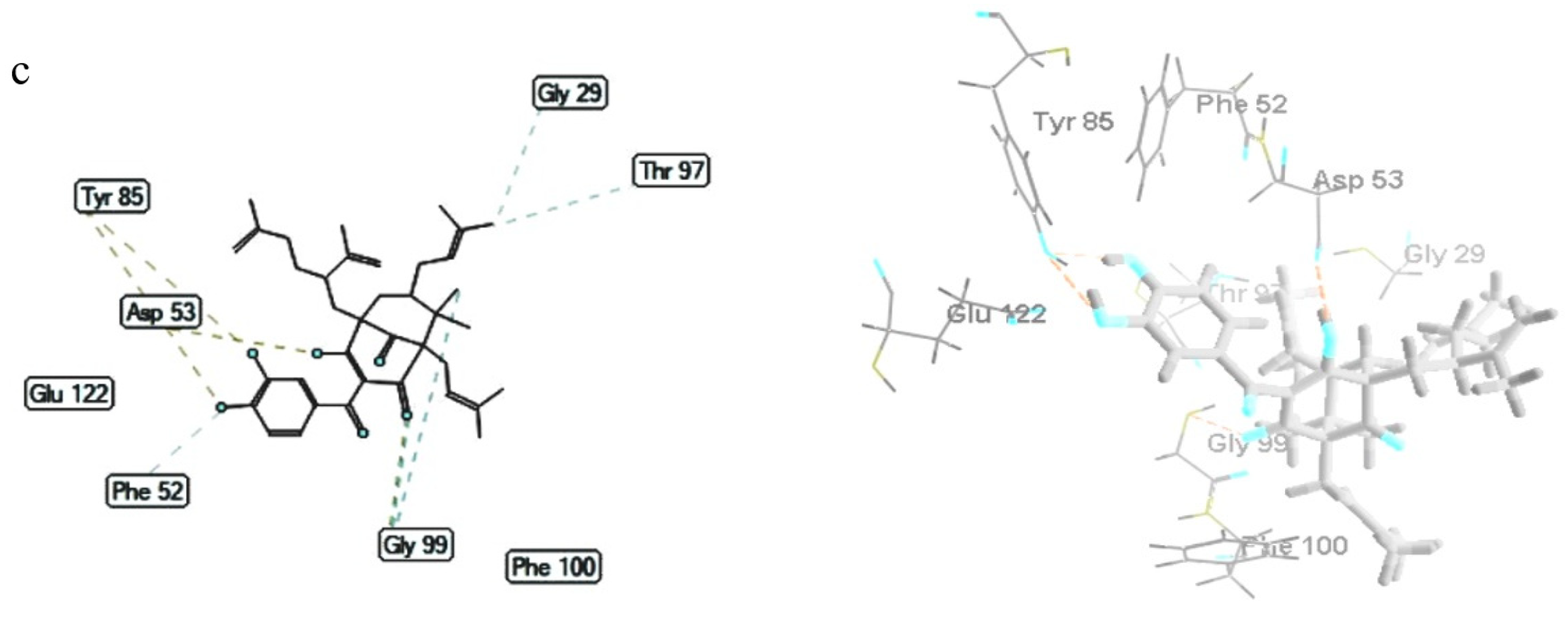
3.6.2. Plasmodium falciparum Lactate Dehydrogenase Enzyme

The molecular docking of polyprenylated benzophenones 1 to 3 was also studied on P. falciparum lactate dehydrogenase enzyme (PDB ID: 1CET). The MDS of compounds 1 to 3 were discovered to be −154.72 ± 0.48, −147.43 ± 1.16, and −145.56 ± 0.84 kcal/mol, respectively (Table 7). Compound 1 possessed the least bonding energy with an MSD value of −154.72 ± 0.48 kcal/mol, and therefore, exhibited the most stable molecular binding with the P. falciparum lactate dehydrogenase receptor. The polyprenylated benzophenones 1 to 3 bound to 1CET through hydrogen bonding of the oxygen atom of the carbonyl group (C10) with Thr101 and Gly99 residues, the oxygen atom of hydroxyl groups (C13 and C14) with Tyr85 residue, the oxygen atom of the hydroxyl group (C1) with Asp53, and the oxygen atom of the pyran ring with Gly99 (Table 7 and Figure 4). Furthermore, compound 1 exhibited steric interactions of the prenyl groups with Asp53 (2) residue, and the benzophenone skeleton with Asp53, Gly29 (2), Gly99 (2), Val55, Thr101, and Ile119 residues. Meanwhile, the steric interactions of compound 2 were established by the oxygen atom of the carbonyl group (C3) with Ile54 residue and the hydroxyl group with Phe52 residue, prenyl groups with Thr101 and Val55 residues, and the benzophenone skeleton with Val55 and Asp53 residues. The Phe52 and Gly99 residues of 1CET were bound to hydroxyl and carbonyl groups, while the Gly29 and Thr97 residues were bound to the prenyl group, and the Gly99 residue was bound to the benzophenone skeleton of compound 3 by steric interactions.

3.6.3. ADMET Profiles

According to the in silico study of the absorption, distribution, metabolism, excretion, and toxicity properties of isolated compounds 1 to 3, as derived from the pkCSM online tool, all compounds had a molecular weight of 602.812 (>500) g/mol. The Caco-2 permeability for the prediction of orally administered drug of compounds 1 to 3 was above 0.90 (Table 8), indicating all compounds possessed high permeability. Furthermore, all benzophenone compounds had high intestinal absorption, and are, therefore, well absorbed in the small intestine. Higher human intestinal absorption implies the compounds tend to be better absorbed within the gastrointestinal tract after oral administration [56]. Additionally, the transdermal drug efficacy illustrated by the compounds’ skin permeability, ranged from −2.75 to −2.73 cm/h (<−2.5). Consequently, compounds 1 to 3 are expected to penetrate the skin properly [57].
Compounds 1 to 3 also possess log VDss values below −0.15, indicating the compounds disperse in blood plasma. The ability of a drug to permeate the brain is indicated by the blood–brain barrier (BBB) permeability. Compounds are able to permeate the blood–brain barrier in cases where the logBB is above 0.3, while compounds with logBB below −1 are hardly dispersed to the brain. In this study, compounds 1 and 3 were discovered to have logBB values between −1 and 0.3 while compound 2 had a logBB value below −1. This indicates that compounds 1 and 3 are somewhat able to permeate the blood–brain barrier but compound 2 is not [57].
Table 8 shows compounds 1 to 3 were non inhibitors for CYP2D6 and CYP3A4. Generally, the compounds do not interfere with the cytochrome CYP450 biotransformation. The cytochrome P450 is a crucial detoxification enzyme in the human body.
Drug excretion from the human body is represented by total clearance and renal OCT2 substrate. The drug clearance is determined by the log (CLtot) of compound (ml/min/kg), from a combination of hepatic clearance and renal clearance. In this study, the total clearance of compounds 1 to 3 ranged from −0.02 to 0.29, and this is categorized as low clearance (Log CLtot < 2) [58]. Therefore, the three compounds are expected to be discharged slowly. Additionally, the adverse interactions of compounds 1 to 3 with OCT2 inhibitors as denoted by the OCT2 substrate parameter showed that the three compounds exhibit no potential contraindication.
The toxicity of promising medicinal compounds is a highly crucial factor. In this study, the acute toxicity (LD50) of compounds 1 to 3 ranged from 3.36 to 3.80 mol/kg, while the chronic toxicity of the compounds (LOAEL) ranged from 1.72 to 1.75 (52.48 to 56.23 mg/kg bw/day). Compound 2 was discovered to be the most toxic of the three benzophenones. The hepatotoxicity descriptor declared that compounds 1 to 3 were not hepatotoxic.

4. Conclusions

This study performed novel extractions of three known polyprenylated benzophenones from the root bark of G. celebica. The compounds were isolated from the EtOAc extract, and structural elucidation was carried out through spectroscopic analysis and literature data comparison. According to the results, the highest antioxidant activities were recorded for the CH2Cl2 extract by DPPH assay (7.45 ± 0.04 μg/mL), followed by the n-hexane extract by ABTS assay (5.96 ± 0.00 μg/mL), and EtOAc extract by FRAP assay. Compound 3 exhibited the highest antioxidant activity by DPPH, ABTS, and FRAP assay, with an IC50 value of 3.87 ± 0.10 μM, 0.88 ± 0.02 μM, and 44.28 ± 0.79 μM Fe2+/g, respectively. Meanwhile, compound 2 exhibited the strongest cytotoxicity against HeLa, MCF-7, and B16 cell lines, with IC50 values of 10.18, 10.73, and 12.39 μM, respectively, and was also active against P. falciparum strain 3D7, with an IC50 value of 2.99 ± 0.20 μM at 10 μg/mL. Based on the molecular docking result, compound 3 possessed the most stable molecular binding with an alpha-estrogen receptor (MDS −162,832 ± 2238 kcal/mol) and compound 1 with P. falciparum lactate dehydrogenase receptor (MDS −154.72 ± 0.48 kcal/mol). Additionally, the ADMET prediction indicated compounds 13 were potential oral drug candidates. These findings imply the value of G. celebica as a natural resource in the medicinal area of malaria and cancer.

Author Contributions

Conceptualization, T.E.; methodology, Y.P.P., A.F., S.F. and T.E.; formal analysis, Y.P.P.; resources, T.E.; data curation, S.F.; writing—original draft preparation, Y.P.P.; writing—review and editing, A.F.; supervision, T.E., A.F., S.F. All authors have read and agreed to the published version of the manuscript.

Funding

This research was funded by Penelitian Keilmuan Grant DRPM ITS (Grant Number: 1247/PKS/ITS/2021).

Institutional Review Board Statement

Not applicable.

Informed Consent Statement

Not applicable.

Data Availability Statement

Not applicable.

Acknowledgments

The authors would like to thank Rector of Musamus University for doctoral program study permission (Y.P.P.), Directorate General of Higher Education Ministry of Education, Culture, Research, and Technology of The Republic of Indonesia for providing Doctoral Scholarship Fund (BPPDN), Edwin Risky Sukandar (Center of Excellence in Natural Products Chulalongkorn University) for structural elucidation assistance, Siswandono (Faculty of Pharmacy Universitas Airlangga) for molecular docking data collection, La Hisa (Balai Taman Nasional Wasur) for plant material collection, and Elvira Hermawati (Department of Chemistry Institut Teknologi Bandung) for NMR measurement.

Conflicts of Interest

The authors declare no conflict of interest.

Abbreviations

ABTS2,2″-azinobis(3-ethyl benzothiazoline-6-sulfonic acid)
ADMETAbsorption Distribution Metabolism Excretion Toxicity
DPPH1,1-diphenyl-2-picrylhydrazyl
FRAPferric reducing antioxidant power
PDBprotein data bank
pkCSMsmall-molecule pharmacokinetics prediction
TPTZ2,4,6-Tris(2-pyridyl)-s-triazine

References

  1. World Health Organization. Available online: https://www.who.int/news-room/fact-sheets/detail/malaria (accessed on 5 June 2021).
  2. Kementerian Kesehatan Republik Indonesia. Available online: https://www.kemkes.go.id/article/view/19020100003/hari-kanker-sedunia-2019.html (accessed on 14 July 2021).
  3. Fajariyani, S.B. Laporan Situasi Terkini Perkembangan Program Pengendalian Malaria di Indonesia Tahun 2019; Kementerian Kesehatan Republik Indonesia: Jakarta, Indonesia, 2020; pp. 1–4. [Google Scholar]
  4. Newman, D.J.; Crag, G.M. Natural Products as Sources of New Drugs over the Nearly Four Decades from 01/1981 to 09/2019. J. Nat. Prod. 2020, 83, 770–803. [Google Scholar] [CrossRef]
  5. Srivastava, N.; Singh, A.; Kumari, P.; Nishad, J.H.; Gautam, V.S.; Yadav, M.; Bharti, R.; Kumar, D.; Kharwar, R.N. Advanced in extraction technologies: Isolation and purification of bioactive compounds from biological materials. In Natural Bioactive Compounds, 1st ed.; Sinha, R.P., Hader, D.-P., Eds.; Elsevier: London, UK, 2021; pp. 409–433. [Google Scholar]
  6. Hay, A.-E.; Aumond, M.-C.; Mallet, S.; Dumontet, V.; Litaudon, M.; Rondeau, D.; Richomme, P. Antioxidant Xanthones from Garcinia vieillardii. J. Nat. Prod. 2004, 67, 707–709. [Google Scholar] [CrossRef]
  7. Yamaguchi, F.; Ariga, T.; Yoshimura, Y.; Nakazawa, H. Antioxidative and Anti-Glycation Activity of Garcinol from Garcinia indica Fruit Rind. J. Agric. Food Chem. 2000, 48, 180–185. [Google Scholar] [CrossRef] [PubMed]
  8. Fouotsa, H.; Lannang, A.; Dzoyem, J.; Tatsimo, S.; Neumann, B.; Mbazoa, C.; Razakarivony, A.; Nkengfack, A.; Eloff, J.; Sewald, N. Antibacterial and Antioxidant Xanthones and Benzophenone from Garcinia smeathmannii. Planta Med. 2015, 81, 594–599. [Google Scholar] [CrossRef] [Green Version]
  9. Liu, C.; Ho, P.C.-L.; Wong, F.C.; Sethi, G.; Wang, L.Z.; Goh, B.C. Garcinol: Current status of its anti-oxidative, anti-inflammatory and anti-cancer effects. Cancer Lett. 2015, 362, 8–14. [Google Scholar] [CrossRef] [PubMed]
  10. Tan, W.-N.; Khairuddean, M.; Wong, K.-C.; Tong, W.-Y.; Ibrahim, D. Antioxidant compounds from the stem bark of Garcinia atroviridis. J. Asian Nat. Prod. Res. 2016, 18, 804–811. [Google Scholar] [CrossRef] [PubMed]
  11. Mohamed, G.A.; Ibrahim, S.R.M. New benzophenones and a dihydroflavanonol from Garcinia mangostana pericarps and their antioxidant and cytotoxic activities. Phytochem. Lett. 2020, 39, 43–48. [Google Scholar] [CrossRef]
  12. Tang, Y.-X.; Fu, W.-W.; Wu, R.; Tan, H.-S.; Shen, Z.-W.; Xu, H.-X. Bioassay-Guided Isolation of Prenylated Xanthone Derivatives from the Leaves of Garcinia oligantha. J. Nat. Prod. 2016, 79, 1752–1761. [Google Scholar] [CrossRef]
  13. Bui, T.Q.; Bui, A.T.; Nguyen, K.T.; Nguyen, V.T.; Trinh, B.T.D.; Nguyen, L.-H.D. A depsidone and six triterpenoids from the bark of Garcinia celebica. Tetrahedron Lett. 2016, 57, 2524–2529. [Google Scholar] [CrossRef]
  14. Youn, U.J.; Sripisut, T.; Miklossy, G.; Turkson, J.; Laphookhieo, S.; Chang, L.C. Bioactive polyprenylated benzophenone derivatives from the fruits extracts of Garcinia xanthochymus. Bioorg. Med. Chem. Lett. 2017, 27, 3760–3765. [Google Scholar] [CrossRef]
  15. Sukandar, E.R.; Kaennakam, S.; Rassamee, K.; Siripong, P.; Fatmawati, S.; Ersam, T.; Tip-pyang, S. Xanthones and biphenyls from the stems of Garcinia cylindrocarpa and their cytotoxicity. Fitoterapia 2018, 130, 112–117. [Google Scholar] [CrossRef] [PubMed]
  16. Zhang, B.-J.; Fu, W.-W.; Wu, R.; Yang, J.-L.; Yao, C.-Y.; Yan, B.-X.; Tan, H.-S.; Zheng, C.-W.; Song, Z.-J.; Xu, H.-X. Bioactive scalemic caged xanthones from the leaves of Garcinia bracteata. Bioorg. Chem. 2019, 82, 274–283. [Google Scholar] [CrossRef] [PubMed]
  17. Yang, J.; Fu, W.; Xiang, Q.; Tang, Y.; Wu, R.; Zheng, C.; Lu, Y.; Zhou, H.; Xu, H. Cytotoxic xanthone derivatives from the twigs of Garcinia oligantha. Phytochemistry 2020, 174, 112329. [Google Scholar] [CrossRef]
  18. Hatami, E.; Jaggi, M.; Chauhan, S.C.; Yallapu, M.M. Gambogic acid: A shining natural compound to nanomedicine for cancer therapeutics. Biochim. Biophys. Acta BBA Rev. Cancer 2020, 1874, 188381. [Google Scholar] [CrossRef]
  19. Protiva, P.; Hopkins, M.E.; Baggett, S.; Yang, H.; Lipkin, M.; Holt, P.R.; Kennelly, E.J.; Bernard, W.I. Growth inhibition of colon cancer cells by polyisoprenylated benzophenones is associated with induction of the endoplasmic reticulum response: Garcinia xanthochymus Benzophenones Induce Stress. Int. J. Cancer 2008, 123, 687–694. [Google Scholar] [CrossRef]
  20. Sukandar, E.R.; Kaennakam, S.; Aree, T.; Nöst, X.; Rassamee, K.; Bauer, R.; Siripong, P.; Ersam, T.; Tip-pyang, S. Picrorhizones A–H, Polyprenylated Benzoylphloroglucinols from the Stem Bark of Garcinia picrorhiza. J. Nat. Prod. 2020, 83, 2102–2111. [Google Scholar] [CrossRef]
  21. Auranwiwat, C.; Laphookhieo, S.; Rattanajak, R.; Kamchonwongpaisan, S.; Pyne, S.G.; Ritthiwigrom, T. Antimalarial polyoxygenated and prenylated xanthones from the leaves and branches of Garcinia mckeaniana. Tetrahedron 2016, 72, 6837–6842. [Google Scholar] [CrossRef] [Green Version]
  22. Lyles, J.; Negrin, A.; Khan, S.; He, K.; Kennelly, E. In Vitro Antiplasmodial Activity of Benzophenones and Xanthones from Edible Fruits of Garcinia Species. Planta Med. 2014, 80, 676–681. [Google Scholar] [CrossRef] [Green Version]
  23. Wairata, J.; Sukandar, E.R.; Fadlan, A.; Purnomo, A.S.; Taher, M.; Ersam, T. Evaluation of the antioxidant, antidiabetic, and antiplasmodial activities of xanthones 2 isolated from Garcinia forbesii and their in silico studies. Biomedicines 2021, 9, 1380. [Google Scholar] [CrossRef]
  24. Elfita, E.; Muharni, M.; Latief, M.; Darwati, D.; Widiyantoro, A.; Supriyatna, S.; Bahti, H.H.; Dachriyanus, D.; Cos, P.; Maes, L. Antiplasmodial and other constituents from four Indonesian Garcinia spp. Phytochemistry 2009, 70, 907–912. [Google Scholar] [CrossRef]
  25. Subarnas, A.; Diantini, A.; Abdulah, R.; Zuhrotun, A.; Nugraha, P.A.; Hadisaputri, Y.E.; Puspitasari, I.M.; Yamazaki, C.; Kuwano, H.; Koyama, H. Apoptosis-mediated antiproliferative activity of friedolanostane triterpenoid isolated from the leaves of Garcinia celebica against MCF-7 human breast cancer cell lines. Biomed. Rep. 2016, 4, 79–82. [Google Scholar] [CrossRef] [Green Version]
  26. Armania, N.; Yazan, L.S.; Musa, S.N.; Ismail, I.S.; Foo, J.B.; Chan, K.W.; Noreen, H.; Hisyam, A.H.; Zulfahmi, S.; Ismail, M. Dillenia suffruticosa exhibited antioxidant and cytotoxic activity through induction of apoptosis and G2/M cell cycle arrest. J. Ethnopharmacol. 2013, 146, 525–535. [Google Scholar] [CrossRef]
  27. Abubakar, A.R.; Haque, M. Preparation of Medicinal Plants: Basic Extraction and Fractionation Procedures for Experimental Purposes. J. Pharm. Bioallied Sci. 2020, 12, 1–10. [Google Scholar] [CrossRef] [PubMed]
  28. Brighente, I.M.C.; Dias, M.; Verdi, L.G.; Pizzolatti, M.G. Antioxidant Activity and Total Phenolic Content of Some Brazilian Species. Pharm. Biol. 2007, 45, 156–161. [Google Scholar] [CrossRef]
  29. Dudonné, S.; Vitrac, X.; Coutière, P.; Woillez, M.; Mérillon, J.-M. Comparative Study of Antioxidant Properties and Total Phenolic Content of 30 Plant Extracts of Industrial Interest Using DPPH, ABTS, FRAP, SOD, and ORAC Assays. J. Agric. Food Chem. 2009, 57, 1768–1774. [Google Scholar] [CrossRef]
  30. Lulan, T.Y.K.; Fatmawati, S.; Santoso, M.; Ersam, T. α-VINIFERIN as a potential antidiabetic and antiplasmodial extracted from Dipterocarpus littoralis. Heliyon 2020, 6, e04102. [Google Scholar] [CrossRef]
  31. Benzie, I.F.F.; Devaki, M. The ferric reducing/antioxidant power (FRAP) assay for non-enzymatic antioxidant capacity: Concepts, procedures, limitations and applications. In Measurement of Antioxidant Activity & Capacity, 1st ed.; Apak, R., Capanoglu, E., Shahidi, F., Eds.; John Wiley & Sons, Ltd.: Chichester, UK, 2017; pp. 77–106. [Google Scholar] [CrossRef]
  32. Nurlelasari, R.I.; Salam, S.; Safari, A.; Harneti, D.; Maharani, R.; Hidayat, A.T.; Tanjung, M.; Retnowati, R.; Shiono, Y.; Supratman, U. A new havanensin-type limonoid from Chisocheton macrophyllus. Appl. Biol. Chem. 2021, 64, 35. [Google Scholar] [CrossRef]
  33. Wang, X.; Miyazaki, Y.; Inaoka, D.K.; Hartuti, E.D.; Watanabe, Y.-I.; Shiba, T.; Harada, S.; Saimoto, H.; Burrows, J.N.; Benito, F.J.G.; et al. Identification of Plasmodium falciparum Mitochondrial Malate: Quinone Oxidoreductase Inhibitors from the Pathogen Box. Genes 2019, 10, 471. [Google Scholar] [CrossRef] [Green Version]
  34. Zhang, L.-J.; Chiou, C.-T.; Cheng, J.-J.; Huang, H.-C.; Kuo, L.-M.Y.; Liao, C.-C.; Bastow, K.F.; Lee, K.-H.; Kuo, Y.-H. Cytotoxic Polyisoprenyl Benzophenonoids from Garcinia subelliptica. J. Nat. Prod. 2010, 73, 557–562. [Google Scholar] [CrossRef] [Green Version]
  35. Iinuma, M.; Tosa, H.; Tanaka, T.; Kanamaru, S.; Asai, F.; Kobayashi, Y.; Miyauchi, K.; Shimano, R. Antibacterial Activity of Some Garcinia Benzophenone Derivatives against Methicilin-Resistant Staphylococcus aureus. Biol. Pharm. Bull. 1996, 19, 311–314. [Google Scholar] [CrossRef] [Green Version]
  36. Baggett, S.; Mazzola, E.P.; Kennelly, E.J. The benzophenones: Isolation, structural elucidation and biological activities. In Studies in Natural Products Chemistry; Elsevier: Amsterdam, The Netherlands, 2005; pp. 721–771. [Google Scholar] [CrossRef]
  37. Ramirez, C.; Gil, J.H.; Marín-Loaiza, J.C.; Rojano, B.; Durango, D. Chemical constituents and antioxidant activity of Garcinia madruno (Kunth) Hammel. J. King Saud. Univ. Sci. 2019, 31, 1283–1289. [Google Scholar] [CrossRef]
  38. Menon, L.N.; Satheesh, S.K.K.; Panicker, S.P.; Rameshkumar, K.B. Antiproliferative activity of caged xanthones from the leaves of Garcinia wightii T. Anderson. Fitoterapia 2020, 143, 104592. [Google Scholar] [CrossRef] [PubMed]
  39. Baggett, S.; Protiva, P.; Mazzola, E.P.; Yang, H.; Ressler, E.T.; Basile, M.J.; Weinstein, I.B.; Kennely, E.J. Bioactive Benzophenones from Garcinia xanthochymus Fruits. J. Nat. Prod. 2005, 68, 354–360. [Google Scholar] [CrossRef] [PubMed]
  40. Floegel, A.; Kim, D.-O.; Chung, S.-J.; Koo, S.I.; Chun, O.K. Comparison of ABTS/DPPH assays to measure antioxidant capacity in popular antioxidant-rich US foods. J. Food Compos. Anal. 2011, 24, 1043–1048. [Google Scholar] [CrossRef]
  41. Kim, D.-O.; Lee, K.W.; Lee, H.J.; Lee, C.Y. Vitamin C Equivalent Antioxidant Capacity (VCEAC) of Phenolic Phytochemicals. J. Agric. Food Chem. 2002, 50, 3713–3717. [Google Scholar] [CrossRef]
  42. Liang, N.; Kitts, D. Antioxidant Property of Coffee Components: Assessment of Methods that Define Mechanisms of Action. Molecules 2014, 19, 19180–19208. [Google Scholar] [CrossRef] [PubMed] [Green Version]
  43. Essien, E.E.; Thomas, P.S.; Ekanem, I.R.; Choudhary, M.I. Isolation and characterization of 5-hydroxymethylfurfural, antiglycation, antihyperglycaemic, antioxidant, and cytotoxic effects of Garcinia kola Heckel roots extract and fractions. S. Afr. J. Bot. 2021, 140, 62–67. [Google Scholar] [CrossRef]
  44. Prasad, K.N.; Chew, L.Y.; Khoo, H.E.; Kong, K.W.; Azlan, A.; Ismail, A. Antioxidant Capacities of Peel, Pulp, and Seed Fractions of Canarium odontophyllum Miq. Fruit. J. Biomed. Biotechnol. 2010, 2010, 871379. [Google Scholar] [CrossRef] [PubMed] [Green Version]
  45. Lin, X.; Tian, D.; Fu, Y.; Li, Y.; Huang, L.; Gu, W.; Song, J.; Li, Y.; Ben-David, Y.; Wen, M.; et al. Synthesis of novel guttiferone E and xanthochymol derivatives with cytotoxicities by inducing cell apoptosis and arresting the cell cycle phase. Eur. J. Med. Chem. 2019, 162, 765–780. [Google Scholar] [CrossRef]
  46. Matsumoto, K.; Akao, Y.; Kobayashi, E.; Ito, T.; Ohguchi, K.; Tanaka, T.; Iinuma, M.; Nozawa, Y. Cytotoxic Benzophenone Derivatives from Garcinia Species Display a Strong Apoptosis-Inducing Effect against Human Leukemia Cell Lines. Biol. Pharm. Bull. 2003, 26, 569–571. [Google Scholar] [CrossRef] [PubMed] [Green Version]
  47. Kumar, S.; Chattopadhyay, S.; Darokar, M.; Garg, A.; Khanuja, S. Cytotoxic Activities of Xanthochymol and Isoxanthochymol Substantiated by LC-MS/MS. Planta Med. 2007, 73, 1452–1456. [Google Scholar] [CrossRef]
  48. Kuete, V.; Tchakam, P.D.; Wiench, B.; Ngameni, B.; Wabo, H.K.; Tala, M.F.; Moungang, M.L.; Ngadjui, B.T.; Murayama, T.; Efferth, T. Cytotoxicity and modes of action of four naturally occuring benzophenones: 2,2′,5,6′-Tetrahydroxybenzophenone, guttiferone E, isogarcinol and isoxanthochymol. Phytomedicine 2013, 20, 528–536. [Google Scholar] [CrossRef]
  49. Wu, S.-B.; Long, C.; Kennelly, E.J. Structural diversity and bioactivities of natural benzophenones. Nat. Prod. Rep. 2014, 31, 1158–1174. [Google Scholar] [CrossRef]
  50. Phang, Y.; Wang, X.; Lu, Y.; Fu, W.; Zheng, C.; Xu, H. Bicyclic polyprenylated acylphloroglucinols and their derivatives: Structural modification, structure-activity relationship, biological activity and mechanism of action. Eur. J. Med. Chem. 2020, 205, 112646. [Google Scholar] [CrossRef]
  51. Lannang, A.; Louh, G.; Biloa, B.; Komguem, J.; Mbazoa, C.; Sondengam, B.; Naesens, L.; Pannecouque, C.; De Clercq, E.; Sayed El Ashry, E. Cytotoxicity of Natural Compounds Isolated from the Seeds of Garcinia afzelii. Planta Med. 2010, 76, 708–712. [Google Scholar] [CrossRef]
  52. Mathew, S.; Abraham, T.E.; Zakaria, Z.A. Reactivity of phenolic compounds towards free radicals under in vitro conditions. J. Food Sci. Technol. 2015, 52, 5790–5798. [Google Scholar] [CrossRef] [PubMed] [Green Version]
  53. Saha Roy, S.; Vadlamudi, R.K. Role of Estrogen Receptor Signaling in Breast Cancer Metastasis. Int. J. Breast Cancer 2012, 2012, 654698. [Google Scholar] [CrossRef] [PubMed] [Green Version]
  54. Hayes, E.L.; Lewis-Wambi, J.S. Mechanisms of endocrine resistance in breast cancer: An overview of the proposed roles of noncoding RNA. Breast Cancer Res. 2015, 17, 40. [Google Scholar] [CrossRef] [Green Version]
  55. Hua, H.; Zhang, H.; Kong, Q.; Jiang, Y. Mechanisms for estrogen receptor expression in human cancer. Exp. Hematol. Oncol. 2018, 7, 24. [Google Scholar] [CrossRef] [Green Version]
  56. Srivastava, V.; Yadav, A.; Sarkar, P. Molecular docking and ADMET study of bioactive compounds of Glycyrrhiza glabra against main protease of SARS-CoV2. Mater. Today Proc. 2020. [Google Scholar] [CrossRef]
  57. Pires, D.E.V.; Blundell, T.L.; Ascher, D.B. pkCSM: Predicting Small-Molecule Pharmacokinetic and Toxicity Properties Using Graph-Based Signatures. J. Med. Chem. 2015, 58, 4066–4072. [Google Scholar] [CrossRef]
  58. Da Paixão, V.G.; da Rocha Pita, S.S. In silico identification and evaluation of new Trypanosoma cruzi trypanothione reductase (TcTR) inhibitors obtained from natural products database of the Bahia semi-arid region (NatProDB). Comput. Biol. Chem. 2019, 79, 36–47. [Google Scholar] [CrossRef] [PubMed]
Figure 1. The scheme of the general experimental procedures.
Biomedicines 09 01654 g001
Figure 2. The structures of the polyprenylated benzophenones (compounds 13) isolated from the root bark of G. celebica.
Biomedicines 09 01654 g002
Figure 3. The 2D and 3D interaction of 1 (a), 2 (b), and 3 (c) with human alpha-estrogen receptor (3ERT). Hydrogen bonds are shown as yellow dotted lines while steric interactions are depicted as blue dotted lines. Carbon atoms are represented in gray, oxygen in blue, and hydrogen in black.
Biomedicines 09 01654 g003
Figure 4. The 2D and 3D interaction of 1 (a), 2 (b), and 3 (c) with Plasmodium falciparum lactate dehydrogenase enzyme (1CET). Hydrogen bonds are shown as yellow dotted lines while steric interactions are depicted as blue dotted lines. Carbon atoms are represented in gray, oxygen in blue, and hydrogen in black.
Biomedicines 09 01654 g004aBiomedicines 09 01654 g004b
Table 1. 1H (500 MHz) and 13C (125 MHz) NMR spectroscopic data of Compounds 13 (in ppm).
Position1 (CD3OD)2 (CD3OD)3 (CD3OD)
δCδH (J in Hz)δCδH (J in Hz)δCδH (J in Hz)
1173.7 172.9 195.8 b
2126.8 128.0 117.9
3196.4 196.0 - c
469.5 71.7 - c
547.1 46.9 50.2
647.41.52, m42.41.98, m47.91.50 a
739.72.27, m 43.32.17, dd (14.1, 4.5)43.72.05, m
2.04, m 1.56, t (13.0) 2.05, m
852.9 54.2 - c
9207.9 207.2 210.6
10194.2 195.1 195.8
11131.1 130.7 129.5
12115.97.28, d (2.1)116.37.29, d (2.1)117.37.19, d (2.2)
13146.7 146.7 146.3
14152.6 152.8 152.5
15115.46.70, d (8.0)115.66.75, d (8.3)115.16.71, d (8.3)
16124.66.95, dd (8.0, 2.1)124.87.06, dd (8.2, 2.1)125.26.98, dd (8.3, 2.2)
1726.62.46, dd (13.5, 6.0)25.82.44, dd (13.8, 5.8)27.02.71, dd (13.30, 9.45)
2.64, dd (14.0, 6.0) 2.63, dd (13.6, 8.0) 2.57, m
18121.24.91 a121.44.86 a121.45.05, m
19135.5 135.1 135.8
2026.61.59, s26.41.61, s26.51.74, s
2118.31.59, s18.31.60, s18.31.69, s
2222.91.15, s22.61.09, s23.21.15, s
2327.10.99, s16.30.77, s27.31.00, s
2430.52.15, m30.61.85, m30.32.05, m
2.67, m 2.07, m 2.05, m
25126.34.92 a122.95.21, t125.64.90 a
26134.0 134.7 133.6
2726.11.69, s26.01.79, s26.01.66, s
2818.61.67, s18.11.65, s18.21.50, s
2928.81.07, br d (13.5) 28.51.04, t (13.6)37.71.90, m
3.09, dd (14.0, 3.4) 3.03, dd (14.2, 3.65) 1.90, m
3042.91.33, m44.61.40, tt (13.5)44.72.57, m
3188.5 88.5 148.9
3228.80.84, s21.51.24, s113.64.50, br s
3321.51.24, s29.00.98, s17.81.59, s
3429.31.15 a28.71.79 a32.71.50 a
1.52, m 2.23, d (14.5) 1.42, m
3536.22.15, m123.95.16, t36.81.83, m
2.27, m 1.83, m
36146.0 134.2 146.9
37111.94.79, s26.01.73, s110.54.64, d (7.3)
3822.31.73, s18.11.62, s22.81.69, s
a Overlapping signals; b Data was extracted from the HMBC spectrum; c Not detected; s: singlet; d: doublet; t: triplet; m: multiplet; dd: doublet of doublet; br: broad; tt: triplet of triplet.
Table 2. The TPC and TFC values of G. celebica root bark extracts.
ExtractTPC (mg GAE/g Extract)TFC (mg QE/g Extract)
n-Hexane188.00 ± 1.7833.07 ± 0.76
CH2Cl2164.35 ± 1.1931.03 ± 2.28
EtOAc180.06 ± 1.0735.73 ± 0.13
MeOH112.84 ± 1.8927.11 ± 0.24
Data are expressed as mean ± SD of triplicate experiments.
Table 3. The antioxidant activities of G. celebica root bark extracts and isolated compounds 13.
Extracts/CompoundsAntioxidant Capacity
DPPHABTSFRAP (μM Fe2+/g)
IC50 (μg/mL)IC50 (μM)IC50 (μg/mL)IC50 (μM)
n-Hexane7.86 ± 0.07-5.96 ± 0.00-35.64 ± 1.56
CH2Cl27.45 ± 0.04-6.43 ± 0.02-34.58 ± 1.67
EtOAc7.55 ± 0.06-6.01 ± 0.01-66.36 ± 5.05
MeOH11.40 ± 0.00-7.63 ± 0.01-38.75 ± 2.41
13.36 ± 0.035.58 ± 0.050.78 ± 0.021.30 ± 0.0422.61 ± 0.06
22.98 ± 0.054.95 ± 0.080.70 ± 0.021.16 ± 0.0424.58 ± 0.29
32.33 ± 0.063.87 ± 0.100.53 ± 0.010.88 ± 0.0244.28 ± 0.79
Gallic acid0.55 ± 0.003.22 ± 0.020.12 ± 0.000.68 ± 0.00Nt
Quercetin1.34 ± 0.024.43 ± 0.060.06 ± 0.000.19 ± 0.00Nt
Ascorbic acidNtNt30.62 ± 0.27
Nt: not tested.
Table 4. The cytotoxic activity of the compounds after 72 h of treatment.
CompoundsIC50 (μM)
HeLaMCF-7A549B16
113.77 ± 0.0713.54 ± 0.0113.77 ± 0.2012.46 ± 0.08
210.18 ± 0.5110.73 ± 0.1413.99 ± 0.0912.39 ± 0.11
315.48 ± 0.1314.93 ± 0.5216.10 + 0.1816.94 ± 0.15
Cisplatin19531843
Table 5. Antiplasmodial activity against the chloroquine-sensitive P. falciparum strain 3D7.
Compound% Inhibition ± SD (10 μg/mL)IC50 ± SD (μM)
1<10Nt
283.97 ± 0.472.99 ± 0.20
3<10Nt
chloroquine98.8 ± 0.250.006 ± 0.01
% Inhibition <10% = inactive, Nt = Not tested.
Table 6. MolDock score and interacting residues of 1, 2, and 3 to human alpha-estrogen receptor (3ERT).
LigandsMolDock Score (kcal/mol)Hydrogen BondingSteric InteractionElectrostatic Interaction
1−144.638 ± 1.41Thr347Asp351 (3); Leu539 (2); Ala350; Met343; Leu354; Thr347; Leu525; Met522
2−139.590 ± 0.37Val534 (2)Leu525 (4); Trp383 (4); Thr347; Leu346; Asp351; Leu539; Leu536; Met528
3−162.832 ± 2.24Thr347Thr347 (2); Ala350; Asp351; Leu354; Leu525; Met522; Met528
4-hydroxtamoxifen−165.042 ± 0,48Arg394; Glu353Ala350Asp351
Table 7. MolDock score and interacting residues of 1, 2, and 3 to P. falciparum lactate dehydrogenase enzyme (1CET).
LigandsMolDock Score (kcal/mol)Hydrogen BondingSteric Interaction
1−154.72 ± 0.48Gly99; Thr101Gly29 (3); Gly99 (2); Asp53 (3); Val55; Ile119, Thr101
2−147.43 ± 1.16Tyr85 (2); Gly99; Glu122Ile54 (3); Asp53; Thr101; Val55 (2), Phe52
3−145.56 ± 0.84Tyr85 (2); Gly99; Asp53Gly29; Gly99 (2); Thr97; Phe52
Chloroquine−108.99 ± 0.48Gly29; Gly99; Ser28Gly29; Gly99; Ala98; Asp53
Table 8. ADMET properties by the pkSCM online tool.
LigandsAbsorptionDistributionMetabolismExcretionToxicity
Caco-2 PermeabilityIntestinal Absorption (Human)Skin PermeabilityVDss (Human)BBB PermeabilityCYP2D6 InhibitorCYP3A4 InhibitorTotal ClearanceRenal OCT2 SubstrateOral Rat Acute Toxicity (LD50)Oral Rat Chronic Toxicity (LOAEL)Hepatotoxicity
11.54100.00−2.74−0.16−0.51NoNo0.04No3.361.74No
21.2692.65−2.73−0.46−1.39NoNo0.29No3.801.72No
31.60100.00−2.75−0.19−0.48NoNo−0.02No3.371.75No
4−Hidroxytamoxifen1.0393.54−2.740.31−0.29YesNo0.59No2.181.37No
Chloroquine1.25989.44−2.5641.7570.410YesNo0.993Yes2.8880.423Yes
Publisher’s Note: MDPI stays neutral with regard to jurisdictional claims in published maps and institutional affiliations.

