Article

DAPTA, a C-C Chemokine Receptor 5 (CCR5), Leads to the Downregulation of Notch/NF-κB Signaling and Proinflammatory Mediators in CD40+ Cells in Experimental Autoimmune Encephalomyelitis Model in SJL/J Mice

1
Department of Pharmacology and Toxicology, College of Pharmacy, King Saud University, Riyadh 11451, Saudi Arabia
2
Department of Pharmaceutics, College of Pharmacy, King Saud University, Riyadh 11451, Saudi Arabia
3
Department of Botany and Microbiology, College of Science, King Saud University, Riyadh 11451, Saudi Arabia
*
Author to whom correspondence should be addressed.
Biomedicines 2023, 11(6), 1511; https://doi.org/10.3390/biomedicines11061511
Submission received: 9 April 2023 / Revised: 21 May 2023 / Accepted: 22 May 2023 / Published: 23 May 2023
(This article belongs to the Special Issue Targeted Drug Delivery in Immune Diseases)

Abstract

:
Multiple sclerosis (MS) is an autoimmune inflammatory disease of the central nervous system characterized by motor deficits, cognitive impairment, fatigue, pain, and sensory and visual dysfunction. CD40, highly expressed in B cells, plays a significant role in MS pathogenesis. The experimental autoimmune encephalomyelitis (EAE) mouse model of MS has been well established, as well as its relevance in MS patients. This study aimed to evaluate the therapeutic potential of DAPTA, a selective C-C chemokine receptor 5 (CCR5) antagonist in the murine model of MS, and to expand the knowledge of its mechanism of action. Following the induction of EAE, DAPTA was administrated (0.01 mg/kg, i.p.) daily from day 14 to day 42. We investigated the effects of DAPTA on NF-κB p65, IκBα, Notch-1, Notch-3, GM-CSF, MCP-1, iNOS, and TNF-α in CD40+ spleen B cells using flow cytometry. Furthermore, we also analyzed the effect of DAPTA on NF-κB p65, IκBα, Notch-1, Notch-3, GM-CSF, MCP-1, iNOS, and TNF-α mRNA expression levels using qRT-PCR in brain tissue. EAE mice treated with DAPTA showed substantial reductions in NF-κB p65, Notch-1, Notch-3, GM-CSF, MCP-1, iNOS, and TNF-α but an increase in the IκBα of CD40+ B lymphocytes. Moreover, EAE mice treated with DAPTA displayed decreased NF-κB p65, Notch-1, Notch-3, GM-CSF, MCP-1, iNOS, and TNF-α and but showed increased IκBα mRNA expression levels. This study showed that DAPTA has significant neuroprotective potential in EAE via the downregulation of inflammatory mediators and NF-κB/Notch signaling. Collectively, DAPTA might have potential therapeutic targets for use in MS treatment.

Graphical Abstract

1. Introduction

Multiple sclerosis (MS) is an inflammatory disease affecting the central nervous system (CNS) [1,2]. The clinical features of MS are diverse and mainly include limb weakness, blurred vision, ataxia, paresthesia, fatigue, and cognitive deficits [3]. In addition, there are progressive neuronal and oligodendroglial losses in MS [4]. EAE mimics many of the critical physiopathological features of MS in humans [5]. The etiology of MS is still unknown, but converging lines of evidence suggest that it is a multifactorial disease of complex tract caused by a dysregulation of the immune system [6]. The role of B cells has recently become a central issue in MS. B cells’ involvement in MS has primarily been considered in terms of the potential role of pathogenic CNS-reactive antibodies [7,8]. To date, immunosuppressive and immunomodulatory medications are the major therapeutic strategies for the treatment of MS, but they carry severe side effects [9]. None of the drugs have proven helpful in treating MS. Due to the high prevalence of MS patients worldwide and poor therapeutic outcomes, most MS patients eventually develop a disability [10,11]. Hence, seeking an effective therapeutic strategy for MS treatment is timely and essential.
The transcription factor NF-κB activation is considered to be a major source of inflammatory cascades in several neuroinflammatory disorders that exacerbate the disease pathogenesis in various CNS diseases, including autoimmune encephalomyelitis [12]. NF-κB activation occurs in microglia, astrocytes, oligodendrocytes, macrophages, and neurons, initiating initiate inflammatory reactions in EAE [13]. Previous results also indicated that NF-κB activation in inflammatory cells enhances inflammation and facilitates MS and EAE development [14]. It has been reported that CD40-mediated NF-κB activation in B cells is increased in MS [15]. Several studies have focused on evaluating Notch signaling during EAE [16,17]. Notch signaling has been implicated as a significant contributor to remyelination failure in MS [16]. Previous results showed that the Notch pathway plays a vital role in controlling inflammatory reactions in the CNS [18].
GM-CSF has been demonstrated to be critical for EAE pathogenesis due to the role it plays in driving the mobilization and activation of neutrophils [19]. A previous study has demonstrated that GM-CSF blockade in the relapsing-remitting (RR)-EAE model prevented disease relapses [20]. It has been previously demonstrated that GM-CSF activates microglia within the CNS and recruits and stimulates peripheral macrophages and dendritic cells during EAE [21]. It has been reported that GM-CSF promotes chronic disability in EAE by altering the composition of CNS myeloid cells [22]. Several studies have described the expression of MCP1 in the CNS of patients with MS using autopsy tissue [23,24]. Previous results from the use of reactive astrocytes also suggested a significant role of MCP-1 in demyelinating MS lesions [25]. MCP-1 was also identified within hypertrophic astrocytes in a noninflamed submeningeal region adjacent to chronic active lesions [26].
TNF-α plays a diverse role in autoimmune and inflammatory disorders [27]. A previous study highlighted that TNF-α increases brain-derived neurotrophic factor expression in trigeminal ganglion neurons [28]. A previous study also indicated that TNF-α is elevated in MS patients and correlated with disease progression [29]. TNF-α is also upregulated in demyelinating lesions in EAE in order to promote neuronal excitotoxicity and oligodendrocyte death [30]. A previous study exhibited that the inducible nitric oxide synthase (iNOS) expression significantly increased in the spinal cords of EAE mice [31]. It has also been reported that the activated microglia revealed iNOS-positive immunoreactions in the perivascular regions of spinal cords in animals with EAE [32]. Additionally, the suppression of iNOS is associated with the amelioration of EAE [33].
Chemokines and their receptors are expressed in endothelial cells, microglia, astrocytes, and neurons [34]. Previous studies reported the pathogenic role of chemokine receptors in EAE [35,36]. The levels of EAE induced in CCR5-knockout mice reveal a decrease in infiltrating immune cells, indicating the important role of CCR5 in cell recruitment into the CNS [37]. Moreover, CCR5 is considered important for leukocyte extravasation, thus facilitating the migration of these cells into the CNS [38]. Peripheral blood CCR5+ cells secrete high levels of IFN-γ, and IFN-γ is observed in demyelinating lesions [39]. The frequency of CCR5+ cells is elevated in EAE and MS [39,40]. Chemokine receptor antagonists ameliorate EAE progression [41]. D-ala-peptide T-amide (DAPTA) is a selective CCR5 antagonist [42]. It has been shown that the administration of DAPTA reduces microglia and astrocyte activation within the hippocampus [43]. In addition, DAPTA treatment appears to be a therapeutic target for the treatment of treating inflammation-related diseases [44]. Our previous study demonstrated that DAPTA administration attenuates immune aberrations by downregulating Th9/Th17 immune responses in the BTBR mouse model of autism [45]. Recently, we also demonstrated that DAPTA treatment alleviates inflammation by regulating IFN-γ/IL-10 and STAT4/Smad3 signaling in a mouse model of EAE [46].
EAE, the most widely used experimental model for MS, is a universal animal model resembling human RR-MS [47]. This model has been extensively used to discover new therapies for application to MS. Recently, we reported that treatment with DAPTA exerts an excellent therapeutic effect in the EAE mouse model [46]. Therefore, we hypothesized that the administration of DAPTA could downregulate proinflammatory mediators and Notch/NF-κB signaling in B cells in the SJL/J mouse model of RR-MS. In this study, we explore the possible molecular mechanisms underlying these effects.

2. Materials and Methods

2.1. Chemicals

DAPTA was purchased from Tocris Bioscience (Bristol, UK). Fluorescein isothiocyanate, PE/Cyanine7, Allophycocyanin, Alexa Fluor® 488, PE/Dazzle™ 594, Alexa Fluor® 647, and phycoerythrin-labeled CD40, NF-κB p65, IκBα, Notch1, Notch3, GM-CSF, iNOS, RORγT, MCP-1, TNF-α, lysis, and permeabilization solution, and fixation buffers were purchased from BD Biosciences and BioLegend (San Diego, CA, USA). SYBR Green and cDNA kits were purchased from Applied Biosystems (Foster City, CA, USA). Primers were purchased from GenScript (Piscataway, NJ, USA). TRizol was purchased from Invitrogen (Carlsbad, CA, USA).

2.2. Animals

The Bioethics Committee of the College of Pharmacy, King Saud University, approved all experimental procedures. Female SJL/J mice, aged 7–8 weeks, were purchased from the Jackson Laboratories (Bar Harbor, ME, USA). The mice were housed in specific pathogen-free conditions in an appropriate environment, maintained in a 12 h light/dark cycle, and given ad libitum access to food and water. Mice were allowed to acclimatize for two weeks before the initiation of the experiment.

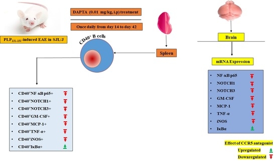
2.3. EAE Induction and DAPTA Treatment

In order to establish the EAE model, SJL/J mice were immunized subcutaneously with 200 µg of proteolipid protein (PLP139-151) peptide (Hooke Laboratories, Lawrence, MA, USA). Each mouse received an additional 200 ng of pertussis toxin (PTX) intraperitoneally (i.p.) on the immunization day [46,48]. DAPTA injection was started from day 14 after immunization and continued once daily until day 42 at a dose of DAPTA (0.01 mg/kg, i.p.). These parameters were selected based on the previous studies [43,44,45,46,47]. The mice in the standard control and EAE model groups were injected daily with the same volume of saline. Mice were sacrificed at the end of the treatment period by deep inhalational anesthesia (isoflurane). Different tissues (brain/spleen) were collected for use in various molecular analyses, i.e., flow cytometry and RT-PCR.

2.4. Flow Cytometry Analysis

The following conjugated monoclonal antibodies were used for the flow cytometry analysis of spleen cells: anti-CD40 PE/Dazzle, anti-CD40 APC, anti-CD40 FITC anti-NF-κB p65 FITC, anti-IκBα PE/Cyanine7, anti-Notch1 PE/Cyanine7, anti-Notch3 APC, anti-GM-CSF Alexa Fluor® 488, anti-iNOS PE/Dazzle, anti-MCP-1 APC, and anti-TNF-α PE/Cyanine7. Cells were washed, and surface staining of CD40 was performed. Intracellular staining was performed after fixing and permeabilizing for anti-NF-κB p65, anti-IκBα, anti-Notch1, anti-Notch3, anti-GM-CSF, anti-iNOS, anti-MCP-1, and anti-TNF-α fluorescent antibodies. The proportions of CD40+NF-κB p65+, CD40+IκBα+, CD40+Notch1+, CD40+Notch3+, CD40+GM-CSF+, CD40+iNOS+, CD40+MCP-1+, and CD40+TNF-α+ cells were determined in the lymphocyte gate. Sample analysis was performed with FACS (FC500), and data analysis was conducted using CXP software (Beckman CoulterIndianapolis, Indianapolis, IN, USA) [46].

2.5. Real-Time PCR Analysis

According to the manufacturer’s protocol, RNA from the brain was extracted using a TRIzol reagent (Invitrogen, Carlsbad, CA, USA). NanoDrop 2000 measured the concentration of RNA. cDNA was prepared using a cDNA reverse transcription kit, followed by RT-PCR with SYBR Green PCR reagents (Applied Biosystems, Foster City, CA, USA). The primer sequences used for the assay were as follows: Nf-κB p65, forward 5′-CTGCCGAGTAAACCGGAACT-3′, reverse 5′-CCCTGTGACATCACCTGCTT-3′; IκBα, forward 5′-AAGGCTACTCCCCCTACCAG-3′, reverse 5′-CAAGAAGGCGACACAGACCT-3′; Notch1, forward 5′-GGTGCTCTGATGGACGACAA-3′, reverse 5′-CATGAGGGGTGTGAAGCCAT-3′; Notch3, forward 5′-AGGCCATGGTCTTCCCCTAT-3′, reverse 5′-ACCTCCCCCATCAGACTCTC-3′; Gm-csf, forward 5′-AGAGGCCATCAAAGAAGCCC-3′, reverse 5′AAATTGCCCCGTAGACCCTG-3′; Mcp-1, forward 5′-GATTCTCCGGCCCATGAGAG-3′, reverse 5′-AAGGATGTTCTTCCCAGCGG-3′; Inos, forward 5′-GTGACCATGGAGCATCCCAA-3′, reverse 5′-CGATGTCATGAGCAAAGGCG-3′; Tnf-α, forward 5′-TGTCTACTCCTCAGAGCCCC-3′, reverse 5′-GGGGGAGAGGTAGGGATGTT-3′; and Gapdh, forward 5′-TGTGAACGGATTTGGCCGTA-3′, reverse 5′-ACTGTGCCGTTGAATTTGCC-3′. The samples were normalized to the internal control Gapdh, and relative expression was calculated using the 2−ΔΔCT method [49].

2.6. Statistical Analysis

The raw data were first tested for homogeneity (F and Bartlett’s test) and normality (Kolmogorov–Smirnov test) of variance and were found to be normally distributed and homogenous. Thus, statistical analysis was performed with parametric tests without data transformation. Data are presented as the mean ±SD. One-way analysis of variance (ANOVA) followed by Tukey’s multiple comparisons post hoc test or Student’s t test was performed to determine the difference between the two groups where appropriate. Statistical analysis was performed using GraphPad-Prism 5.0 (GraphPad Software, San Diego, CA, USA). A p value below 0.05 was considered statistically significant.

3. Results

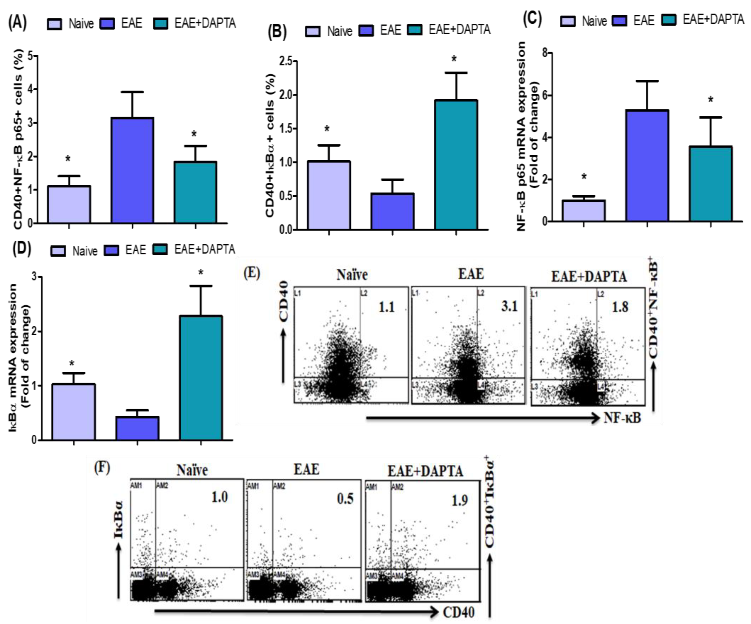
3.1. DAPTA Treatment Regulates NF-κB p65/IκBα Expression in EAE Mice

In order to investigate the effect of DAPTA on NF-κB p65- and IκBα-expressing CD40+ B cells, we performed flow cytometric analysis of splenic cells from untreated and DAPTA-treated EAE mice (Figure 1A,B) to confirm whether DAPTA affects Nf-κB p65 and IκBα mRNA expression in the brain tissue. Our results showed that NF-κB p65-expressing CD40+ B cells were significantly decreased in DAPTA-treated EAE mice, whereas the presence of IκBα-expressing CD40+ B cells increased greatly compared to the levels displayed in untreated EAE mice (Figure 1A,B). Our results showed that NF-κB p65 mRNA expression was decreased in DAPTA-treated EAE mice, whereas IκBα expression was increased compared to untreated EAE mice (Figure 1C,D). Our results demonstrated that DAPTA treatment attenuates immune imbalances, a function is likely performed by regulating NF-κB/IκBα activation.

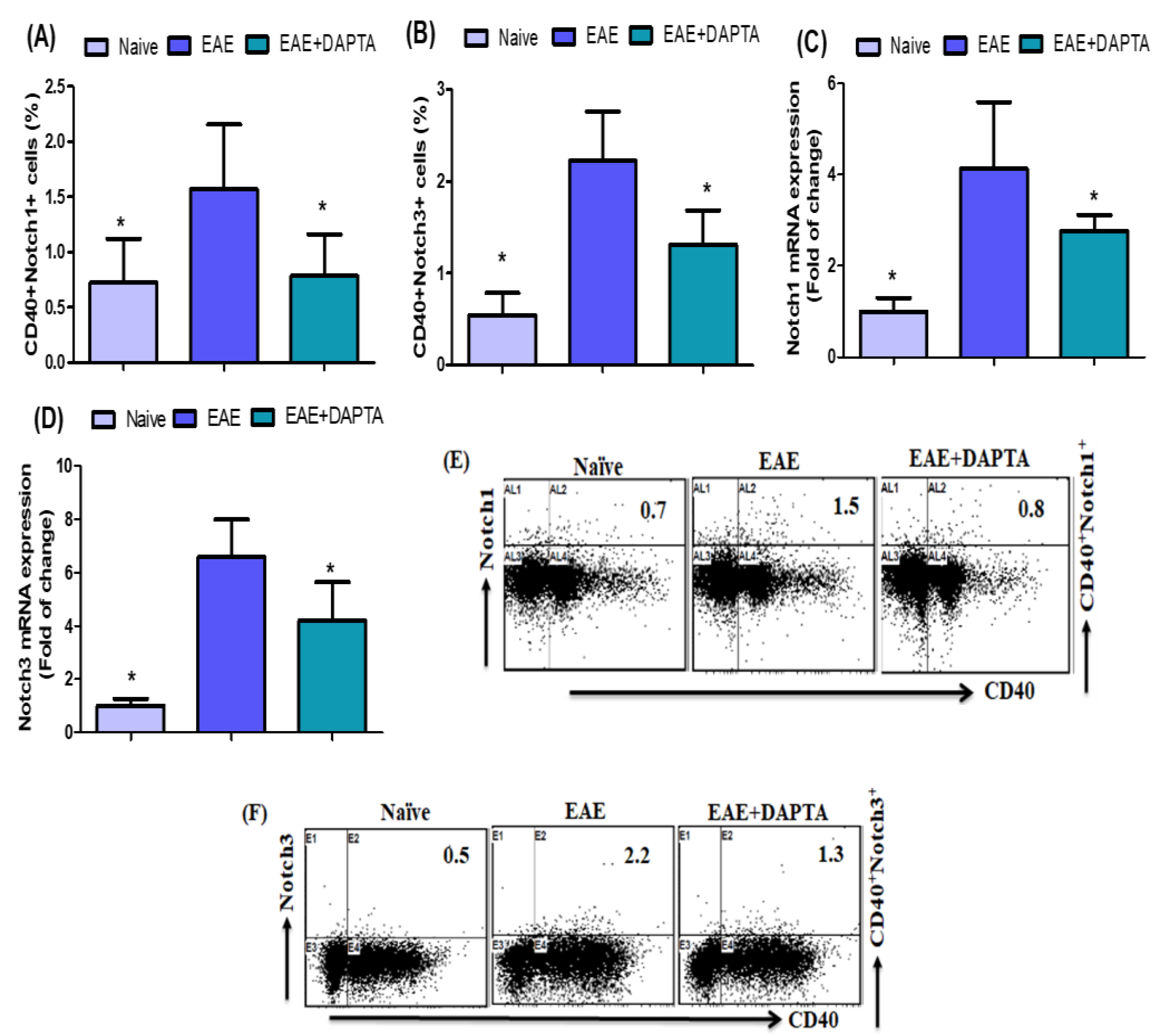
3.2. Effect of DAPTA on Notch1 and Notch3 Signaling

To further investigate the effect of DAPTA on EAE mice, flow cytometric analysis was performed to evaluate Notch1- and Notch3-expressing CD40+ B cells in the spleen. Our results revealed that EAE mice treated with DAPTA harbored fewer Notch1- and Notch3-expressing CD40+ B cells than untreated EAE mice (Figure 2A,B). We further investigated the mRNA expression levels of the potent inflammatory signaling Notch1 and Notch3 in the brain tissue. We observed a significant reduction in Notch1 and Notch3 mRNA expression levels in DAPTA-treated EAE mice compared to the levels exhibited in untreated EAE mice (Figure 2C,D). These results suggested that DAPTA administration can restore immune abnormalities during MS.

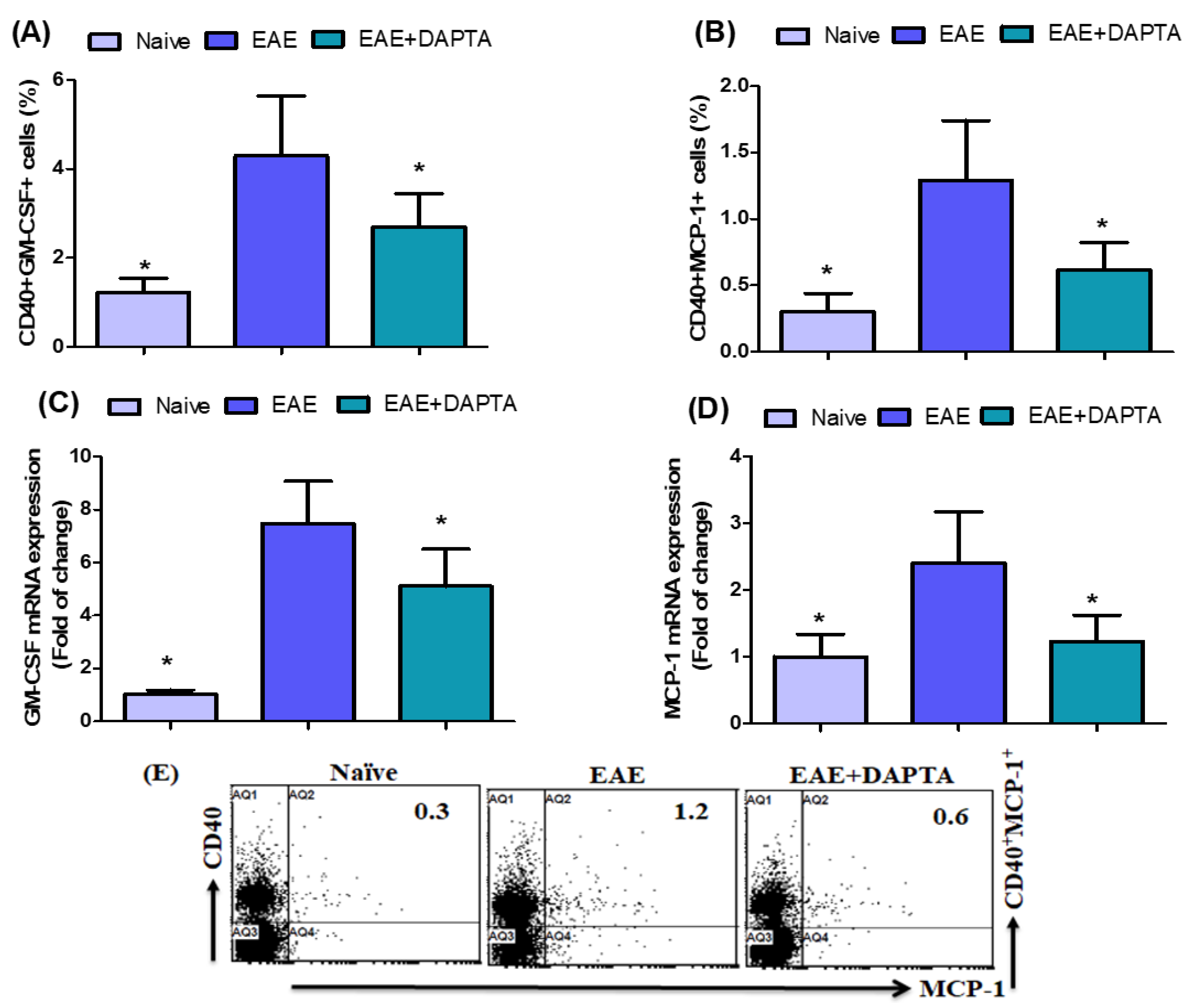
3.3. DAPTA Downregulates GM-CSF and MCP-1 Expression in EAE Mice

We further revealed the effect of DAPTA on GM-CSF- and MCP-1-expressing CD40+ B cells in the spleen. We showed that the proportion of GM-CSF- and MCP-1-expressing CD40+ B cells decreased in EAE mice treated with DAPTA compared to that visible in untreated EAE mice (Figure 3A,B). To further explain the mechanism of action of DAPTA, we examined changes in Gm-csf and Mcp-1 mRNA expression levels in brain tissue. DAPTA treatment in EAE mice caused a significant decrease in Gm-csf and Mcp-1 at the mRNA levels compared to untreated EAE mice (Figure 3C,D). These data suggest that DAPTA administration may be anti-inflammatory in the EAE mouse model.

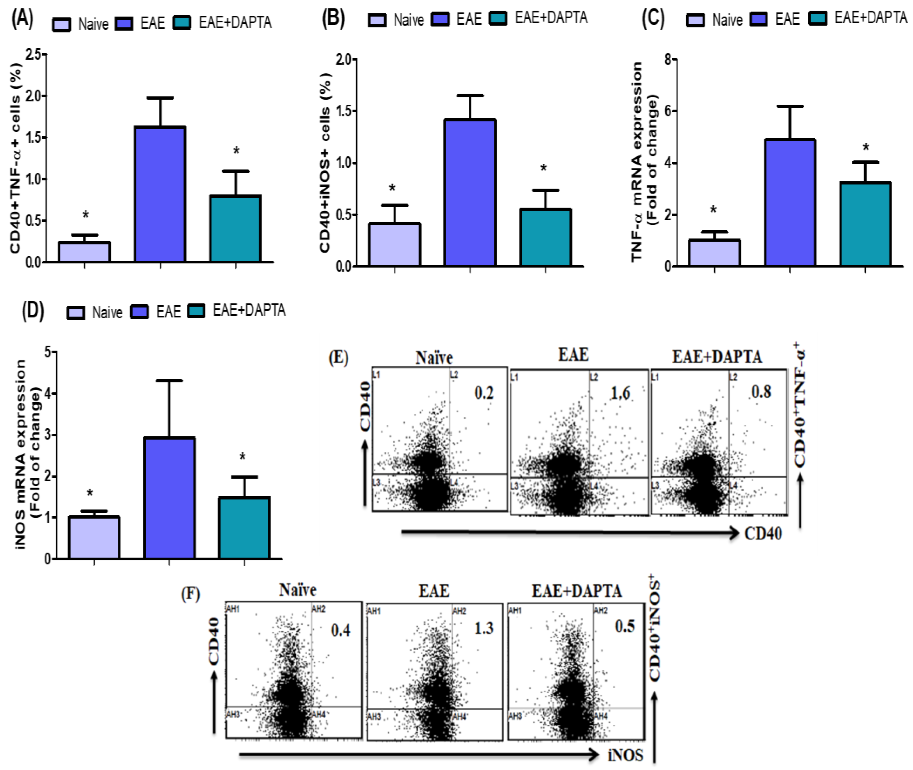
3.4. DAPTA Downregulates Proinflammatory Mediators in EAE Mice

We further evaluated the number of TNF-α- and iNOS-expressing CD40+ B cells in the spleen. We found a significant increase in TNF-α- and iNOS-expressing CD40+ B cells in untreated EAE mice. Our data indicate that DAPTA treatment in EAE mice significantly decreased TNF-α- and iNOS-expressing CD40+ B cells compared to untreated EAE mice (Figure 4A,B). We also measured the mRNA expression to confirm decreased Tnf-α and Inos mRNA levels in the brain tissue. DAPTA-treated EAE mice showed a significant reduction in Tnf-α and Inos mRNA levels compared to the EAE untreated mice (Figure 4C,D). These results prove that DAPTA of EAE has been involved in the anti-inflammatory response and suppressed the expressions of both TNF-α and iNOS.

4. Discussion

MS is a chronic autoimmune inflammatory disease of the CNS. MS is observed to result from an intricate interplay between inflammatory effector cells [50,51]. The animal model for exploring neuroinflammation is EAE, which shares significant characteristics with MS [52]. B cells play an essential role in the pathogenesis of MS. [53]. B cell repertory in the CNS and the periphery are closely associated, suggesting that disease-relevant B cell networks interact at both sides of the blood–brain barrier [54,55]. There is evidence that the B cells of MS patients exhibited aberrant proinflammatory cytokine responses [56]. In MS, CD40 is expressed in different cell types, including B cells, macrophages, endothelial cells, and resident CNS [57]. Previously, it has been demonstrated that CD40 plays a key role during neuroinflammation progression [58]. The CD40 signaling pathway has been well documented as an immune checkpoint and humoral and cellular immunity stimulator. The CD40 signaling pathway in APCs contributes to numerous cellular functions, such as Th17 cell polarization, proinflammatory cytokine release, and immunoglobulin isotype switching [59]. Furthermore, CD40 deficiency inhibits leukocyte infiltration into the CNS [60]. Therefore, this study explored the overall inflammatory potential of APCs expressing CD40 in EAE mice.
NF-κB is the major transcription factor that regulates inflammation in inflammatory diseases, including MS and EAE [14,61]. The NF-κB signaling cascade plays a vital role in the modulation of immune and inflammatory responses, which has been associated with developing autoimmune demyelinating diseases [12]. Many studies have found that NF-κB is activated in the brain tissue of patients with MS [13]. Previous studies suggested that NF-κB activation promotes oligodendrocyte survival during inflammatory insults [62]; some findings determined that NF-κB plays a critical role in the resident cells of the CNS during disease progression [63,64]. The current study aimed to evaluate the therapeutic potential and underlying mechanisms of CCR5 antagonist in an animal model of MS. We demonstrated that EAE mice, treated with DAPTA, significantly exhibited decreased CD40+NF-κB p65+ and increased CD40+IκBα+ cells in the spleen. RT-PCR was used to analyze the molecular mechanisms related to inflammation in the brain. Therefore, we found that EAE mice treated with DAPTA downregulated Nf-κB p65 and upregulated IκBα mRNA expression in brain tissue. This study showed that DAPTA has significant neuroprotective potential in EAE via modulation of NF-κB/IκBα signaling. Therefore, we supposed that DAPTA might effectively treat or improve MS.
Notch signaling plays multiple roles in health and disease [65]. Notch signaling is an essential regulator of type 2 immunity [66]. A recent report indicated that Notch signaling regulates T-cell accumulation and function in the CNS during EAE [67]. In addition, substantial evidence showed that inhibition of Notch signaling ameliorates the clinical course of EAE [17]. Existing evidence suggests that the absence of Notch expression in murine myeloid cells attenuates the development of EAE [68]. Another study also showed that the inhibition of Notch signaling attenuates the development of EAE [69]. The aims of this study were to further evaluate the cellular and molecular mechanisms of CCR5 antagonists in the EAE mouse model. In our present study, DAPTA treatment decreased Notch1- and Notch3-expressing CD40+ B cells in the spleen of EAE mice. In addition, the mRNA expression levels of Notch1 and Notch3 were significantly decreased by DAPTA treatment in EAE brain tissue, playing a crucial role in preventing MS/EAE. These results support our hypothesis that DAPTA reduces Notch1/Notch3 production/expression in EAE mice. Our data suggest that decreases in Notch1/Notch3 signaling in EAE mice could be one of the mechanisms by which DAPTA attenuates MS progression.
The critical role of GM-CSF in EAE and MS was recently reported [70]. The expression of GM-CSF is significantly increased in MS [71]. A previous study has shown that GM-CSF is critical in MS pathogenesis and EAE development since GM-CSF-deficient mice resist EAE induction [72]. GM-CSF signaling was shown to be sufficient to initiate EAE and was produced by myeloid cells infiltrating the CNS sustained neuroinflammation [73]. GM-CSF promotes chronic disability in EAE patients by altering the composition of CNS-infiltrating cells [22]. MCP-1 expression correlated with clinical severity and attacks in relapsing EAE [74]. MCP-1 expression was followed by further infiltration of monocytes into the CNS and the onset of clinical signs [75]. Infiltrating neutrophils, macrophages, and CD8 T cells can produce MCP1 in EAE [76]. CCL2 expression was upregulated in gamma interferon knockout (GKO) mice with EAE [77]. The use of anti-MCP1 using DNA vaccination or antibodies during relapsing EAE reduced the severity of the disease [74,75,76,77,78]. We further explored the therapeutic role of the CCR5 antagonist DAPTA in EAE mice. In this study, we showed that GM-CSF- and MCP-1-expressing CD40+ B cells were significantly decreased in mice with EAE by the treatment of DAPTA. We further observed that Gm-csf and Mcp-1 mRNA expression levels were considerably reduced in DAPTA-treated mice with EAE. DAPTA may represent a new target for inhibiting inflammatory processes in MS development. Therefore, these results support our hypotheses that a CCR5 antagonist could be a potential therapeutic target for treating neuroimmune disorders, including MS.
iNOS is expressed in microglial cells, astrocytes, and neurons in the CNS [79]. It has been shown that there are increased levels of iNOS protein in the CNS in MS [80]. A previous study also observed that iNOS expression was raised in the EAE mice brain [81]. A clinical association between iNOS and pathogenesis has been reported in MS and EAE [82,83]. During chronic demyelination, a pathogenic phenotype of microglial cells is associated with iNOS expression [84]. A previous study showed TNF-α expression by resident microglia and infiltrating leukocytes in the CNS of mice with EAE [85]. In the present study, it has been revealed that EAE mice treated with DAPTA downregulate proinflammatory mediators, namely, iNOS- and TNF-α-expressing CD40+ B cells. DAPTA treatment inEAE mice also caused a reduction in the Inos and Tnf-α mRNA expression levels. This study has demonstrated that DAPTA treatment may downregulate the expression of proinflammatory mediators in the spleen and brain of the EAE mice, eventually attenuating EAE development. These observations suggested that DAPTA may potentially alleviate MS. Therefore, these findings may lead to the development of a new drug for treating neurodegenerative diseases, including MS. Further research is warranted to explore the complex molecular pathways of DAPTA in the EAE model.

5. Conclusions

Our results demonstrate the critical role played by inflammatory mediators and NF-κB/Notch signaling in RR-EAE and provide a novel therapeutic approach. CCR5 antagonist leads to a significant reduction in NF-κB/Notch signaling in EAE mice. Additionally, CCR5 antagonist reduces inflammatory mediators such as GM-CSF/iNOS and MCP-1/TNF-α in EAE; this points to the potential benefits of CCR5 antagonist as a novel therapeutic target for the treatment of MS. Therefore, these findings provide additional evidence that DAPTA has possible effects on the treatment of MS and expand our knowledge of the mechanism of action of DAPTA in the EAE model.

Author Contributions

Conceptualization, H.A., M.A.A. (Mushtaq A. Ansari), A.N., M.A.A. (Majed Ali Algonaiah), S.M.A., S.A.B., T.H.A., S.A., M.S., A.S.A., M.Y.A., Y.A.A. and S.F.A.; Data curation, T.H.A., S.A., M.S., A.S.A., M.Y.A., Y.A.A. and S.F.A.; Formal analysis, M.A.A. (Mushtaq A. Ansari), M.A.A. (Majed Ali Algonaiah), S.M.A., T.H.A., S.A., M.S., A.S.A., M.Y.A., Y.A.A. and S.F.A.; Funding acquisition, A.N. and S.F.A.; Investigation, H.A., M.A.A. (Mushtaq A. Ansari), A.N., M.A.A. (Majed Ali Algonaiah), S.M.A., S.A.B. and S.F.A.; Methodology, H.A., M.A.A. (Mushtaq A. Ansari), A.N., M.A.A. (Majed Ali Algonaiah), S.M.A., S.A.B., T.H.A., S.A., M.S., A.S.A., M.Y.A., Y.A.A. and S.F.A.; Project administration, S.F.A.; Resources, T.H.A., S.A., M.S., A.S.A., M.Y.A., Y.A.A. and S.F.A.; Software, M.A.A. (Majed Ali Algonaiah), T.H.A., S.A., M.S., A.S.A., M.Y.A., Y.A.A. and S.F.A.; Supervision, H.A., A.N., S.M.A. and S.F.A.; Validation, A.N., T.H.A., S.A., M.S., A.S.A., M.Y.A., Y.A.A. and S.F.A.; Visualization, S.A.B. and S.F.A.; Writing—original draft, H.A., M.A.A. (Mushtaq A. Ansari), A.N., M.A.A. (Majed Ali Algonaiah), S.M.A., S.A.B. and S.F.A.; Writing—review and editing, H.A., M.A.A. (Mushtaq A. Ansari), A.N., S.M.A., S.A.B. and S.F.A. All authors have read and agreed to the published version of the manuscript.

Funding

The authors extend their appreciation to the Deputyship for Research & Innovation, Ministry of Education in Saudi Arabia for funding this research work through the project no. (IFKSUOR3-065-1).

Institutional Review Board Statement

The animal study protocol was approved by the Ethical Committee of the College of Pharmacy, King Saud University (Ethical Reference No: KSU-SE-21-50).

Data Availability Statement

All data presented in this study are available on reasonable request from the corresponding author.

Acknowledgments

The authors extend their appreciation to the Deputyship for Research & Innovation, Ministry of Education in Saudi Arabia for funding this research work through the project no. (IFKSUOR3-065-1).

Conflicts of Interest

The authors declare no conflict of interest.

Abbreviations

Multiple sclerosis (MS); experimental autoimmune encephalomyelitis (EAE); C-C chemokine receptor 5 (CCR5); central nervous system (CNS); inducible nitric oxide synthase (iNOS); D-Ala-peptide T-amide (DAPTA); proteolipid protein (PLP); complementary DNA (cDNA); nuclear factor kappa-light-chain-enhancer of activated B cells (NF-κB); granulocyte-macrophage colony-stimulating factor (GM-CSF); tumor necrosis factor alpha (TNF-α); monocyte chemoattractant protein-1 (MCP-1); intraperitoneally (i.p.); phycoerythrin (PE); allophycocyanin (APC); fluoroisothiocyanate (FITC); glyceraldehyde 3-phosphate dehydrogenase (GAPDH); messenger RNA (mRNA); reverse transcription polymerase chain reaction (RT-PCR). Roswell Park Memorial Institute (RPMI) 1640; Proteolipid protein (PLP).

References

  1. Lassmann, H.; Bruck, W.; Lucchinetti, C.F. The immunopathology of multiple sclerosis: An overview. Brain Pathol. 2007, 17, 210–218. [Google Scholar] [CrossRef] [PubMed]
  2. Milo, R.; Kahana, E. Multiple sclerosis: Geoepidemiology, genetics, and the environment. Autoimmun. Rev. 2010, 9, A387–A394. [Google Scholar] [CrossRef] [PubMed]
  3. Gu, C. KIR4.1: K+ Channel Illusion or Reality in the Autoimmune Pathogenesis of Multiple Sclerosis. Front. Mol. Neurosci. 2016, 9, 90. [Google Scholar] [CrossRef] [PubMed]
  4. Zhornitsky, S.; Johnson, T.A.; Metz, L.M.; Weiss, S.; Yong, V.W. Prolactin in combination with interferon-β reduces disease severity in an animal model of multiple sclerosis. J. Neuroinflammation 2015, 12, 55. [Google Scholar] [CrossRef]
  5. Constantinescu, C.S.; Farooqi, N.; O’Brien, K.; Gran, B. Experimental autoimmune encephalomyelitis (EAE) as a model for multiple sclerosis (MS). Br. J. Pharmacol. 2011, 164, 1079–1106. [Google Scholar] [CrossRef]
  6. Handel, A.E.; Williamson, A.J.; Disanto, G.; Handunnetthi, L.; Giovannoni, G.; Ramagopalan, S.V. An updated meta-analysis of risk of multiple sclerosis following infectious mononucleosis. PLoS ONE 2010, 5, e12496. [Google Scholar] [CrossRef]
  7. Cepok, S.; von Geldern, G.; Grummel, V.; Hochgesand, S.; Celik, H.; Hartung, H.; Hemmer, B. Accumulation of class switched IgD-IgM-memory B cells in the cerebrospinal fluid during neuroinflammation. J. Neuroimmunol. 2006, 180, 33–39. [Google Scholar] [CrossRef]
  8. von Budingen, H.C.; Harrer, M.D.; Kuenzle, S.; Meier, M.; Goebels, N. Clonally expanded plasma cells in the cerebrospinal fluid of MS patients produce myelin-specific antibodies. Eur. J. Immunol. 2008, 38, 2014–2023. [Google Scholar] [CrossRef]
  9. Hartung, D.M.; Bourdette, D.N.; Ahmed, S.M.; Whitham, R.H. The cost of multiple sclerosis drugs in the US and the pharmaceutical industry: Too big to fail? Neurology 2015, 84, 2185–2192. [Google Scholar] [CrossRef]
  10. Baird, J.F.; Sandroff, B.M.; Motl, R.W. Therapies for mobility disability in persons with multiple sclerosis. Expert Rev. Neurother. 2018, 18, 493–502. [Google Scholar] [CrossRef]
  11. Tur, C.; Carbonell-Mirabent, P.; Cobo-Calvo, Á.; Otero-Romero, S.; Arrambide, G.; Midaglia, L.; Castilló, J.; Vidal-Jordana, Á.; Rodríguez-Acevedo, B.; Zabalza, A.; et al. Association of Early Progression Independent of Relapse Activity With Long-term Disability After a First Demyelinating Event in Multiple Sclerosis. JAMA Neurol. 2023, 80, 151–160. [Google Scholar] [CrossRef] [PubMed]
  12. Mc Guire, C.; Prinz, M.; Beyaert, R.; van Loo, G. Nuclear factor kappa B (NF-κB) in multiple sclerosis pathology. Trends Mol. Med. 2013, 19, 604–613. [Google Scholar] [CrossRef] [PubMed]
  13. Bonetti, B.; Stegagno, C.; Cannella, B.; Rizzuto, N.; Moretto, G.; Raine, C.S. Activation of NF-kappaB and c-jun transcription factors in multiple sclerosis lesions. Implications for oligodendrocyte pathology. Am. J. Pathol. 1999, 155, 1433–1438. [Google Scholar] [CrossRef] [PubMed]
  14. Yue, Y.; Stone, S.; Lin, W. Role of nuclear factor κB in multiple sclerosis and experimental autoimmune encephalomyelitis. Neural. Regen. Res. 2018, 13, 1507–1515. [Google Scholar]
  15. Chen, D.; Ireland, S.J.; Remington, G.; Alvarez, E.; Racke, M.K.; Greenberg, B.; Frohman, E.M.; Monson, N.L. CD40-Mediated NF-κB Activation in B Cells Is Increased in Multiple Sclerosis and Modulated by Therapeutics. J. Immunol. 2016, 197, 4257–4265. [Google Scholar] [CrossRef]
  16. Jurynczyk, M.; Jurewicz, A.; Bielecki, B.; Raine, C.S.; Selmaj, K. Inhibition of Notch signaling enhances tissue repair in an animal model of multiple sclerosis. J. Neuroimmunol. 2005, 170, 3–10. [Google Scholar] [CrossRef]
  17. Jurynczyk, M.; Jurewicz, A.; Raine, C.S.; Selmaj, K. Notch3 inhibition in myelin-reactive T cells down-regulates protein kinase Cθ and attenuates experimental autoimmune encephalomyelitis. J. Immunol. 2008, 180, 2634–2640. [Google Scholar] [CrossRef]
  18. Grandbarbe, L.; Michelucci, A.; Heurtaux, T.; Hemmer, K.; Morga, E.; Heuschling, P. Notch signaling modulates the activation of microglial cells. Glia 2007, 55, 1519–1530. [Google Scholar] [CrossRef]
  19. El-Behi, M.; Ciric, B.; Dai, H.; Yan, Y.; Cullimore, M.; Safavi, F.; Zhang, G.X.; Dittel, B.N.; Rostami, A. The encephalitogenicity of T(H)17 cells is dependent on IL-1- and IL-23-induced production of the cytokine GM-CSF. Nat. Immunol. 2011, 12, 568–755. [Google Scholar] [CrossRef]
  20. Ifergan, I.; Davidson, T.S.; Kebir, H.; Xu, D.; Palacios-Macapagal, D.; Cann, J.; Rodgers, J.M.; Hunter, Z.N.; Pittet, C.L.; Beddow, S.; et al. Targeting the GM-CSF receptor for the treatment of CNS autoimmunity. J. Autoimmun. 2017, 84, 1–11. [Google Scholar] [CrossRef]
  21. Ponomarev, E.D.; Shriver, L.P.; Maresz, K.; Pedras-Vasconcelos, J.; Verthelyi, D.; Dittel, B.N. GM-CSF production by autoreactive T cells is required for the activation of microglial cells and the onset of experimental autoimmune encephalomyelitis. J. Immunol. 2007, 178, 39–48. [Google Scholar] [CrossRef] [PubMed]
  22. Duncker, P.C.; Stoolman, J.S.; Huber, A.K.; Segal, B.M. GM-CSF Promotes Chronic Disability in Experimental Autoimmune Encephalomyelitis by Altering the Composition of Central Nervous System-Infiltrating Cells, but Is Dispensable for Disease Induction. J. Immunol. 2018, 200, 966–973. [Google Scholar] [CrossRef] [PubMed]
  23. Simpson, J.E.; Newcombe, J.; Cuzner, M.L.; Woodroofe, M.N. Expression of monocyte chemoattractant protein-1 and other beta-chemokines by resident glia and inflammatory cells in multiple sclerosis lesions. J. Neuroimmunol. 1998, 84, 238–249. [Google Scholar] [CrossRef] [PubMed]
  24. Van Der Voorn, P.; Tekstra, J.; Beelen, R.H.; Tensen, C.P.; Van Der Valk, P.; De Groot, C.J. Expression of MCP-1 by reactive astrocytes in demyelinating multiple sclerosis lesions. Am. J. Pathol. 1999, 154, 45–51. [Google Scholar] [CrossRef]
  25. Huang, D.R.; Wang, J.; Kivisakk, P.; Rollins, B.J.; Ransohoff, R.M. Absence of monocyte chemoattractant protein 1 in mice leads to decreased local macrophage recruitment and antigen-specific T helper cell type 1 immune response in experimental autoimmune encephalomyelitis. J. Exp. Med. 2001, 193, 713–726. [Google Scholar] [CrossRef]
  26. McManus, C.; Berman, J.W.; Brett, F.M.; Staunton, H.; Farrell, M.; Brosnan, C.F. MCP-1, MCP-2 and MCP-3 expression in multiple sclerosis lesions: An immunohistochemical and in situ hybridization study. J. Neuroimmunol. 1998, 86, 20–29. [Google Scholar] [CrossRef]
  27. Alam, M.S.; Gaida, M.M.; Bergmann, F.; Lasitschka, F.; Giese, T.; Giese, N.A.; Hackert, T.; Hinz, U.; Hussain, S.P.; Kozlov, S.V.; et al. Selective inhibition of the p38 alternative activation pathway in infiltrating T cells inhibits pancreatic cancer progression. Nat. Med. 2015, 21, 1337–1343. [Google Scholar] [CrossRef]
  28. Bałkowiec-Iskra, E.; Vermehren-Schmaedick, A.; Balkowiec, A. Tumor necrosis factor-α increases brain-derived neurotrophic factor expression in trigeminal ganglion neurons in an activity-dependent manner. Neuroscience 2011, 180, 322–333. [Google Scholar] [CrossRef]
  29. Sharief, M.K.; Hentges, R. Association between tumor necrosis factor-alpha and disease progression in patients with multiple sclerosis. N. Engl. J. Med. 1991, 325, 467–472. [Google Scholar] [CrossRef]
  30. Fresegna, D.; Bullitta, S.; Musella, A.; Rizzo, F.R.; De Vito, F.; Guadalupi, L.; Caioli, S.; Balletta, S.; Sanna, K.; Dolcetti, E.; et al. Re-examining the role of TNF in MS pathogenesis and therapy. Cells 2020, 9, 2290. [Google Scholar] [CrossRef]
  31. Jung, K.; Kim, J.; Ahn, G.; Matsuda, H.; Akane, T.; Ahn, M.; Shin, T. Alendronate alleviates the symptoms of experimental autoimmune encephalomyelitis. Int. Immunopharmacol. 2020, 84, 106534. [Google Scholar] [CrossRef] [PubMed]
  32. Shin, T.; Kim, S.; Moon, C.; Wie, M.; Kim, H. Aminoguanidine-induced amelioration of autoimmune encephalomyelitis is mediated by reduced expression of inducible nitric oxide synthase in the spinal cord. Immunol. Investig. 2000, 29, 233–241. [Google Scholar] [CrossRef] [PubMed]
  33. Moon, C.; Ahn, M.; Jee, Y.; Heo, S.; Kim, S.; Kim, H.; Sim, K.B.; Koh, C.S.; Shin, Y.G.; Shin, T. Sodium salicylate-induced amelioration of experimental autoimmune encephalomyelitis in Lewis rats is associated with the suppression of inducible nitric oxide synthase and cyclooxygenases. Neurosci. Lett. 2004, 356, 123–126. [Google Scholar] [CrossRef]
  34. Banisadr, G.; Gosselin, R.D.; Mechighel, P.; Kitabgi, P.; Rostene, W.; Parsadaniantz, S.M. Highly regionalized neuronal expression of monocyte chemoattractant protein-1 (MCP-1/CCL2) in rat brain: Evidence for its colocalization with neurotransmitters and neuropeptides. J. Comp. Neurol. 2005, 489, 275–292. [Google Scholar] [CrossRef] [PubMed]
  35. Godiska, R.; Chantry, D.; Dietsch, G.N.; Gray, P.W. Chemokine expression in murine experimental allergic encephalomyelitis. J. Neuroimmunol. 1995, 58, 167–176. [Google Scholar] [CrossRef]
  36. Muller, D.M.; Pender, M.P.; Greer, J.M. Chemokines and chemokine receptors: Potential therapeutic targets in multiple sclerosis. Curr. Drug Targets Inflamm. Allergy 2004, 3, 279–290. [Google Scholar] [CrossRef]
  37. Gu, S.M.; Park, M.H.; Yun, H.M.; Han, S.B.; Oh, K.W.; Son, D.J.; Yun, J.S.; Hong, J.T. CCR5 knockout suppresses experimental autoimmune encephalomyelitis in C57BL/6 mice. Oncotarget 2016, 7, 15382–15393. [Google Scholar] [CrossRef]
  38. dos Santos, A.C.; Barsante, M.M.; Arantes, R.M.E.; Bernard, C.C.; Teixeira, M.M.; Carvalho-Tavares, J. CCL2 and CCL5 mediate leukocyte adhesion in experimental autoimmune encephalomyelitis–an intravital microscopy study. J. Neuroimmunol. 2005, 162, 122–129. [Google Scholar] [CrossRef]
  39. Balashov, K.E.; Rottman, J.B.; Weiner, H.L.; Hancock, W.W. CCR5(+) and CXCR3(+) T cells are increased in multiple sclerosis and their ligands MIP-1alpha and IP-10 are expressed in demyelinating brain lesions. Proc. Natl. Acad. Sci. USA 1999, 96, 6873–6878. [Google Scholar] [CrossRef]
  40. Fischer, F.R.; Santambrogio, L.; Luo, Y.; Berman, M.A.; Hancock, W.W.; Dorf, M.E. Modulation of experimental autoimmune encephalomyelitis: Effect of altered peptide ligand on chemokine and chemokine receptor expression. J. Neuroimmunol. 2000, 110, 195–208. [Google Scholar] [CrossRef]
  41. Moriguchi, K.; Miyamoto, K.; Tanaka, N.; Ueno, R.; Nakayama, T.; Yoshie, O.; Kusunoki, S. C-C chemokine receptor type 4 antagonist Compound ameliorates experimental autoimmune encephalomyelitis. J. Neuroimmunol. 2016, 291, 54–58. [Google Scholar] [CrossRef]
  42. Redwine, L.S.; Pert, C.B.; Rone, J.D.; Nixon, R.; Vance, M.; Sandler, B.; Lumpkin, M.D.; Dieter, D.J.; Ruff, M.R. Peptide T blocks GP120/CCR5 chemokine receptor-mediated chemotaxis. Clin. Immunol. 1999, 93, 124–131. [Google Scholar] [CrossRef]
  43. Rosi, S.; Pert, C.B.; Ruff, M.R.; McGann-Gramling, K.; Wenk, G.L. Chemokine receptor 5 antagonist D-Ala-peptide T-amide reduces microglia and astrocyte activation within the hippocampus in a neuroinflammatory rat model of Alzheimer’s disease. Neuroscience 2005, 134, 671–676. [Google Scholar] [CrossRef] [PubMed]
  44. Lee, Y.K.; Choi, D.Y.; Jung, Y.Y.; Yun, Y.W.; Lee, B.J.; Han, S.B.; Hong, J.T. Decreased pain responses of C-C chemokine receptor 5 knockout mice to chemical or inflammatory stimuli. Neuropharmacology 2013, 67, 57–65. [Google Scholar] [CrossRef] [PubMed]
  45. Ahmad, S.F.; Ansari, M.A.; Nadeem, A.; Bakheet, S.A.; Alotaibi, M.R.; Alasmari, A.F.; Alshammari, M.A.; Al-Mazroua, H.A.; Attia, S.M. DAPTA, a C-C chemokine receptor 5 (CCR5) antagonist attenuates immune aberrations by downregulating Th9/Th17 immune responses in BTBR T+ Itpr3tf/J mice. Eur. J. Pharmacol. 2019, 846, 100–108. [Google Scholar] [CrossRef]
  46. Ahmad, S.F.; Nadeem, A.; Ansari, M.A.; Bakheet, S.A.; Shahid, M.; Al-Mazroua, H.A.; Sobeai, H.M.A.; Alasmari, A.F.; Alanazi, M.M.; Alhamed, A.S.; et al. CC chemokine receptor 5 antagonist alleviates inflammation by regulating IFN-γ/IL-10 and STAT4/Smad3 signaling in a mouse model of autoimmune encephalomyelitis. Cell Immunol. 2022, 379, 104580. [Google Scholar] [CrossRef] [PubMed]
  47. Furlan, R.; Cuomo, C.; Martino, G. Animal models of multiple sclerosis. Methods Mol. Biol. 2009, 549, 157–173. [Google Scholar]
  48. Jolivel, V.; Luessi, F.; Masri, J.; Kraus, S.H.; Hubo, M.; Poisa-Beiro, L.; Klebow, S.; Paterka, M.; Yogev, N.; Tumani, H.; et al. Modulation of dendritic cell properties by laquinimod as a mechanism for modulating multiple sclerosis. Brain 2013, 136 Pt 4, 1048–1066. [Google Scholar] [CrossRef]
  49. Schmittgen, T.D.; Livak, K.J. Analyzing real-time PCR data by the comparative CT method. Nat. Protoc. 2008, 3, 1101–1108. [Google Scholar] [CrossRef]
  50. Baecher-Allan, C.; Kaskow, B.J.; Weiner, H.L. Multiple sclerosis: Mechanisms and immunotherapy. Neuron 2018, 97, 742–768. [Google Scholar] [CrossRef]
  51. Hohlfeld, R.; Dornmair, K.; Meinl, E.; Wekerle, H. The search for the target antigens of multiple sclerosis, part 2: CD8+ T cells, B cells, and antibodies in the focus of reverse-translational research. Lancet Neurol. 2016, 15, 317–331. [Google Scholar] [CrossRef]
  52. Barclay, W.; Shinohara, M.L. Inflammasome activation in multiple sclerosis and experimental autoimmune encephalomyelitis (EAE). Brain Pathol. 2017, 27, 213–219. [Google Scholar] [CrossRef] [PubMed]
  53. Farh, K.K.; Marson, A.; Zhu, J.; Kleinewietfeld, M.; Housley, W.J.; Beik, S.; Shoresh, N.; Whitton, H.; Ryan, R.J.; Shishkin, A.A.; et al. Genetic and epigenetic fine mapping of causal autoimmune disease variants. Nature 2015, 518, 337–343. [Google Scholar] [CrossRef] [PubMed]
  54. Eggers, E.L.; Michel, B.A.; Wu, H.; Wang, S.Z.; Bevan, C.J.; Abounasr, A.; Pierson, N.S.; Bischof, A.; Kazer, M.; Leitner, E.; et al. Clonal relationships of CSF B cells in treatment-naive multiple sclerosis patients. JCI Insight 2017, 2, e92724. [Google Scholar] [CrossRef]
  55. Greenfield, A.L.; Dandekar, R.; Ramesh, A.; Eggers, E.L.; Wu, H.; Laurent, S.; Harkin, W.; Pierson, N.S.; Weber, M.S.; Henry, R.G.; et al. Longitudinally persistent cerebrospinal fluid B cells can resist treatment in multiple sclerosis. JCI Insight 2019, 4. [Google Scholar] [CrossRef]
  56. Bar-Or, A.; Fawaz, L.; Fan, B.; Darlington, P.J.; Rieger, A.; Ghorayeb, C.; Calabresi, P.A.; Waubant, E.; Hauser, S.L.; Zhang, J.; et al. Abnormal B-cell cytokine responses: A trigger of T-cell–mediated disease in MS? Ann. Neurol. 2010, 67, 452–461. [Google Scholar] [CrossRef] [PubMed]
  57. Aarts, S.A.B.M.; Seijkens, T.T.P.; van Dorst, K.J.F.; Dijkstra, C.D.; Kooij, G.; Lutgens, E. The CD40-CD40L dyad in experimental autoimmune encephalomyelitis and multiple sclerosis. Front. Immunol. 2017, 8, 1791. [Google Scholar] [CrossRef]
  58. Aarts, S.A.; Seijkens, T.T.; Kusters, P.J.; van Tiel, C.M.; Reiche, M.E.; den Toom, M.; Beckers, L.; van Roomen, C.P.; de Winther, M.P.; Kooij, G.; et al. Macrophage CD40 signaling drives experimental autoimmune encephalomyelitis. J. Pathol. 2019, 247, 471–480. [Google Scholar] [CrossRef]
  59. Iezzi, G.; Sonderegger, I.; Ampenberger, F.; Schmitz, N.; Marsland, B.J.; Kopf, M. CD40-CD40L cross-talk integrates strong antigenic signals and microbial stimuli to induce development of IL-17-producing CD4+ T cells. Proc. Natl. Acad. Sci. USA 2009, 106, 876–881. [Google Scholar] [CrossRef]
  60. Becher, B.; Durell, B.G.; Miga, A.V.; Hickey, W.F.; Noelle, R.J. The clinical course of experimental autoimmune encephalomyelitis and inflammation is controlled by the expression of CD40 within the central nervous system. J. Exp. Med. 2001, 193, 967–974. [Google Scholar] [CrossRef]
  61. Herrington, F.D.; Carmody, R.J.; Goodyear, C.S. Modulation of NF-kB signaling as a therapeutic target in autoimmunity. J. Biomol. Screen. 2016, 21, 223–242. [Google Scholar] [CrossRef] [PubMed]
  62. Lin, W.; Lin, Y.; Li, J.; Fenstermaker, A.G.; Way, S.W.; Clayton, B.; Jamison, S.; Harding, H.P.; Ron, D.; Popko, B. Oligodendrocyte-specific activation of PERK signaling protects mice against experimental autoimmune encephalomyelitis. J. Neurosci. 2013, 33, 5980–5991. [Google Scholar] [CrossRef]
  63. Leibowitz, S.M.; Yan, J. NF-κB Pathways in the Pathogenesis of Multiple Sclerosis and the Therapeutic Implications. Front. Mol. Neurosci. 2016, 9, 84. [Google Scholar] [CrossRef] [PubMed]
  64. Yan, J.; Greer, J.M.; NF-kappa, B. A potential therapeutic target for the treatment of multiple sclerosis. CNS Neurol. Disord. Drug Targets 2008, 7, 536–557. [Google Scholar] [CrossRef]
  65. Kopan, R.; Ilagan, M.X. The canonical Notch signaling pathway: Unfolding the activation mechanism. Cell 2009, 137, 216–233. [Google Scholar] [CrossRef] [PubMed]
  66. Tu, L.; Fang, T.C.; Artis, D.; Shestova, O.; Pross, S.E.; Maillard, I.; Pear, W.S. Notch signaling is an important regulator of type 2 immunity. J. Exp. Med. 2005, 202, 1037–1042. [Google Scholar] [CrossRef]
  67. Sandy, A.R.; Stoolman, J.; Malott, K.; Pongtornpipat, P.; Segal, B.M.; Maillard, I. Notch signaling regulates T cell accumulation and function in the central nervous system during experimental autoimmune encephalomyelitis. J. Immunol. 2013, 191, 1606–1613. [Google Scholar] [CrossRef]
  68. Fernández, M.; Monsalve, E.M.; López-López, S.; Ruiz-García, A.; Mellado, S.; Caminos, E.; García-Ramírez, J.J.; Laborda, J.; Tranque, P.; Díaz-Guerra, M.J.M. Absence of Notch1 in murine myeloid cells attenuates the development of experimental autoimmune encephalomyelitis by affecting Th1 and Th17 priming. Eur. J. Immunol. 2017, 47, 2090–2100. [Google Scholar] [CrossRef]
  69. Dishowitz, M.I.; Mutyaba, P.L.; Takacs, J.D.; Barr, A.M.; Engiles, J.B.; Ahn, J.; Hankenson, K.D. Systemic inhibition of canonical Notch signaling results in sustained callus inflammation and alters multiple phases of fracture healing. PLoS ONE 2013, 8, e68726. [Google Scholar] [CrossRef]
  70. Uyttenhove, C.; Gaignage, M.; Donckers, D.; Nasr, Z.; Cheou, P.; van Snick, J.; D’Auria, L.; van Pesch, V. Prophylactic treatment against GM-CSF, but not IL-17, abolishes relapses in a chronic murine model of multiple sclerosis. Eur. J. Immunol. 2018, 48, 1883–1891. [Google Scholar] [CrossRef]
  71. Rasouli, J.; Ciric, B.; Imitola, J.; Gonnella, P.; Hwang, D.; Mahajan, K.; Mari, E.R.; Safavi, F.; Leist, T.P.; Zhang, G.X.; et al. Expression of GM-CSF in T Cells Is Increased in Multiple Sclerosis and Suppressed by IFN-beta Therapy. J. Immunol. 2015, 194, 5085–5093. [Google Scholar] [CrossRef]
  72. McQualter, J.L.; Darwiche, R.; Ewing, C.; Onuki, M.; Kay, T.W.; Hamilton, J.A.; Reid, H.H.; Bernard, C.C. Granulocyte macrophage colony-stimulating factor: A new putative therapeutic target in multiple sclerosis. J. Exp. Med. 2001, 194, 873–882. [Google Scholar] [CrossRef]
  73. Codarri, L.; Gyulveszi, G.; Tosevski, V.; Hesske, L.; Fontana, A.; Magnenat, L.; Suter, T.; Becher, B. RORgammat drives production of the cytokine GM-CSF in helper T cells, which is essential for the effector phase of autoimmune neuroinflammation. Nat. Immunol. 2011, 12, 560–567. [Google Scholar] [CrossRef] [PubMed]
  74. Kennedy, K.J.; Strieter, R.M.; Kunkel, S.L.; Lukacs, N.W.; Karpus, W.J. Acute and relapsing experimental autoimmune encephalomyelitis are regulated by differential expression of the CC chemokines macrophage inflammatory protein-1alpha and monocyte chemotactic protein-1. J. Neuroimmunol. 1998, 92, 98–108. [Google Scholar] [CrossRef] [PubMed]
  75. Berman, J.W.; Guida, M.P.; Warren, J.; Amat, J.; Brosnan, C.F. Localization of monocyte chemoattractant peptide-1 expression in the central nervous system in experimental autoimmune encephalomyelitis and trauma in the rat. J. Immunol. 1996, 156, 3017–3023. [Google Scholar] [CrossRef]
  76. Nygårdas, P.T.; Määttä, J.A.; Hinkkanen, A.E. Chemokine expression by central nervous system resident cells and infiltrating neutrophils during experimental autoimmune encephalomyelitis in the BALB/c mouse. Eur. J. Immunol. 2000, 30, 1911–1918. [Google Scholar] [CrossRef]
  77. Glabinski, A.R.; Ransohoff, R.M. Chemokines and chemokine receptors in CNS pathology. J. Neurovirol. 1999, 5, 3–12. [Google Scholar] [CrossRef]
  78. Youssef, S.; Wildbaum, G.; Karin, N. Prevention of experimental autoimmune encephalomyelitis by MIP-1alpha and MCP-1 naked DNA vaccines. J. Autoimmun. 1999, 13, 21–29. [Google Scholar] [CrossRef] [PubMed]
  79. Sheng, W.; Zong, Y.; Mohammad, A.; Ajit, D.; Cui, J.; Han, D.; Hamilton, J.L.; Simonyi, A.; Sun, A.Y.; Gu, Z.; et al. Pro-inflammatory cytokines and lipopolysaccharide induce changes in cell morphology, and upregulation of ERK1/2, iNOS and sPLA(2)-IIA expression in astrocytes and microglia. J. Neuroinf. 2011, 8, 121. [Google Scholar] [CrossRef]
  80. Bagasra, O.; Michaels, F.H.; Zheng, Y.M.; Bobroski, L.E.; Spitsin, S.V.; Fu, Z.F.; Tawadros, R.; Koprowski, H. Activation of the inducible form of nitric oxide synthase in the brains of patients with multiple sclerosis. Proc. Natl. Acad. Sci. USA 1995, 92, 12041–12045. [Google Scholar] [CrossRef] [PubMed]
  81. He, Y.; Du, M.; Gao, Y.; Liu, H.; Wang, H.; Wu, X.; Wang, Z. Astragaloside IV Attenuates Experimental Autoimmune Encephalomyelitis of Mice by Counteracting Oxidative Stress at Multiple Levels. PLoS ONE 2013, 8, e76495. [Google Scholar] [CrossRef] [PubMed]
  82. Danilov, A.I.; Andersson, M.; Bavand, N.; Wiklund, N.P.; Olsson, T.; Brundin, L. Nitric oxide metabolite determinations reveal continuous inflammation in multiple sclerosis. J. Neuroimmunol. 2003, 136, 112–118. [Google Scholar] [CrossRef]
  83. Willenborg, D.O.; Staykova, M.; Fordham, S.; O’Brien, N.; Linares, D. The contribution of nitric oxide and interferon gamma to the regulation of the neuro-inflammation in experimental autoimmune encephalomyelitis. J. Neuroimmunol. 2007, 191, 16–25. [Google Scholar] [CrossRef] [PubMed]
  84. Lu, F.; Selak, M.; O’Connor, J.; Croul, S.; Lorenzana, C.; Butunoi, C.; Kalman, B. Oxidative damage to mitochondrial DNA and activity of mitochondrial enzymes in chronic active lesions of multiple sclerosis. J. Neurol. Sci. 2000, 177, 95–103. [Google Scholar] [CrossRef] [PubMed]
  85. Renno, T.; Krakowski, M.; Piccirillo, C.; Lin, J.Y.; Owens, T. TNF-alpha expression by resident microglia and infiltrating leukocytes in the central nervous system of mice with experimental allergic encephalomyelitis: Regulation by Th1 cytokines. J. Immunol. 1995, 154, 944–953. [Google Scholar] [CrossRef]
Figure 1. (A,B) Effects of DAPTA on NF-κB p65- and IκBα-expressing CD40+ B cells in the spleen cells were analyzed using flow cytometry. (C,D) Effects of DAPTA on NF-κB p65 and IκBα mRNA expression levels were analyzed using RT-PCR in brain tissue. (E,F) Flow cytometry representative dot plots of CD40+NF-κB p65+ and CD40+IκBα+ expressions are shown in one mouse from each group. Naive SJL/J mice were treated with saline (naive), EAE mice were treated with saline (EAE), and EAE mice were treated with DAPTA (EAE+DAPTA) at 0.01 mg/kg, i.p., once daily from day 14 to day 42. All data are shown as mean ± SD (n = 6). * p < 0.05.
Biomedicines 11 01511 g001
Figure 2. (A,B) Effects of DAPTA on Notch1- and Notch3-expressing CD40+ B cells in the spleen cells were analyzed using flow cytometry. (C,D) Effects of DAPTA on Notch1 and Notch3 mRNA expression levels were analyzed using RT-PCR in brain tissue. (E,F) Flow cytometry representative dot plots of CD40+Notch1+ and CD40+ Notch3+ expressions are shown in one mouse from each group. Naive SJL/J mice were treated with saline (naive), EAE mice were treated with saline (EAE), and EAE mice were treated with DAPTA (EAE+DAPTA), at 0.01 mg/kg, i.p., once daily from day 14 to day 42. All data are shown as mean ± SD (n = 6). * p < 0.05.
Biomedicines 11 01511 g002
Figure 3. (A,B) Effects of DAPTA on GM-CSF- and MCP-1-expressing CD40+ B cells in the spleen cells were analyzed using flow cytometry. (C,D) Effects of DAPTA on Gm-csf and Mcp-1 mRNA expression levels were analyzed using RT-PCR in brain tissue. (E) Flow cytometry representative dot plots of CD40+MCP-1+ expression are shown in one mouse from each group. Naive SJL/J mice were treated with saline (naive), EAE mice were treated with saline (EAE), and EAE mice were treated with DAPTA (EAE+DAPTA), 0.01 mg/kg, i.p., once daily from day 14 to day 42. All data are shown as mean ± SD (n = 6). * p < 0.05.
Biomedicines 11 01511 g003
Figure 4. (A,B) Effects of DAPTA on TNF-α-and iNOS-expressing CD40+ B cells in the spleen cells were analyzed using flow cytometry. (C,D) Effects of DAPTA on Tnf-α and Inos mRNA expression levels were analyzed using RT-PCR in brain tissue. (E,F) Flow cytometry representative dot plots of CD40+TNF-α+ and CD40+iNOS+ expressions are shown in one mouse from each group. Naive SJL/J mice were treated with saline (naive), EAE mice were treated with saline (EAE), and EAE mice were treated with DAPTA (EAE+DAPTA), 0.01 mg/kg, i.p., once daily from day 14 to day 42. All data are shown as mean ± SD (n = 6). * p < 0.05.
Biomedicines 11 01511 g004
Disclaimer/Publisher’s Note: The statements, opinions and data contained in all publications are solely those of the individual author(s) and contributor(s) and not of MDPI and/or the editor(s). MDPI and/or the editor(s) disclaim responsibility for any injury to people or property resulting from any ideas, methods, instructions or products referred to in the content.

