Review

Extracorporeal Carbon Dioxide Removal: From Pathophysiology to Clinical Applications; Focus on Combined Continuous Renal Replacement Therapy

1
Unit of Nephrology, Dialysis and Transplantation, Ospedale Policlinico San Martino, 16132 Genova, Italy
2
Department of Internal and Medical Specialities (DIMI), University of Genoa, 16132 Genova, Italy
3
Unit of Nephrology, Ospedale San Luca, 55100 Lucca, Italy
4
Neonatal and Pediatric Intensive Care Unit, Emergency Department, IRCCS Istituto Giannina Gaslini, 16146 Genova, Italy
*
Author to whom correspondence should be addressed.
Biomedicines 2023, 11(1), 142; https://doi.org/10.3390/biomedicines11010142
Submission received: 30 November 2022 / Revised: 29 December 2022 / Accepted: 31 December 2022 / Published: 5 January 2023
(This article belongs to the Special Issue Biomedicines: 10th Anniversary)

Abstract

:
Lung-protective ventilation (LPV) with low tidal volumes can significantly increase the survival of patients with acute respiratory distress syndrome (ARDS) by limiting ventilator-induced lung injuries. However, one of the main concerns regarding the use of LPV is the risk of developing hypercapnia and respiratory acidosis, which may limit the clinical application of this strategy. This is the reason why different extracorporeal CO2 removal (ECCO2R) techniques and devices have been developed. They include low-flow or high-flow systems that may be performed with dedicated platforms or, alternatively, combined with continuous renal replacement therapy (CRRT). ECCO2R has demonstrated effectiveness in controlling PaCO2 levels, thus allowing LPV in patients with ARDS from different causes, including those affected by Coronavirus disease 2019 (COVID-19). Similarly, the suitability and safety of combined ECCO2R and CRRT (ECCO2R–CRRT), which provides CO2 removal and kidney support simultaneously, have been reported in both retrospective and prospective studies. However, due to the complexity of ARDS patients and the limitations of current evidence, the actual impact of ECCO2R on patient outcome still remains to be defined. In this review, we discuss the main principles of ECCO2R and its clinical application in ARDS patients, in particular looking at clinical experiences of combined ECCO2R–CRRT treatments.

1. Introduction

Respiratory failure is defined as a failure of the lung to oxygenate the arterial blood adequately and/or to prevent carbon dioxide (CO2) retention [1]. Different types of respiratory failure are associated with various degrees of hypoxemia and CO2 retention; hypercapnia usually is associated with hypoventilation and ventilation–perfusion inequality. Most patients with respiratory failure require mechanical ventilation (MV), and in some cases, extracorporeal respiratory support as well [2]. These therapies encompass extracorporeal membrane oxygenation (ECMO) and the extracorporeal CO2 removal system (ECCO2R). ECMO takes over the gas exchange function of the lungs, ensuring full oxygenation and CO2 removal. ECCO2R is a CO2 removal system that does not affect oxygenation, whose principal aim is enabling lung-protective MV (LPV) by limiting the risks of ventilator-induced lung injuries (VILIs) [3]. In this review, we discuss the principles of ECCO2R and its main clinical applications, focusing on experiences of the use of ECCO2R in combination with continuous renal replacement therapy (CRRT) in patients with or without renal failure.

2. Carbon Dioxide and Acid–Base Balance

CO2 is produced in mitochondria as the ‘end product’ of aerobic metabolism, and it is carried in the blood in different forms. The normal arterial partial pressure of CO2 (PaCO2) is 37 to 43 mmHg. A part of CO2 is dissolved in the blood (about 5%), and it is the fraction available for removal with an extracorporeal system. Other ways of CO2 carriage are through bicarbonate (HCO3) and carbamino compounds [4]. Carbamino compounds, comprising about 20% of the total CO2, are formed by the combination of CO2 with terminal amine groups of blood proteins, of which the most important is the globin of hemoglobin (carbaminohemoglobin). Bicarbonate is the principal storage of CO2 (about 70%), and it is formed by the following reaction:
CO2 + H2O ← → H2CO3 ← → HCO3 + H+
The combination of CO2 with free water (H2O) to form carbonic acid (H2CO3) is catalyzed in red blood cells and on pulmonary capillaries’ membranes by carbonic anhydrase, which is not present in plasma. At physiologic pH ranges, 96% of carbonic acid is dissociated in HCO3 and hydrogen ion (H+) [5]. The reverse reaction, which generates CO2 from HCO3, follows linear kinetics and does not saturate; therefore, CO2 diffuses more efficiently than O2 and is almost not affected by the hemoglobin concentration [2,3]. HCO3 and CO2 are the main components driving pH and follow the formula:
pH = 6.1 + log [HCO3]/[CO2] = 6.1 + log [HCO3]/0.03 × [PaCO2]
The lungs eliminate over 10.000 mEq of carbon acid every day, and they are the main system that compensates for the metabolic alteration of the acid–base status. Respiratory acidosis often develops in cases of hypercapnic respiratory failure, driven by the augmentation of CO2 and the reduction in the HCO3/CO2 ratio. This alteration in the pH is even more important in the case of both acute kidney injuries (AKIs) and chronic kidney damage (CKD) because the capacity of the kidney for HCO3 reabsorption is blunted or ineffective.

3. ECCO2R: Principles

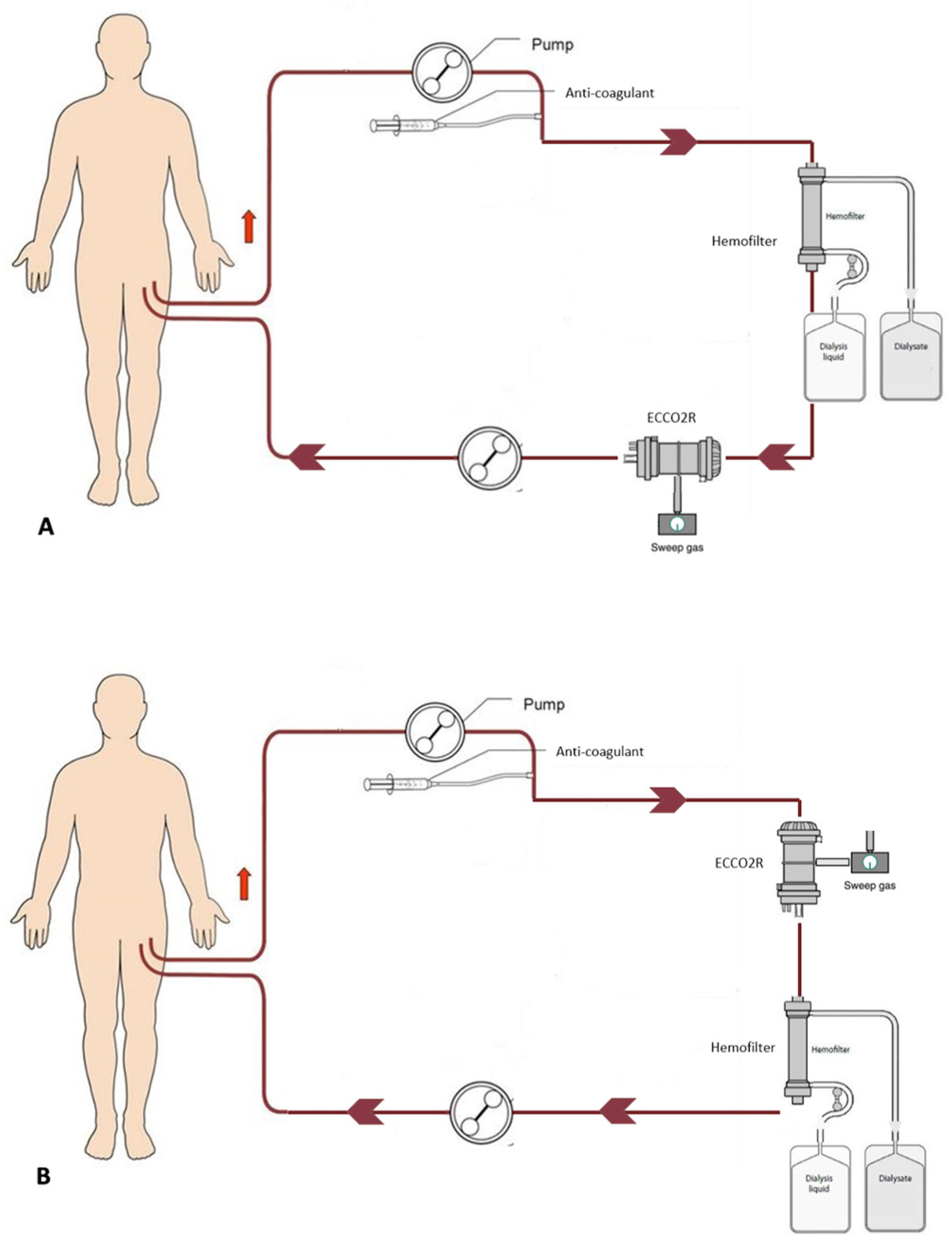
3.1. Principles and Systems

A working ECCO2R system requires vascular access, a blood pump, a membrane lung, an exchange gas, and anticoagulation [6]. ECCO2R devices have two different configurations: venovenous (VV-ECCO2R) and artero-venous (AV-ECCO2R) [7,8]. AV-ECCO2R is performed via arterial and venous cannulation, usually femoral, with 15 French cannulas, using the arterial blood pressure to pump the blood inside the circuit. So, the blood flow depends exclusively on the cardiac output of the patient [9]. This technique is invasive, less effective in hypotension, and can have many complications, so it is not widely used. In VV-ECCO2R, blood is drawn from a central vein by a draining cannula using centrifugal, roller, non-occlusive, or diagonal flow magnetic rotary pumps, which generates a pressure gradient and permits a flow across the circuit. This approach allows ECCO2R by utilizing small central venous catheters, commonly introduced via the right internal jugular vein [10]. The core of the ECCO2R circuit is the gas exchange membrane, a device with a complex geometry based on hollow fibers. The membrane material is poly-4-methyl-1-pentene (PMP), which represents the most used configuration because it reduces plasma leakage and permits gas transfer by diffusion, avoiding direct blood–gas contact. The exchange surfaces of the membranes differ in size from 0.32 to 0.65 m2 for low-flow VV systems and 1.3 m2 for high-flow VV and AV systems. Circuits and membranes are coated with heparin to improve biocompatibility and gas exchange, as well as too limit capillary leakage. The extraction of carbon dioxide is performed through the sweeping of the membrane by a fresh gas (O2 or medical air) devoid of CO2 [1,2,3,4,5]. The main determinants of CO2 removal in ECCO2R are extracorporeal blood flow, the PaCO2 gradient, sweep gas flow, and membrane size and characteristics [11]. According to the blood flow rate, we can distinguish between low-flow VV-ECCO2R systems operating with a blood flow rate between 200 and 400 mL/min, and high-flow systems (i.e., blood flow rate higher than 500 mL/min). The potential advantages of low-flow systems include the possibility of using conventional CRRT platforms and dual-lumen dialysis catheters, whereas high-flow systems require dedicated devices. Regarding the CO2 removal efficiency, in theory, an augmentation of blood flow should result in a linear increase in CO2 removal. So, considering that 1 L of blood transports around 500 mL of CO2, and that an average adult produces 250 mL/min of CO2, a blood flow rate of 200–300 mL/min may permit the removal of about 50% of the total CO2 produced, while an increase in the blood flow rate > 500 mL/min may remove all the produced CO2. However, experimental evidence suggests that, due to the limitations of blood flow and membrane efficiency, the actual removal capacity is inferior and, in particular, low-flow systems may remove up to 25% of the carbon dioxide produced [12]. Blood flow is only one of the determinants of CO2 removal [13]. Indeed, the CO2 transfer follows a diffusion gradient according to Fick’s law, so the difference between the blood flow and sweep gas in terms of CO2 pressure has a crucial role. In the sweep gas, the PaCO2 tends to be as low as possible (or even absent). Then, the venous blood partial pressure sustains the diffusion gradient. As the CO2 diffuses and achieves equilibrium almost instantaneously, the sweep gas flow rate is crucial to keeping the CO2 low on the gas side of the membrane [14]. The CO2 removal has a linear relationship with the sweep gas flow until a threshold of 4–5 L/min; after that there is no augmentation of the CO2 removal [15]. In addition, the membrane surface also has a relevant impact on the CO2 diffusion, and it is proportional to the quantity of the gas exchange and CO2 removal. Furthermore, large membranes carry a higher thrombotic risk, while small ones have an increased risk of haemolysis. Interestingly, as an innovative approach, there are some experimental studies on the application of hollow fibers coated with immobilized carbonic anhydrase to enhance the conversion of carbonic acid to CO2 [16].

3.2. Anticoagulation

As for other extracorporeal circuits, anticoagulation is required to prevent thromboembolic complications, especially for low-flux ECCO2R systems that are at a high risk of circuit clotting. No standard anticoagulation strategy for ECCO2R has been established yet. Among the different options, the most used in clinical trials and daily practice is systemic anticoagulation with heparin, which may be provided with unfractionated heparin or low-molecular-weight heparin. The major adverse effects of this strategy are bleeding and heparin-induced thrombocytopenia [17]. A promising alternative is citrate-based regional anticoagulation. Trisodium citrate infused at the beginning of the extracorporeal circuit binds to calcium, inhibiting the activation of calcium-dependent coagulation factors. The infusion of calcium chlorate at the circuit end reverses the citrate effect before the blood returns to the patient [18]. This strategy may reduce the incidence of hemorrhagic complications and can also improve the ECCO2R circuit survival time [19]. Moreover, the administration of trisodium citrate leads to the formation of sodium bicarbonate, an end-product of citrate metabolism, which might buffer the excess acid [20].

3.3. Complications

There are many complications related to the use of ECCO2R. They can be divided into three groups: patient-related, catheter-related, and device-related [10]. The most frequent adverse event is the occurrence of bleeding events (cerebral, gastrointestinal, and nasopharyngeal), mainly caused by the necessity of systemic anticoagulation [21]. Other commonly observed complications are thrombocytopenia and hemolysis. Distal limb ischemia and compartment syndrome of the lower limb (requiring fasciotomy or limb amputation) are associated with arterial cannulation [22]. Otherwise, venous catheterization can present more common complications, such as catheter-site bleeding, malposition, and infection. Vascular thrombosis occurs more often during low-flow VV-ECCO2R because of the increased exposure time to the membrane lung and circuit. Finally, device alterations can lead to pump or oxygenator failure, heat exchanger malfunction, or clot formation [21].

4. ECCO2R: Clinical Applications

While ECMO can ensure blood oxygenation and decarboxylation, ECCO2R provides partial respiratory support by removing CO2 with minimal impact on blood oxygenation [23]. The optimization of CO2 removal may allow for a proper ventilatory strategy in patients with respiratory failure. Indeed, these patients often require invasive MV (IMV), which may present some harmful effects, such as VILI, especially in cases in which a high tidal volume (TV) and plateau pressure (Pplat) are used [24]. The most recognized strategy to avoid VILI is lung-protective ventilation (LPV), which has the advantage of a low TV and Pplat. The introduction of these strategies in clinical practice has constituted a significant advance in the care of patients with respiratory failure [25]. However, one of the main concerns regarding the use of LPV is the risk of developing hypercapnia and respiratory acidosis, which are independently associated with increased adverse effects, including increased mortality [26]. In particular, hypercapnia may increase intracranial pressure and exert vasoconstrictive effects on pulmonary circulation, leading to pulmonary hypertension and augmented right ventricular afterload [9]. These are why ECCO2R techniques and devices have been developed, thus allowing LPV in cases of respiratory failure [27]. Moreover, ECCO2R may also be used to sustain the reduction in ventilation pressures in cases of non-intubated patients, thus preventing the demand for intubation. Given these objectives, the treatment of hypercapnic respiratory acidosis consequent to chronic obstructive pulmonary disease (COPD) and acute respiratory distress syndrome (ARDS) constitutes the main clinical indication for ECCO2R.

4.1. ECCO2R in COPD

COPD represents a condition of chronic hypercapnia that may worsen during acute exacerbations (ae-COPD). In this case, hypercapnia may be generated because of the reduced CO2 removal due to alveolar overdistension and the ventilation/perfusion imbalance, as well as increased CO2 production secondary to respiratory muscle work [28]. Although non-invasive ventilation (NIV) represents the first-choice treatment for ae-COPD [29], NIV failures often occur, and endotracheal intubation and IMV may be required [23]. ECCO2R therapy is an emerging option for managing hypercapnia while allowing LPV in these cases [27]. The use of ECCO2R in patients with ae-COPD enhances CO2 removal, lowers the respiratory rate, prolongs the expiratory time, and minimizes positive end-expiratory pressure (PEEP) [9]. Moreover, there is a reduction in the use of respiratory muscles with a consequent decrease in CO2 production [23]. Thus, ECCO2R devices can reduce NIV failure, preventing the need for IMV, or can facilitate weaning from MV [30,31]. However, it should be recognized that there is no evidence of survival benefits; additionally, ECCO2R does not seem to be risk-free in this setting. In a case–control ECLAIR study involving twenty-five COPD patients with acute hypercapnic respiratory failure refractory to NIV, the initiation of the VV-ECCO2R treatment was associated with a PaCO2 reduction of 17.5 mm Hg at 1 h and 29.5 mm Hg at 24 h accompanied by a 56% reduction in the intubation rate and a 60% reduction in the time on IMV. However, there were no significant effects on the length of patients’ ICU stay and mortality rates; moreover, the treatment was complicated by major adverse events in 11 patients (44%), including 9 patients (36%) with bleeding events [32]. In 2020, a consensus proposed the principal criteria for starting ECCO2R in patients with ae-COPD (no decrease in PaCO2 and no decrease in respiratory rate while on NIV), as well as patients recently initiated on mechanical ventilation after NIV failure to allow for early extubating. Accordingly, treatment targets for ae-COPD patients receiving ECCO2R therapy include comfortable patients, a pH of >7.30/7.35, a respiratory rate of <20–25 breaths/min, a decrease in PaCO2 by 10/20%, weaning from NIV, a decrease in HCO3, and the maintenance of hemodynamic stability [33].

4.2. ECCO2R in ARDS

ARDS is a life-threatening syndrome in which the respiratory system fails in the gas exchange function of oxygenation and/or carbon dioxide elimination. The mortality rate from ARDS is approximately 40 to 50%, and IMV is required in almost all patients [34,35]. However, in some patients, hypoxia and/or hypercapnia are refractory to MV despite maximal tolerable ventilation settings. A landmark trial by the ARDSNet group demonstrated that ventilating ARDS patients with an LPV modality with a low TV of 6 mL/kg for their predicted body weight (PBW) compared with a traditional TV of 12 mL/kg PBW significantly decreased mortality [36]. However, subsequent results showed lung hyperinflation still occurs in approximately 30% of ARDS patients ventilated with the ARDSNet strategy [37,38]. Therefore, the reduction in the TV to 3–4 mL/kg PBW and the Pplat to ≤25 cmH2O, otherwise known as ultraprotective ventilation (uLVP), has been proposed to further minimize the risk of VILI [39]. This strategy entails a significant risk of severe hypercapnic respiratory acidosis [40], a condition independently associated with worse outcomes [41,42]. So, the development of hypercapnia may constitute a limitation for the use of LPV and provide the reason why a validated ECCO2R method may help provide proper ventilation in ARDS patients. In the SUPERNOVA study, a prospective multicenter study, Combes et al. have shown that ECCO2R can minimize respiratory acidosis while applying a uLVP strategy in patients with moderate ARDS (PaO2/FiO2 100–200 mmHg, with PEEP ≥ 5 cmH2O) [43]. In this study, ninety-five patients were treated with the Hemolung Respiratory Assist System (ALung Technologies, Pittsburgh, PA, USA), the iLA active (Novalung, Heilbronn, Germany), and the Cardiohelp® HLS 5.0 (Getinge Cardiopulmonary Care, Rastatt, Germany) devices. The primary outcome was the number of patients who successfully achieved a TV of 4 mL/kg PBW with their PaCO2 not increasing more than 20% from the baseline with the value of the arterial pH > 7.30. Secondary endpoints included the assessment of physiological variables and ECCO2R settings as well as the frequency of adverse events. The proportions of patients who achieved ultra-protective settings by 8 h and 24 h were 78% and 82%, respectively. The TV, respiratory rate, minute ventilation, and Pplat were significantly lower at 8 h and 24 h compared to the baseline (p = 0.001). Moreover, the PaCO2 and PaO2/FiO2 ratio remained stable, while the pH significantly increased at 8 h (p < 0.001). ECCO2R was maintained for 5 (range 3–8) days. During the ECCO2R treatment, adverse events were reported in 39% of the patients, including two severe adverse events directly attributed to ECCO2R (brain hemorrhage and pneumothorax). Overall, 69 patients (73%) were alive on day 28, while fifty-nine patients (62%) were alive at hospital discharge. In conclusion, the authors stated that, despite the effectiveness of ECCO2R, the relatively high number of adverse events may call into question the risk/benefit balance of this approach, which should be confirmed in randomized clinical trials. The necessity for more robust evidence has been recently highlighted by the results of the REST trial, a multicenter, randomized, open-label, pragmatic clinical trial, which enrolled 412 adult patients receiving mechanical ventilation for acute hypoxemic respiratory failure [44]. The participants were randomized to receive lower TV ventilation facilitated by ECCO2R for at least 48 h (n = 202) or standard care with conventional low-TV ventilation (n = 210). The primary outcome was the all-cause mortality 90 days after randomization. Among the patients with acute hypoxemic respiratory failure, ECCO2R associated with a low TV did not significantly reduce the 90-day mortality when compared with the standard low-TV ventilation. However, due to the early termination, the study may have been underpowered to detect clinically relevant differences (the initial target enrolment was 1120 patients). Overall, these data highlight that beyond the strong rationale for using ECCO2R in ARDS patients, the available evidence is inconclusive, and there is space for expanding the research on this issue. Finally, it should be mentioned that ECCO2R has been used also in patients affected by Coronavirus disease-19 (COVID-19). Akkanti et al. described a cohort of 29 mechanically ventilated patients with ARDS secondary to COVID-19 complicated by severe hypercapnia and respiratory acidosis. In this cohort, ECCO2R treatments with the Hemolung Respiratory Assist System (ALung Technologies, Pittsburgh, PA, USA) were associated with an improvement in the acid–base parameters while providing LVP. No treatment-related adverse effects were reported, but the prognosis of these patients remained severe, with an overall survival of 38% [45].

5. Respiratory and Renal Failure: A Dangerous Interconnection

In critically ill patients, pulmonary and renal damage are often associated, providing evidence of lung–kidney crosstalk [46]. It has been estimated that ventilated patients have a three-fold increase in the risk of AKI; up to 30% of patients with ARDS may present kidney damage to some extent [47]. The mechanisms of lung–kidney interactions are bidirectional and multifaceted. First, during ARDS, renal function may be impaired by hemodynamic alterations, driven by venous congestion, neurohormonal activation, and ischemic injury [48]. It has been proved that MV directly impacts renal perfusion, while blood gas disturbances, and, in particular, hypercapnia, may act as a direct renal vasoconstrictor. In addition, toxic factors, oxidative stress, and MV-induced systemic inflammation may promote renal damage [49]. On the other side of the coin, kidney injury can aggravate pulmonary damage through different mechanisms [50]. Fluid overload and metabolic acidosis can increase respiratory work by inducing alveolar flooding and impairing pulmonary gas exchange. The systemic release of mediators expands pulmonary vascular permeability, lung inflammation, and apoptosis. Finally, the downregulation of the transepithelial electrolyte and water transport leads to respiratory failure [5]. Lung–kidney crosstalk has relevant clinical and therapeutical implications. The combination of AKI and ARDS aggravates the mortality rate by as high as 80% [51]. Furthermore, about 35% to 60% of patients with respiratory failure also need renal replacement therapies (RRTs) [52]. This observation underlines the potential clinical utility of providing simultaneous multiple extracorporeal supports, which may also include the combination of ECCO2R circuits with the CRRT platform. The most rational indication for ECCO2R coupled with CRRT is the association of hypercapnic respiratory acidosis with renal damage requiring CRRT [53]. However, in practice, the ECCO2R–CRRT combination has been also used in patients without renal failure, aiming to provide ECCO2R by the standard CRRT system, thus reducing the cost and the complexity of the ECCO2R treatment [54].

6. Experiences with ECCO2R Integrated into CRRT Platforms

The advantages of integrating a hollow-fiber gas exchanger in a CRRT platform include its simplicity and its potential applicability in non-specialized centers, such as the fact that no additional venous catheter placements are needed. Interestingly, even though ECCO2R combined with CRRT had already been under investigation years before the current Coronavirus disease-19 (COVID-19) pandemic, this approach began to attract even more attention during the pandemic due to its potential utility in improving resource allocation. In a pivotal paper in 2009, Terragni et al. tested the possibility of integrating a membrane lung in a modified renal replacement circuit [55]. They used a neonatal membrane with a total membrane surface of 0.33 m2 set in a series with a hemofilter to facilitate uLVP in 32 ARDS patients without AKI. They found that the extracorporeal treatment normalized their PaCO2 and pH and allowed the use of VT < 6 mL/kg for 144 (84–168) h, which in turn was associated with an improvement in the lung structure and a reduction in the pulmonary cytokines concentration. Following these results, in 2013, Forster et al. reported their experience with a low-flow hollow-fiber gas exchanger implemented in a CRRT circuit in 10 patients with combined respiratory and renal failure [56]. They used a CRRT platform and, after the hemofilter, a small standard hollow-fiber gas exchanger (D902 Liliput 2 ECMO; Sorin Group Milan, Italy; surface area of 0.67 m2). The RRT mode was continuous venovenous hemodialysis (CVVHD). The data showed an average PaCO2 reduction of 17.3 mmHg in about 4 h with a concomitant increase in pH. In parallel to the pH correction, a marked stabilization of hemodynamics was observed. At 24 h, the mean TV was reduced from 8.4 to 7.3 mL/kg PBW and the Pplat was reduced from 19.8 to 18.8 cmH2O. All the patients tolerated the intervention, and no complications occurred during the therapy. Two episodes of clotting were observed, but no serious adverse events attributed to the hollow-fiber gas exchanger or the CRRT occurred. Seven out of ten patients were successfully weaned from the low-flow CO2 removal system, their pulmonary function was improved, and they recovered from critical illnesses. In 2014, Quintard et al. conducted a very similar investigation on 16 patients affected by ARDS treated with CRRT for oliguric AKI [57]. They used a standard device in CVVHD or continuous venovenous hemofiltration (CVVH) modality. An oxygenation membrane, initially designed for pediatric ECMO (HILITE 2400 LT, Medos), was introduced upstream from the hemofilter. The average PaCO2 reduction was 24.4 mmHg after 6 h and 30 mmHg after 12 h (31% and 39%, respectively), associated with a pH increase of 0.16 at 6 h and 0.23 at 12 h, respectively. The mean TV was reduced from 5.9 mL/kg PBW before the treatment to 5.5 mL/kg PBW at 12 h. The mean Pplat before the treatments were 27.7 cmH2O and 25.6 cmH2O at 12 h. No complications or adverse events attributable to the treatment were reported. Seven of the sixteen patients died, but the timing, cause, and place of death were not specified. In 2015, Allardet-Servent et al. conducted a prospective human observational study on eleven patients with ARDS and AKI. CRRT was delivered with a PrismaFlex v6.0 monitor (Gambro, Lund, Sweden) in the CVVH modality and the membrane oxygenator was inserted either upstream or downstream of the hemofilter [58]. On average, the oxygenator blood flow and CO2 removal rate were higher when the membrane was put upstream of the hemofilter, but the differences were not statistically significant (PaCO2 relative reduction 22 ± 7% upstream vs 18 ± 6% downstream). At the beginning of the treatment, the TV ventilation was fixed at 6 mL/kg PBW, but then it was possible to reduce it to 4 mL/kg PBW. Thereafter, the TV was reduced to 4 mL/kg PBW for the remainder of the study (72 h). However, even in this cohort, the ICU mortality rate remained elevated (nine patients-82%). A point of strength of this study is that, unlike previous investigations, the authors used a standardized protocol of ventilation based on the ARDSNet protocol. In 2018, Fanelli et al. compared thirteen patients treated with ECCO2R–CRRT with propensity-score-matched patients treated only with CRRT [59]. They found that after 24h of the combined treatment, it was possible to achieve a significant reduction in TV (from 7.04 ± 0.5 to 4.84 ± 0.4 mL/kg PBW) while maintaining a stable PaCO2 level. Interestingly, the authors also observed a significant decrease in inflammation and apoptosis markers in patients undergoing the combined treatment. In 2018 in a multicenter study, Schmidt et al. evaluated twenty patients with mild-to-moderate ARDS, treated with a low-flow CO2-removal device, Prismalung® (Baxter Gambro Renal, Deerfield, IL, USA), which consisted of a 0.32m2 membrane oxygenator that was integrated into the Prismaflex® platform (Baxter Gambro Renal, USA) [60]. None of the patients had AKI, and so the ECCO2R was provided standalone (without concomitant RRT). Additionally, in this case, the ECCO2R was helpful in sustaining uLVP and limiting the increase in PaCO2. Interestingly, the same ECCO2-CRRT configuration was used by Nentwich et al., who, in a multicenter observational study, evaluated twenty hypercapnic patients with concomitant renal failure requiring CRRT [61]. The RRT modality was CVVH, and ventilation parameters were set according to the ARDSNet recommendations. The data showed an average PaCO2 reduction of 7.4 mmHg, a concomitant 0.4 increase in pH, and a slight decrease in the VT (from 6.0 ± 0.7 to 5.5 ± 0.8 mL/kg PWB) and Pplat (from 30 ± 4 to 28.9 ± 3.6 cmH2O) after 24 h. The combined treatment ameliorated respiratory acidosis and effectively reduced the invasiveness of MV while delivering an efficient renal replacement therapy and reducing the vasopressor requirements. Notably, this study provided the first description of a certified and labeled combination therapy on a commercially available organ support platform. Finally, in 2021, Consales et al. published their retrospective observational study (the CICERO study) carried out between 2016 and 2019 in 22 patients with either mild-to-moderate ARDS or aeCOPD associated with AKI treated with combined ECCO2R–CRRT [53]. Similarly to Netwitch et al., they used the PrismaLung®-Prismaflex® platform. The average PaCO2 was efficiently reduced from 73.8 to 46.6 mmHg in 24 h, and the pH concomitantly increased from 7.20 to 7.40. The treatment allowed 12/17 patients on mechanical ventilation to shift to protective ventilation within 24 h. No complications related to ECCO2R–CRRT were recorded. Overall, 21 out of the 22 patients recovered from AKI during their hospitalization, while one patient was on intermittent hemodialysis due to underlying end-stage renal disease before their admission to the ICU. In Figure 1 we present schematic representations of the different configurations of the combined ECCO2R–CRRT circuits. Table 1 and Table 2 summarize the main clinical characteristics and operational parameters of the devices used in these studies.

Experiences with ECCO2R Integrated into CRRT Platform in COVID-19 Patients

As already stated, the COVID-19 pandemic has offered the possibility to reevaluate the suitability of ECCO2R provided on CRRT platforms. Indeed, the high number of COVID-19 patients suffering from ARDS highlighted the need for a simple and widely available solution to provide the best ventilatory strategy option for these patients, regardless of their renal function [62]. Therefore, unlike previous experience, during the pandemic, ECCO2R–CRRT combined treatment was mostly used in patients without AKI or renal failure. Moving beyond single-case reports [63], in 2020, Husain-Syed et al. treated four COVID-19 patients complicated by ARDS with an ECCO2R (multiECCO2R, Eurosets) in conjunction with multiFiltrate CRRT platforms (Fresenius Medical Care, Bad Homburg, Germany) [64]. Three patients received ECCO2R standalone with the multiFiltrate set in the hemoperfusion mode, and one patient, who suffered from AKI, received ECCO2R coupled with CRRT in the CVVHD mode. The multiECCO2R was inserted in a series after the hemofilter (Ultraflux AV 1000S, Fresenius Medical Care, Bad Homburg, Germany). The ECCO2R–CRRT was commenced at a blood flow of 200 mL/min. Regional citrate anticoagulation plus systemic heparinization were used as an anticoagulation strategy. In two patients, it was necessary to implement the blood flow rate to 400 mL/min to achieve good PaCO2 clearance. The average PaCO2 reduction was 15.4 mmHg after 24 h of treatment, and the pH increased from 7.33 ± 0.07 to 7.45 ± 0.07. After 24 h, it was possible to decrease the TV and Pplat. No ECCO2R–CRRT-related adverse events occurred. The ECCO2R treatment was terminated after a median of 5.5 (4.5–7.5) days due to a sustained improvement in hypercapnia. In the AKI patient, CRRT was continued for another four days because of oliguria. During the first wave of the pandemic, Ding et al. conducted a single-center study on 12 patients affected by COVID-19 ARDS with refractory hypercapnia (PaCO2 > 50 mmHg) admitted in the ICU of Wuhan [65]. They used a low-flow gas-exchanger oxygenator integrated into the Prismaflex platform (Gambro-Baxter) to decrease the PaCO2 level and permit a low Pplat and driving pressure ventilation. In this case, the patients did not suffer from AKI, so the CRRT machine was set in the slow continuous ultrafiltration (SCUF) mode with an ultrafiltration rate of 0 mL. The mean blood flow was 342.5 ± 49.20 mL/min, and the CO2 clearance reached the best efficiency (45.91 ± 7.70 mL/min) at a sweep gas flow of 10 L/min. After the application of the ECCO2R device, the PaCO2 in all the patients decreased. The treatment led to an 8.48 cmH2O reduction in the Pplat in 24 h. Even in this cohort, the combined ECCO2R–CRRT treatment was safe, and no major adverse events were reported. Nevertheless, the 28-day mortality was high (67%). Finally, in 2022, Alessandri et al. retrospectively reported their experience in the treatment of 27 patients with ARDS and AKI requiring invasive mechanical ventilation undergoing ECCO2R–CRRT [66]. The initiation of the treatment reduced the TV from 6.0 ± 0.6 mL/kg to 4.3 ± 0.3 mL/ mL/kg PWB and the Pplat from 28.9 ± 2.7 to 21.6 ± 2.8 cmH2O with a reduction in the respiratory rate. Throughout the course of the ECCO2R, these changes were accompanied by the stabilization of PaCO2 and an increase in pH. Simultaneously, the combined treatment was associated with a significant reduction in the serum creatinine levels. No major adverse effects occurred, but 17 patients (63%) died within 28 days. Table 3 and Table 4 summarize the main clinical characteristics and operational parameters of the devices used in COVID-19 patients with ARDS.

7. Critical Considerations

Here, we have reviewed the principal available experiences and evidence of combined ECCO2R–CRRT in various clinical settings, such as ARDS of different etiologies, including COVID-19, and aeCOPD in patients with or without associated renal failure. The critical analysis of these data allows us to make some generalizations. As a point of strength, all the studies agree in that they suggest that ECCO2 R alone or set on a CRRT platform effectively controls hypercapnia and respiratory acidosis in MV patients. This is a crucial issue because the regulation of PaCO2 levels is essential to permitting the adoption of LPV strategies [67]. Furthermore, during ECCO2R–CRRT, CO2 removal may be obtained using low blood flow, thus facilitating clinical management and reducing treatment-related adverse effects. On the other hand, the reported findings present many weaknesses. The most relevant limitation is that, currently, there is no evidence of the effects of ECCO2R and ECCO2R–CRRT in improving patient outcomes and reducing mortality [68]. This is a result often found in studies investigating critically ill patients, which may be a consequence of the clinical complexity of these patients but also of the small sample size and short time of treatment that characterizes these studies [69]. Moreover, they present a high heterogeneity since different patient populations, outcomes, devices, and operative parameters were investigated. Similarly, with some exceptions, there is a lack of a standardized ventilation protocol without prefixed objectives. These aspects significantly reduce the generalizability of the reported data. Furthermore, it should be noted that in studies involving patients with renal failure, the renal outcome and recovery were been poorly reported; thus, the adequacy of renal support provided by ECCO2R–CRRT systems is unclear. Finally, many other issues have not been sufficiently explored. For example, we need to investigate the most efficient circuit configuration (i.e., the position of the membrane oxygenator and hemofilter may impact the circuit performance), the effects of a dialysis buffer on the systemic acid–base balance, and the management of anticoagulation with the possible use of citrate. All these considerations underline the need for further studies and suggest caution in translating experimental evidence into clinical practice.

8. Conclusions

Extracorporeal CO2 removal techniques offer several advantages for ventilatory strategy optimization in patients with respiratory failure. However, although the different studies demonstrated the efficacy of ECCO2R in improving hypercapnia and metabolic acidosis, this treatment is not risk-free, and its impact on the prognosis of critically ill patients is undefined. Notably, these patients are characterized by high complexity, with multiorgan involvement, often requiring a multidisciplinary approach [70]. In this sense, the combination of different extracorporeal support techniques could offer clinical benefits in terms of the reduction in complications, as well as organizational and economic advantages. The possibility of using ECCO2R coupled with CRRT platforms provides an example of this approach. First, the ECCO2R–CRRT combination is flexible since it can be employed in patients with respiratory failure, including those with COVID-19, with or without concomitant renal disease. Furthermore, exploiting widely available equipment, such as those required for CRRT, it can also be used in non-highly specialized centers, as it does not require specific training. For the same reason, ECCO2R–CRRT could save time and costs compared to equipment specifically designed for ECCO2R. The disadvantage is that the ECCO2R circuit integrated into the CRRT only allows for low-flow techniques, which may be insufficient for some patients. On the other hand, the low-flux treatment with ECCO2R–CRRT seems to be well tolerated and not burdened by significant adverse events, except for the risk of circuit coagulation. Nevertheless, as discussed above, the available evidence presents many limitations, so an ideal approach would be to wait for specifically designed randomized clinical trials to determine the actual clinical impact of ECCO2R and ECCO2R–CRRT. However, admittedly, in critically ill patients, large clinical trials are not easy to implement. So, the active reporting of clinical experiences and cohort studies is essential to defining and confirming the suitability and safety of this approach as well as identifying patients who can benefit the most from this therapy.

Author Contributions

Conceptualization, F.C.; E.C. and L.M.; methodology, V.Z. and L.N.; formal analysis, E.R. and F.S.; writing—original draft preparation, S.B. and P.E.; writing—review and editing, F.V. All authors have read and agreed to the published version of the manuscript.

Funding

This research received no external funding.

Institutional Review Board Statement

Not applicable.

Informed Consent Statement

Not applicable.

Data Availability Statement

Not applicable.

Conflicts of Interest

The authors declare no conflict of interest.

References

  1. Roussos, C.; Koutsoukou, A. Respiratory Failure. Eur. Respir. J. Suppl. 2003, 47, 3s–14s. [Google Scholar] [CrossRef] [PubMed] [Green Version]
  2. Shekar, K.; Mullany, D.V.; Thomson, B.; Ziegenfuss, M.; Platts, D.G.; Fraser, J.F. Extracorporeal Life Support Devices and Strategies for Management of Acute Cardiorespiratory Failure in Adult Patients: A Comprehensive Review. Crit. Care 2014, 18, 219. [Google Scholar] [CrossRef] [PubMed] [Green Version]
  3. Camporota, L.; Barrett, N. Current Applications for the Use of Extracorporeal Carbon Dioxide Removal in Critically Ill Patients. BioMed Res. Int. 2016, 2016, 9781695. [Google Scholar] [CrossRef] [PubMed] [Green Version]
  4. Doyle, J.; Cooper, J.S. Physiology, Carbon Dioxide Transport; StatPearls Publishing: Treasure Island, FL, USA, 2022. [Google Scholar]
  5. Ronco, C.; Bellomo, R.; Kellum, J.A.; Ricci, Z. Critical Care Nephrology; Elsevier Health Sciences: Amsterdam, The Netherlands, 2019; ISBN 9780323449427. [Google Scholar]
  6. Boyle MBChB, A.J.; McNamee FCICM, J.J.; McAuley, D.F.; Boyle, A.J.; McNamee, J.J.; Boyle, A.J.; Sklar, M.C.; McNamee, J.J.; Brodie, D.; Slutsky, A.S.; et al. Extracorporeal Carbon Dioxide Removal for Lowering the Risk of Mechanical Ventilation: Research Questions and Clinical Potential for the Future. Lancet Respir. Med. 2018, 6, 874–884. [Google Scholar] [CrossRef] [PubMed] [Green Version]
  7. May, A.G.; Omecinski, K.S.; Frankowski, B.J.; Federspiel, W.J. Effect of Hematocrit on the CO2 Removal Rate of Artificial Lungs. ASAIO J. 2020, 66, 1161–1165. [Google Scholar] [CrossRef]
  8. Yu, T.Z.; Tatum, R.T.; Saxena, A.; Ahmad, D.; Yost, C.C.; Maynes, E.J.; O’Malley, T.J.; Massey, H.T.; Swol, J.; Whitson, B.A.; et al. Utilization and Outcomes of Extracorporeal CO2 Removal (ECCO2R): Systematic Review and Meta-Analysis of Arterio-Venous and Veno-Venous ECCO2R Approaches. Artif. Organs 2022, 46, 763–774. [Google Scholar] [CrossRef]
  9. Giraud, R.; Banfi, C.; Assouline, B.; de Charrière, A.; Cecconi, M.; Bendjelid, K. The Use of Extracorporeal CO2 Removal in Acute Respiratory Failure. Ann. Intensive Care 2021, 11, 43. [Google Scholar] [CrossRef]
  10. Morales-Quinteros, L.; Del Sorbo, L.; Artigas, A. Extracorporeal Carbon Dioxide Removal for Acute Hypercapnic Respiratory Failure. Ann. Intensive Care 2019, 9, 79. [Google Scholar] [CrossRef]
  11. Terragni, P.P.; Birocco, A.; Faggiano, C.; Ranieri, V.M. Extracorporeal CO2; Removal. In Cardiorenal Syndromes in Critical Care; Karger Publishers: Basel, Switzerland, 2010; Volume 165, pp. 185–196. ISBN 978-3-8055-9472-1. [Google Scholar]
  12. Terragni, P.; Maiolo, G.; Ranieri, V.M. Role and Potentials of Low-Flow CO(2) Removal System in Mechanical Ventilation. Curr. Opin. Crit. Care 2012, 18, 93–98. [Google Scholar] [CrossRef]
  13. Del Sorbo, L.; Cypel, M.; Fan, E. Extracorporeal Life Support for Adults with Severe Acute Respiratory Failure. Lancet Respir. Med. 2014, 2, 154–164. [Google Scholar] [CrossRef]
  14. Ficial, B.; Vasques, F.; Zhang, J.; Whebell, S.; Slattery, M.; Lamas, T.; Daly, K.; Agnew, N.; Camporota, L. Physiological Basis of Extracorporeal Membrane Oxygenation and Extracorporeal Carbon Dioxide Removal in Respiratory Failure. Membranes 2021, 11, 225. [Google Scholar] [CrossRef]
  15. Barrett, N.A.; Hart, N.; Camporota, L. In Vivo Carbon Dioxide Clearance of a Low-Flow Extracorporeal Carbon Dioxide Removal Circuit in Patients with Acute Exacerbations of Chronic Obstructive Pulmonary Disease. Perfusion 2020, 35, 436–441. [Google Scholar] [CrossRef]
  16. Arazawa, D.T.; Kimmel, J.D.; Finn, M.C.; Federspiel, W.J. Acidic Sweep Gas with Carbonic Anhydrase Coated Hollow Fiber Membranes Synergistically Accelerates CO2 Removal from Blood. Acta Biomater. 2015, 25, 143–149. [Google Scholar] [CrossRef] [Green Version]
  17. Menajovsky, L.B. Heparin-Induced Thrombocytopenia: Clinical Manifestations and Management Strategies. Am. J. Med. 2005, 118 (Suppl. 8A), 21S–30S. [Google Scholar] [CrossRef]
  18. Tolwani, A.; Wille, K.M. Advances in Continuous Renal Replacement Therapy: Citrate Anticoagulation Update. Blood Purif. 2012, 34, 88–93. [Google Scholar] [CrossRef]
  19. Bagshaw, S.M.; Laupland, K.B.; Boiteau, P.J.E.; Godinez-Luna, T. Is Regional Citrate Superior to Systemic Heparin Anticoagulation for Continuous Renal Replacement Therapy? A Prospective Observational Study in an Adult Regional Critical Care System. J. Crit. Care 2005, 20, 155–161. [Google Scholar] [CrossRef]
  20. Sharma, A.S.; Weerwind, P.W.; Bekers, O.; Wouters, E.M.; Maessen, J.G. Carbon Dioxide Dialysis in a Swine Model Utilizing Systemic and Regional Anticoagulation. Intensive Care Med. Exp. 2016, 4, 2. [Google Scholar] [CrossRef] [Green Version]
  21. Sklar, M.C.; Beloncle, F.; Katsios, C.M.; Brochard, L.; Friedrich, J.O. Extracorporeal Carbon Dioxide Removal in Patients with Chronic Obstructive Pulmonary Disease: A Systematic Review. Intensive Care Med. 2015, 41, 1752–1762. [Google Scholar] [CrossRef]
  22. Cove, M.E.; MacLaren, G.; Federspiel, W.J.; Kellum, J.A. Bench to Bedside Review: Extracorporeal Carbon Dioxide Removal, Past Present and Future. Crit. Care 2012, 16, 232. [Google Scholar] [CrossRef] [Green Version]
  23. Azzi, M.; Aboab, J.; Alviset, S.; Ushmorova, D.; Ferreira, L.; Ioos, V.; Memain, N.; Issoufaly, T.; Lermuzeaux, M.; Laine, L.; et al. Extracorporeal CO2removal in Acute Exacerbation of COPD Unresponsive to Non-Invasive Ventilation. BMJ Open Respir. Res. 2021, 8, e001089. [Google Scholar] [CrossRef]
  24. Ricard, J.D.; Dreyfuss, D.; Saumon, G. Ventilator-Induced Lung Injury. Eur. Respir. J. Suppl. 2003, 42, 2s–9s. [Google Scholar] [CrossRef] [PubMed] [Green Version]
  25. Fan, E.; Brodie, D.; Slutsky, A.S. Acute Respiratory Distress Syndrome. JAMA 2018, 319, 698. [Google Scholar] [CrossRef] [PubMed]
  26. Tiruvoipati, R.; Pilcher, D.; Buscher, H.; Botha, J.; Bailey, M. Effects of Hypercapnia and Hypercapnic Acidosis on Hospital Mortality in Mechanically Ventilated Patients*. Crit. Care Med. 2017, 45, e649–e656. [Google Scholar] [CrossRef] [PubMed]
  27. İnal, V.; Efe, S. Extracorporeal Carbon Dioxide Removal (Ecco2r) in Copd and Ards Patients with Severe Hypercapnic Respiratory Failure. a Retrospective Case-Control Study. Turk. J. Med. Sci. 2021, 51, 2127–2135. [Google Scholar] [CrossRef] [PubMed]
  28. Dave, C.; Wharton, S.; Mukherjee, R.; Faqihi, B.M.; Stockley, R.A.; Turner, A.M. Development and Relevance of Hypercapnia in COPD. Can. Respir. J. 2021, 2021, 6623093. [Google Scholar] [CrossRef]
  29. Brochard, L.; Mancebo, J.; Wysocki, M.; Lofaso, F.; Conti, G.; Rauss, A.; Simonneau, G.; Benito, S.; Gasparetto, A.; Lemaire, F.; et al. Noninvasive Ventilation for Acute Exacerbations of Chronic Obstructive Pulmonary Disease. N. Engl. J. Med. 1995, 333, 817–822. [Google Scholar] [CrossRef]
  30. del Sorbo, L.; Pisani, L.; Filippini, C.; Fanelli, V.; Fasano, L.; Terragni, P.; Dell’Amore, A.; Urbino, R.; Mascia, L.; Evangelista, A.; et al. Extracorporeal Co2 Removal in Hypercapnic Patients at Risk of Noninvasive Ventilation Failure: A Matched Cohort Study with Historical Control. Crit. Care Med. 2015, 43, 120–127. [Google Scholar] [CrossRef]
  31. Morelli, A.; D’Egidio, A.; Orecchioni, A.; Alessandri, F.; Mascia, L.; Ranieri, V.M. Extracorporeal Co2 Removal in Hypercapnic Patients Who Fail Noninvasive Ventilation and Refuse Endotracheal Intubation: A Case Series. Intensive Care Med. Exp. 2015, 3, A824. [Google Scholar] [CrossRef] [Green Version]
  32. Braune, S.; Sieweke, A.; Brettner, F.; Staudinger, T.; Joannidis, M.; Verbrugge, S.; Frings, D.; Nierhaus, A.; Wegscheider, K.; Kluge, S. The Feasibility and Safety of Extracorporeal Carbon Dioxide Removal to Avoid Intubation in Patients with COPD Unresponsive to Noninvasive Ventilation for Acute Hypercapnic Respiratory Failure (ECLAIR Study): Multicentre Case–Control Study. Intensive Care Med. 2016, 42, 1437–1444. [Google Scholar] [CrossRef]
  33. Combes, A.; Auzinger, G.; Capellier, G.; du Cheyron, D.; Clement, I.; Consales, G.; Dabrowski, W.; de Bels, D.; de Molina Ortiz, F.J.G.; Gottschalk, A.; et al. ECCO2R Therapy in the ICU: Consensus of a European Round Table Meeting. Crit. Care 2020, 24, 490. [Google Scholar] [CrossRef]
  34. Sloane, P.J.; Gee, M.H.; Gottlieb, J.E.; Albertine, K.H.; Peters, S.P.; Robert Burns, J.; Machiedo, G.; Fish, J.E. A Multicenter Registry of Patients with Acute Respiratory Distress Syndrome. Am. Rev. Respir. Dis. 1992, 146, 419–426. [Google Scholar] [CrossRef]
  35. Zilberberg, M.D.; Epstein, S.K.; Ep-Stein, S.K. Acute Lung Injury in the Medical ICU Comorbid Conditions, Age, Etiology, and Hospital Outcome. Am. J. Respir. Crit. Care Med. 1998, 157, 1159–1164. [Google Scholar] [CrossRef] [Green Version]
  36. Yndrome, S.; Etwork, N. The new england journal of medicine ventilation with lower tidal volumes as compared with traditional tidal volumes for acute lung injury and the acute respiratory distress syndrome a bstract background traditional approaches to mechanical. N. Engl. J. Med. 2000, 342, 1301–1308. [Google Scholar]
  37. Terragni, P.P.; Rosboch, G.; Tealdi, A.; Corno, E.; Menaldo, E.; Davini, O.; Gandini, G.; Herrmann, P.; Mascia, L.; Quintel, M.; et al. Tidal Hyperinflation during Low Tidal Volume Ventilation in Acute Respiratory Distress Syndrome. Am. J. Respir. Crit. Care Med. 2007, 175, 160–166. [Google Scholar] [CrossRef]
  38. Bellani, G.; Guerra, L.; Musch, G.; Zanella, A.; Patroniti, N.; Mauri, T.; Messa, C.; Pesenti, A. Lung Regional Metabolic Activity and Gas Volume Changes Induced by Tidal Ventilation in Patients with Acute Lung Injury. Am. J. Respir. Crit. Care Med. 2011, 183, 1193–1199. [Google Scholar] [CrossRef] [Green Version]
  39. Rozencwajg, S.; Guihot, A.; Franchineau, G.; Lescroat, M.; Bréchot, N.; Hékimian, G.; Lebreton, G.; Autran, B.; Luyt, C.E.; Combes, A.; et al. Ultra-Protective Ventilation Reduces Biotrauma in Patients on Venovenous Extracorporeal Membrane Oxygenation for Severe Acute Respiratory Distress Syndrome. Crit. Care Med. 2019, 47, 1505–1512. [Google Scholar] [CrossRef]
  40. Fanelli, V.; Costamagna, A.; Ranieri, V.M. Extracorporeal Support for Severe Acute Respiratory Failure. Semin. Respir. Crit. Care Med. 2014, 35, 519–527. [Google Scholar] [CrossRef]
  41. Almanza-Hurtado, A.; Polanco Guerra, C.; Martínez-Ávila, M.C.; Borré-Naranjo, D.; Rodríguez-Yanez, T.; Dueñas-Castell, C. Hypercapnia from Physiology to Practice. Int. J. Clin. Pract. 2022, 2022, 2635616. [Google Scholar] [CrossRef]
  42. Nin, N.; Muriel, A.; Peñuelas, O.; Brochard, L.; Lorente, J.A.; Ferguson, N.D.; Raymondos, K.; Ríos, F.; Violi, D.A.; Thille, A.W.; et al. Severe Hypercapnia and Outcome of Mechanically Ventilated Patients with Moderate or Severe Acute Respiratory Distress Syndrome. Intensive Care Med. 2017, 43, 200–208. [Google Scholar] [CrossRef]
  43. Combes, A.; Fanelli, V.; Pham, T.; Ranieri, V.M.; Goligher, E.C.; Brodie, D.; Pesenti, A.; Beale, R.; Brochard, L.; Chiche, J.D.; et al. Feasibility and Safety of Extracorporeal CO2 Removal to Enhance Protective Ventilation in Acute Respiratory Distress Syndrome: The SUPERNOVA Study. Intensive Care Med. 2019, 45, 592–600. [Google Scholar] [CrossRef] [Green Version]
  44. McNamee, J.J.; Gillies, M.A.; Barrett, N.A.; Perkins, G.D.; Tunnicliffe, W.; Young, D.; Bentley, A.; Harrison, D.A.; Brodie, D.; Boyle, A.J.; et al. Effect of Lower Tidal Volume Ventilation Facilitated by Extracorporeal Carbon Dioxide Removal vs Standard Care Ventilation on 90-Day Mortality in Patients with Acute Hypoxemic Respiratory Failure: The REST Randomized Clinical Trial. JAMA J. Am. Med. Assoc. 2021, 326, 1013–1023. [Google Scholar] [CrossRef] [PubMed]
  45. Akkanti, B.; Jagpal, S.; Darwish, R.; Saavedra Romero, R.; Scott, L.K.; Dinh, K.; Hussain, S.; Radbel, J.; Saad, M.A.; Enfield, K.B.; et al. Physiologic Improvement in Respiratory Acidosis Using Extracorporeal Co 2 Removal With Hemolung Respiratory Assist System in the Management of Severe Respiratory Failure From Coronavirus Disease 2019. Crit. Care Explor. 2021, 3, e0372. [Google Scholar] [CrossRef] [PubMed]
  46. Husain-Syed, F.; Slutsky, A.S.; Ronco, C. Lung–Kidney Cross-Talk in the Critically Ill Patient. Am. J. Respir. Crit. Care Med. 2016, 194, 402–414. [Google Scholar] [CrossRef] [PubMed]
  47. van den Akker, J.P.; Egal, M.; Groeneveld, J.A. Invasive Mechanical Ventilation as a Risk Factor for Acute Kidney Injury in the Critically Ill: A Systematic Review and Meta-Analysis. Crit. Care 2013, 17, R98. [Google Scholar] [CrossRef] [PubMed] [Green Version]
  48. Murray, P.T. The Kidney in Respiratory Failure and Mechanical Ventilation. Cardiorenal Syndr. Crit. Care 2010, 165, 159–165. [Google Scholar]
  49. Kuiper, J.W.; Groeneveld, A.B.J.; Slutsky, A.S.; Plötz, F.B. Mechanical Ventilation and Acute Renal Failure. Crit. Care Med. 2005, 33, 1408–1415. [Google Scholar] [CrossRef]
  50. Alge, J.; Dolan, K.; Angelo, J.; Thadani, S.; Virk, M.; Akcan Arikan, A. Two to Tango: Kidney-Lung Interaction in Acute Kidney Injury and Acute Respiratory Distress Syndrome. Front. Pediatr. 2021, 9, 1046. [Google Scholar] [CrossRef]
  51. Mehta, R.L.; Pascual, M.T.; Gruta, C.G.; Zhuang, S.; Chertow, G.M. Refining Predictive Models in Critically Ill Patients with Acute Renal Failure. J. Am. Soc. Nephrol. 2002, 13, 1350–1357. [Google Scholar] [CrossRef] [Green Version]
  52. Uchino, S. Acute Renal Failure in Critically Ill Patients—A Multinational, Multicenter Study. JAMA 2005, 294, 813. [Google Scholar] [CrossRef] [Green Version]
  53. Consales, G.; Zamidei, L.; Turani, F.; Atzeni, D.; Isoni, P.; Boscolo, G.; Saggioro, D.; Resta, M.V.; Ronco, C. Combined Renal-Pulmonary Extracorporeal Support with Low Blood Flow Techniques : A Retrospective Observational Study (CICERO Study). Blood Purif. 2022, 51, 299–308. [Google Scholar] [CrossRef]
  54. De Bels, D.; Pierrakos, C.; Spapen, H.D.; Honore, P.M. A Double Catheter Approach for Extracorporeal CO 2 Removal Integrated within a Continuous Renal Replacement Circuit. J. Transl. Int. Med. 2018, 6, 157–158. [Google Scholar] [CrossRef] [Green Version]
  55. Terragni, P.P.; Del Sorbo, L.; Mascia, L.; Urbino, R.; Martin, E.L.; Birocco, A.; Faggiano, C.; Quintel, M.; Gattinoni, L.; Ranieri, V.M. Tidal Volume Lower than 6 Ml/Kg Enhances Lung Protection: Role of Extracorporeal Carbon Dioxide Removal. Anesthesiology 2009, 111, 826–835. [Google Scholar] [CrossRef]
  56. Forster, C.; Schriewer, J.; John, S.; Eckardt, K.U.; Willam, C. Low-Flow CO2 Removal Integrated into a Renal-Replacement Circuit Can Reduce Acidosis and Decrease Vasopressor Requirements. Crit. Care 2013, 17, R154. [Google Scholar] [CrossRef] [Green Version]
  57. Quintard, J.M.; Barbot, O.; Thevenot, F.; de Matteis, O.; Benayoun, L.; Leibinger, F. Partial Extracorporeal Carbon Dioxide Removal Using a Standard Continuous Renal Replacement Therapy Device: A Preliminary Study. ASAIO J. 2014, 60, 564–569. [Google Scholar] [CrossRef]
  58. Allardet-Servent, J.; Castanier, M.; Signouret, T.; Soundaravelou, R.; Lepidi, A.; Seghboyan, J.M. Safety and Efficacy of Combined Extracorporeal Co2 Removal and Renal Replacement Therapy in Patients with Acute Respiratory Distress Syndrome and Acute Kidney Injury: The Pulmonary and Renal Support in Acute Respiratory Distress Syndrome Study. Crit. Care Med. 2015, 43, 2570–2581. [Google Scholar] [CrossRef] [Green Version]
  59. Fanelli, V.; Cantaluppi, V.; Alessandri, F.; Costamagna, A.; Cappello, P.; Brazzi, L.; Pugliese, F.; Biancone, L.; Terragni, P.; Marco Ranieri, V. Extracorporeal CO2 Removal May Improve Renal Function of Patients with Acute Respiratory Distress Syndrome and Acute Kidney Injury: An Open-Label, Interventional Clinical Trial. Am. J. Respir. Crit. Care Med. 2018, 198, 687–690. [Google Scholar] [CrossRef]
  60. Schmidt, M.; Jaber, S.; Zogheib, E.; Godet, T.; Capellier, G.; Combes, A. Feasibility and Safety of Low-Flow Extracorporeal CO2 Removal Managed with a Renal Replacement Platform to Enhance Lung-Protective Ventilation of Patients with Mild-to-Moderate ARDS. Crit. Care 2018, 22, 122. [Google Scholar] [CrossRef] [Green Version]
  61. Nentwich, J.; Wichmann, D.; Kluge, S.; Lindau, S.; Mutlak, H.; John, S. Low-Flow CO2 Removal in Combination with Renal Replacement Therapy Effectively Reduces Ventilation Requirements in Hypercapnic Patients: A Pilot Study. Ann. Intensive Care 2019, 9, 3. [Google Scholar] [CrossRef]
  62. Richardson, S.; Hirsch, J.S.; Narasimhan, M.; Crawford, J.M.; McGinn, T.; Davidson, K.W.; Barnaby, D.P.; Becker, L.B.; Chelico, J.D.; Cohen, S.L.; et al. Presenting Characteristics, Comorbidities, and Outcomes among 5700 Patients Hospitalized with COVID-19 in the New York City Area. JAMA J. Am. Med. Assoc. 2020, 323, 2052–2059. [Google Scholar] [CrossRef]
  63. Gacitúa, I.; Frías, A.; Sanhueza, M.E.; Bustamante, S.; Cornejo, R.; Salas, A.; Guajardo, X.; Torres, K.; Figueroa, E.; Eduardo, C.; et al. Extracorporeal CO2 Removal and Renal Replacement Therapy in Acute Severe Respiratory Failure in COVID-19 Pneumonia: Case Report. Semin. Dial. 2021, 34, 257–262. [Google Scholar] [CrossRef]
  64. Husain-Syed, F.; Birk, H.W.; Wilhelm, J.; Ronco, C.; Ranieri, V.M.; Karle, B.; Kuhnert, S.; Tello, K.; Hecker, M.; Morty, R.E.; et al. Extracorporeal Carbon Dioxide Removal Using a Renal Replacement Therapy Platform to Enhance Lung-Protective Ventilation in Hypercapnic Patients With Coronavirus Disease 2019-Associated Acute Respiratory Distress Syndrome. Front. Med. 2020, 7, 757. [Google Scholar] [CrossRef] [PubMed]
  65. Ding, X.; Chen, H.; Zhao, H.; Zhang, H.; He, H.; Cheng, W.; Wang, C.; Jiang, W.; Ma, J.; Qin, Y.; et al. ECCO2R in 12 COVID-19 ARDS Patients With Extremely Low Compliance and Refractory Hypercapnia. Front. Med. 2021, 8, 654658. [Google Scholar] [CrossRef] [PubMed]
  66. Alessandri, F.; Tonetti, T.; Pistidda, L.; Busani, S.; Borrazzo, C.; Fanelli, V.; Polzoni, M.; Piazza, O.; Lorini, L.; Cattaneo, S.; et al. Extracorporeal CO2 Removal During Renal Replacement Therapy to Allow Lung-Protective Ventilation in Patients with COVID-19–Associated Acute Respiratory Distress Syndrome. ASAIO J. 2023, 69, 36–42. [Google Scholar] [CrossRef] [PubMed]
  67. Chonghaile, M.N.; Higgins, B.; Laffey, J.G. Permissive Hypercapnia: Role in Protective Lung Ventilatory Strategies. Curr Opin Crit. Care 2005, 11, 56–62. [Google Scholar] [CrossRef] [Green Version]
  68. Bein, T.; Weber-Carstens, S.; Goldmann, A.; Müller, T.; Staudinger, T.; Brederlau, J.; Muellenbach, R.; Dembinski, R.; Graf, B.M.; Wewalka, M.; et al. Lower Tidal Volume Strategy (≈3 Ml/Kg) Combined with Extracorporeal CO2 Removal versus ‘Conventional’ Protective Ventilation (6 Ml/Kg) in Severe ARDS. Intensive Care Med. 2013, 39, 847–856. [Google Scholar] [CrossRef] [Green Version]
  69. Granholm, A.; Alhazzani, W.; Derde, L.P.G.; Angus, D.C.; Zampieri, F.G.; Hammond, N.E.; Sweeney, R.M.; Myatra, S.N.; Azoulay, E.; Rowan, K.; et al. Randomised Clinical Trials in Critical Care: Past, Present and Future. Intensive Care Med. 2022, 48, 164–178. [Google Scholar] [CrossRef]
  70. Ronco, C.; Ratanarat, R.; Bellomo, R.; Salvatori, G.; Petras, D.; de Cal, M.; Nalasso, F.; Bonello, M.; Brendolan, A. Multiple Organ Support Therapy for the Critically Ill Patient in Intensive Care. J. Organ. Dysfunct. 2005, 1, 57–68. [Google Scholar] [CrossRef]
Figure 1. Exemplificative schemes of combined ECCO2R–CRRT configurations. In the example CRRT is provided according to CVVHD modality. Membrane oxygenator for ECCO2R may be inserted either downstream (A) or upstream (B) of the hemofilter.
Biomedicines 11 00142 g001
Table 1. Design, patient characteristics, and outcome of studies reporting use of combined ECCO2R–CRRT treatment.
Study, RefStudy DesignPatients, nPatient Characteristics (%)Patients with Renal Failure,
n (%)
PaCO2 (mmHg)/
pH Baseline
PaCO2 (mmHg)/
pH End
Main OutcomesAE
Terragni 2009 [39]Prospective cohort study32Pneumonia (34)
Sepsis (50)
Trauma (16)
073.6 ± 11
7.2 ± 0.02
47.2 ± 8.6
7.38 ± 0.04
Reduction in TV in patients with initial high PplatMembrane clotting in three pts
Forster 2013 [56]Pilot study10H1N1 pneumonia (30)
Bacterial pneumonia (50)
aeCOPD (20)
AKI: 10 (100)69 ± 10.5
7.18 ± 0.8
53.6 ± 13.5
7.29 ± 0.07
Seven pts weaning from MV
Two pts died in ICU
System clotting in two pts
Quintard
2014 [57]
Retrospective single-center study16ARDS with
Pneumonia (56)
Shock (19)
Other (25)
AKI: 16 (100)77.4 ± 13.4
7.17 ± 0.1
47.4 ± 9.7
7.40 ± 0.07
Reduction in TV
Seven pts (43%) died in ICU
None
Allardet-Servent 2015 [58]Prospective
observational study
11ARDS with
Pneumonia (27)
Urinary infection (36)
Peritonitis (18)
Other (18)
AKI: 11 (100)47 ± 11
7.28 ± 0.12
37 ± 4
7.42 ± 4.8
PaCO2 reduction during LPV
High mortality in ICU (82%)
Hemofilter clotting in one pt
Fanelli 2018 [59]Prospective cohort study—propensity score matching13 ECCO2R–CRRT
Vs
13 CRRT standalone
ARDS,
not specified
AKI: 26 (100)NANA
(reported as stable)
In ECCO2R–CRRT group: uLPV reduced inflammatory and apoptosis markerNone
Schmidt 2018 [60]Prospective observational study20Mild/Moderate ARDS
Pneumonia (80)
Other (20)
043 ± 8
7.39 ± 0.1
53 ± 9
7.32 ± 0.1
Limited PaCO2 increase during LPV
28-day mortality 15%.
Membrane clotting in ten pts
Two cases hemoptysis
Nentwich 2019 [61]Multicenter observational pilot study20ARDS (65)
arCOPD (35)
AKI: 14 (70)
CIHD: 6 (30)
68.3 ± 11.8
7.18 ± 0.09
53.2 ± 14.7
7.22 ± 0.08
Improvement of ventilatory parameters and reduction in norepinephrineCircuit clotting in five pts
Consales 2021 [53]Retrospective single-center observational study22ARDS (36)
aeCOPD (64)
AKI: 18 (82)
CKD: 4 (18)
73.8 ± 11.3
7.20 ± 0.02
43.5 ± 4
7.40 ± 0.02
Shift to LPV in 62% of MV pts
21 pts recover from AKI
Mortality 27%
None
Data are expressed as Mean ± SD. Abbreviations: ECCO2R = Extracorporeal CO2 Removal; CRRT = Continuous Renal Replacement Therapy; ARDS = Acute Respiratory Distress Syndrome; aeCOPD = acute exacerbation of Chronic Obstructive Pulmonary Disease; AKI = Acute Kidney Injury; CIHD = Chronic Intermittent Haemodialysis; CKD = Chronic Kidney Failure; TV = Tidal Volume; MV = Mechanical Ventilation; LPV = lung-protective ventilation; ICU = Intensive Unit Care; pts= patients; NA = data not available; AE = adverse effects.
Table 2. Devices and operative parameters of studies reporting use of combined ECCO2R–CRRT treatment.
Study,
Ref
ECCO2R
Device
CRRT
Platform
CRRT ModalityECCO2R
Position *
Circuit Duration
(h)
AnticoagulantBlood Flow (mL/min)Membrane Oxigenator Area (m2)Sweep Gas Flow (L/min)CO2
Removal
(mL/min)
Terragni, 2009 [39]Decap®, HemodecHemofilter
MedicaD200, Medolla, Ita
NAPre144
(84–168)
Heparin191–4220.338NA
Forster
2013 [56]
D902 Liliput 2 ECMO; Sorin Groupbm11/14; Edwards-Lifescience, IrvineCVVHDPost24Heparin378 ± 85.30.675.2 ± 0.98NA
Quintard
2014, [57]
HILITE 2400 LT, MEDOSMultifiltrate, Fresenius MedicalCareCVVHD/
CVVH
Pre5.9 ± 3.8 daysHeparin400–5000.6510NA
Allardet-Servent
2015 [58]
HILITE 2400 LT, MEDOSPrismaFlex v6.0 monitor Baxter GambroCVVHFPre: 7 pts
Post: 5 pts
72HeparinPre: 432 ± 25
Post: 382 ± 29
0.658Pre: 91 ± 49
Post: 72 ± 59
Fanelli
2018 [59]
NADiapact;
B. Braun
Avitum
NAPreNAHeparin 6 pts
Citrate 7 pts
276 ± 53NA8.1 ± 0.5NA
Schmidt 2018 [60]Prismalung™, Baxter Gambro PrismaFlex v6.0 Baxter GambroNot appliedNA31 ± 22Heparin421 ± 400.3210 ± 0.351 ± 26
Nentwich 2019 [61]Prismalung™, Baxter Gambro PrismaFlex v6.0 Baxter GambroCVVHFPost95.8 ± 47.7Heparin400–5000.32NA43.4 ± 14.1
Consales
2021 [53]
Prismalung™, Baxter Gambro PrismaFlex v6.0 Baxter GambroCVVHDFNA82.9 ± 31.2Heparin217 ± 88.20.326.4 ± 4.9NA
Data are expressed as Mean ± SD or Median (ranges). Abbreviations: ECCO2R = Extracorporeal CO2 Removal; CRRT = Continuous Renal Replacement Therapy; CVVHD = Continuous Venovenous Haemodialysis; CVVH = Continuous Venovenous Haemofiltration; CVVHF = Continuous Venovenous Haemofiltration; pts = patients; NA = data not available; AE = adverse effects. * ECCO2R position is indicated as Pre or Post for when membrane oxygenator is placed upstream or downstream of the hemofilter, respectively.
Table 3. Design, patient characteristics, and outcome of studies reporting use of combined ECCO2R–CRRT treatment in COVID-19 patients with ARDS.
Study, RefStudy DesignPatients, nPatients with Renal Failure, n (%)PaCO2
(mmHg)/
pH Baseline
PaCO2
(mmHg)/
pH End
Main OutcomesAE (%)
Husain-Syed
2020 [64]
Single-center, prospective41 (25)60.7 ± 5.4
7.33 ± 0.07
47 ± 3.7
7.42 ± 0.05
TV and Pplat reduction, no effect on hemodynamicsNone
Ding
2021 [65]
Single-center, prospective12064.5 (56–88.75)
7.33 (7.22–7.41)
66.4 (44.3–95.9)
NA
TV and Pplat reduction,
28-day mortality 67%
None
Alessandri
2022 [66]
Multicenter retrospective study27AKI: 27 (100)68.1 ± 11.2
7.30 ± 0.08
NA (stable)
7.39 ± 0.08
TV reduction.
Renal function improvement
28-day mortality 63%.
Circuit clotting in four pts
Data are expressed as Mean ± SD or Median (ranges). Abbreviations: ECCO2R = Extracorporeal CO2 Removal; CRRT = Continuous Renal Replacement Therapy; ARDS = Acute Respiratory Distress Syndrome; AKI = Acute Kidney Injury; TV = Tidal Volume; Pplat = Plateau Pressure; pts= patients; NA = data not available; AE = adverse effects.
Table 4. Devices and operative parameters of studies reporting use of combined ECCO2R–CRRT treatment in COVID-19 patients with ARDS.
Study, RefECCO2R
Device
CRRT
Platform
CRRT ModalityECCO2R
Position *
Circuit Duration (h)AnticoagulantBlood Flow (mL/min)Membrane Oxigenator Area (m2)Sweep Gas Flow (L/min)CO2
Removal
(mL/min)
Husain-Syed
2020 [64]
MultiECCO2R; EurosetsMultifiltrate, Fresenius Medical CareHemoperfusion
(3 pts)
CVVHD (1 pt)
Post5.5 daysHeparin + Regional Citrate350 ± 871.355.4 ± 1NA
Ding
2021 [65]
QUADROX-I pediatric HMO30000, MAQUETPrismaflex platform, Gambro-BaxterSCUF
(with UF = 0)
Pre24 hHeparin342.5 ± 490.81045.91 ± 7.70
Alessandri
2022 [66]
OMNI blood purification
System, B.Braun Avitum
OMNI blood purification
system
CVVHDF (15 pts)
CVVHD (6 pts)
CVVH (6 pts)
Pre>48 hHeparin186–3931.819–11NA
Data are expressed as Mean ± SD. Abbreviations: ECCO2R = Extracorporeal CO2 Removal; CRRT = Continuous Renal Replacement Therapy; CVVHD = Continuous Venovenous Haemodialysis; SCUF = Slow Continuous Ultrafiltration; UF = ultrafiltration; CVVH = Continuous Venovenous Haemofiltration; pts = patients; NA = data not available; AE = adverse effects. * ECCO2R position is indicated as Pre or Post for when membrane oxygenator is placed upstream or downstream of the hemofilter, respectively.
Disclaimer/Publisher’s Note: The statements, opinions and data contained in all publications are solely those of the individual author(s) and contributor(s) and not of MDPI and/or the editor(s). MDPI and/or the editor(s) disclaim responsibility for any injury to people or property resulting from any ideas, methods, instructions or products referred to in the content.

