Communication

Isolation of Hepatocytes from Liver Tissue by a Novel, Semi-Automated Perfusion Technology

1
Miltenyi Biotec B.V. & Co. KG, R&D Department, 51429 Bergisch Gladbach, Germany
2
Department of Gastroenterology, Justus-Liebig-University Giessen, 35392 Giessen, Germany
3
Institute of Molecular Pathobiochemistry, Experimental Gene Therapy and Clinical Chemistry (IFMPEGKC), RWTH University Hospital Aachen, 52074 Aachen, Germany
*
Author to whom correspondence should be addressed.
Biomedicines 2022, 10(9), 2198; https://doi.org/10.3390/biomedicines10092198
Submission received: 26 July 2022 / Revised: 25 August 2022 / Accepted: 30 August 2022 / Published: 6 September 2022
(This article belongs to the Special Issue New Techniques and Materials for Biomedical Applications)

Abstract

:
Primary hepatocytes are a major tool in biomedical research. However, obtaining high yields of variable hepatocytes is technically challenging. Most protocols rely on the two-step collagenase perfusion protocol introduced by Per Ottar Seglen in 1976. In this procedure, the liver is perfused in situ with a recirculating, constant volume of calcium-free buffer, which is maintained at 37 °C and continuously oxygenated. In a second step, the liver is removed from the carcass and perfused with a collagenase solution in order to dissociate the extracellular matrix of the liver and liberate individual cells. Finally, the dissected hepatocytes are further purified and concentrated by density-based centrifugation. However, failure in proper cannulation, incomplete enzymatic digestion or over-digestion can result in low cell yield and viability. Here we present a novel semi-automated perfusion device, which allows gentle, rapid and efficient generation of a single-cell suspension from rodent livers. In combination with prefabricated buffers, the system allows reliable and highly reproducible isolation of primary hepatocytes.

Graphical Abstract

1. Introduction

Primary hepatocytes are a well-established in vitro model widely used to investigate numerous aspects of liver physiology and pathology. Cultured hepatocytes are still considered as the gold standard for many applications because they carry out most of the hepatic functions including protein synthesis, protein storage, carbohydrate metabolism and synthesis of cholesterol, bile salts and phospholipids [1,2]. They have become a standard model to evaluate hepatic drug metabolism and there is a promise that hepatocyte organoids derived from either fetal or adult livers will have great impact on all areas of regenerative medicine [3,4]. In this regard, hepatocyte transplantation with in vitro-expanded hepatocytes has been intensively explored to treat liver diseases.
During the last decade, various approaches have been employed to promote the viability and functionality of primary hepatocytes in culture. In particular, two-dimensional (2D) sandwich cultures, diverse co-culture systems with non-parenchymal liver cells, and aggregation into three-dimensional (3D) organoids have been established and have improved the ability to expand hepatocytes in culture for longer periods [4,5]. In addition, primary hepatocytes are an integral part of several “liver-on-a-chip” and “liver-disease-on-a-chip” models that have become valuable tools to study liver disease, facilitate drug discovery, and enable toxicity testing [6]. Nevertheless, all these encouraging methods are still in progress and further research aiming to improve techniques for isolation of viable primary hepatocytes is necessary.
The landmark study by Seglen has shown that the collagenase perfusion method is likely the ultimate method for preparation of hepatocytes [7]. In the respective procedure, the liver is first pre-perfused with a Ca2+-free buffer to wash out blood and circulating cells and further disperse the intracellular matrix of the liver tissue by loosening the cell-cell connections. Thereafter, the liver is perfused with a buffer containing Ca2+ and collagenase in order to dissociate the extracellular matrix and liberate individual cells from the liver tissue.
Although the method is straightforward, there are several potential pitfalls that might result in overall low cell yield and viability. First, the cannulation process is challenging, particular when working with young animals that have smaller veins. Second, batch-to-batch variation of collagenase might result in unpredictable enzymatic efficiencies and low reproducibility, requiring fine-tuning of individual collagenase lots for the standardized cell isolation process. Third, the isolation of primary cells using a perfusion apparatus and self-made buffers is prone to biological contamination.
Consequently, the isolation of viable primary hepatocytes is technically demanding and requires trained and qualified staff. Moreover, anesthetized animals subjected to in situ perfusion by cannulation of the portal vein are placed in the USDA pain/distress category D in the US as it is considered a painful procedure, and as a surgical model performed under general anesthesia, it is classified as a “moderate procedure” in the EU; in both cases, this requires review and approval by responsible animal welfare committees [8,9,10]. Nevertheless, the pioneering protocol of Seglen for isolation of rat hepatocytes is widely applied and has been adapted to mice and humans [11,12,13,14,15,16].

2. Results

To increase the reproducibility and to simplify the cell isolation procedure, we have developed a new disposable semi-automated perfusion tube (i.e., the gentleMACSTM Perfuser) and Perfusion Sleeves for the gentleMACS Octo Dissociator with Heaters (Miltenyi Biotec, Bergisch Gladbach, Germany), which can be run with preformatted, standardized reagents to allow simple and reliable isolation of murine primary hepatocytes.
The system is composed out of four individual parts (i.e., Lid, Clamp, Grid, and Base). The liver tissue is clamped on the Grid via the Clamp, and the Lid is connected to the Grid by snap-fit before being fixed to the Base by thread-lock (Figure 1A–C).
The usage of the gentleMACS Perfusion Technology system is rather simple and semi-automated. In brief, the gentleMACS Perfuser comes with a Lid attached to a Base and a disassembled Grid and Clamp (Figure 2A). At the beginning of a perfusion, the gentleMACS Perfusion Sleeves are installed on the gentleMACS Octo Dissociator with Heaters, the gentleMACS Perfuser Base-Lid (w/o Clamp and Grid) is placed on one of the eight positions of the instrument and the gentleMACS Heating Unit is put on the Perfuser. Then, the liver lobe is placed onto the Grid and fixed with the Clamp (Figure 2B,C). Afterwards, the Lid containing the Grid holder is connected to the Grid-Tissue-Clamp assembly by simply clicking the two ends of the Grid holder into the Grid (Figure 2D). In the next step, the Lid-Grid-Tissue-Clamp assembly is transferred to the gentleMACS Perfuser Base. By screwing the Lid via the thread into the Base, the four needles of the Base automatically penetrate the liver tissue.
Following this, the gentleMACS Octo Dissociator with Heaters runs an automated program (37C_m_LIPK_1) optimized for digestion of mouse liver which consists of different steps including priming, initial perfusion, washing, equilibration, and enzymatic perfusion (Figure 2E,F). At different steps, the program pauses allowing the operator to manually add or remove buffers or enzyme solutions using a disposable glass Pasteur pipette with elongated tips (Figure 2G,H). After each step, the program is resumed via the display of the gentleMACS Octo Dissociator with Heaters (Figure 2I).
After completion of the perfusion process, the used enzyme solution and the perfused liver lobe are transferred to the gentleMACS C Tube, an established tube format for dissociation purposes, which can be run with standard sleeves on the gentleMACS Octo Dissociator with Heaters (Figure 2J–N). In this disposable tube, the hepatocytes are released from the perfused liver tissue by soft stirring, leading to turbidity of the solution (Figure 2O). Finally, the cell suspension is filtered through a MACS SmartStrainer and hepatocytes are enriched and collected by a low-spin centrifugation (Figure 2P,Q).
Using this procedure, the typical yield isolated from one liver lobe as estimated from 18 individual experiments was ~1.4 × 10⁷ hepatocytes with a ~83% viability as assessed by trypan blue exclusion staining.
Another round of independent experiments using the left lateral liver lobes of female mice (n = 33) with variable genetic backgrounds as starting materials resulted in 1.1 × 10⁷ ± 3.7 × 10⁶ hepatocytes (corresponding to 3.4 × 10⁷ ± 1.3 × 10⁷ hepatocytes per gram of liver tissue) with a purity of 88.96% ± 3.00% as assessed by flow cytometry (Table 1).
Similar to the conventional isolation procedure, the success of washing and enzymatic digestion can be followed by the typical color change of the liver tissue (Figure 3).
The isolated hepatocytes have the characteristic flattened, hexagonal appearance after 24 h following plating (Figure 4).
Immunofluorescence analysis and Western blot analysis revealed that hepatocytes isolated with the gentleMACS Perfusion Technology express typical parenchymal markers including the hepatocyte cytokeratin 8, Zonula occludens-1 required for tight junctions and apical polarity in hepatocytes and hepatocyte nuclear factor 4α acting as a master regulator of hepatic differentiation [17,18,19] (Figure 5).
Similar to hepatocytes that were isolated by the conventional perfusion protocol, cells isolated with the gentleMACS Perfusion Technology are capable of expressing mRNA for albumin (Alb) and hepatocyte nuclear factor 4α (Hnf4a) (Figure 6A). Moreover, the cells express and secrete Lipocalin 2 (LCN2), a protein that can be further triggered by the pro-inflammatory cytokine interleukin-1β (IL-1β) (Figure 6B)—a finding that was previously reported by us and others—and help to counteract IL-1β-induced stress [20,21]. In addition, the isolated cells express hepatic estrogen receptor α (ER-α) which plays a key role in the maintenance of gluconeogenesis and lipid metabolism [22]. Similar to cells isolated by the conventional isolation procedure, the cells can be grown for several days in-culture. There were also no differences between the hepatocytes isolated by the two different isolation methods in regard to sensitivity towards IL-1β (Figure 6C).
The phenotypic appearance and the marker expression indicate that the cells isolated with the gentleMACS Perfusion Technology are similar to those isolated by usage of a conventional perfusion apparatus.

3. Discussion

The automated procedure is easy, operator-independent, and does not require extensive training of personnel. The system allows parallelization of up to eight perfusions at the same time on one instrument. In addition, the disposable format of the Perfuser in combination with optimized reagents and enzyme solutions offers an easy aseptic workflow with strict lot-to-lot consistency. Finally, the Perfuser allows isolation of cells from a liver or parts thereof taken from dead animals (ideally within 5 min of sacrifice). This reduces the suffering of animals, and in many countries, there is no need to obtain permission for perfusion from any animal ethics committee. Importantly, remaining liver parts can be used further for other applications, reducing the overall number of animals used for scientific purposes. Therefore, the device is a significant add-on to foster the 3R principle proposed by Russell and Burch to avoid animal experiments (Replacement) and to limit the number of animals (Reduction) and their suffering (Refinement) [23].
We are convinced that the Perfuser, which allows the ex vivo perfusion of any tissue using enzymes, will also find other wide-ranging applications. In fact, we are working on protocols that will allow us to isolate viable hepatocytes from diseased liver tissue. Moreover, it is reasonable to assume that the device will allow the isolation of non-parenchymal hepatic cell subtypes including hepatic stellate cells, liver sinusoidal endothelial cells, and Kupffer cells from other species and human materials. Accurate adaptation of enzyme concentrations will certainly also allow isolation of cells from fibrotic or even cirrhotic tissue.
The automation of the workflow has several additional advantages compared to conventional isolation procedures. In biomedical research, the overall reproducibility of experiments is important and automated protocols might help to prevent human failures. In this regard, a survey of 1576 researchers who took a brief online questionnaire on reproducibility in research showed an alarming trend: more than 70% of researchers failed to reproduce another scientist’s research, and more than half failed to reproduce their own experiments [24]. In light of the fact that cell-based systems have increasingly replaced in vivo research and the application of in vitro models has gained an ever-growing popularity [25], it is essential that primary cells isolated in different laboratories should be as equal as possible. However, the individual steps for procuring primary cells are experimentally challenging. In particular, failures in the cannulation of the vena cava and low cell viability resulting from either incomplete or over-digestion with collagenase are major pitfalls that might occur during conventional cell isolation [12]. Importantly, the gentleMACS Perfusion Technology uses pre-formatted buffers, standardized enzyme solution, and precisely controlled incubation times for priming, initial perfusion, washing, equilibration and enzymatic perfusion. The method further offers the possibility of using young animals as starting material because there is no need for in situ cannulation. Instead, the liver is taken from sacrificed animals, which simplifies the ethical framework for animal experimentation because in most countries animal ethics approval is not necessary for work with materials from dead animals.
Noteworthy is the fact that different lengths of tubing and flow rates in conventional isolation protocols affect the temperature during the perfusion procedure. All these experimental variations are excluded by the use of the standardized gentleMACS Perfuser developed in this study. In addition, primary liver cells are highly sensitive to hypoxia, mechanical stress and other factors that impact cell viability. The usage of the disposable C Tube in which the cells are released from the perfused liver by slow and soft stirring in the gentleMACS Octo Dissociator with Heaters reduces the mechanical stress that occurs in the conventional isolation procedure when stretching and tearing the tissue with tweezers for tissue disaggregation.
Contaminants are other factors that interfere with the reproducibility of experiments. The conventional isolation method proposed by Seglen requires a technically demanding in situ circulating perfusion step prone to contamination, while the combination of the two disposables (i.e., Perfuser and C Tube) reduces the risk of unwanted microbial contamination during cell isolation. As such, the device fosters adhering to good aseptic cell-culture techniques, which in turn increases the reproducibility of the results obtained with the respective cells.
The fact that the cells can be isolated from resected tissue makes this technology also interesting for those that will isolate hepatocytes from parts of livers or other sources, including human resected liver specimens. Finally, the automated gentleMACS Perfusion Technology offers the possibility to perform up to eight individual perfusion processes in parallel, which might be interesting in the case that cells from livers of animals differing in genotype, gender or age are simultaneously isolated.
Altogether, there are marked differences between standard perfusion and the gentleMACS Perfusion Technology that makes the new device attractive for many laboratories working with primary liver cells (Table 2).

4. Conclusions

In sum, the novel device developed here may have the potential to become a new standard technique for isolating all kinds of liver cells from almost all sources. It reduces the variability of primary liver cells, thereby increasing the overall reproducibility of experiments conducted by different persons or laboratories. Combined with preformatted buffers and enzyme solutions, it allows the establishment of standardized protocols (SOPs) which will allow researchers to isolate hepatocytes and other hepatic cell types from different sources in a more consistent way.

Author Contributions

Conceptualization, C.P., R.J., A.H., O.H., M.R. and R.W.; methodology, T.A., S.K.S. and C.G.T.; validation, T.A., S.K.S. and C.G.T.; formal analysis, C.P., E.R., M.R. and R.W.; investigation, T.A., S.K.S., C.G.T., M.R. and R.W.; resources, E.R. and R.W.; writing—original draft preparation, R.W.; writing—review and editing, C.P., M.R. and R.W.; visualization, C.P., M.R., S.K.S. and R.W.; supervision, C.P. and R.W.; project administration, C.P., M.R. and R.W.; funding acquisition, E.R. and R.W. All authors have read and agreed to the published version of the manuscript.

Funding

R.W. and M.R. are supported by grants from the Deutsche Forschungsgemeinschaft (WE2554/13-1, WE2554/15-1, WE2554/17-1, and RO3714/4-1). The funder had no role in study design, data collection and analysis, decision to publish, or preparation of the manuscript.

Institutional Review Board Statement

The experiments were carried out in full compliance with the Animal Research: Reporting of In Vivo Experiments (ARRIVE) guidelines developed by the National Centre for the Replacement, Refinement and Reduction of Animals in Research (NC3Rs). The study was approved by the respective authority, which is the Landesamt für Natur, Umwelt und Verbraucherschutz Nordrhein-Westfalen (LANUV, Recklinghausen, Germany) under permit number 81-02.04.2020.A228.

Informed Consent Statement

Not applicable.

Data Availability Statement

The data that support the findings of this study are available from the corresponding author upon reasonable request.

Acknowledgments

The authors are grateful to Sabine Weiskirchen for preparing the graphical abstract for this paper.

Conflicts of Interest

C.P., T.A,. R.J., A.H. and O.H. are employees of Miltenyi Biotec B.V. & Co. KG. All other authors have nothing to declare.

Appendix A. Step-by-Step Protocol for the Isolation of Primary Hepatocytes with the gentleMACS Perfusion Technology

  • Kit Components: The Liver Perfusion Kit, mouse and rat (Miltenyi Biotec; # 130-128-030) contains the following six components, which are suitable for 25 digestions:
  • 1 vial of Enzyme D (lyophilized powder)
  • 1 vial of Enzyme R (lyophilized powder)
  • 1 vial of Enzyme A (lyophilized powder)
  • 100 mL Buffer P (20×)
  • 1 mL Reagent C
  • 1 mL Reagent E
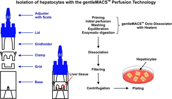
  • Principle: The gentleMACS Perfusion Technology allows the disaggregation of mouse liver tissue into highly viable single-cell suspensions. The perfusion is performed on the gentleMACS Octo Dissociator with Heaters equipped with gentleMACS Perfusion Sleeves in combination with gentleMACS Perfusers. The liver tissue is enzymatically digested using the components of the Liver Perfusion Kit, thereby loosening the structural integrity of the extracellular matrix in the tissue during the perfusion process. Afterwards, single cells are liberated from the tissue by a short mechanical disruption of the perfused tissue using the gentleMACS Octo Dissociator with Heaters and a gentleMACS C Tube. The sample is then applied to a MACS SmartStrainer to remove any remaining larger particles from the single-cell suspension. Cells should be processed immediately for downstream applications, such as cell separation, cell culture, and cellular or molecular analyses.
  • Background: The Liver Perfusion Kit in combination with the gentleMACS Perfusion Technology is suitable for the gentle, rapid, and efficient generation of single-cell suspensions from mouse liver. It is optimized for a high yield of hepatocytes, while preserving most cell surface epitopes.
  • Additional Reagents
  • Sterile, distilled water
  • Phosphate-buffered saline (PBS), pH 7.2
  • Culture medium (Dulbecco’s modified Eagle Medium containing 5% fetal bovine serum (FBS), 2 mM L-glutamine, 100 IU/mL penicillin, and 100 µg/mL streptomycin)
  • Additional Disposables
  • Disposable glass Pasteur pipettes with elongated tip (230 mm)
  • 6 cm and 10 cm petri dishes
  • Cell culture dishes (in desired format)
  • 15 and 50 mL reagent tubes
  • MACS SmartStrainers (100 μm) (Miltenyi, #130-098-463)
  • gentleMACS C Tubes (Miltenyi, #130-093-237)
  • gentleMACS Perfusers (Miltenyi, #130-128-151)
  • gentleMACS Perfusion Sleeves (Miltenyi, #130-128-752)
  • Additional Instruments
  • Surgical instrument set
  • Heatable water bath
  • Liquid suction pump
  • Cooling centrifuge with a swinging bucket rotor
  • gentleMACS Octo Dissociator with Heaters (Miltenyi, #130-096-427)
  • Preparation of Reagents from the Liver Perfusion Kit
  • Pre-heat water bath at 37 °C.
  • Warm sterile Buffer P (20×) at 37 °C for at least 45 min before use.
  • Prepare Reconstitution buffer: The reconstitution buffer is prepared by diluting 400 μL Buffer P (20×) with 7.6 mL sterile water. Add 16 μL Reagent C and mix well.
  • Reconstitute Enzyme D solution: Enzyme D solution is prepared by reconstitution of the lyophilized powder in the vial with 3 mL reconstitution buffer. After closing the lid, the vial is inverted to dissolve the enzyme for 5–10 min. The solution is sterile-filtered and aliquots can be stored at –20 °C for up to 6 months.
  • Reconstitute Enzyme R solution: Enzyme R solution is prepared by reconstitution of the lyophilized powder in the vial with 3 mL reconstitution buffer. After closing the lid, the vial is inverted to dissolve the enzyme for 5–10 min. After the enzyme is dissolved, sterile-filter the solution. Aliquots can be stored at −20 °C for up to 6 months.
  • Reconstitute Enzyme A solution: Enzyme A solution is prepared by sterile reconstitution of the lyophilized powder in the vial with 1 mL reconstitution buffer. Aliquots can be stored at −20 °C for up to 6 months.
  • Prepare pre-digestion buffer: For each isolation, prepare 62 mL Buffer P (1×) by diluting 3.1 mL Buffer P (20×) with 58.9 mL sterile, distilled water. Store buffer at 37 °C.
  • Prepare equilibration buffer: Add 36 µL Reagent C in 18 mL pre-digestion buffer. Transfer 9 mL of that buffer to a separate tube. This will be later used to prepare the enzyme digestion mix. Store both 9 mL buffer aliquots at 37 °C.
  • Experimental Setup and Preparation of Instrument and gentleMACS Perfuser
  • Exchange standard sleeves with gentleMACS Perfusion Sleeves on the gentleMACS Octo Dissociator with Heaters.
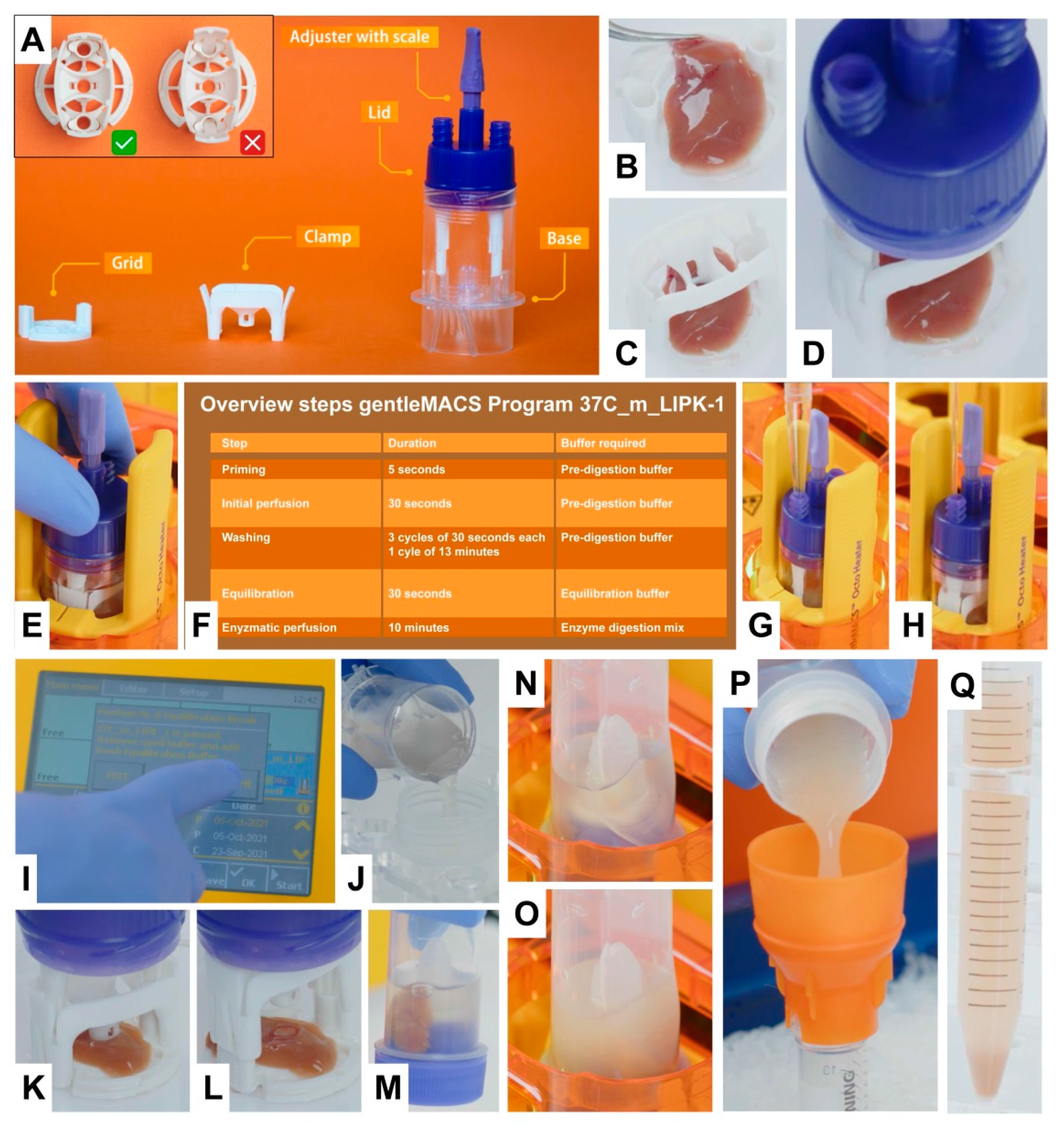
  • Remove Grid and Clamp from the gentleMACS Perfuser (Figure 2A) and place the Base with the Lid of the Perfuser on the instrument. Leave out the Grid and Clamp. Note: Ensure the Adjuster is in the correct (delivered) position according to the tissue to be perfused (must not be changed in the case of mouse perfusion).
  • Transfer 8 mL of warm pre-digestion buffer via one of the Luer lock openings using a 10 mL pipette (Figure 2G,H).
  • Place a Heating Unit onto the gentleMACS Perfuser and start the program 37C_m_LIPK_1 (Priming: 5 s; Initial perfusion: 30 s; Washing: 3 × 30 s, 1 × 13 min; Equilibration: 30 s; Enzymatic perfusion: 10 min) (Figure 2F,I). Note: The program will automatically pause after the priming step.
  • Put the Grid of the Perfuser into a sterile 10 cm petri dish.
  • Perfusion Procedure
  • Sacrifice the mouse.
  • Carefully dissect an appropriate liver lobe (left lateral lobe is preferred) and rinse with PBS buffer. Note: Make sure that the liver lobe is not injured during dissection.
  • Place the liver lobe in the center of the Grid using tweezers (see Figure 2B).
  • Squeeze the Clamp and attach it into the Grid by pushing it all the way down to the petri dish (see Figure 2C).
  • Remove the Lid from the Base and attach its Grid-holder channels through the Clamp into the Grid (Figure 2D). Note: A click is audible on each side of the two channels.
  • Place the assembly of Lid, Grid, Clamp, and liver tissue onto the installed Base.
  • Slowly turn the Lid counterclockwise without pressure until it reaches a lower position. Then, close Lid turning clockwise without tilting (Figure 2E).
  • Click the respective position and resume the program to start the 30 s initial perfusion.
  • After the initial perfusion step, select the flashing pause position.
  • Remove used buffer from the Perfuser with a disposable elongated Pasteur pipette (230 mm) connected to a suction pump. This washing phase consists of 3 short cycles (30 s) and one long cycle (1 × 13 min) with each 8 mL pre-digestion buffer. For buffer exchange, the program will pause after each cycle. During the pause steps, remove the used pre-digestion buffer manually with the glass Pasteur pipette, add new pre-digestion buffer and resume the program. At the last washing step, remove the used pre-digestion buffer.
  • Add 8 mL of warm equilibration buffer and resume the program.
  • Prepare the enzyme digestion mix. For one perfusion add 110 μL Enzyme D, 110 μL Enzyme R, and 30 μL Enzyme A to the 9 mL pre-warmed equilibration buffer (see Preparation of Reagents from the Liver Perfusion Kit, step 8).
  • Select paused position and remove the used buffer with the Pasteur pipette.
  • Add 8 mL of warm enzyme digestion mix and resume program.
  • After the enzymatic perfusion step (10 min), resume to finish the gentleMACS Program.
  • Remove Heating Unit and Perfuser from instrument.
  • Place Lid, Grid, and Clamp assembly in a petri dish.
  • Transfer used enzyme digestion mix to a gentleMACS C Tube (Figure 2J–L).
  • Lift Clamp with tweezers to remove perfused liver.
  • Transfer the perfused tissue to the gentleMACS C Tube containing the used enzyme digestion mix and close the C Tube tightly (Figure 2M).
  • Place gentleMACS C Tube upside-down in one position of the gentleMACS Dissociator equipped with a standard sleeve (Figure 2N).
  • Run the gentleMACS program LIPK_HR_1 that releases the hepatocytes from the perfused tissue within 5 min (Figure 2O).
  • Put a 15 mL tube on ice and place a MACS SmartStrainer (100 μm) on it.
  • After termination of the program, remove the gentleMACS C Tube from the gentleMACS Dissociator.
  • Open the gentleMACS C Tube and filter cell suspension through a MACS 100 µm SmartStrainer (Figure 2P). Note: Avoid producing air bubbles.
  • Wash the C Tube with 6 mL culture medium and pour this solution on the MACS SmartStrainer.
  • Centrifuge the cell suspension at 30× g for 5 min at 4 °C. A pellet containing hepatocytes will form (Figure 2Q).
  • Remove the supernatant completely. Note: This solution is enriched in non-parenchymal cells.
  • Carefully re-suspend the cell pellet consisting of hepatocytes by slowly pipetting up and down in an appropriate buffer for downstream application. Note: At this step you can use 5 mL cold culture medium.
  • Count cells in a Neubauer chamber and plate required number of cells into an appropriate culture dish.
  • Remarks
  • A maximum of 1.2 g of liver tissue can be perfused in one gentleMACS Perfuser.
  • The time from the dissection of the liver to the start of the perfusion should not exceed 10 min.
  • It is recommended to use the biggest mouse liver lobe (left lateral lobe) instead of the whole mouse liver.
  • The most critical step is screwing the Lid onto the Base. Do not use too much pressure during this step as the Clamp must not be lifted unless perfusion has been finished.
  • A video showing the complete procedure for isolation of hepatocytes from mouse liver using the gentleMACS Perfusion Technology was launched at [26].

References

  1. Rose, S.; Ezan, F.; Cuvellier, M.; Bruyère, A.; Legagneux, V.; Langouët, S.; Baffet, G. Generation of proliferating human adult hepatocytes using optimized 3D culture conditions. Sci. Rep. 2021, 11, 515. [Google Scholar] [CrossRef] [PubMed]
  2. Ward, L.; Wilcock, J.; Butler, P. Drug metabolism assessment: Hepatocytes. In The ADME Encyclopedia; Talevi, A., Ed.; Springer: Cham, Switzerland, 2021. [Google Scholar] [CrossRef]
  3. Sahi, J.; Grepper, S.; Smith, C. Hepatocytes as a tool in drug metabolism, transport and safety evaluations in drug discovery. Curr. Drug Discov. Technol. 2010, 7, 188–198. [Google Scholar] [CrossRef] [PubMed]
  4. Peng, W.C.; Kraaier, L.J.; Kluiver, T.A. Hepatocyte organoids and cell transplantation: What the future holds. Exp. Mol. Med. 2021, 53, 1512–1528. [Google Scholar] [CrossRef] [PubMed]
  5. Gamboa, C.M.; Wang, Y.; Xu, H.; Kalemba, K.; Wondisford, F.E.; Sabaawy, H.E. Optimized 3D culture of hepatic cells for liver organoid metabolic assays. Cells 2021, 10, 3280. [Google Scholar] [CrossRef] [PubMed]
  6. Hassan, S.; Sebastian, S.; Maharjan, S.; Lesha, A.; Carpenter, A.M.; Liu, X.; Xie, X.; Livermore, C.; Zhang, Y.S.; Zarrinpar, A. Liver-on-a-Chip Models of Fatty Liver Disease. Hepatology 2020, 71, 733–740. [Google Scholar] [CrossRef] [PubMed]
  7. Seglen, P.O. Preparation of isolated rat liver cells. Methods Cell. Biol. 1976, 13, 29–83. [Google Scholar] [CrossRef] [PubMed]
  8. USDA. Animal and Plant Health Inspection Service. Research Facility Annual Summary & Archive Reports. Available online: https://www.aphis.usda.gov/aphis/ourfocus/animalwelfare/sa_obtain_research_facility_annual_report/ct_research_facility_annual_summary_reports (accessed on 25 August 2022).
  9. National Research Council (US) Committee on Recognition and Alleviation of Pain in Laboratory Animals. Recognition and Alleviation of Pain in Laboratory Animals; US Regulations and Guidelines Regarding Recognition and Alleviation of Pain in Laboratory Animals; National Academies Press (US): Washington, DC, USA, 2009. Available online: https://www.ncbi.nlm.nih.gov/books/NBK32657/ (accessed on 25 August 2022).
  10. EUR-Lex. Access to European Union Law. Directive 2010/63/EU of the European Parliament and of the Council of 22 September 2010 on the Protection of Animals Used for Scientific Purposes Text with EEA Relevance. Available online: https://eur-lex.europa.eu/legal-content/EN/TXT/PDF/?uri=CELEX:32010L0063&from=EN (accessed on 25 August 2022).
  11. Borkham-Kamphorst, E.; Huss, S.; Van de Leur, E.; Haas, U.; Weiskirchen, R. Adenoviral CCN3/NOV gene transfer fails to mitigate liver fibrosis in an experimental bile duct ligation model because of hepatocyte apoptosis. Liver Int. 2012, 32, 1342–1353. [Google Scholar] [CrossRef] [PubMed]
  12. Jung, Y.; Zhao, M.; Svensson, K.J. Isolation, culture, and functional analysis of hepatocytes from mice with fatty liver disease. STAR Protoc. 2020, 1, 100222. [Google Scholar] [CrossRef] [PubMed]
  13. Charni-Natan, M.; Goldstein, I. Protocol for primary mouse hepatocyte isolation. STAR Protoc. 2020, 1, 100086. [Google Scholar] [CrossRef] [PubMed]
  14. Mitry, R.R. Isolation of human hepatocytes. Methods Mol. Biol. 2009, 481, 17–23. [Google Scholar] [CrossRef] [PubMed]
  15. Green, C.J.; Charlton, C.A.; Wang, L.M.; Silva, M.; Morten, K.J.; Hodson, L. The isolation of primary hepatocytes from human tissue: Optimising the use of small non-encapsulated liver resection surplus. Cell Tissue Bank 2017, 18, 597–604. [Google Scholar] [CrossRef] [PubMed]
  16. Lee, S.M.; Schelcher, C.; Demmel, M.; Hauner, M.; Thasler, W.E. Isolation of human hepatocytes by a two-step collagenase perfusion procedure. J. Vis. Exp. 2013, 79, 50615. [Google Scholar] [CrossRef] [PubMed]
  17. Kawai, M.; Saegusa, Y.; Kemmochi, S.; Harada, T.; Shimamoto, K.; Shibutani, M.; Mitsumori, K. Cytokeratin 8/18 is a useful immunohistochemical marker for hepatocellular proliferative lesions in mice. J. Vet. Med. Sci. 2010, 72, 263–269. [Google Scholar] [CrossRef] [PubMed]
  18. Itoh, M.; Terada, M.; Sugimoto, H. The zonula occludens protein family regulates the hepatic barrier system in the murine liver. Biochim. Biophys Acta Mol. Basis Dis. 2021, 1867, 165994. [Google Scholar] [CrossRef] [PubMed]
  19. Walesky, C.; Apte, U. Role of hepatocyte nuclear factor 4α (HNF4α) in cell proliferation and cancer. Gene Expr. 2015, 16, 101–108. [Google Scholar] [CrossRef] [PubMed]
  20. Borkham-Kamphorst, E.; Drews, F.; Weiskirchen, R. Induction of lipocalin-2 expression in acute and chronic experimental liver injury moderated by pro-inflammatory cytokines interleukin-1β through nuclear factor-κB activation. Liver Int. 2011, 31, 656–665. [Google Scholar] [CrossRef] [PubMed]
  21. Hu, Y.; Xue, J.; Yang, Y.; Zhou, X.; Qin, C.; Zheng, M.; Zhu, H.; Liu, Y.; Liu, W.; Lou, G.; et al. Lipocalin 2 upregulation protects hepatocytes from IL1-β-induced stress. Cell. Physiol. Biochem. 2015, 36, 753–762. [Google Scholar] [CrossRef] [PubMed]
  22. Qiu, S.; Vazquez, J.T.; Boulger, E.; Liu, H.; Xue, P.; Hussain, M.A.; Wolfe, A. Hepatic estrogen receptor α is critical for regulation of gluconeogenesis and lipid metabolism in males. Sci. Rep. 2017, 7, 1661. [Google Scholar] [CrossRef] [PubMed]
  23. Russell, W.M.S.; Burch, R.L. The Principles of Humane Experimental Technique; Reprinted 1992; Universities Federation for Animal Welfare: Wheathampstead, UK, 1959; Available online: https://caat.jhsph.edu/principles/the-principles-of-humane-experimental-technique (accessed on 25 August 2022).
  24. Baker, M. 1500 scientists lift the lid on reproducibility. Nature 2016, 533, 452–454. [Google Scholar] [CrossRef] [PubMed] [Green Version]
  25. Hirsch, C.; Schildknecht, S. In vitro research reproducibility: Keeping up high standards. Front. Pharmacol. 2019, 10, 1484. [Google Scholar] [CrossRef] [PubMed]
  26. Miltenyi Biotec’s Video Library. How to Perfuse Mouse Liver with the gentleMACS Perfusion Technology; Isolating Hepatocytes from Mouse Liver Has Never Been Easier! gentleMACSTM Perfusion Technology Makes It a Breeze to Isolate Hepatocytes from Rodent Liver. Available online: https://www.miltenyibiotec.com/tutorial-rodent-liver-perfusion (accessed on 25 August 2022).
Figure 1. The gentleMACS Perfusion Technology. (AC) The Perfuser can be disassembled into four parts, i.e., the Lid, Clamp, Grid, and Base. The Base contains an in-built peristaltic pump which delivers liquids via a needle array into the penetrated liver sample. To aspirate buffer from the inside of the Perfuser, a Pasteur pipette with an elongated tip is introduced through one of the Luer-lock connectors to the Base bottom. The Adjuster on top of the Lid can be used to adapt the penetration depth of the needle array into the liver tissue.
Biomedicines 10 02198 g001
Figure 2. Selected working steps for the isolation of primary mouse hepatocytes using the gentleMACS Perfusion Technology. (A) The gentleMACS Perfuser components. The inlet shows how the Clamp should be inserted into the Grid. (B,C) Prior to perfusion, the liver lobe is placed on the Grid and fixed with the Clamp. (D) The Lid connected via the Grid holder to the Grid-Clamp assembly. (E) Lid-Clamp-Grid structure screwed into the Base of the gentleMACS Perfuser. (F) Overview of program steps of the gentleMACS Program 37C_m_LIPK_1. (G,H) Addition and removal of buffers after each cycle can be done via one of the Luer locks on top of the Lid. (I) Display of the gentleMACS Octo Dissociator with Heaters. (J) Filling of the used enzyme digestion mix from the Base into a gentleMACS C Tube. (K,L) Lifting of the Clamp after completion of the perfusion to enable transfer of the perfused lobe into the gentleMACS C Tube. (M) Tissue after transfer from the Grid into the C Tube containing the used enzyme digestion mix. (N) gentleMACS C Tube containing perfused liver placed on the gentleMACS Dissociator. (O) Cell suspension in C Tube after running Program LIPK_HR_1. The release of hepatocytes from the tissue results in cloudiness of the solution. (P) Filtering of cell suspension through the MACS SmartStrainer. (Q) Hepatocyte pellet after low-spin centrifugation. The detailed protocol is given in Appendix A.
Biomedicines 10 02198 g002
Figure 3. Color changes of the liver tissue during perfusion with the gentleMACS Perfusion Technology. (A) Four different liver lobes before perfusion are depicted. (B) The upper two liver lobes from (A) after perfusion with the pre-digestion buffer. (C) The lower two liver lobes from (A) after enzymatic digestion.
Biomedicines 10 02198 g003
Figure 4. Light microscopic appearance of hepatocytes isolated with the gentleMACS Perfusion Technology. Primary hepatocytes were isolated from a 3-month-old male mouse and seeded in HepatoZYME-SFM medium (#17705021, ThermoFisher, Dreieich, Germany). The medium was changed to Dulbecco’s Modified Eagle’s Medium containing 10% fetal bovine serum, 2 mM L-glutamine, 100 IU/mL penicillin, and 100 µg/mL streptomycin (all from ThermoFisher) two hours after initial plating. Pictures were taken at indicated times and magnifications.
Biomedicines 10 02198 g004
Figure 5. Marker protein expression in hepatocytes. (A) Primary hepatocytes were isolated with the gentleMACS Perfusion Technology and subjected to fluorescence immunostaining using rabbit monoclonal anti-cytokeratin 8 (CK8) (#ab53280, Abcam, Berlin, Germany), polyclonal rabbit anti-Zonula occludens-1 (ZO-1) (#85047, Novus Biologicals, Wiesbaden-Nordenstadt, Germany), and polyclonal goat hepatocyte nuclear factor 4α (HNF4α) (#sc-6556, Santa Cruz Biotech., Santa Cruz, CA, USA) for staining. Nuclei were counterstained with 4′,6-diamidino-2-phenylindole (DAPI, Sigma-Aldrich, Taufkirchen, Germany) and images were taken at 1000× magnification using a Leica DMRB fluorescence microscope (Leica, Wetzlar, Germany) equipped with a Nikon Coolpix 5400 camera (Nikon Germany, Düsseldorf, Germany). Brightness and contrast were adjusted for clarity without changing the content. (B) Protein extracts from hepatocytes isolated with the gentleMACS Perfusion Technology or conventional isolation procedure were tested using Western blot analysis for expression of ZO-1 (#40-2300, ThermoFisher), HNF4α (#sc-6556, Santa Cruz Biotech.), Lipocalin 2 (LCN2) (#AF3508, R&D Systems, Wiesbaden, Germany) and Glyceraldehyde-3-phosphate dehydrogenase (GAPDH) (#sc-32233, Santa Cruz). The mouse hepatocyte cell line AML12 was taken as a positive control for expression of ZO-1 and HNF4α.
Biomedicines 10 02198 g005
Figure 6. Biochemical features of hepatocytes isolated with the gentleMACS Perfusion Technology. (A) Comparative analysis of Albumin (Alb) and hepatocyte nuclear factor 4α (Hnf4a) mRNA expression in hepatocytes isolated either by the gentleMACS Perfusion Technology or conventional isolation procedure. The RNA was subjected to a standard RT-PCR using the following primers: Alb-for: 5′-ggt ctc atc tgt ccg tca gag-3′, Alb-rev: 5′-gga aga cat cct tgg cct cag-3′, Hnf4a-for: 5′-cca aga ggt cca tgg tgt tt-3′, Hnf4a-rev: 5′-ccg agg gac gat gta gtc at-3′, Actb-for: 5′-ctc tag act tcg agc agg aga tgg-3′, and Actb-rev: 5′-atg cca cag gat tcc ata ccc aag a-3′. Cycle conditions were: 5 min initial denaturation at 95 °C, 1 min at 95 °C, 1 min annealing at 60 °C (Actb, 25 cycles; Hnf4a, 35 cycles) or 64 °C (Alb, 30 cycles), 3 min extension at 72 °C, and final elongation at 72 °C for 10 min. Amplicons were separated in 1.6% (Alb) or 2% (Actb, Hnf4a) 1 × TBE-buffered agarose gels containing ethidium bromide and visualized using gel imager Gel iX20 (Intas Science Imaging Instruments GmbH, Göttingen, Germany). (B) Western blot analysis of protein extracts and supernatants isolated from cultured hepatocytes isolated with the gentleMACS Perfusion Technology that were stimulated with recombinant human IL-1β (#130-093-895, Miltenyi Biotec) or left untreated (control). (C) Comparative analysis of IL-1β sensitivity of hepatocytes isolated with the gentleMACS Perfusion Technology or the conventional isolation protocol. Antibodies used were directed against estrogen receptor-α (ER-α) (#MA1-27107, ThermoFisher), Lipocalin 2 (LCN2) (#AF3508, R&D Systems), or Glyceraldehyde-3-phosphate dehydrogenase (GAPDH) (#sc-32233, Santa Cruz Biotech.).
Biomedicines 10 02198 g006
Table 1. Representative hepatocyte isolations from female mice (n = 33) *.
Number of
Animals Used
StrainAge (Weeks)Total
Hepatocyte Yield **
Hepatocyte Yield/g **Viability (%) ***
n = 2CD157.0 × 1061.5 × 10781
1.9 × 1075.8 × 10791
n = 2BALB/c91.5 × 1074.5 × 10785
5.5 × 1061.7 × 10787
n = 2BALB/c81.1 × 1073.9 × 10790
6.7 × 1061.9 × 10788
n = 2C57BL/6121.4 × 1072.9 × 10788
9.6 × 1062.0 × 10788
n = 2BALB/c101.2 × 1073.0 × 10793
1.2 × 1073.5 × 10790
n = 4BALB/c109.5 × 1063.2 × 10793
3.2 × 1061.1 × 10786
1.1 × 1073.8 × 10793
1.5 × 1074.8 × 10792
n = 4C57BL/691.1 × 1073.5 × 10791
6.2 × 1061.7 × 10785
1.4 × 1074.1 × 10791
9.8 × 1062.6 × 10795
n = 8BALB/c81.2 × 1073.4 × 10791
1.0 × 1072.5 × 10791
1.6 × 1075.4 × 10790
1.1 × 1073.3 × 10793
7.8 × 1062.4 × 10790
1.3 × 1074.1 × 10785
1.5 × 1074.3 × 10790
7.2 × 1061.9 × 10784
n = 3BALB/c69.3 × 1062.3 × 10784
1.1 × 1073.0 × 10783
7.4 × 1062.2 × 10786
n = 4C57BL/691.4 × 1074.6 × 10790
1.7 × 1075.2 × 10794
1.0 × 1075.9 × 10790
1.7 × 1075.6 × 10788
Notes: * For this set of experiments, hepatocyte isolation was exclusively done from the left lateral lobe; ** Yield is given either as total yield or alternatively as yield per g liver tissue; *** For determination of cell viability, the cells were stained with propidium iodide (final concentration: 1 µg/mL) and directly analyzed by flow cytometry. Cells with high forward and side scatter were selected, gated on simultaneous autofluorescence in the UV and FITC channels representing typical features of hepatocytes and evaluated for the existence of propidium iodide intensity.
Table 2. Differences between the standard perfusion and the gentleMACS Perfusion Technology.
FeatureConventional In Vivo PerfusiongentleMACS Perfusion Technology
Approachin situex vivo
Perfusion environmentanaesthetized animalresected tissue
Animalsbest working with adult animals
(cannulation of veins is necessary)
flexible in regard to age of animals
(no cannulation of veins necessary)
Animal ethics approvalnecessarynot needed
Sample material flexibilityno
(whole organ)
yes
(lobe rather than whole organ is recommended)
Perfusion enzymesnot standardized
(lot-to-lot variations, activities must be determined by customer)
standardized
(consistent enzyme activities contained in the corresponding Liver Perfusion Kit)
(Difficult) manual cannulation of liver vessels neededyesnot needed
Sample processed in a closed systemnoyes
Perfusion steps and volumes (mL)highly variable7 × 8
Collection of used perfusion liquidnoyes
Other lobes of the same liver available for other experimentsno
(at least even more difficult to process)
yes
Robustness of needle insertion into the tissuelow-high
(depending on user skills)
high
(automatically done by the consumable)
Flow rate during perfusionto be set by userpredefined by the device
Sterility of equipmentno
(sterilization needs preparation time)
yes
(usage of disposable items)
Sample size limitationno
(open system)
yes
(closed system)
Needed instrumentationperistaltic pump with tubinggentleMACS Octo Dissociator with Heaters and gentleMACS Perfusion Sleeve
Needed one-way consumablessyringe needle, buffer reservoirsgentleMACS Perfuser (for perfusion) and gentleMACS C Tube (for dissociation of perfused liver)
Ease of parallelization of perfusion processlow
(would require additional peristaltic pumps and complex nesting of work steps)
high
(Up to 8 cell preparations can be performed simultaneously on one instrument)
Disruption of perfused liver tissuemanually
(mincing of tissue with forceps)
automated
(within C Tube driven by instrument)
Publisher’s Note: MDPI stays neutral with regard to jurisdictional claims in published maps and institutional affiliations.

