
Optical Behavior of Human Skin Substitutes: Absorbance in the 200–400 nm UV Range

 , , , and
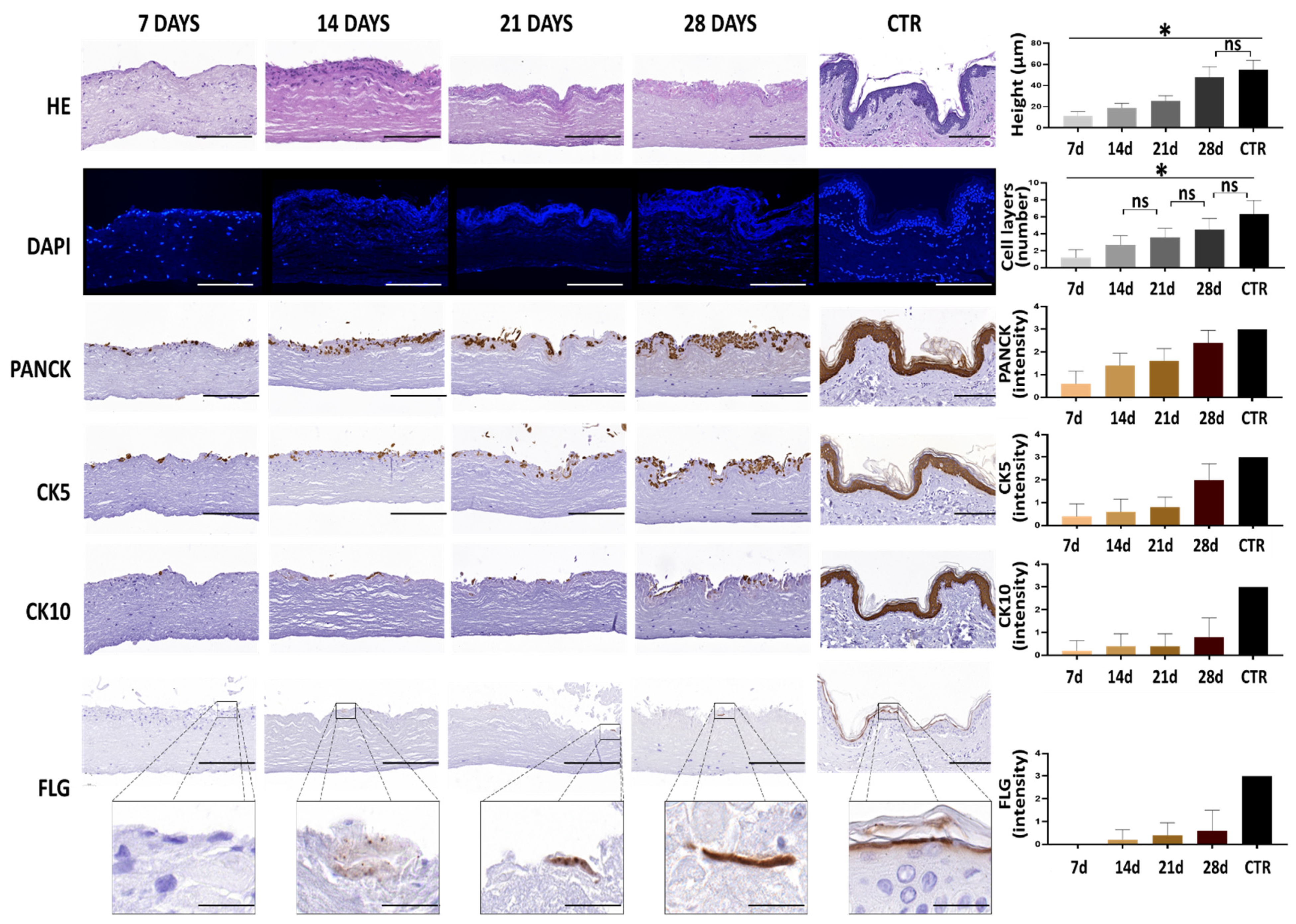
Abstract
:1. Introduction
2. Materials and Methods
2.1. Tissue Samples and Cell Isolation
2.2. Generation of Human Skin Substitutes by Tissue Engineering
2.3. Histological Analysis
2.4. Optical Properties
2.5. Quantification of Histological Parameters and Statistical Analysis
3. Results
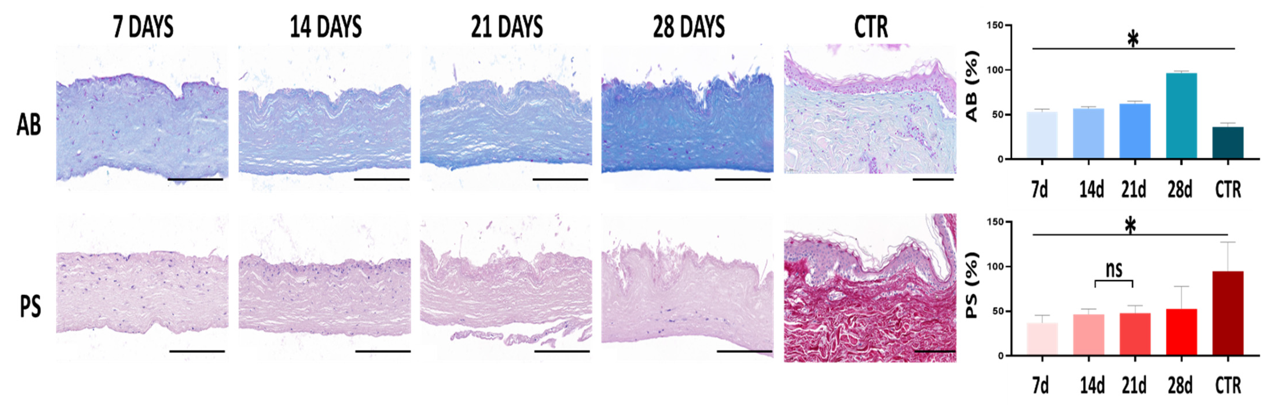
3.1. Histological Characterization of Human Skin Substitutes
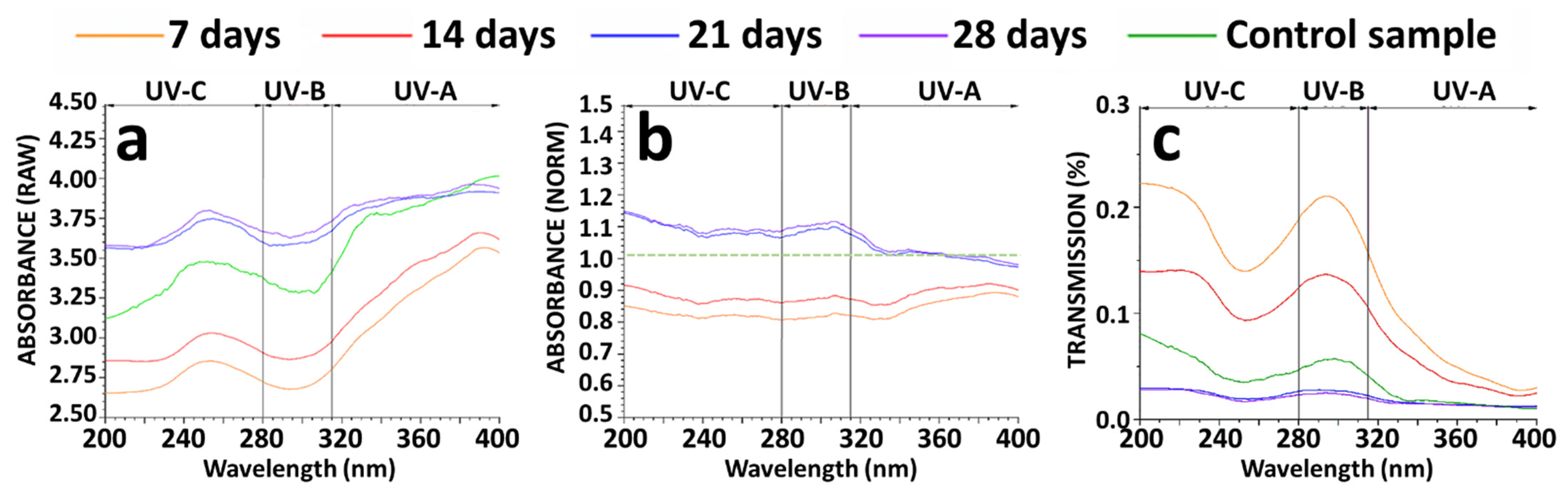
3.2. Optical Characterization of the Human Skin Substitutes
4. Discussion
5. Conclusions
Author Contributions
Funding
Institutional Review Board Statement
Informed Consent Statement
Data Availability Statement
Conflicts of Interest
References
- D’Orazio, J.; Jarrett, S.; Amaro-Ortiz, A.; Scott, T. UV Radiation and the Skin. Int. J. Mol. Sci. 2013, 14, 12222–12248. [Google Scholar] [CrossRef] [PubMed] [Green Version]
- Bernerd, F.; Marionnet, C.; Duval, C. Solar Ultraviolet Radiation Induces Biological Alterations in Human Skin in Vitro: Relevance of a Well-Balanced UVA/UVB Protection. Indian J. Dermatol. Venereol. Leprol. 2012, 78 (Suppl. S1), S15–S23. [Google Scholar] [CrossRef] [PubMed]
- Mohania, D.; Chandel, S.; Kumar, P.; Verma, V.; Digvijay, K.; Tripathi, D.; Choudhury, K.; Mitten, S.K.; Shah, D. Ultraviolet Radiations: Skin Defense-Damage Mechanism. Adv. Exp. Med. Biol. 2017, 996, 71–87. [Google Scholar] [CrossRef] [PubMed]
- Matsumura, Y.; Ananthaswamy, H.N. Toxic Effects of Ultraviolet Radiation on the Skin. Toxicol. Appl. Pharmacol. 2004, 195, 298–308. [Google Scholar] [CrossRef]
- Shukla, A.; Mukherjee, S.; Sharma, S.; Agrawal, V.; Radha Kishan, K.V.; Guptasarma, P. A Novel UV Laser-Induced Visible Blue Radiation from Protein Crystals and Aggregates: Scattering Artifacts or Fluorescence Transitions of Peptide Electrons Delocalized through Hydrogen Bonding? Arch. Biochem. Biophys. 2004, 428, 144–153. [Google Scholar] [CrossRef] [PubMed]
- Xu, G.; Marcusson, J.A.; Hemminki, K. DNA Photodamage Induced by UV Phototherapy Lamps and Sunlamps in Human Skin in Situ and Its Potential Importance for Skin Cancer. J. Investig. Dermatol. 2001, 116, 194–195. [Google Scholar] [CrossRef] [Green Version]
- Tyrrell, R.M.; Keyse, S.M. New Trends in Photobiology the Interaction of UVA Radiation with Cultured Cells. J. Photochem. Photobiol. B 1990, 4, 349–361. [Google Scholar] [CrossRef]
- Jensen, J.M.; Proksch, E. The Skin’s Barrier. G. Ital. Dermatol. Venereol. 2009, 144, 689–700. [Google Scholar]
- MacNeil, S. Progress and Opportunities for Tissue-Engineered Skin. Nature 2007, 445, 874–880. [Google Scholar] [CrossRef]
- Lazic, T.; Falanga, V. Bioengineered Skin Constructs and Their Use in Wound Healing. Plast. Reconstr. Surg. 2011, 127 (Suppl. S1), 75S–90S. [Google Scholar] [CrossRef]
- Ijaola, A.O.; Akamo, D.O.; Damiri, F.; Akisin, C.J.; Bamidele, E.A.; Ajiboye, E.G.; Berrada, M.; Onyenokwe, V.O.; Yang, S.-Y.; Asmatulu, E. Polymeric Biomaterials for Wound Healing Applications: A Comprehensive Review. J. Biomater. Sci. Polym. Ed. 2022, 1–53. [Google Scholar] [CrossRef] [PubMed]
- Berthiaume, F.; Hsia, H.C. Regenerative Approaches for Chronic Wounds. Annu. Rev. Biomed. Eng. 2022, 24, 61–83. [Google Scholar] [CrossRef] [PubMed]
- Nilforoushzadeh, M.A.; Amirkhani, M.A.; Khodaverdi, E.; Razzaghi, Z.; Afzali, H.; Izadpanah, S.; Zare, S. Tissue Engineering in Dermatology-from Lab to Market. Tissue Cell 2022, 74, 101717. [Google Scholar] [CrossRef] [PubMed]
- Xu, J.; Zheng, S.; Hu, X.; Li, L.; Li, W.; Parungao, R.; Wang, Y.; Nie, Y.; Liu, T.; Song, K. Advances in the Research of Bioinks Based on Natural Collagen, Polysaccharide and Their Derivatives for Skin 3D Bioprinting. Polymers 2020, 12, 1237. [Google Scholar] [CrossRef]
- Final Report Summary-EUROSKINGRAFT (A Novel Generation of Skin Substitutes to Clinically Treat a Broad Spectrum of Severe Skin Defects)|FP7. Available online: https://cordis.europa.eu/project/id/279024/reporting (accessed on 4 July 2022).
- Oliveira, A.; Simões, S.; Ascenso, A.; Reis, C.P. Therapeutic Advances in Wound Healing. J. Dermatol. Treat. 2022, 33, 2–22. [Google Scholar] [CrossRef]
- Holmes, J.H.; Schurr, M.J.; King, B.T.; Foster, K.; Faucher, L.D.; Lokuta, M.A.; Comer, A.R.; Rooney, P.J.; Barbeau, K.F.; Mohoney, S.T.; et al. An Open-Label, Prospective, Randomized, Controlled, Multicenter, Phase 1b Study of StrataGraft Skin Tissue versus Autografting in Patients with Deep Partial-Thickness Thermal Burns. Burns J. Int. Soc. Burn Inj. 2019, 45, 1749–1758. [Google Scholar] [CrossRef]
- Takami, Y.; Yamaguchi, R.; Ono, S.; Hyakusoku, H. Clinical Application and Histological Properties of Autologous Tissue-Engineered Skin Equivalents Using an Acellular Dermal Matrix. J. Nippon Med. Sch. 2014, 81, 356–363. [Google Scholar] [CrossRef] [Green Version]
- Oostendorp, C.; Meyer, S.; Sobrio, M.; van Arendonk, J.; Reichmann, E.; Daamen, W.F.; van Kuppevelt, T.H. Evaluation of Cultured Human Dermal- and Dermo-Epidermal Substitutes Focusing on Extracellular Matrix Components: Comparison of Protein and RNA Analysis. Burns 2017, 43, 520–530. [Google Scholar] [CrossRef]
- Carriel, V.; Garzón, I.; Jiménez, J.-M.; Oliveira, A.-C.-X.; Arias-Santiago, S.; Campos, A.; Sánchez-Quevedo, M.-C.; Alaminos, M. Epithelial and Stromal Developmental Patterns in a Novel Substitute of the Human Skin Generated with Fibrin-Agarose Biomaterials. Cells Tissues Organs 2012, 196, 1–12. [Google Scholar] [CrossRef]
- Ionescu, A.M.; Chato-Astrain, J.; Cardona Pérez, J.d.l.C.; Campos, F.; Pérez Gómez, M.; Alaminos, M.; Garzón Bello, I. Evaluation of the Optical and Biomechanical Properties of Bioengineered Human Skin Generated with Fibrin-Agarose Biomaterials. J. Biomed. Opt. 2020, 25, 055002. [Google Scholar] [CrossRef]
- Garzón, I.; Miyake, J.; González-Andrades, M.; Carmona, R.; Carda, C.; Sánchez-Quevedo, M.d.C.; Campos, A.; Alaminos, M. Wharton’s Jelly Stem Cells: A Novel Cell Source for Oral Mucosa and Skin Epithelia Regeneration. Stem Cells Transl. Med. 2013, 2, 625–632. [Google Scholar] [CrossRef] [PubMed]
- Martin-Piedra, M.A.; Alfonso-Rodriguez, C.A.; Zapater, A.; Durand-Herrera, D.; Chato-Astrain, J.; Campos, F.; Sanchez-Quevedo, M.C.; Alaminos, M.; Garzon, I. Effective Use of Mesenchymal Stem Cells in Human Skin Substitutes Generated by Tissue Engineering. Eur. Cell. Mater. 2019, 37, 233–249. [Google Scholar] [CrossRef] [PubMed]
- Cuende, N.; Boniface, C.; Bravery, C.; Forte, M.; Giordano, R.; Hildebrandt, M.; Izeta, A.; Dominici, M. Legal and Regulatory Affairs Committee—Europe, International Society for Cellular Therapy the Puzzling Situation of Hospital Exemption for Advanced Therapy Medicinal Products in Europe and Stakeholders’ Concerns. Cytotherapy 2014, 16, 1597–1600. [Google Scholar] [CrossRef] [PubMed] [Green Version]
- Egea-Guerrero, J.J.; Carmona, G.; Correa, E.; Mata, R.; Arias-Santiago, S.; Alaminos, M.; Gacto, P.; Cuende, N. Transplant of Tissue-Engineered Artificial Autologous Human Skin in Andalusia: An Example of Coordination and Institutional Collaboration. Transplant. Proc. 2019, 51, 3047–3050. [Google Scholar] [CrossRef] [PubMed]
- Chato-Astrain, J.; Sánchez-Porras, D.; García-García, Ó.D.; Vairo, C.; Villar-Vidal, M.; Villullas, S.; Sánchez-Montesinos, I.; Campos, F.; Garzón, I.; Alaminos, M. Improvement of Cell Culture Methods for the Successful Generation of Human Keratinocyte Primary Cell Cultures Using EGF-Loaded Nanostructured Lipid Carriers. Biomedicines 2021, 9, 1634. [Google Scholar] [CrossRef]
- Chato-Astrain, J.; Chato-Astrain, I.; Sánchez-Porras, D.; García-García, Ó.-D.; Bermejo-Casares, F.; Vairo, C.; Villar-Vidal, M.; Gainza, G.; Villullas, S.; Oruezabal, R.-I.; et al. Generation of a Novel Human Dermal Substitute Functionalized with Antibiotic-Loaded Nanostructured Lipid Carriers (NLCs) with Antimicrobial Properties for Tissue Engineering. J. Nanobiotechnology 2020, 18, 174. [Google Scholar] [CrossRef]
- Sánchez-Porras, D.; Caro-Magdaleno, M.; González-Gallardo, C.; García-García, Ó.D.; Garzón, I.; Carriel, V.; Campos, F.; Alaminos, M. Generation of a Biomimetic Substitute of the Corneal Limbus Using Decellularized Scaffolds. Pharmaceutics 2021, 13, 1718. [Google Scholar] [CrossRef]
- Juzeniene, A.; Brekke, P.; Dahlback, A.; Andersson-Engels, S.; Reichrath, J.; Moan, K.; Holick, M.F.; Grant, W.B.; Moan, J. Solar Radiation and Human Health. Rep. Prog. Phys. 2011, 74, 066701. [Google Scholar] [CrossRef] [Green Version]
- Carriel, V.; Garzón, I.; Campos, A.; Cornelissen, M.; Alaminos, M. Differential Expression of GAP-43 and Neurofilament during Peripheral Nerve Regeneration through Bio-Artificial Conduits. J. Tissue Eng. Regen. Med. 2017, 11, 553–563. [Google Scholar] [CrossRef]
- Ortiz-Arrabal, O.; Carmona, R.; García-García, Ó.-D.; Chato-Astrain, J.; Sánchez-Porras, D.; Domezain, A.; Oruezabal, R.-I.; Carriel, V.; Campos, A.; Alaminos, M. Generation and Evaluation of Novel Biomaterials Based on Decellularized Sturgeon Cartilage for Use in Tissue Engineering. Biomedicines 2021, 9, 775. [Google Scholar] [CrossRef]
- Rodriguez-Pozo, J.A.; Ramos-Lopez, J.F.; Gonzalez-Gallardo, M.C.; Campos, F.; Sanchez-Porras, D.; Oyonarte, S.; Oruezabal, R.I.; Campos, A.; Martin-Piedra, M.A.; Alaminos, M. Evaluation of Myopic Cornea Lenticules. A Histochemical and Clinical Correlation. Exp. Eye Res. 2020, 196, 108066. [Google Scholar] [CrossRef] [PubMed]
- Carriel, V.; Garzón, I.; Alaminos, M.; Campos, A. Evaluation of Myelin Sheath and Collagen Reorganization Pattern in a Model of Peripheral Nerve Regeneration Using an Integrated Histochemical Approach. Histochem. Cell Biol. 2011, 136, 709–717. [Google Scholar] [CrossRef] [PubMed]
- Vela-Romera, A.; Carriel, V.; Martín-Piedra, M.A.; Aneiros-Fernández, J.; Campos, F.; Chato-Astrain, J.; Prados-Olleta, N.; Campos, A.; Alaminos, M.; Garzón, I. Characterization of the Human Ridged and Non-Ridged Skin: A Comprehensive Histological, Histochemical and Immunohistochemical Analysis. Histochem. Cell Biol. 2019, 151, 57–73. [Google Scholar] [CrossRef] [Green Version]
- Rico-Sánchez, L.; Garzón, I.; González-Andrades, M.; Ruíz-García, A.; Punzano, M.; Lizana-Moreno, A.; Muñoz-Ávila, J.I.; Sánchez-Quevedo, M.D.C.; Martínez-Atienza, J.; Lopez-Navas, L.; et al. Successful Development and Clinical Translation of a Novel Anterior Lamellar Artificial Cornea. J. Tissue Eng. Regen. Med. 2019, 13, 2142–2154. [Google Scholar] [CrossRef] [PubMed] [Green Version]
- Tsumura, N.; Haneishi, H.; Miyake, Y. Independent Component Analysis of Spectral Absorbance Image in Human Skin. Opt. Rev. 2000, 7, 479–482. [Google Scholar] [CrossRef] [Green Version]
- Austin, E.; Geisler, A.N.; Nguyen, J.; Kohli, I.; Hamzavi, I.; Lim, H.W.; Jagdeo, J. Visible Light. Part I: Properties and Cutaneous Effects of Visible Light. J. Am. Acad. Dermatol. 2021, 84, 1219–1231. [Google Scholar] [CrossRef]
- Nguyen, J.; Hayakawa, C.K.; Mourant, J.R.; Spanier, J. Perturbation Monte Carlo Methods for Tissue Structure Alterations. Biomed. Opt. Express 2013, 4, 1946–1963. [Google Scholar] [CrossRef] [Green Version]
- Castellano-Pellicena, I.; Uzunbajakava, N.E.; Mignon, C.; Raafs, B.; Botchkarev, V.A.; Thornton, M.J. Does Blue Light Restore Human Epidermal Barrier Function via Activation of Opsin during Cutaneous Wound Healing? Lasers Surg. Med. 2019, 51, 370–382. [Google Scholar] [CrossRef] [Green Version]
- Meyer, T.; Stockfleth, E. Light and Skin. Curr. Probl. Dermatol. 2021, 55, 53–61. [Google Scholar] [CrossRef]
- Fernanda de Mello Costa, M.; Weiner, A.I.; Vaughan, A.E. Basal-like Progenitor Cells: A Review of Dysplastic Alveolar Regeneration and Remodeling in Lung Repair. Stem Cell Rep. 2020, 15, 1015–1025. [Google Scholar] [CrossRef]
- Cascella, R.; Strafella, C.; Germani, C.; Manzo, L.; Marsella, L.T.; Borgiani, P.; Sobhy, N.; Abdelmaksood, R.; Gerou, S.; Ioannides, D.; et al. FLG (Filaggrin) Null Mutations and Sunlight Exposure: Evidence of a Correlation. J. Am. Acad. Dermatol. 2015, 73, 528–529. [Google Scholar] [CrossRef] [Green Version]
- Mildner, M.; Jin, J.; Eckhart, L.; Kezic, S.; Gruber, F.; Barresi, C.; Stremnitzer, C.; Buchberger, M.; Mlitz, V.; Ballaun, C.; et al. Knockdown of Filaggrin Impairs Diffusion Barrier Function and Increases UV Sensitivity in a Human Skin Model. J. Investig. Dermatol. 2010, 130, 2286–2294. [Google Scholar] [CrossRef] [Green Version]
- Morrison, H. Photochemistry and Photobiology of Urocanic Acid. Photo-dermatology 1985, 2, 158–165. [Google Scholar] [CrossRef]
- Zenisek, A.; Kral, J.A.; Hais, I.M. Sun-Screening Effect of Urocanic Acid. Biochim. Biophys. Acta 1955, 18, 589–591. [Google Scholar] [CrossRef]
- Ji, S.; Zhu, Z.; Sun, X.; Fu, X. Functional Hair Follicle Regeneration: An Updated Review. Signal Transduct. Target. Ther. 2021, 6, 66. [Google Scholar] [CrossRef]
- Antezana, P.E.; Municoy, S.; Álvarez-Echazú, M.I.; Santo-Orihuela, P.L.; Catalano, P.N.; Al-Tel, T.H.; Kadumudi, F.B.; Dolatshahi-Pirouz, A.; Orive, G.; Desimone, M.F. The 3D Bioprinted Scaffolds for Wound Healing. Pharmaceutics 2022, 14, 464. [Google Scholar] [CrossRef]
- Olejnik, A.; Semba, J.A.; Kulpa, A.; Dańczak-Pazdrowska, A.; Rybka, J.D.; Gornowicz-Porowska, J. 3D Bioprinting in Skin Related Research: Recent Achievements and Application Perspectives. ACS Synth. Biol. 2022, 11, 26–38. [Google Scholar] [CrossRef]


Publisher’s Note: MDPI stays neutral with regard to jurisdictional claims in published maps and institutional affiliations. |
© 2022 by the authors. Licensee MDPI, Basel, Switzerland. This article is an open access article distributed under the terms and conditions of the Creative Commons Attribution (CC BY) license (https://creativecommons.org/licenses/by/4.0/).
Share and Cite
Ruiz-López, J.; Cardona, J.C.; Garzón, I.; Pérez, M.M.; Alaminos, M.; Chato-Astrain, J.; Ionescu, A.M. Optical Behavior of Human Skin Substitutes: Absorbance in the 200–400 nm UV Range. Biomedicines 2022, 10, 1640. https://doi.org/10.3390/biomedicines10071640
Ruiz-López J, Cardona JC, Garzón I, Pérez MM, Alaminos M, Chato-Astrain J, Ionescu AM. Optical Behavior of Human Skin Substitutes: Absorbance in the 200–400 nm UV Range. Biomedicines. 2022; 10(7):1640. https://doi.org/10.3390/biomedicines10071640
Chicago/Turabian StyleRuiz-López, Javier, Juan C. Cardona, Ingrid Garzón, María M. Pérez, Miguel Alaminos, Jesus Chato-Astrain, and Ana M. Ionescu. 2022. "Optical Behavior of Human Skin Substitutes: Absorbance in the 200–400 nm UV Range" Biomedicines 10, no. 7: 1640. https://doi.org/10.3390/biomedicines10071640