Article

Deletion of Toxoplasma Rhoptry Protein 38 (PruΔrop38) as a Vaccine Candidate for Toxoplasmosis in a Murine Model

1
National Animal Protozoa Laboratory, College of Veterinary Medicine, China Agricultural University, Beijing 100193, China
2
Key Laboratory of Animal Epidemiology of the Ministry of Agriculture, College of Veterinary Medicine, China Agricultural University, Beijing 100193, China
*
Author to whom correspondence should be addressed.
Biomedicines 2022, 10(6), 1336; https://doi.org/10.3390/biomedicines10061336
Submission received: 29 April 2022 / Revised: 3 June 2022 / Accepted: 4 June 2022 / Published: 6 June 2022
(This article belongs to the Topic Infectious Diseases)

Abstract

:
Toxoplasmosis is a serious zoonotic disease that threatens human and animal health. Here, we evaluated the vaccine potential of the deletion of Toxoplasma rhoptry protein 38 (PruΔrop38) through its pathogenicity and immunoprotective efficacy in mice. Mice inoculated intraperitoneally with 1 × 10³, 2 × 10³, or 4 × 10³ PruΔrop38 showed no visible signs, whereas mice inoculated with 1 × 10³ parental Pru strain showed obvious wasting and bow-back, suggesting a significantly lower pathogenicity of PruΔrop38 in mice. Vaccination with 1 × 10² PruΔrop38 triggered a mixed Th1/Th2 response (Th1 response predominant), with higher IgG, IgG2a, and IgG1 levels in serum from week 3 to week 12, and a significant increase in IFN-γ, IL-12, and IL-10 in suspensions of splenocytes at 30 or 60 days post-immunization. All vaccinated mice survived when infected intraperitoneally with tachyzoites (RH, Pru, VEG, or TgcatBJ1) or when infected orally with cysts (Pru or ME49). The brain parasite burden during Pru tachyzoite, Pru cyst and ME49 cyst challenges were significantly reduced in vaccinated mice. The duration of immunization showed that vaccination with PruΔrop38 could protect mice from challenge with different varied genotypes of Toxoplasma strains against different routes of infection. Collectively, these findings indicate that PruΔrop38 is an attenuated strain that provides long-term protective efficacy against acute or chronic toxoplasmosis in mice.

1. Introduction

Toxoplasma gondii is an obligate intracellular protozoan with a wide host range that can infect almost all warm-blooded animals and is capable of causing toxoplasmosis in both humans and animals [1,2]. Toxoplasmosis remains one of the world’s leading causes of illness and death, resulting in more than 200 million clinical cases of human zoonotic infections each year, which develop as fatal encephalitis or pneumonia in immunodeficient patients undergoing AIDS treatment [3]. Infected pregnant women may suffer a miscarriage or their newborn babies may suffer from congenital toxoplasmosis [4]. Toxoplasma infection is acquired by ingestion of water and food, contaminated with T. gondii oocysts or cysts. Ingestion of these life stages rapidly converts them into tachyzoites in the host for mass reproduction, leading to acute infection [5].
Pyrimethamine and sulfadiazine are commonly available treatments for the control of toxoplasmosis, while failure rates and severe side effects of the drugs remain significant, targeting only the active stage of infection [6]. Vaccines are expected to be the most promising method to prevent and cure toxoplasmosis [7]. The development of toxoplasmosis vaccines has evolved from traditional vaccines (e.g., live and inactivated vaccines) to molecular biotechnology vaccines (e.g., recombinant live vector vaccines, recombinant subunit vaccines, and DNA vaccines), but none of these have yet been able to eliminate the formation of cysts effectively. Although live-attenuated vaccines are assumed to induce a stronger immune response against infection and provide more stable protection for experimental animals [8], only the S48 live-attenuated strain (Toxovax) is a licensed vaccine. S48 is restricted to sheep to reduce abortion and congenital toxoplasmosis, which remains a risk for vulnerable hosts and provides partial protection for sheep [9]. The development of a safe and effective vaccine to prevent the transmission of toxoplasmosis is of great importance.
The progress in the development of live-attenuated vaccines has always been facilitated by CRISPR/Cas9-mediated homologous recombination [10], and several gene-deletion strains have been evaluated for their potential use in vaccine research against toxoplasmosis in mice [11,12,13,14]. T. gondii secretory proteins from three obligate secretory organs (rhoptries, micronemes and dense granules) have been shown to manipulate the host immune response [15]. We found that deletion of TgROP38 promoted production of pro-inflammatory cytokines (IL-18 secretion) in early infection with the Pru strain, and PruΔrop38 exhibited attenuated virulence in mice [16]. Another comparison of the roles of rhoptry proteins in establishing chronic infection indicated that TgROP38 is essential for the chronic stage, with severely reduced brain cyst burdens in PruΔrop38/29/19 [17] compared to the parental Pru strain. Considering that PruΔrop38 may have the potential to be a live-attenuated vaccine candidate for toxoplasmosis, we assessed the pathogenicity, immunogenicity and protective efficacy of PruΔrop38 in mice.

2. Materials and Methods

2.1. Animals and Ethical Approval

Female CD-1 mice (6 to 7 weeks) were purchased from Beijing Vital River Laboratory Animal Technology Co., Ltd. (Beijing, China, LICENCE No. SCXK (Jing) 2021-0006). Immunogenicity and protection protocols were in accordance with the recommendations of the Guide for the Care and Use of Laboratory Animals, Ministry of Science and Technology of China. The procedures were reviewed and approved by the Laboratory Animal Welfare and Animal Experimental Ethical Committee of China Agricultural University (Certificate No.: CAU-AW31901202-2-1).

2.2. Parasites

Tachyzoites of PruΔrop38, T. gondii type I strain RH, type II strain Pru, type III strain VEG, and ToxoDB #9 strain TgCatBj1 [18] were propagated in human foreskin fibroblast cells (obtained from ATCC, Manassas, VA, USA), which were cultured in Dulbecco’s modified Eagle’s medium (DMEM, Macgene, CM17206, Beijing, China) supplemented with 2% fetal bovine serum (FBS, Gibco, Rockville, MD, USA) and incubated at 37 °C and 5% CO2.
Cysts of T. gondii type II Pru and ME49 strain were maintained in CD-1 mice by oral passaging of cysts in mice.

2.3. Virulence Comparison of PruΔrop38 and Pru Strain

To determine the virulence of PruΔrop38 in vivo, different doses of freshly egressed Pru (1 × 10³ and 2 × 10³) or PruΔrop38 (1 × 10³, 2 × 10³, 4 × 10³, and 8 × 10³) tachyzoites were injected intraperitoneally (i.p.) into mice (five mice per group), followed by daily monitoring of body weight gain status, clinical symptoms [19], and survival of the mice monitored for another 30 days. Pru strain is less virulent than genotype I strains; mice infected with 1 × 10³ Pru caused obvious clinical signs [16], so mice were infected with 1 × 10³, 2 × 10³, 4 × 10³, and 8 × 10³ PruΔrop38 to determine the degree of reduced pathogenicity. Initial screening at different doses can also provide an understanding of the safety range for vaccine strain.

2.4. Dynamic Distribution of PruΔrop38 In Vivo

To determine the dynamic distribution of immunized PruΔrop38 strains, five mice per group were infected by i.p. injections of 1 × 10², 5 × 10², and 1 × 10³ PruΔrop38 tachyzoites, respectively. Three mice from each group were selected for DNA extraction from spleen, kidney, liver, lung, heart, brain tissue, mesenteric lymph nodes and peritoneal fluid collection weekly after inoculation, in accordance with the instructions of Genomic DNA Extraction Kit (Aidlab Biotech, Beijing, China). To screen for T. gondii, amplification was performed using the 529 [20], ITS-1 [21] and B1 [22] genes, reaction conditions were 7 min incubation at 94 °C, followed by 35 cycles of 1 min at 94 °C, 1 min at 55 °C, 1 min at 72 °C and a final 10 min incubation at 72 °C as described previously (Supplementary Table S1).

2.5. Brain Parasite Burden Assay

Groups of 5 mice were infected by i.p. injection of 1 × 10², 5 × 10², and 1 × 10³ PruΔrop38 tachyzoites, and a group of 5 mice infected by i.p. injection of 1 × 10³ Pru were used as control. Thirty days post-infection, the brains of mice infected with Pru and PruΔrop38 strains were harvested and homogenized in 2 mL volumes of sterile PBS using homogenizers. Cyst counts were performed on 200 μL of brain homogenates by DBA-FITC (Vector Laboratories, Burlingame, CA, USA) staining [23]. Cyst images were observed using a fluorescence microscope (Olympus Co., Tokyo, Japan). To quantify the brain parasite burden of mice, the number of parasites and DNA concentration in each brain tissue sample were detected by calculating the 529 gene of Toxoplasma and the 28S rRNA gene of the brain, respectively. Reaction conditions were 7 min incubation at 94 °C, followed by 45 cycles of 1 min at 94 °C, 1 min at 60 °C, 1 min at 72 °C and a final 10 min incubation at 72 °C using quantitative real-time PCR with the SYBR Green Master Mix (Vazyme Biotech, Nanjing, China) [24] (Supplementary Table S1). The DNA concentration was measured by spectrophotometry. Data were analyzed by a Roche LightCycler 480 (Roche, Basel, Switzerland).

2.6. Immunization and Challenges

CD-1 mice were randomly divided into two equal groups, and vaccinated by i.p. inoculation with 1 × 10² tachyzoites of PruΔrop38 or PBS.
To evaluate the protective efficacy against toxoplasmosis, 30 and 60 days after vaccination, mice were challenged with different genotypes of T. gondii strains (five mice per group), and mice were infected type I strain RH, type II strain Pru, type III strain VEG, and ToxoDB #9 strain TgCatBj1 tachyzoites by i.p. inoculation. To mimic the natural infection route, mice were also orally infected with Pru and ME49 tissue cysts. Four weeks after challenge, the number of brain cysts in mice challenged with Pru tachyzoites, Pru cysts and ME49 cysts was determined under microscopic examination.

2.7. Toxoplasma IgG Antibody Assay

Three mice from the vaccination and control groups were selected for sera extraction every week after inoculation, and the serum samples were stored at −20 °C until assayed by ELISA to measure the levels of Toxoplasma-specific IgG and its two subtypes IgG1 and IgG2a. Briefly, 96-well plates were coated with 100 μL of T. gondii soluble antigens from RH strain [25] (TSA; 5 µg/mL) diluted overnight at 4 °C in coating buffer, and the plates were washed three times with PBST and blocked with 5% skim milk at room temperature. After washing 3 times with PBST, 100 μL serum samples (diluted 1:50 with 5% skim milk) were added to the wells and incubated at 37 °C for 1 h. The plates were then washed 3 times with PBST and incubated with horseradish peroxidase (HRP)-conjugated goat anti-mouse IgG, IgG1, or IgG2a secondary antibodies (1:5000 dilutions, Proteintech Group Inc., Chicago, CA, USA) at 37 °C for 1 h. After washing 3 times with PBST, the immune complexes were revealed by incubation with TMB as substrate, and the results were measured at OD 450 nm using a microplate reader (Synergy H(1), Biotek, Winooski, VT, USA), and all assays were performed in triplicate.

2.8. Cytokine Production Assay

Mice spleens from the vaccination and control groups were collected to compare cytokine production of splenocytes. Briefly, spleens were aseptically removed from three mice 30 and 60 days post-immunization and pressed through a 70 μm cell strainer to remove tissue debris. Splenocytes were extracted and purified using Lymphocyte Separation Medium (Dakewe Biotech, Beijing, China). Purified splenocytes were plated in 96-well plates (3 × 10⁵ per well) and cultured in RPMI 1640 (Macgene, CM302001, Beijing, China) supplemented with 8% FBS. Finally, the suspensions were stimulated with TSA (final concentration of 50 μg/mL), Concanavalin A (Solarbio, C8110, Beijing, China) as a positive control (ConA; final concentration 5 µg/mL) or RPMI 1640 medium alone as a negative control at 37 °C and 5% CO2 environment. Culture supernatants were collected at different time points, where levels of interleukin (IL)-10, interferon gamma (IFN-γ), and IL-12 were detected at 72, 96, and 96 h, respectively [12], following the manufacturer’s instructions for commercial ELISA kits (SEA111Mu, SEA049Mu, and SEA056Mu; Cloud-Clone Corp, Wuhan, China).

2.9. Statistical Analysis

Graphs were created and statistical analyses were performed by Prism 8 (GraphPad, San Diego, CA, USA). Statistical analyses were performed using unpaired Student’s t-test or one-way ANOVA analysis. A p value < 0.05 was considered statistically significant, * p < 0.05, ** p < 0.01, and *** p < 0.001.

3. Results

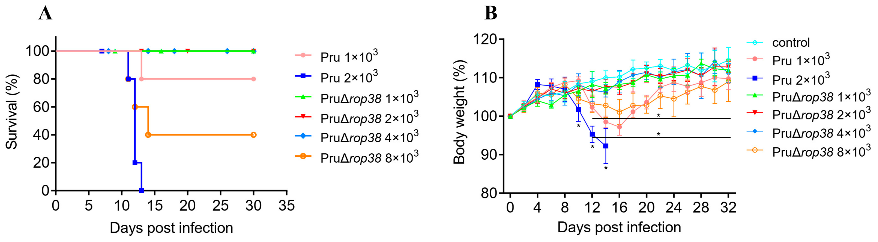
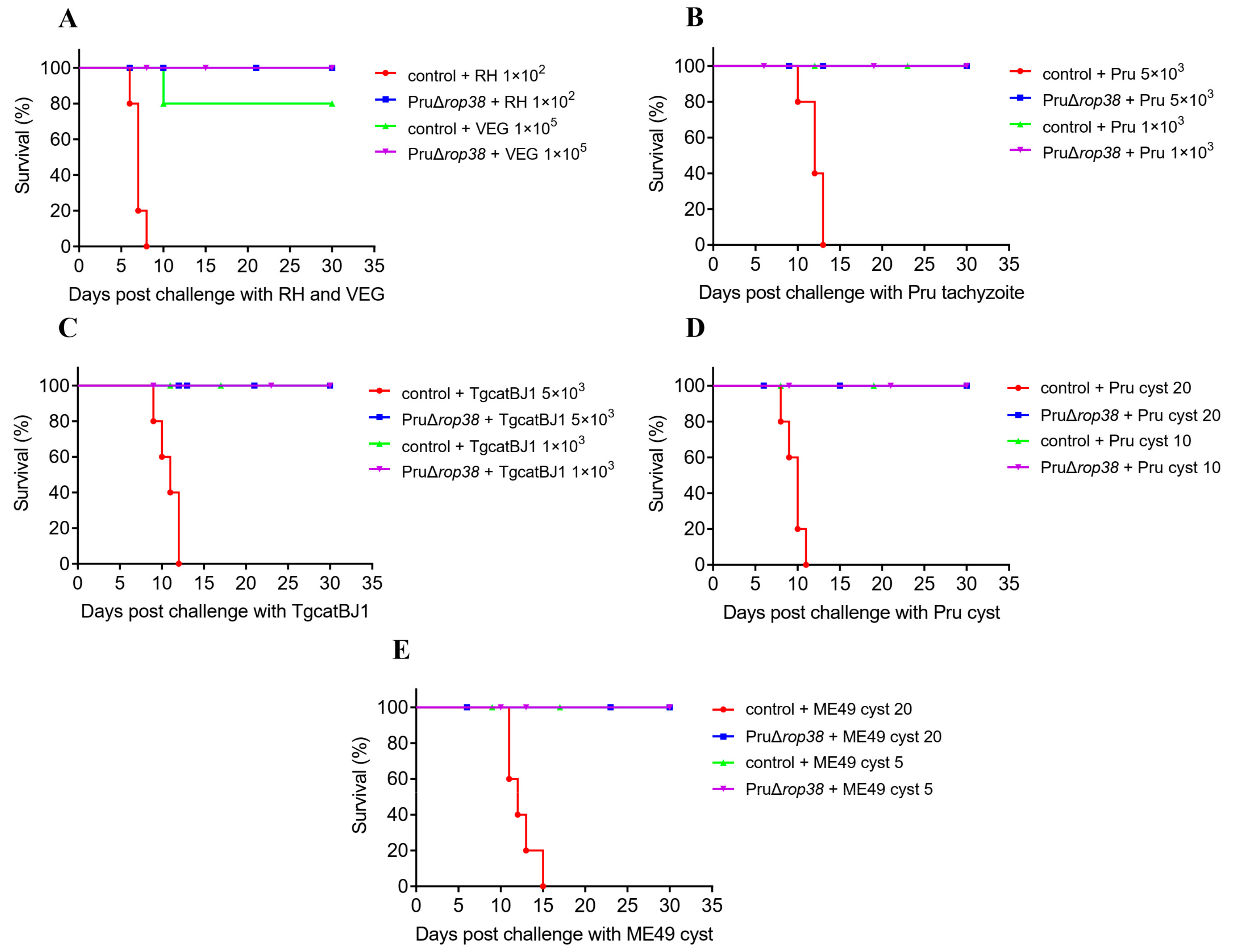
3.1. PruΔrop38 Exhibits Significantly Reduced Pathogenicity in Mice

Mice inoculated intraperitoneally with the parental Pru strain showed varying degrees of symptoms, mice inoculated at a dose of 1 × 10³ showed obvious wasting and bow-back, with a survival rate of 80%, and mice inoculated at a dose of 2 × 10³ showed obvious signs with the death of all mice (Figure 1A,B). However, mice inoculated with doses 1 × 10³, 2 × 10³, and 4 × 10³ of PruΔrop38 showed no obvious clinical symptoms. Mice showed obvious wasting after inoculation with PruΔrop38 at a dose of 8 × 10³, with a survival rate of 40%. Based on the survival status, clinical symptoms and body weight gain data, the virulence of PruΔrop38 in mice was significantly attenuated compared to the parental Pru strain, indicating its availability as a candidate strain for vaccine potential studies.

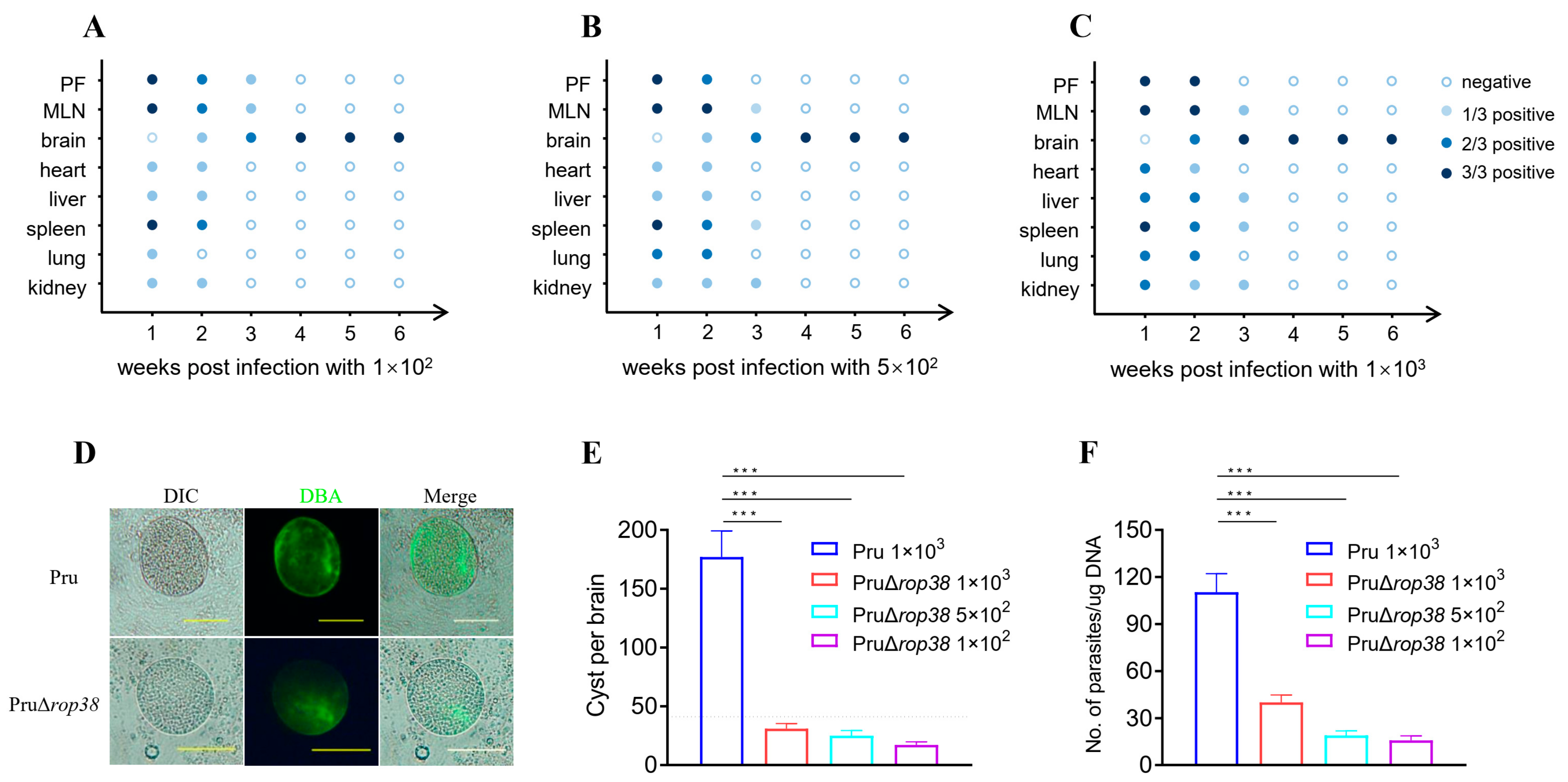
3.2. Dynamic Distribution of PruΔrop38 in Mice

To further assess the pathogenicity in mice, we first observed the dynamic distribution and histological changes in mice inoculated intraperitoneally with PruΔrop38 at doses of 1 × 10², 5 × 10², and 1 × 10³. The amplification results showed that PruΔrop38 was widely distributed in all organ tissues except the brain in the first week after inoculation, and PruΔrop38 was also detected in the brain from the second week. The third week results revealed that T. gondii-specific genes were detected only in the peritoneal fluid, Mesenteric lymph nodes (MLN), and brain tissues with 1 × 10² inoculation (Figure 2A), and in the MLN, brain, spleen, and kidney with 5 × 10² inoculation (Figure 2B), as well as in the MLN, brain, liver, spleen, and kidney with 1 × 10³ inoculation (Figure 2C), however, no T. gondii-specific genes were detected from organs in three groups of organs from week 4 onwards, but rather from brain tissue. There was some degree of enlargement in the spleen and MLN without pathological changes, and no obvious histological lesions were seen on observation of the remaining organ and tissues. The dynamic distribution of PruΔrop38 in mice and histological observations further indicate that PruΔrop38 significantly reduces pathogenicity in mice.

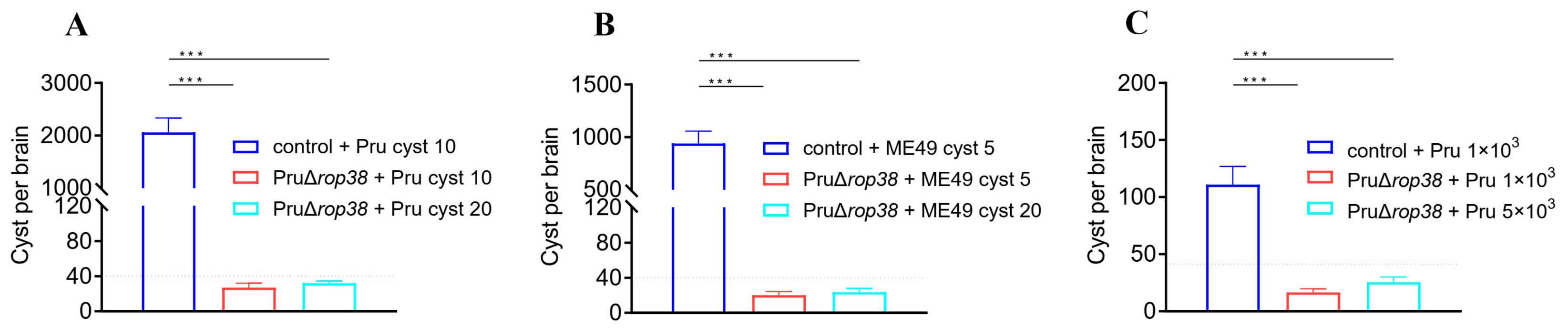
3.3. Deletion of TgROP38 Significantly Reduces Brain Parasite Burden

To determine whether deletion of TgROP38 affects chronic infection in vivo at 30 days post-infection, we enumerated brain cysts in mice inoculated with 1 × 10², 5 × 10², and 1 × 10³ of PruΔrop38, and we also infected CD-1 mice with Pru stain at a dose of 1 × 10³ as a control (Figure 2D). The average number of brain cysts in mice inoculated with Pru was 177, while the average number of brain cysts in mice inoculated with 1 × 10², 5 × 10² and 1 × 10³ of PruΔrop38 was 17 (p < 0.001), 25 (p < 0.001) and 31 (p < 0.001), respectively (Figure 2E). The parasite load in the brains of mice inoculated with 1 × 10², 5 × 10², and 1 × 10³ of PruΔrop38 was 15.84, 19.22, and 25.94 parasites per μg of brain DNA, respectively (Figure 2F), being significantly lower than that of Pru-inoculated mice (110.47 parasites per μg of brain DNA; p < 0.001). These results suggest that PruΔrop38 significantly reduces the brain parasite burden compared to the parental Pru strain.

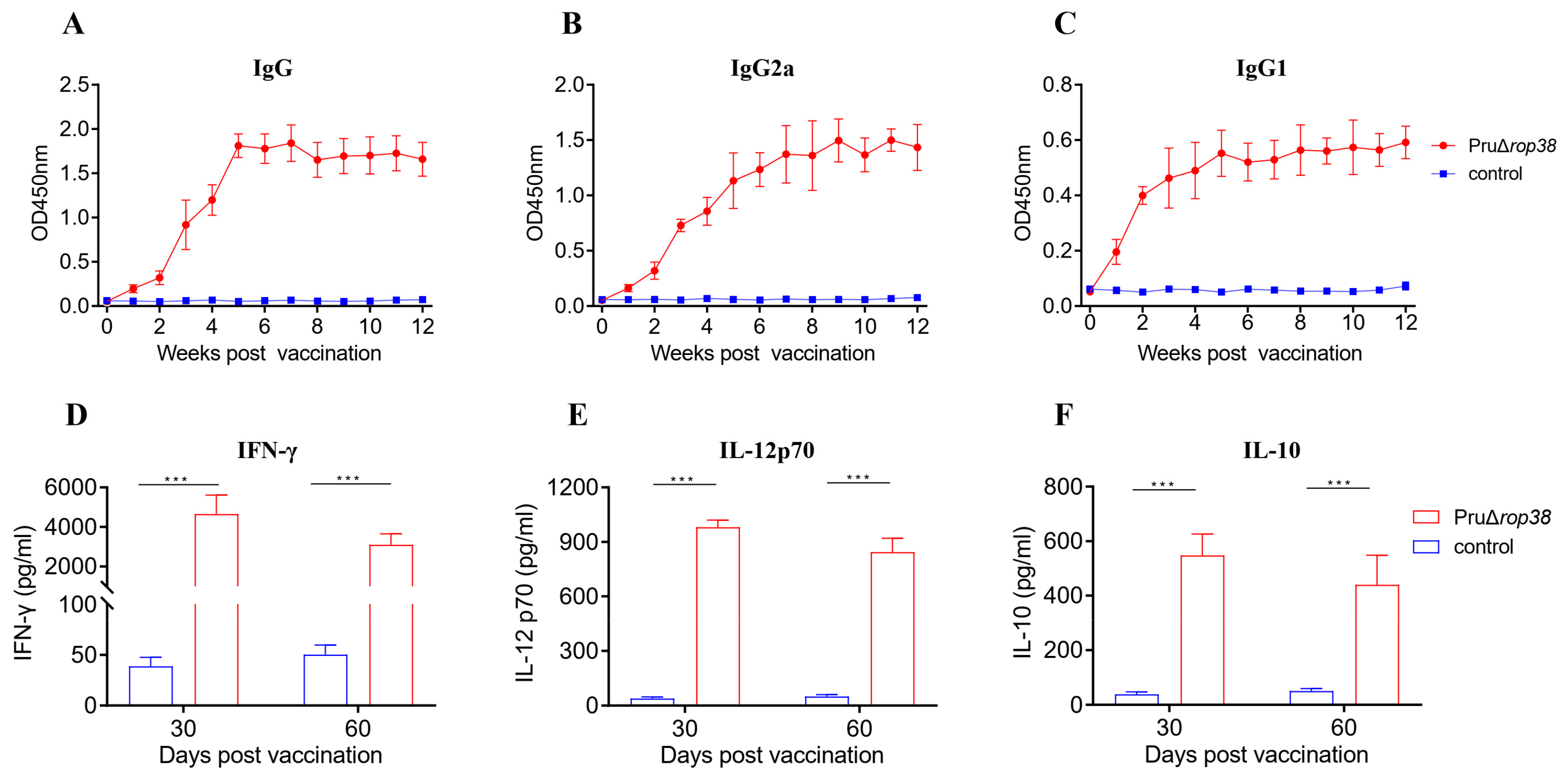
3.4. Humoral Immune Responses Induced by PruΔrop38

We next evaluated the potential of PruΔrop38 as an effective vaccine. To determine the humoral immune responses derived from PruΔrop38 immunization, we measured the antibody levels of specific anti-T. gondii IgG and IgG isotypes in the mouse serum. The safe immunization dose chosen was 1 × 10² tachyzoites by i.p. inoculation from the immunoprotective efficacy comparison of mice from four immunization doses in mice (Supplementary Figure S1a–d). With weekly serum collection after immunization, ELISA results showed a continuous increase in the level of IgG antibodies from the first to the third week, and revealed significantly high levels from the third to the twelfth week after vaccination (Figure 3A).
To determine whether a Th1 and/or a Th2 humoral response was induced by vaccination with PruΔrop38, sera from unvaccinated mice were also used as controls, and serum levels of both IgG2a (Figure 3B) and IgG1(Figure 3C) increased significantly from the second week onwards. In addition, the IgG1/IgG2a ratio was less than 1, demonstrating that PruΔrop38 induced a combined Th1/Th2 immune response after vaccination, with a predominant Th1 response.

3.5. Cellular Immune Responses Induced by PruΔrop38

Splenocytes harvested from vaccinated or control mice at 30 or 60 days post-immunization were stimulated with TSA, ConA or RPMI 1640 medium. The suspensions (stimulated with TSA) were measured by ELISA to assess the immunological memory and cellular immune responses induced by PruΔrop38. The levels of IFN-γ (Figure 3D), IL-12 (Figure 3E) and IL-10 (Figure 3F) were markedly increased in immunized mice compared to those in control mice (p < 0.001) at both time points, indicating that immunizing mice with PruΔrop38 induces a mixed Th1/Th2 response and that Th1-type cell-mediated immune responses trigger leading protection against challenge.

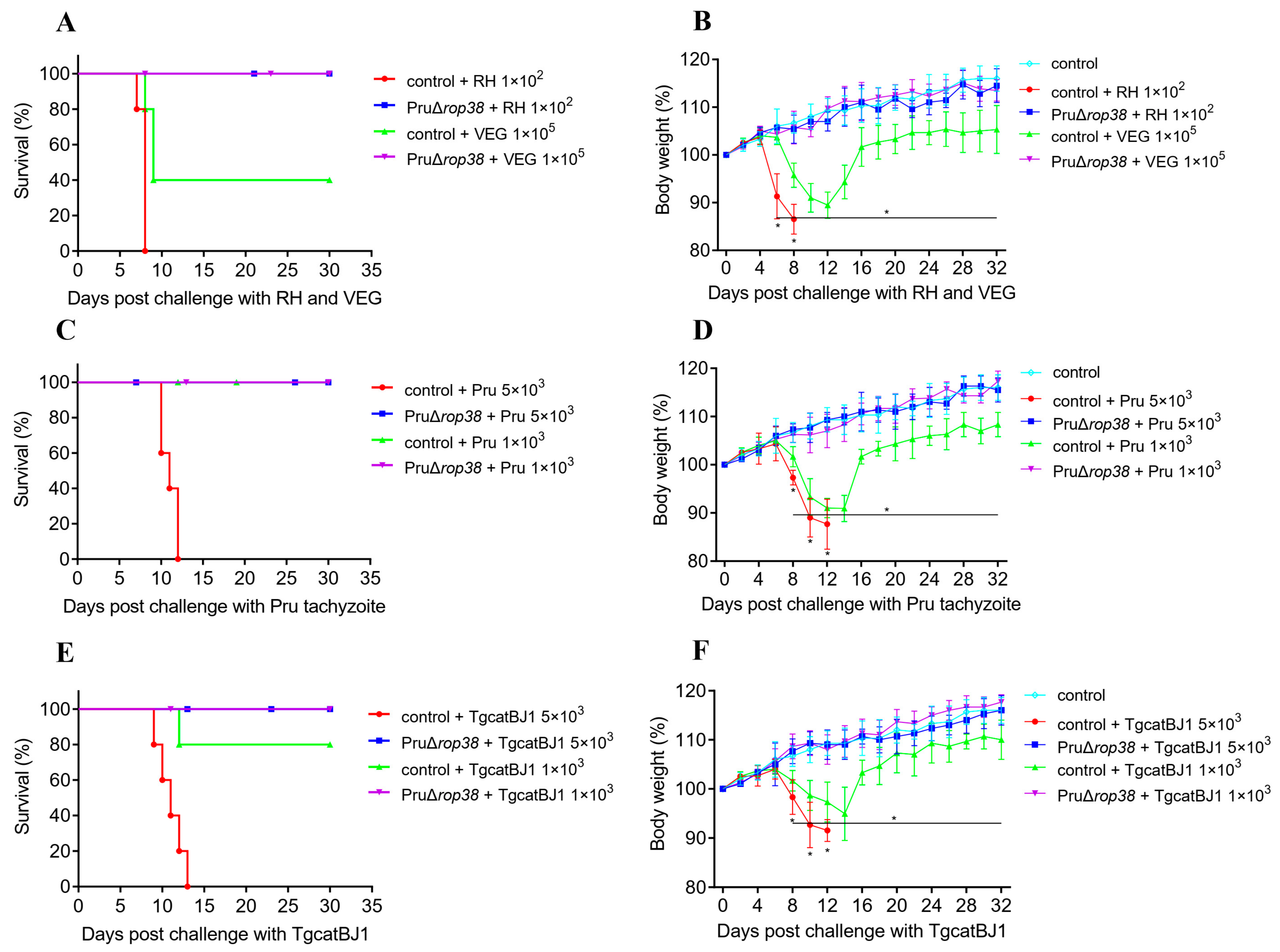
3.6. Protection against Varied Genotypes of Tachyzoites

To evaluate the protective efficacy against toxoplasmosis in mice, varied genotypes of tachyzoites (RH, Pru, VEG, and TgCatBj1) were used to infect mice intraperitoneally at corresponding doses. Control mice, challenged with lethal doses of 1 × 10² RH, 1 × 10⁵ VEG, 5 × 10³ Pru, or 5 × 10³ TgCatBj1 tachyzoites, exhibited significant weight loss and bow-back, and all mice died within the second week after challenge, with only 40% of mice surviving the 1 × 10⁵ VEG infection (Figure 4A,B). Besides, control mice infected with 1 × 10³ Pru (Figure 4C,D) or 1 × 10³ TgCatBj1 (Figure 4E,F) tachyzoites (non-lethal dose) revealed continued weight loss at 8–14 days post-challenge, with body weight dropping to a minimum of 90.93% (p < 0.001) and 94.53% (p < 0.001) of their initial weight, respectively, and only one mice died with 1 × 10³ TgCatBj1. In contrast, vaccinated mice challenged with the four genotypes of tachyzoites showed normal weight gain (p > 0.05) and the same mental status as blank control mice. Taken together, PruΔrop38 may provide efficient protective efficacy against varied genotypes of RH, Pru, VEG, and TgCatBj1 tachyzoites challenge in mice.

3.7. Protection against Pru and ME49 Cyst

We also evaluated the protective efficacy of PruΔrop38 upon oral re-challenge with cysts, to simulate the natural transmission route; two groups of mice were infected with two doses of Pru and ME49 cysts, respectively. During the 30-day observation of survival, body weight and clinical signs, all control mice died within 8–13 days of infection with lethal doses of 20 Pru (Figure 5A,B) or ME49 cysts (Figure 5C,D), with a more severe weight loss (p < 0.001). Mice were also infected with 10 Pru or 5 ME49 cysts (pathogenic dose) to assess the protective efficacy against chronic infection, and all control mice survived the observation period while their body weight dropped to a minimum of 95.17% (p < 0.001) (Figure 5B) and 94.36% (p < 0.001) (Figure 5D) of their initial weight, respectively. Instead, all vaccinated mice challenged with Pru or ME49 cysts survived, and no obvious clinical signs were observed, and none of the weight gains were significant compared to blank control mice (p > 0.05). Taken together, PruΔrop38 may also provide efficient protective efficacy in mice that survive during acute or chronic infection with Pru and ME49 cysts.

3.8. Significant Reduction in Brain Parasite Burden in Challenging Type II Strains

Another aspect of evaluation of the protective efficacy against chronic infection is the brain parasite burden, so we counted the number of brain cysts in mice challenged with type II strains. The average number of cysts in control mice challenged with 10 Pru cysts, 5 ME49 cysts, and 1 × 10³ Pru tachyzoites was 2062, 940, and 111, respectively (Figure 6A–C). However, the number of cysts in vaccinated mice was about 27 (p < 0.001), 20 (p < 0.001), and 17 (p < 0.001), respectively. Additionally, the number of cysts in vaccinated mice challenged with 20 Pru cysts, 20 ME49 cysts, and 5 × 10³ Pru tachyzoites (lethal doses in control mice) was 32 (p < 0.001), 24 (p < 0.001), and 25 (p < 0.001), respectively. Vaccination with PruΔrop38 has the ability to significantly reduce the brain parasite burden of the challenging type II strains and provide protection against T. gondii chronic infection.

3.9. Vaccination with PruΔrop38 Provides Long-Time Protection in Mice

To evaluate the duration of immunity with PruΔrop38, we finally assessed whether mice were able to resist T. gondii challenge 60 days after vaccination with PruΔrop38. When challenged with lethal doses of tachyzoites (1 × 10² RH, 1 × 10⁵ VEG, 5 × 10³ Pru, or 5 × 10³ TgCatBj1) or cysts (20 Pru or 20 ME49), control mice exhibited serious clinical signs (wasting, bow-back and depression), and all mice died at the second week (Supplementary Figure S2a–e), despite all vaccinated mice surviving challenges with varied genotypes of T. gondii strains (Figure 7A–E). When challenged with pathogenic doses of tachyzoites (1 × 10³ Pru, or 1 × 10³ TgCatBj1) or cysts (10 Pru or 5 ME49), all control mice survived (Figure 7B–E), but body weight dropped to 92.96% (p < 0.001), 93.16% (p < 0.001), 94.33% (p < 0.001), and 92.61% (p < 0.001) of the initial weight, respectively (Supplementary Figure S2b–e). Similarly, all vaccinated mice exhibited relatively stable weight and with no obvious clinical signs.
The average number of cysts in control mice infected with 1 × 10³ Pru tachyzoites, 10 Pru cysts and 5 ME49 cysts was 1690, 850, and 157, respectively (Supplementary Figure S3a–c). However, the number of cysts in vaccinated mice was about 29 (p < 0.001) and 35 (p < 0.001), and 24 (p < 0.001), respectively. In addition, the number of cysts in vaccinated mice challenged with 20 Pru cysts, 20 ME49 cysts, and 5 × 10³ Pru tachyzoites (lethal doses in control mice) was 31 (p < 0.001), 36 (p < 0.001), and 26 (p < 0.001), respectively. Taken together, vaccination with PruΔrop38 remains able to significantly reduce the brain parasite burden of the challenging type II strains, providing long-term efficacy against acute or chronic toxoplasmosis in mice.

4. Discussion

Toxoplasmosis is a serious zoonotic disease that threatens human and animal health, and vaccination strategies are considered ideal to prevent infection. The objectives are to prevent congenital toxoplasmosis in both women and livestock, prevent/reduce T. gondii tissue cysts in food-producing animals and oocyst shedding in cats [8]. While various approaches have been attempted to develop and achieve effective anti-T. gondii vaccines with varying degrees of protective immunity, the field of live-attenuated vaccine development has been carried out for safer vaccines for advantages in terms of protective efficacy [7]. Due to the essential roles of TgROP38 in the secretion of pro-inflammatory cytokines [16] and the attenuated virulence and reduced cyst burdens exhibited by PruΔrop38 in mice [17], the objective of this study was to evaluate the potential PruΔrop38 as an applicable vaccine candidate and assess its pathogenicity, immunogenicity and protective efficacy.
Survival status was the primary factor determining the pathogenicity of PruΔrop38 in vivo, besides, we also focused on clinical symptoms and body weight gain of mice after inoculation with different doses of Pru (1 × 10³ and 2 × 10³) or PruΔrop38 (1 × 10³, 2 × 10³, 4 × 10³, and 8 × 10³) tachyzoites. No obvious signs (wasting and bow-back) or deaths were seen even when inoculated with 4 × 10³ PruΔrop38, demonstrating the attenuated pathogenicity of the candidate to mice. T. gondii-specific genes were only detected in the brain from week 4 onwards, and deletion of TgROP38 significantly reduced the brain parasite burden compared to the parental Pru strain. These data indicate that PruΔrop38 conforms to a number of characteristics that make it an ideal live-attenuated vaccine. Pathological changes in organs and tissues were also observed, with some degree of enlargement in the spleen and MLN, and no obvious histological lesions observed in the remaining organ and tissue. The final vaccination dose (1 × 10² per mouse) was considered to significantly reduce pathogenicity and provide high biosafety in mice.
As with live-attenuated vaccine candidates [12], immunization with PruΔrop38 induces strong humoral and cellular immune responses. The level of specific anti-T. gondii IgG and IgG two isotypes (IgG1 and IgG2a) revealed a significantly high level from week 3 to week 12 after vaccination, since the IgG1/IgG2a ratio was less than 1, PruΔrop38 induces a combined Th1/Th2 immune response, in which the Th1 response predominates. In contrast, IgG activated by B-cell response can regulate immune cells and complement to inhibit the infective capacity of T. gondii [26].
At 30 or 60 days post-vaccination, the secretion of IFN-γ, IL-12 and IL-10 in the immunized mice splenocytes significantly increased under the TSA stimulation, indicating a Th1-type cell-mediated immune response was induced predominantly. IL-12 and IFN-γ trigger multiple intracellular responses to clear T. gondii by activating cytotoxic lymphocytes and natural killer cells, leading to protection against acute T. gondii infection [27,28]. However, excessive pro-inflammatory cytokine (IFN-γ and IL-12) responses usually lead to death in mice during acute infection, and as an anti-inflammatory cytokine, IL-10 can downregulate the expression of IFN-γ and IL-12, which in turn reduces the inflammatory response [29]. Immunogenicity assessment showed that vaccination with PruΔrop38 rapidly activated humoral and cellular immune responses to specifically recognize the Toxoplasma antigen.
PruΔrop38 provides long-term efficacy against acute and chronic toxoplasmosis in mice. For Toxoplasma it is now apparent that, besides the three typical genotypes (type I, type II and type III) described worldwide, there exist many atypical T. gondii genotypes, with a high degree of genetic diversity [30,31]. As a result, we used varied genotypes of tachyzoites (RH, Pru, VEG, and TgcatBJ1) to assess the protective efficacy of PruΔrop38, with TgcatBJ1 being a ToxoDB#9 genotype strain isolated from stray cats [18]. Several atypical isolates are also commonly used in experiments to evaluate the efficacy of vaccine strains, such as the ToxoDB#9 genotype tachyzoites PYS [7], TgC7 [32], and C7719 [13], as well as the ToxoDB#3 tachyzoites WH-1 [33]. PruΔrop38 could provide effective protection to mice against challenge by tachyzoites of varied genotypes. However, intraperitoneal injection does not represent the natural route of infection, so Pru and ME49 cysts were orally challenged to objectively assess the protective efficacy, and PruΔrop38 could provide efficient efficacy in mice that survived during acute or chronic infection with Pru and ME49 cysts. Additionally, vaccination with PruΔrop38 was able to significantly reduce the brain parasite burden of the challenging type II strains in chronic infections. Taken together, PruΔrop38 is a potential live-attenuated vaccine candidate. However, the current study is limited to the mouse model, so more studies are needed to evaluate the effectiveness and safety of the vaccine candidate for food-producing animals or cats.

5. Conclusions

In summary, virulence, dynamic distribution, and brain parasite burden assays indicated that PruΔrop38 was significantly less pathogenic in mice compared to the parental Pru strain. Vaccination with 1 × 10² PruΔrop38 triggered high levels of serum IgG, IgG2a, and IgG1 from week 3 to week 12, and a significant increase in IFN-γ, IL-12, and IL-10 in splenocyte suspensions at 30 or 60 days post-immunization. PruΔrop38 provides long-term efficacy against acute and chronic toxoplasmosis in mice and significantly reduces the brain parasite burden of the challenging T. gondii type II strains.

Supplementary Materials

The following supporting information can be downloaded at: https://www.mdpi.com/article/10.3390/biomedicines10061336/s1. Figure S1: Protective efficacy comparison of four doses of vaccination; Figure S2: Body weight of mice in evaluation of duration of immunity with PruΔrop38; Figure S3: Effect on brain parasite burden in the evaluation of duration of immunity with PruΔrop38; Table S1: The primers used in this study.

Author Contributions

Conceptualization, Q.L. and Y.W.; methodology, Q.L., J.L., Y.W., Z.Z. and Y.X.; software, Y.W. and Z.Y.; formal analysis, Y.W. and Z.Z.; writing—original draft preparation, Y.W.; writing—review and editing, Q.L.; funding acquisition, Q.L. All authors have read and agreed to the final version of the manuscript.

Funding

This research was funded by the National Natural Science Foundation of China (31730096 and 81971964).

Institutional Review Board Statement

Immunogenicity and protection protocols were in accordance with the recommendations of the Guide for the Care and Use of Laboratory Animals, Ministry of Science and Technology of China. The Procedures were reviewed and approved by the Laboratory Animal Welfare and Animal Experimental Ethical Committee of China Agricultural University (Certificate No.: CAU-AW31901202-2-1).

Informed Consent Statement

Not applicable.

Data Availability Statement

Not applicable.

Acknowledgments

All datasets generated for this study are included in the manuscript/Supplementary Files.

Conflicts of Interest

The authors declare no conflict of interest.

References

  1. Sanchez, S.G.; Besteiro, S. The pathogenicity and virulence of Toxoplasma gondii. Virulence 2021, 12, 3095–3114. [Google Scholar] [CrossRef]
  2. Yarovinsky, F. Innate immunity to Toxoplasma gondii infection. Nat. Rev. Immunol. 2014, 14, 109–121. [Google Scholar] [CrossRef] [PubMed]
  3. Izadi, M.; Jafari, N.J.; Poornaki, A.M.; Sadraei, J.; Rezavand, B.; Mirzaei, H.R.; Zarrinfar, H.; Khedive, A. Detection of Toxoplasma gondii from Clinical Specimens of Patients Receiving Renal Transplant Using ELISA and PCR. Nephro-Urol. Mon. 2013, 5, 983–987. [Google Scholar] [CrossRef] [Green Version]
  4. Matta, S.K.; Rinkenberger, N.; Dunay, I.R.; Sibley, L.D. Toxoplasma gondii infection and its implications within the central nervous system. Nat. Rev. Microbiol. 2021, 19, 467–480. [Google Scholar] [CrossRef] [PubMed]
  5. Tenter, A.M.; Heckeroth, A.R.; Weiss, L.M. Toxoplasma gondii: From animals to humans. Int. J. Parasitol. 2000, 30, 1217–1258. [Google Scholar] [CrossRef] [Green Version]
  6. Dunay, I.R.; Gajurel, K.; Dhakal, R.; Liesenfeld, O.; Montoya, J.G. Treatment of toxoplasmosis: Historical perspective, animal models, and current clinical practice. Clin. Microbiol. Rev. 2018, 31, e00057-17. [Google Scholar] [CrossRef] [Green Version]
  7. Wang, J.; Zhang, N.; Li, T.; He, J.; Elsheikha, H.M.; Zhu, X. Advances in the development of anti-Toxoplasma gondii vaccines: Challenges, opportunities, and perspectives. Trends Parasitol. 2019, 35, 239–253. [Google Scholar] [CrossRef] [PubMed]
  8. Mévélec, M.-N.; Lakhrif, Z.; Dimier-Poisson, I. Key limitations and new insights into the parasite stage switching for future vaccine development in human, livestock, and cats. Front. Cell. Infect. Microbiol. 2020, 10, 607198. [Google Scholar] [CrossRef]
  9. Buxton, D.; Thomson, K.; Maley, S.; Wright, S.; Bos, H.J. Vaccination of sheep with a live incomplete strain (S48) of Toxoplasma gondii and their immunity to challenge when pregnant. Vet. Rec. 1991, 129, 89–93. [Google Scholar] [CrossRef]
  10. Shen, B.; Brown, K.M.; Lee, T.D.; Sibley, L.D. Efficient gene disruption in diverse strains of Toxoplasma gondii using CRISPR/CAS9. mBio 2014, 5, e01114-14. [Google Scholar] [CrossRef] [Green Version]
  11. Wang, J.; Li, T.; Elsheikha, H.M.; Chen, K.; Cong, W.; Yang, W.; Bai, M.; Huang, S.; Zhu, X. Live attenuated Pru:Δcdpk2 strain of Toxoplasma gondii protects against acute, chronic, and congenital toxoplasmosis. J. Infect. Dis. 2018, 218, 768–777. [Google Scholar] [CrossRef] [Green Version]
  12. Lagal, V.; Dinis, M.; Cannella, D.; Bargieri, D.; Gonzalez, V.; Andenmatten, N.; Meissner, M.; Tardieux, I. AMA1-deficient Toxoplasma gondii parasites transiently colonize mice and trigger an innate immune response that leads to long-lasting protective immunity. Infect. Immun. 2015, 83, 2475–2486. [Google Scholar] [CrossRef] [Green Version]
  13. Xia, N.; Zhou, T.; Liang, X.; Ye, S.; Zhao, P.; Yang, J.; Zhou, Y.; Zhao, J.; Shen, B. A lactate fermentation mutant of Toxoplasma stimulates protective immunity against acute and chronic toxoplasmosis. Front. Immunol. 2018, 9, 1814. [Google Scholar] [CrossRef] [Green Version]
  14. Fox, B.A.; Bzik, D.J. Avirulent uracil auxotrophs based on disruption of orotidine-5′-monophosphate decarboxylase elicit protective immunity to Toxoplasma gondii. Infect. Immun. 2010, 78, 3744–3752. [Google Scholar] [CrossRef] [Green Version]
  15. Hunter, C.A.; Sibley, L.D. Modulation of innate immunity by Toxoplasma gondii virulence effectors. Nat. Rev. Microbiol. 2012, 10, 766–778. [Google Scholar] [CrossRef] [PubMed] [Green Version]
  16. Xu, Y.; Wang, X.; Liu, J.; Fu, Y.; Xu, J.; Liu, Q. Toxoplasma gondii rhoptry protein38 (TgROP38) affects parasite invasion, egress, and induces IL-18 secretion during early infection. Acta Biochim. Biophys. Sin. 2018, 50, 766–775. [Google Scholar] [CrossRef] [Green Version]
  17. Fox, B.A.; Rommereim, L.M.; Guevara, R.B.; Falla, A.; Hortua Triana, M.A.; Sun, Y.; Bzik, D.J. The Toxoplasma gondii rhoptry kinome is essential for chronic infection. mBio 2016, 7, e00193-16. [Google Scholar] [CrossRef] [Green Version]
  18. Qian, W.; Wang, H.; Su, C.; Shan, D.; Cui, X.; Yang, N.; Lv, C.; Liu, Q. Isolation and characterization of Toxoplasma gondii strains from stray cats revealed a single genotype in Beijing, China. Vet. Parasitol. 2012, 187, 408–413. [Google Scholar] [CrossRef]
  19. Mahmoud, M.E.; Ihara, F.; Fereig, R.M.; Nishimura, M.; Nishikawa, Y. Induction of depression-related behaviors by reactivation of chronic Toxoplasma gondii infection in mice. Behav. Brain Res. 2016, 298, 125–133. [Google Scholar] [CrossRef]
  20. Homan, W.L.; Vercammen, M.; De Braekeleer, J.; Verschueren, H. Identification of a 200- to 300-fold repetitive 529 bp DNA fragment in Toxoplasma gondii, and its use for diagnostic and quantitative PCR. Int. J. Parasitol. 2000, 30, 69–75. [Google Scholar] [CrossRef]
  21. Burrells, A.; Bartley, P.M.; Zimmer, I.A.; Roy, S.; Kitchener, A.C.; Meredith, A.; Wright, S.E.; Innes, E.A.; Katzer, F. Evidence of the three main clonal Toxoplasma gondii lineages from wild mammalian carnivores in the UK. Parasitology 2013, 140, 1768–1776. [Google Scholar] [CrossRef]
  22. Jones, C.D.; Okhravi, N.; Adamson, P.; Tasker, S.; Lightman, S. Comparison of PCR detection methods for B1, P30, and 18S rDNA genes of T. gondii in aqueous humor. Invest. Ophthalmol. Vis. 2000, 41, 634–644. [Google Scholar] [CrossRef]
  23. Fox, B.A.; Falla, A.; Rommereim, L.M.; Tomita, T.; Gigley, J.P.; Mercier, C.; Cesbron-Delauw, M.-F.; Weiss, L.M.; Bzik, D.J. Type II Toxoplasma gondii KU80 knockout strains enable functional analysis of genes required for cyst development and latent infection. Eukaryot. cell 2011, 10, 1193–1206. [Google Scholar] [CrossRef] [Green Version]
  24. Song, X.; Lin, M.; Li, M.; Yang, X.; Liu, J.; Liu, Q. Toxoplasma gondii metacaspase 2 is an important factor that influences bradyzoite formation in the Pru strain. Parasitol. Res. 2020, 119, 2287–2298. [Google Scholar] [CrossRef]
  25. Lourenco, E.V.; Pereira, S.R.; Faca, V.M.; Coelho-Castelo, A.A.M.; Mineo, J.R.; Roque-Barreira, M.C.; Greene, L.J.; Panunto-Castelo, A. Toxoplasma gondii micronemal protein MIC1 is a lactose-binding lectin. Glycobiology 2001, 11, 541–547. [Google Scholar] [CrossRef] [Green Version]
  26. Sayles, P.C.; Gibson, G.W.; Johnson, L.L. B cells are essential for vaccination-induced resistance to virulent Toxoplasma gondii. Infect. Immun. 2000, 68, 1026–1033. [Google Scholar] [CrossRef] [Green Version]
  27. Yarovinsky, F.; Zhang, D.; Andersen, J.F.; Bannenberg, G.L.; Serhan, C.N.; Hayden, M.S.; Hieny, S.; Sutterwala, F.S.; Flavell, R.A.; Ghosh, S.; et al. TLR11 activation of dendritic cells by a protozoan profilin-like protein. Science 2005, 308, 1626–1629. [Google Scholar] [CrossRef] [Green Version]
  28. Plattner, F.; Yarovinsky, F.; Romero, S.; Didry, D.; Carlier, M.F.; Sher, A.; Soldati-Favre, D. Toxoplasma profilin is essential for host cell invasion and TLR11-dependent induction of an interleukin-12 response. Cell Host Microbe 2008, 3, 77–87. [Google Scholar] [CrossRef] [Green Version]
  29. Mordue, D.G.; Monroy, F.; La Regina, M.; Dinarello, C.A.; Sibley, L.D. Acute toxoplasmosis leads to lethal overproduction of Th1 cytokines. J. Immunol. 2001, 167, 4574–4584. [Google Scholar] [CrossRef] [Green Version]
  30. Su, C.; Khan, A.; Zhou, P.; Majumdar, D.; Ajzenberg, D.; Dardé, M.-L.; Zhu, X.-Q.; Ajioka, J.W.; Rosenthal, B.M.; Dubey, J.P.; et al. Globally diverse Toxoplasma gondii isolates comprise six major clades originating from a small number of distinct ancestral lineages. Proc. Natl. Acad. Sci. USA 2012, 109, 5844–5849. [Google Scholar] [CrossRef] [PubMed] [Green Version]
  31. Galal, L.; Hamidović, A.; Dardé, M.L.; Mercier, M. Diversity of strains at the global level and its determinants. Food Waterborne Parasitol. 2019, 15, e00052. [Google Scholar] [CrossRef] [PubMed]
  32. Liang, Q.; Sun, L.; Elsheikha, H.M.; Cao, X.; Nie, L.; Li, T.; Li, T.; Zhu, X.; Wang, J. RHΔgra17Δnpt1 strain of Toxoplasma gondii elicits protective immunity against acute, chronic and congenital toxoplasmosis in mice. Microorganisms 2020, 8, 352. [Google Scholar] [CrossRef] [PubMed] [Green Version]
  33. Yang, J.; Yang, C.; Qian, J.; Li, F.; Zhao, J.; Fang, R. Toxoplasma gondii α-amylase deletion mutant is a promising vaccine against acute and chronic toxoplasmosis. Microb. Biotechnol. 2020, 13, 2057–2069. [Google Scholar] [CrossRef]
Figure 1. Pathogenicity of PruΔrop38 in mice. (A) Survival rate and (B) body weight gain of CD-1 mice infected with the parental Pru strain or the PruΔrop38 strain. Pru (1 × 10³ and 2 × 10³) or PruΔrop38 (1 × 10³, 2 × 10³, 4 × 10³, and 8 × 10³) tachyzoites were injected intraperitoneally (i.p.) into mice (n = 5), and daily monitoring of mice for 30 days (presented as the mean of body weight ± SD; * p < 0.05; one-way ANOVA with Tukey).
Biomedicines 10 01336 g001
Figure 2. Dynamic distribution and effect on brain parasite burden of PruΔrop38 in mice. Amplification results of T. gondii-specific genes from organs and tissues in mice. Injections of (A) 1 × 10², (B) 5 × 10², and (C) 1 × 10³ PruΔrop38 tachyzoites, respectively. (D) Cyst counts were performed on brain homogenates by DBA-FITC staining, brain cyst from mice injected with PruΔrop38 and Pru were observed using a fluorescence microscope. (E) Immunofluorescence assays determined the number of brain cysts of Pru and PruΔrop38. Pru (1 × 10³) or PruΔrop38 (1 × 10², 5 × 10², and 1 × 10³) tachyzoites were injected intraperitoneally (i.p.) into mice (n = 5) for 30 days. (Cyst walls of T. gondii were labeled with DBA-FITC (green), Scale bar = 50 µm). (F) The number of parasites and DNA concentration in each brain tissue were detected by calculating the 529 gene of Toxoplasma and the 28S rRNA gene of the brain with quantitative real-time PCR, the load of parasites = number of parasites/DNA concentration of brain tissue (presented as the mean ± SD; *** p < 0.001; one-way ANOVA with Tukey).
Biomedicines 10 01336 g002
Figure 3. Humoral and cellular immune responses induced by PruΔrop38. Levels of IgG and its subtype (IgG2a and IgG1) antibodies in the serum obtained from vaccinated and control mice. (A) IgG, (B) IgG2a, and (C) IgG1, respectively (n = 3; weekly serum collection after immunization). The levels of cytokines in the suspension of splenocyte in vaccinated and control mice. Splenocytes were collected at 30 or 60 days post-vaccination and co-incubated with TSA (50 μg/mL), and the levels of (D) IFN-γ, (E) IL-12, and (F) IL-10 were analyzed by ELISA (n = 3; presented as the mean ± SD; *** p < 0.001; Student’s t-test).
Biomedicines 10 01336 g003
Figure 4. Protective efficacy against varied genotypes of tachyzoite challenge. Survival rate and body weight gain of vaccinated and control mice re-challenged with (A,B) RH (1 × 10²) and VEG (1 × 10⁵) tachyzoites, (C,D) Pru (1 × 10³ and 5 × 10³) tachyzoites, and (E,F) TgCatBj1 (1 × 10³ and 5 × 10³) tachyzoites, respectively. (n = 5; mice were injected intraperitoneally (i.p.); daily monitoring of mice for 30 days; presented as the mean of body weight ± SD; * p < 0.05; one-way ANOVA with Tukey).
Biomedicines 10 01336 g004
Figure 5. Protective efficacy against varied genotypes of cyst challenge. Survival rate and body weight gain of vaccinated and control mice re-challenged with (A,B) Pru (20 and 10) cysts and (C,D) ME49 (20 and 5) cysts (n = 5; mice were injected orally; daily monitoring of mice for 30 days; presented as the mean of body weight ± SD; * p < 0.05; one-way ANOVA with Tukey).
Biomedicines 10 01336 g005
Figure 6. Effect on brain parasite burden when challenged with T. gondii type II strains. The brain from vaccinated and control mice were harvested after challenge with 30 days, and cysts were observed by IFA and then the number of cysts were determined by DBA-FITC staining. (A) Pru cyst challenge (20 and 10), (B) ME49 cyst challenge (20 and 5), and (C) Pru tachyzoite (5 × 10³ and 1 × 10³) challenge, respectively. (n = 5; presented as the mean of cyst number ± SD; *** p < 0.001; one-way ANOVA with Tukey).
Biomedicines 10 01336 g006
Figure 7. The duration of immunity with PruΔrop38. Survival rate of vaccinated and control mice re-challenged with (A) RH (1 × 10²) and VEG (1 × 10⁵) tachyzoites, (B) Pru (1 × 10³ and 5 × 10³) tachyzoites, and (C) TgCatBj1 (1 × 10³ and 5 × 10³) tachyzoites, (D) Pru (20 and 10) cysts, and (E) ME49 (20 and 5) cysts, respectively (n = 5; mice were injected with tachyzoites intraperitoneally (i.p.) or with cysts orally; daily monitoring of mice for 30 days).
Biomedicines 10 01336 g007
Publisher’s Note: MDPI stays neutral with regard to jurisdictional claims in published maps and institutional affiliations.

