Review

Interdisciplinary Approaches to Deal with Alzheimer’s Disease—From Bench to Bedside: What Feasible Options Do Already Exist Today?

by
Irene Ablinger
1,†,
Katharina Dressel
2,†,
Thea Rott
3,†,
Anna Andrea Lauer
4,5,
Michael Tiemann
6,
João Pedro Batista
7,
Tim Taddey
8,
Heike Sabine Grimm
4,5 and
Marcus Otto Walter Grimm
4,5,*
1
Speech and Language Therapy, Campus Bonn, SRH University of Applied Health Sciences, 53111 Bonn, Germany
2
Speech and Language Therapy, Campus Düsseldorf, SRH University of Applied Health Sciences, 40210 Düsseldorf, Germany
3
Interdisciplinary Periodontology and Prevention, Campus Rheinland, SRH University of Applied Health Sciences, 51377 Leverkusen, Germany
4
Nutrition Therapy and Counseling, Campus Rheinland, SRH University of Applied Health Sciences, 51377 Leverkusen, Germany
5
Experimental Neurology, Saarland University, 66424 Homburg, Germany
6
Sport Science, Campus Rheinland, SRH University of Applied Health Sciences, 51377 Leverkusen, Germany
7
Sport Science and Physiotherapy, Campus Rheinland, SRH University of Applied Health Sciences, 51377 Leverkusen, Germany
8
Physiotherapy, Campus Rheinland, SRH University of Applied Health Sciences, 51377 Leverkusen, Germany
*
Author to whom correspondence should be addressed.
These authors contributed equally to this work.
Biomedicines 2022, 10(11), 2922; https://doi.org/10.3390/biomedicines10112922
Submission received: 30 September 2022 / Revised: 3 November 2022 / Accepted: 9 November 2022 / Published: 14 November 2022

Abstract

:
Alzheimer’s disease is one of the most common neurodegenerative diseases in the western population. The incidence of this disease increases with age. Rising life expectancy and the resulting increase in the ratio of elderly in the population are likely to exacerbate socioeconomic problems. Alzheimer’s disease is a multifactorial disease. In addition to amyloidogenic processing leading to plaques, and tau pathology, but also other molecular causes such as oxidative stress or inflammation play a crucial role. We summarize the molecular mechanisms leading to Alzheimer’s disease and which potential interventions are known to interfere with these mechanisms, focusing on nutritional approaches and physical activity but also the beneficial effects of cognition-oriented treatments with a focus on language and communication. Interestingly, recent findings also suggest a causal link between oral conditions, such as periodontitis or edentulism, and Alzheimer’s disease, raising the question of whether dental intervention in Alzheimer’s patients can be beneficial as well. Unfortunately, all previous single-domain interventions have been shown to have limited benefit to patients. However, the latest studies indicate that combining these efforts into multidomain approaches may have increased preventive or therapeutic potential. Therefore, as another emphasis in this review, we provide an overview of current literature dealing with studies combining the above-mentioned approaches and discuss potential advantages compared to monotherapies. Considering current literature and intervention options, we also propose a multidomain interdisciplinary approach for the treatment of Alzheimer’s disease patients that synergistically links the individual approaches. In conclusion, this review highlights the need to combine different approaches in an interdisciplinary manner, to address the future challenges of Alzheimer’s disease.

1. Introduction

Alzheimer’s disease (AD) is the most prevalent form of dementia and is characterized by behavioral and cognitive impairments. It results in a loss of memory and acquired skills, as well as a decrease in participation in daily activities, communication skills, social interactions and quality of life leading to an increasing burden on caregivers [1,2]. The main histopathological hallmarks of AD are the severe accumulation of amyloid-β in extracellular neuritic plaques as well as intracellular neurofibrillary tangles (NFTs) in vulnerable brain regions such as the hippocampus and cortex [3]. Based on the onset of symptoms and pathological changes in the cortex and hippocampus, clinical AD stages can be classified into at least four phases. The pre-symptomatic/pre-clinical stage is asymptomatic despite early pathological changes in the cortex and hippocampal formation [4,5,6], followed by mild cognitive impairment (MCI). MCI shows limitations in some cognitive domains on functional examination without restrictions in the ability to cope with everyday life [7,8,9] but definite laboratory evidence, including biomarkers such as low amyloid-β and increased tau proteins in the cerebrospinal fluid [5]. Mild/early dementia due to AD is characterized by symptoms such as spatial and temporal disorientation, loss of memory and concentration, word-finding difficulties, and the development of depression. In the moderate stage of AD, increased memory loss, impairment of visuospatial abilities leading to difficulties in recognizing family members and friends, and problems with language and communication occur [5,6,10]. The final stage of AD, severe/late AD, is associated with severe accumulation of NTFs and senile plaques in the entire cortex area, resulting in advanced functional and cognitive impairment, incontinence, dysphagia, and complete dependence on caregivers [5].
Currently (2020), AD is estimated to affect 55 million people worldwide. Global AD prevalence is predicted to increase to 139 million people living with AD by 2050 due to the aging population, making AD a major public health concern. Yet, to date, there are only two classes of approved drugs to treat persons with Alzheimer’s disease (PwAD): Cholinesterase enzyme inhibitors and N-methyl-D-aspartate (NMDA) inhibitors. Acetylcholine-producing cells are destroyed in AD by different physiological processes. Treatment with acetylcholinesterase inhibitors such as donepezil, galantamine and rivastigmine blocks the catabolism of acetylcholine, increasing acetylcholine concentration in the synaptic cleft and thus cholinergic transmission in the brain [5,11,12,13]. Overactivation of NMDA-receptors leads to increased intracellular calcium levels, promoting cell death and synaptic dysfunction. The use of partial NMDA antagonists such as memantine, which can be taken in combination with cholinesterase inhibitors, prevents NMDA-receptor overactivation and restores its normal activity [14,15]. However, these medications are only temporary effective in treating the symptoms of AD by improving quality of life but do not cure or prevent the disease.
Most AD cases occur sporadically (known as sporadic Alzheimer’s disease, SAD) with the age of onset above 65 years. Less than 10% of AD cases are caused by genetic familial mutations leading to earlier disease onset, usually between the age of 30 and 60 (known as early onset Alzheimer’s disease, EOAD or familial Alzheimer’s disease, FAD). AD is considered a multifactorial disease as it is characterized by impairments in multiple cellular processes. In addition to cholinergic dysfunction and the well-known Abeta (Aβ) and Tau pathology of AD, inflammation, oxidative stress, as well as, e.g., alterations in lipid and energy metabolism are also involved in the pathogenesis of AD.
The Lancet Commission on dementia prevention, intervention and care [16] recently identified 12 potentially modifiable risk factors for dementia that account for around 40% of dementia cases worldwide. These include, among others, physical inactivity, low social contact, obesity, and associated diabetes. Frequent physical activity, promotion of communication to maintain social interactions, and a healthy diet to avoid the risks of obesity and diabetes may therefore affect neuropathological damage and cognitive reserve [16] and contribute to the prevention or delay of dementia. According to current literature, the intraoral condition could also represent such a modifiable risk factor [17,18,19].
Reducing modifiable risk factors, in addition to pharmacological interventions, is an important approach in dementia treatment. According to Spector and Orrell’s biopsychosocial (BPS) model [20], dementia is a multifactorial disease in which psychosocial and biological processes are interrelated. Both domains include fixed, non-modifiable factors (e.g., age; education) and tractable, modifiable factors (e.g., physical health; social interaction, mental stimulation). Treatment plans should be tailored to the individual’s needs. Factors amenable to change are identified with the aim of influencing them through medical and non-pharmacological interventions [20,21]. In recent years, non-pharmacological interventions have become increasingly important in the management of dementia and in the effort to improve living with dementia and maintain quality of life [1,7,22,23,24]. Non-pharmacological interventions include evidence-based psychological, bodily, nutritional, digital or basic methods and approaches, that are individually selected and adjusted to the persons’ needs in their courses of disease [7,24]. Among these, cognition-oriented treatments (COT) [1,24,25], physical activity [26], and diet [27] form an integral part.

1.1. Aim of This Review

To date, there is no pharmaceutical or antibody-based causal therapy for the treatment of AD. As demographics change, alternative treatments for this devasting neurodegenerative disease will be needed. In this review, we summarize different nutritional approaches and their underlying mechanisms, the effect of physical activity, cognition-oriented treatments with a focus on communication, and the potential advantages of socioenvironmental factors in relation to AD. In addition, the current literature suggests that oral status, particularly periodontitis and edentulism, are also mechanistically linked to AD. This represents another interesting new approach in the treatment of AD. However, any of the aforementioned treatment approaches applied individually show limited benefits in treating PwAD. Therefore, recent efforts are focused on combining different approaches as multidisciplinary interventions. In the second part of our review, we summarize the results of the first multidomain interventions. Finally, we propose a multidisciplinary, individualized treatment, based on an even stronger integration of the different disciplines and already existing treatment methods.

1.2. Molecular Mechanisms Involved in AD Pathogenesis

The amyloid pathology of AD is caused by sequential proteolytic cleavage of the amyloid precursor protein (APP), resulting in Aβ monomers, that aggregate to oligomeric Aβ, discussed as the most neurotoxic form, and finally to Aβ fibrils and plaques [28,29]. The type-I transmembrane protein APP belongs to an evolutionarily conserved protein family and is ubiquitously expressed in mammals [30,31]. However, the function of APP is still debated, with the discussion that it plays a role in cell health, growth, gene transcription and lipid homeostasis [32]. The release of Aβ peptides out of APP is a physiological process, that occurs throughout life and depends on whether APP is primarily processed in the non-amyloidogenic or amyloidogenic pathway. The non-amyloidogenic pathway prevents the release of Aβ peptides out of APP and thus the formation of Aβ plaques. The initial ectodomain shedding of APP in the non-amyloidogenic pathway is realized by α-secretases, cleaving APP within the Aβ domain. The activity of the identified α-secretases ADAM9, ADAM10 and ADAM17 [33,34], belonging to the ADAM family (a disintegrin and metalloprotease), generates soluble α-secreted APP (sAPPα) and the membrane-tethered C-terminal fragment αCTF, further processed by γ-secretase, leading to the non-toxic peptide p3. Furthermore, sAPPα has neuroprotective and memory-enhancing effects [35,36,37]. In the amyloidogenic pathway, APP is first cleaved by β-secretase, generating the N-terminus of Aβ. The membrane-bound aspartyl protease BACE 1 (β-site APP cleaving enzyme) has been identified as the main β-secretase [38], mainly found in intracellular compartments with an acidic pH such as late Golgi-compartments and endosomes [39,40]. Similar to α-secretase cleavage of APP, BACE-cleavage leads to the release of a soluble fragment, β-secreted APP (sAPPβ), and a C-terminal membrane-embedded fragment called βCTF [39,41]. Subsequent cleavage of βCTF by γ-secretase within the transmembrane domain results in the generation of Aβ peptides varying in their length at the C-terminus [42,43]. Aβ40 peptides have been found to be the most abundant Aβ peptides (80–90% of Aβ peptides) whereas the more hydrophobic Aβ42 peptides represent only approximately 10% of Aβ peptides. The heterotetrameric protein complex leading to γ-secretase activity includes the transmembrane proteins presenilin1 (PS1) or presenilin 2 (PS2), identified as the catalytically active components, anterior pharynx defective 1 (APH-1), presenilin enhancer 2 (PEN-2) and nicastrin (NCT) [44,45]. All components of the γ-secretase complex as well as β-secretase BACE1 have been found in lipid rafts, small membrane microdomains enriched in sphingolipids and cholesterol [46,47]. Vice versa, non-amyloidogenic α-secretase processing of APP was reported to be localized in non-raft membrane domains, indicating that APP processing is highly dependent on membrane lipid composition.
In addition to amyloid plaques, neurofibrillary tangles (NFTs) are considered a key pathological feature of AD. Intraneuronal NFTs are composed of hyperphosphorylated tau proteins aggregated as insoluble paired helical fragments inside neurons [48,49,50]. Tau proteins that can bind to tubulin monomers are mainly expressed in neurons [51,52] and are important to stabilize the neuronal microtubule network, being essential for maintaining cell shape and transport along axons. Tau activity and tau function are regulated by phosphorylation, involving protein kinases and protein phosphatases [53,54]. Activation of kinases and/or a decrease in the activity of phosphatases are discussed to result in Tau hyperphosphorylation in AD. Several kinases involved in Tau hyperphosphorylation and tangle-like filament morphology have been identified, including, e.g., glycogen synthase kinase-3β (GSK3β), protein kinase A (PKA), the calcium and calmodulin-dependent protein kinase-II (CaMKII) as well as cyclin-dependent kinase 5 (CDK5) [55]. Furthermore, mitogen-activated protein (MAP) kinases have been reported to result in tau hyperphosphorylation, e.g., ERK1 and ERK2, p38, c-Jun N-terminal kinase (JNK) and p70S6 kinase [55]. Tau dephosphorylation has been found to be catalyzed by the protein phosphatases PP1, PP2A, PP2B and PP5 in vitro and in vivo [56]. Among these phosphatases, PP2A is discussed as one of the main enzymes preventing hyperphosphorylation of tau in AD [57,58]. Interestingly, the activity of the tau kinases ERK1 and ERK2, PKA, CaMKII and p70S6 is regulated by PP2A, which has been found to be reduced in AD brains, emphasizing the role of PP2A in tau hyperphosphorylation in AD.
In addition to the Aβ and tau pathology, the occurrence of a continuous immune response in the brain is considered a third core pathology of AD [59]. Acute inflammation by activated microglia and other immune cells in the brain serves to defend against toxins, brain injury and infections and plays a neuroprotective role during the acute-phase response. In early AD pathogenesis, the enhanced immune response leads to the clearance of Aβ by activated microglia and has been shown to have beneficial effects on AD-related pathologies in several animal models [60,61]. When there is an imbalance between pro-inflammatory and anti-inflammatory signaling, as reported in AD, chronic neuroinflammation occurs [62,63], leading to the release of pro-inflammatory and toxic products, including reactive oxidative species (ROS), nitric oxide and cytokines, e.g., interleukin-1 (IL-1), interleukin 1β (IL-1β), and tumor necrosis factor-α (TNFα). Sustained activation of microglia has been shown to exacerbate both Aβ and Tau pathology, thus linking neuroinflammation to the other two core pathologies. For example, IL-1 has been reported to be responsible for elevated APP production and Aβ load [64], whereas interleukin-1β increases levels of interleukin-6, which is known to stimulate CDK5, a well-known kinase involved in Tau hyperphosphorylation [65].
Similar to the imbalance between pro-inflammatory and anti-inflammatory signaling, the imbalance between the generation and detoxification of ROS leading to oxidative stress is also closely linked to the pathogenesis of AD [66]. This delicate balance between the rate of ROS generation and ROS clearance is warranted by antioxidants and related enzymes. Elevated levels of ROS, resulting from either increased ROS production or impaired antioxidative system, lead to oxidative damage of different biomolecules, including lipid peroxidation, protein oxidation and oxidation of nucleic acids. The important role of oxidative stress in AD is also supported by the findings that oxidative imbalance is an early event in AD pathogenesis. In individuals suffering from MCI, significant oxidative imbalance, increased total protein peroxidation, and oxidative modification of specific proteins have been found in the hippocampus and superior and middle temporal gyri [67,68,69]. Furthermore, significantly decreased levels of non-enzymatic antioxidants, such as vitamin C, vitamin A, vitamin E, and lutein, as well as decreased levels of antioxidant enzymes, such as superoxide dismutase, glutathione peroxidase, and glutathione reductase were found in MCI [67,69]. Additionally, structurally, and functionally damaged mitochondria, which produce ROS more efficiently and ATP less efficiently, were found to be an early and prominent feature of AD [70,71].

2. Individual Approaches

2.1. Nutritional Approaches: Molecular Mechanisms of Dietary Fatty Acids and Vitamins in the Development of Alzheimer’s Disease

As mentioned above, diet is discussed as an AD risk factor. Dietary interventions to prevent or delay AD, including various vitamins, dietary fatty acids (FA) and herbal ingredients are mainly associated with Aβ and Tau pathology of AD and affect inflammation and oxidative stress, including mitochondrial damage [72,73,74,75].
Importantly, the brain is highly enriched in lipids, accounting for at least 50% of dry brain weight [76]. Lipids, as basic structural components of neuronal cell membranes, play an important role in human health and brain function. Disruption of lipid homeostasis is closely associated with neurological disorders and neurodegenerative diseases such as AD. Moreover, aging in general is associated with changes in cerebral lipid content and composition. For example, it has been shown, that ethanolamine plasmalogens are decreased until the age of 70 and sphingomyelin levels were reduced by around 20% until the age of 100 years. Additionally, a progressive decline in polyunsaturated fatty acids (PUFAs) during aging in particular in DHA has been reported. Importantly, a decrease in plasmalogens, PUFA and sphingomyelins has been additionally discussed to be associated with an increased risk of Alzheimer’s disease [76,77,78].
The brain is particularly rich in long-chain polyunsaturated fatty acids (PUFAs) docosahexaenoic acid (DHA; 22:6 n-3) and arachidonic acid (AA; 20:4 n-6), representing precursors for the biosynthesis of lipid mediators that control the inflammatory response. The omega-6 PUFAs such as AA represent precursors of pro-inflammatory eicosanoids, whereas the n-3 PUFA DHA exerts anti-inflammatory, antioxidant and neuroprotective effects [79]. Besides the involvement of lipid membrane composition in brain function and disease, vitamins are discussed to modulate AD progression due to their antioxidative properties and important role in the homocysteine/methionine cycle [80,81].

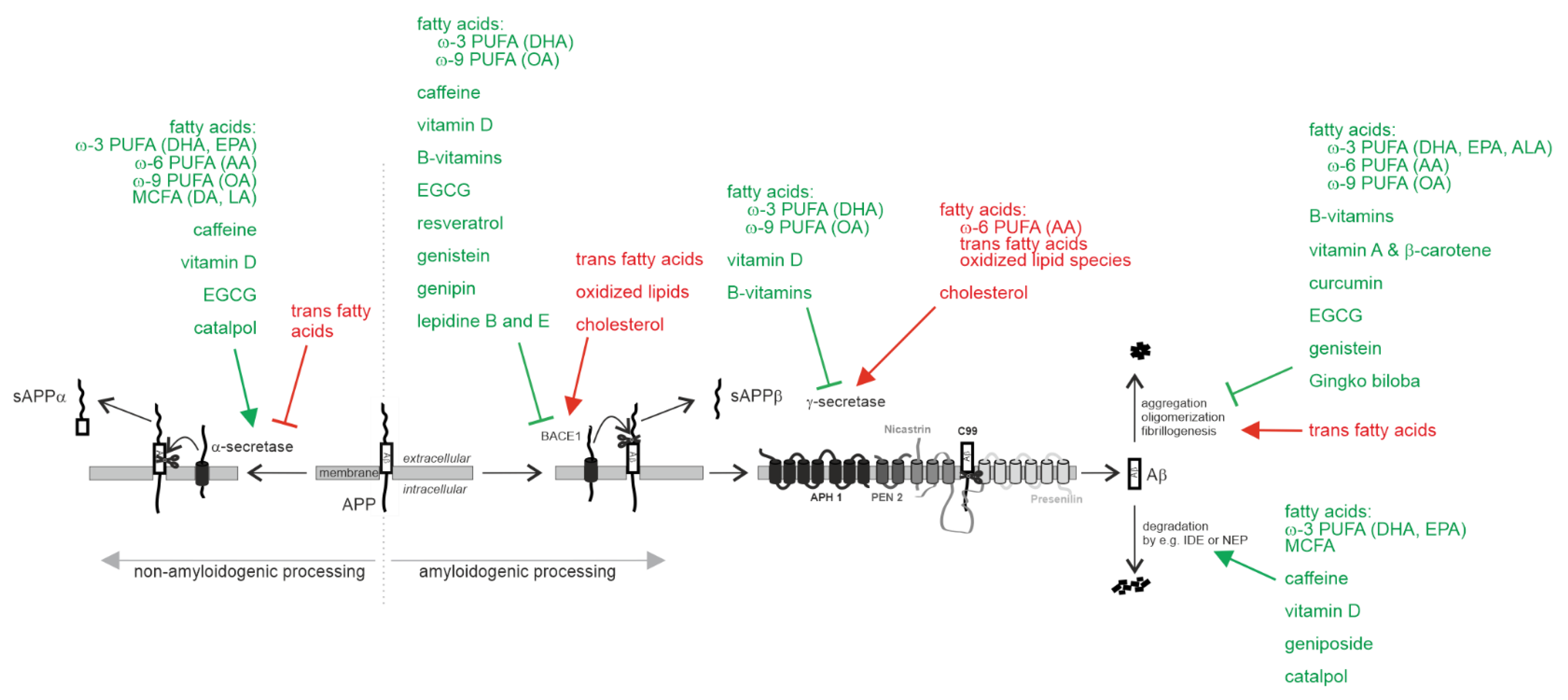
2.1.1. Effect on Aβ Pathology

Non-Amyloidogenic α-Secretase Processing of APP

Dietary interventions modulating the Aβ-preventing α-secretase cleavage of APP are a promising target for the treatment of AD (Figure 1).
DHA is the most abundant n-3 PUFA in all brain regions [82]. It is discussed as one of the most important FAs for the treatment or prevention of AD. It was found that α-secretase processing of APP, which reduces Aβ formation is stimulated in DHA-treated cellular models of AD, including neuroblastoma cells overexpressing APP695wildtype or APP695 with the Swedish double mutation (K670N, M671L) or both the Swedish and the Arctic (E693G) mutations as well as APP695 transfected HEK cells [83,84,85]. The molecular mechanisms leading to increased α-secretase processing of APP in the presence of DHA are due to increased ADAM17 gene expression and increased ADAM17 protein stability [84]. Notably, small amounts of oxidized DHA reversed the protective effect of DHA, decreasing the α-secretase processing of APP [86]. Besides DHA, both the n-3 PUFA eicosapentaenoic acid (EPA) and the n-6 PUFA AA increased α-secreted APP (sAPPα) in differentiated human neuroblastoma SH-SY5Y cells [87]. Increased levels of sAPPα have been also found in an early onset AD transgenic (Tg) mouse model fed with a low-fat, cholesterol-free diet enriched with the n-9 PUFA oleic acid (OA) [88]. The α-secretase stimulating effect is not limited to PUFAs, as phospholipids containing medium-chain saturated fatty acids (MCFAs)—decanoic acid (10:0) and lauric acid (12:0)—also increase α-secretase activity in human neuroblastoma cells [89]. In addition, calcifediol (25OH vitamin D3) has been shown to increase α-secretase activity promoting the non-amyloidogenic processing of APP [90]. In contrast, trans-fatty acids, whose consumption has increased in the 20th century since the first successful hydrogenation of oils [91], have been found to decrease non-amyloidogenic processing of APP by decreasing ADAM10 gene expression, resulting in elevated production of Aβ [92]. This effect is mediated by a decrease in ADAM10 gene expression and ADAM10 protein levels. Furthermore, high concentrations of trans fatty acids have been reported to decrease brain DHA in a 3xTg-AD mouse model of AD [93]. However, no significant effect on major brain neuropathological hallmarks of AD, including levels of Aβ40 and Aβ42 levels, was found in this study.
It has been reported that, in addition to FA and vitamins, dietary phytochemicals reduce the Aβ generation by increasing the α-secretase processing of APP. The polyphenol epigallocatechin-3-gallate (EGCG), a major catechin present in green tea, has been shown to decrease Aβ generation in neurons overexpressing APP695 and to decrease Aβ levels and plaques in Tg2576 AD mice, expressing the Swedish mutant of APP [94,95]. The EGCG-induced reduction in Aβ was attributed to an elevation in α-secretase APP cleavage, leading to an increase in α-CTF generation and sAPPα release, caused by a significant increase in the expression of ADAM10 and TNF-α converting enzyme, a further α-secretase candidate [94,96]. EGCG also significantly decreased brain Aβ production and plaque burden by increasing the levels of α-secretase ADAM10 in high-fat-fed transgenic APPswe/PSdE9 mice [97]. In vivo, intraperitoneal treatment of transgenic APPswedish Tg2576 mice with EGCG for 2 months significantly decreased Aβ levels [95]. Catalpol, an iridoid glycoside extracted from Rehmannia glutinosa roots, elevated the expression of α-secretase, promoting non-amyloidogenic APP processing in neuronal N2a cells overexpressing the Swedish mutant of APP [98]. An increase in protein stability of the α-secretase ADAM10 was identified as the molecular mechanism underlying the α-secretase stimulating effect in cell cultures, treated with methylxanthines, including caffeine, a natural alkaloid stimulant of coffee beans [99].

Amyloidogenic Processing of APP by β-Site Cleaving Enzyme BACE1 and γ-Secretase

Besides the beneficial property of DHA to increase the α-secretase processing of APP, DHA was found to decrease the amyloidogenic processing of APP by β- and γ-secretase [84] (Figure 1). In 15-month-old APP/PS1 mice, DHA supplementation also decreased Aβ deposition. This suggests that a DHA-enriched diet can diminish AD-like pathology [100]. DHA reduces amyloidogenic β-site cleavage of APP by a direct effect on β-secretase activity and by impairing internalization of BACE1 into the endosomal system [84]. Aβ-releasing γ-secretase activity is also directly affected by DHA. Furthermore, DHA reduces γ-secretase processing of APP in lipid rafts by causing a shift of PS1 (and cholesterol) and thus γ-secretase activity from the raft to non-raft fractions of the membrane. The potential of DHA for the treatment or prevention of AD is further underlined by its cholesterol-lowering effect, which further reduces Aβ generation. DHA reduces cholesterol de novo synthesis by directly inhibiting HMGCR activity, the rate-limiting step in cholesterol de novo synthesis, and by disturbing lipid raft integrity. This directs cholesterol out of these membrane microdomains [84,101,102]. These identified molecular mechanisms result in significant reductions in total Aβ levels and Aβ accumulation in DHA-supplemented cell culture experiments or in animal studies [100,103,104,105,106]. Besides DHA, the n-9 PUFA oleic acid (OA), the most abundant dietary FA, exerts anti-amyloidogenic properties. Transgenic AD mice expressing the Swedish double mutation and Indiana mutation fed a low-fat, cholesterol-free diet enriched with OA exhibited reduced levels of β-site APP cleaving enzyme (BACE) and reduced presenilin levels along with reduced amyloid plaques in the brain [88]. The potency of OA to reduce Aβ levels has been also found in APP695 transfected COS7 cells. Supplementation with OA resulted in reduced secreted Aβ levels [88].
It is discussed that the n-6 PUFA AA, in contrast to the n-3 PUFA DHA, elevates γ-secretase processing of APP, since SP-C99 transfected COS7 cells exposed to AA secreted significantly more Aβ40 and Aβ42 peptides [107]. In cells cultured with 0.1 mM or 0.2 mM linoleic acid (18:2), the precursor of AA, elevated levels of the N-terminal fragment of the γ-secretase component PS1 were found along with increased Aβ levels [108]. In line, an early onset AD transgenic mouse model expressing the Swedish double mutation (K670N/M671L) and Indiana mutation (V717F) revealed higher levels of Aβ and amyloid plaques in brains when mice were fed a diet supplemented with 2% AA [107]. Beside AA, trans fatty acids enhance the amyloidogenic processing of APP leading to elevated Aβ levels in cell culture experiments [92]. Compared with the corresponding cis conformation, trans fatty acids directly affect enzyme activities of β- and γ-secretase. Additionally, trans fatty acids significantly increase BACE1 gene expression and gene transcription of all γ-secretase components. Notably, the protective effect of DHA is reverted in presence of oxidized DHA. In cell culture experiments, using human neuroblastoma cells and mouse mixed cortical neurons, five different oxidized DHA derivatives and the lipid peroxidation products of n-3 and n-6 PUFAs, HNE and 4-hydroxy-hexenal, revealed elevated Aβ and soluble β-secreted APP levels [86]. The molecular mechanisms leading to higher Aβ levels in presence of oxidized lipids were identified as an increase in gene expression of BACE1 and the γ-secretase components PS1, Nicastrin and Aph1b, leading to amyloidogenic processing of APP. Additionally, oxidized lipids had a direct stimulating effect on β-secretase activity. Importantly, as little as 1% oxidized DHA was sufficient to reverse the protective effects of DHA and significantly increase Aβ generation [86]. It is therefore necessary, to prevent the oxidation of DHA in dietary approaches or supplements. Supplementation of PUFA must be implemented under conditions that protect from unintentional oxidation.
Due to the anti-oxidative property of vitamin E protecting lipids from oxidation, supplementation with vitamin E is discussed to be beneficial in AD. Additionally, vitamin E has neuroprotective, anti-inflammatory and hypocholesterolemic properties [109]. However, the effect of vitamin E on Aβ generation is still not completely understood and evidence of the beneficial role of vitamin E for the treatment of AD remains inconclusive [110]. While vitamin E supplementation led to reduced cerebral Aβ content and amyloid deposition in young AD transgenic mice, this effect was not observed in aged APP transgenic mice, that received the vitamin E-supplemented diet at an older age [111]. Reduced cerebral levels of Aβ oligomers have been also found in APP/PS1 double transgenic mice fed with α-tocopherol quinine, an oxidative metabolite of α-tocopherol [112]. In contrast, cell culture experiments suggest amyloidogenic potential of different members of the vitamin E family, including α-, γ- and δ-Tocopherol [113]. All tocopherols increased Aβ generation in human neuroblastoma cells by an elevation in gene expression as well as protein level of BACE1 and the components of the γ-secretase complex. In line with the Aβ increasing effect of tocopherols, α-tocotrienol also elevated Aβ levels in human neuroblastoma SH-SY5Y wildtype cells as well as APP695 expressing SH-SY5Y cells. α-Tocotrienol has a direct stimulating effect on the enzyme activities of β- and γ-secretase. Beside these unfavorable properties of members of the vitamin E family with respect to Aβ generation, both tocopherols and α-tocotrienol reduced cholesterol levels, a well-known risk factor for AD [114,115,116]. Due to the partially opposable effects of vitamin E with respect to the molecular mechanisms involved in AD, a recommendation without restriction for the treatment or prevention of AD should be reconsidered. Several epidemiological studies as well as animal studies and cell culture studies show that high cholesterol is linked to elevated Aβ generation and AD pathology [117,118,119,120]. High cholesterol levels have been found to increase β- and γ-secretase activity, especially in cholesterol-rich lipid raft membrane microdomains. In contrast to the E-vitamins, vitamin D has been reported to exert anti-amyloidogenic potential. APP695 transfected SH-SY5Y cells treated with 25(OH) vitamin D3 (calcifediol) or different vitamin D3- and vitamin D2-analogues showed significantly reduced total Aβ levels [90]. Vitamin D3 and its analogues decrease β-secretase activity due to a reduction in BACE1 gene expression along with a decrease in BACE1 protein level. A 24% reduction in BACE1 protein level was also found in aged rats that received a subcutaneous injection of 1,α25-dihydroxyvitamin D3 (42 I.U./kg for 21 days) [121]. In line with these findings, Aβ40 and Aβ42 levels are elevated in hypovitaminosis D mouse brains caused by an elevated BACE1 protein level resulting in elevated β-secretase activity in these mice [122]. Vitamin D3 and its analogues also decrease gene expression of the γ-secretase component nicastrin, leading to reduced γ-secretase activity [90]. In addition to these molecular mechanisms, another study has found that APP promoter activity is suppressed in presence of 1,α25-dihydroxyvitamin D, indicating that Aβ secretion might be reduced after vitamin D3 treatment due to a diminished gene expression of the Aβ-precursor APP [123]. APP transgenic mice fed with a vitamin D3 enriched diet for five months, starting immediately after weaning, also revealed a decrease in the number of amyloid plaques and a reduction in Aβ peptides, further underlining the anti-amyloidogenic potential of vitamin D [124]. However, a recent study reports that vitamin D supplementation worsens the progression of AD [125]. In this study, vitamin D supplementation increased Aβ level and amyloid burden along with elevated BACE1 protein level in the hippocampus of APP/PS1 transgenic mice.
Several cell culture and animal studies dealing with vitamin B supplementation or vitamin B deficiency strongly indicate that B-vitamins exert protective effects with respect to Aβ pathology [126,127,128,129,130]. Tg2576 transgenic mice overexpressing the APP Swedish (APPswe) mutation, resulting in elevated β-secretase processing of APP and therefore Aβ levels, fed with a folate (vitamin B9), vitamin B6 and vitamin B12 deficient diet for 7 months, showed significantly increased levels of Aβ peptides in the hippocampus compared to Tg2576 mice fed with the control diet [126]. In line, elevated Aβ deposition has been reported for TgCRND8 mice (expressing the Indiana mutation in addition to the APPswe mutation), fed with a diet deficient in vitamin B6, vitamin B12 and folate compared to the control diet [127]. In this study elevated Aβ deposition was caused by an increase in PS1 and BACE expression. Besides the identification of alterations in BACE1 and PS1 gene expression in presence of vitamin B deficiency, leading to elevated Aβ levels, Aβ levels are increased caused by an elevation in cholesterol de novo synthesis. Human adipocytes cultured in media containing low vitamin B12 or no vitamin B12 revealed significantly increased cholesterol levels caused by an increase in gene expression of genes involved in cholesterol de novo synthesis, including the rate-limiting enzyme HMGCR (3-hydroxy-3-methylglutarly CoA reductase) [131] compared to cells cultured with adequate vitamin B12 levels. Furthermore, low vitamin B12 increased gene expression of sterol regulatory element-binding proteins, SREBP1 and SREBP2, and the sterol regulatory element-binding transcription factors, SREBF1 and SREBF2, which are involved in the regulation of cholesterol synthesis and low-density lipoprotein receptor (LDLR) gene expression. Again, hypomethylation of the promoter regions is discussed to play a crucial role in the increase in cholesterol biosynthesis under vitamin B12 deficient conditions [131], leading to elevated Aβ levels. Based on these studies B-vitamins are discussed to reduce amyloid pathology by decreasing BACE1 and PS1 gene expression and decreasing cholesterol de novo synthesis [81]. This protective effect of B-vitamins is further underlined by several clinical randomized controlled trials showing the beneficial effects of B-vitamins in persons affected by MCI or AD [81,132,133,134,135].
Among phytochemicals, EGCG is discussed to reduce the amyloidogenic processing of APP by inhibiting β-secretase activity in a cell-free system [136]. In vivo, prolonged administration of EGCG to mice has been shown to down-regulate APP in the hippocampus, suggesting that EGCG might reduce Aβ levels additionally by impairing APP gene expression [96]. Resveratrol, derived from a subclass of non-flavonoid polyphenols termed stilbenes (mainly found in red grapes and red wine), is reported to possess anti-amyloidogenic activity, reducing secreted and intracellular Aβ peptide levels in several cell lines expressing the APPswedish mutation [137]. The anti-amyloidogenic activity of resveratrol is discussed to be attributed to an inhibitory effect on the activity of β-secretase as well as by modulating the proteasome [137,138,139]. Furthermore, in vivo studies have demonstrated that resveratrol decreases amyloid plaque formation, Aβ42 levels, BACE1 and APP levels in Tg6799 mice expressing five familial AD mutations (5xFAD mice) [140] and decreases the amount of insoluble Aβ in the hippocampus of AD rats [141]. Genistein, a naturally occurring isoflavone primarily present in legumes, green peas, soybeans, and peanuts, has been found to inhibit BACE1 through reversible non-competitive mechanism, thus reducing Aβ generation [142]. Recently, Genipin, an aglycone isolated from the extract of Gardenia jasminoides Ellis fruit, has been reported to decrease Aβ production by inhibiting BACE1 expression in N2a cells expressing the Swedish mutant of APP [143]. Furthermore, Lepidine B and E from Lepidium sativum have been suggested as potent inhibitors of β-secretase BACE1 in a recent study [144]. Caffeine, that, like DHA, resulted in elevated α-secretase activity, also decreased amyloidogenic APP processing by down-regulating BACE1 gene expression and directly affecting β-secretase activity [99].

Aβ Degradation

Total Aβ level is not only determined by the production of Aβ out of its precursor APP but also strongly depends on its degradation by Aβ-degrading enzymes. An insulin-degrading enzyme (IDE), a zink-metalloprotease, as well as neprilysin (NEP) have been identified as the main enzymes involved in Aβ catabolism [145,146]. Several micronutrients, including PUFAs, DHA, and EPA as well as medium-chain length fatty acids (MCFAs), have been found to enhance Aβ degradation by affecting IDE [147,148] (Figure 1). DHA has been reported to elevate the exosome release of IDE and to directly stimulate IDE activity, leading to an increased Aβ degradation in the extracellular space [147]. EPA also directly stimulates IDE activity, besides elevating gene expression of IDE. MCFAs also directly elevate the activity of recombinant IDE and increase extracellular IDE levels, whereas longer-chain length FAs resulted in an inhibited IDE activity [148]. Calcifediol (25(OH) vitamin D3) as well as D3- and D2-analogous have been reported to elevate Aβ degradation in mouse neuroblastoma cells [90]. The increase in Aβ degradation in presence of vitamin D3 or analogous could be mainly attributed to an elevated expression of NEP along with an increase in NEP activity. Calcifediol also elevated the protein level of IDE [122]. In line with an increase in NEP expression and NEP activity in vitamin D-supplemented cell culture experiments, a significant decrease in NEP expression and NEP activity is reported for hypovitaminosis D mouse brains [122]. Vitamin D supplementation (1,α25-dihydroxyvitamin D3, 42 I.U./kg for 21 days) also resulted in significantly elevated NEP protein levels in aged rats [121]. In contrast, members of the vitamin E family, tocopherols as well as tocotrienols, have been found to decrease Aβ degradation by affecting IDE, leading to elevated Aβ levels [149,150].
The monoterpene Geniposide, a major iridoid glycoside of Gardenia jasminoides, has been reported to elevate Aβ degradation. The intragastric administration of Geniposide in streptozotocin-induced diabetic rats increased the expression of IDE and decreased Aβ1-42 levels [151]. Similarly, Catalpol increased IDE expression and reduced Aβ levels in mice injected with Aβ and D-galactose [152]. In addition, it has been shown that Catalpol alleviates fibrillar Aβ1-42 induced disruption of the blood–brain barrier and enhances soluble Aβ clearance [153].

Aβ Oligomerization, Aβ Aggregation and Aβ Fibrillogenesis

DHA as well as OA were identified as excellent inhibitors of Aβ40 and Aβ42 fibrillogenesis (~81–84% inhibition) in vitro by the use of fluorescence-based aggregation kinetic experiments, transmission electron microscopy and molecular docking studies [154] (Figure 1). EPA, α-linolenic acid (ALA) and AA also exhibit anti-aggregation properties, although to a much lesser extent than DHA and OA [154]. Accordingly, DHA and AA at micellar concentrations stabilized soluble Aβ42 wild-type protofibrils, preventing their conversion to insoluble fibrils [155]. In contrast, fluorescence thioflavin-T-based assay and electron microscopy studies found that trans fatty acids, which already elevate amyloidogenic processing of APP, increase oligomerization and aggregation of Aβ in cell culture studies [92]. B-vitamins, in addition to reducing amyloid pathology by decreasing PS1 and BACE1 gene expression and cholesterol de novo synthesis, have promising beneficial properties in relation to Aβ fibrillogenesis and aggregation. Vitamin B12 inhibited Aβ42 aggregation in a dose-dependent manner, significantly prevented the conversion of Aβ42 from random coil to the β-sheet formation and reduced the hydrophobicity of Aβ fibrils as well as the site of the aggregates [81,156]. A recent study further supports the inhibitory effect of vitamin B12 on Aβ fibrillation. Vitamin B12 has been found to significantly reduce Aβ fibril content by reducing the transition of Aβ oligomers to mature fibrils [157]. Furthermore, in presence of synthetic neuronal membranes vitamin B12 has been found to disaggregate preformed fibrils [157]. Besides vitamin B12, vitamin A and provitamin A (β-carotene) inhibit oligomerization of Aβ and destabilize preformed Aβ fibrils [158]. Using fluorescence spectroscopy with Thioflavin T and electron microscopy, it was shown that vitamin A and β-carotene inhibited the formation of fibrillar Aβ from fresh Aβ and its extension in vitro in a dose-dependent manner [159]. In addition, retinoic acid (vitamin A) decreased cellular toxicity by inhibition of Aβ42 oligomerization [160]. In vivo, retinoic acid attenuates Aβ deposition and rescues memory deficits in a transgenic mouse model of AD [161]. A recent study by Joshi et al. further underlines the protective effect of vitamin A with respect to Aβ aggregation in vitro and in vivo [162]. The authors found that vitamin A delayed Aβ42 aggregation in an in vitro screen based on Thioflavin T, whereas α-tocopherol, a vitamin E metabolite, promotes the aggregation of Aβ42. Combining vitamin A and vitamin E has no effect on Aβ42 aggregation. Transmission electron microscopy also revealed a reduction in Aβ42 fibrils in presence of retinoic acid and an increase in Aβ42 fibrils in presence of α-tocopherol. As discussed in the section on Aβ pathology, vitamin E, including α-tocopherol, must be considered an ambiguous player in AD pathology. In animal models, protective effects could be found in relation to Aβ pathology, whereas cell culture studies or in vitro studies revealed potential negative properties [113,162]. However, the study by Joshi et al. further shows that vitamin A, as well as vitamin E, have protective effects in a Caenorhabditis elegans model of AD. In this AD model, Aβ42 is expressed in the body wall muscle cells, where it aggregates and results in age-progressive paralysis. Upon treatment with vitamin A and vitamin E, a decrease in Aβ42 aggregates/fibrils along with an increase in the total fitness of the worms compared to untreated worms was detected [162]. Orally administered α-tocopherol also decreased levels of Aβ oligomers in brains of APPswedish/PS1dE9 transgenic mice [112].
Several in vitro and in vivo studies indicate that curcumin, a yellow pigment in the rhizome of turmeric (Curcuma longa) has anti-amyloidogenic properties [163]. In vitro studies have demonstrated that curcumin inhibits the formation and extension of neurotoxic Aβ1-40 and Aβ1-42 fibrils from fresh Aβ in a dose-dependent manner and destabilizes preformed fibrils to regenerate Aβ monomers [164,165,166]. Several molecular mechanisms of the anti-amyloidogenic property of curcumin are discussed: (1) curcumin with its two 3,4-methoxyhydroxyphenyl rings connected by a short carbohydrate chain might be able to specifically bind free Aβ and subsequently inhibit the polymerization of Aβ into Aβ fibrils [164]; (2) curcumin might specifically bind to Aβ fibrils and might destabilize the β-sheet-rich conformation of Aβ in Aβ fibrils [164]; (3) curcumin is able to bind to the N-terminus (residues 5–20) of Aβ42 monomers and low molecular weight oligomers [167] and to induce major structural changes in the Aβ1-42 aggregates [168]; (4) curcumin molecules intercalate among the Aβ chains in the first step of Aβ aggregation and bind tightly to them by hydrogen bonds and hydrophobic interactions, leading to less flexible and more disordered amyloid structures [169]. In vivo, systemic treatment with curcumin reduces pre-existing plaques in ~8-month-old APPswe/PS1dE9 mice, suggesting the ability of curcumin to disaggregate Aβ deposits [170]. Besides stimulating α-secretase activity, EGCG interferes with Aβ aggregation. EGCG binds weakly and non-specifically to Aβ monomers, whereas it exhibits higher affinity to oligomers [171]. Electron microscopy revealed that ECGC interferes with the early step of Aβ aggregation by forming spherical, off-pathway aggregates, thus inhibiting secondary nucleation of Aβ [171,172]. Recently, it has been shown, that EGCG is also able to disassemble preformed Aβ fibrils. ECGC has been reported to disrupt Aβ protofibrils by forming π-π and hydrogen bonding interactions [173,174]. Additionally, a recent meta-analysis of 17 studies in AD animal models showed that EGCG can reduce Aβ pathology through anti-aggregating activity in combination with anti-inflammatory and antioxidant properties [175]. The isoflavone genistein also prevents the formation of Aβ aggregates by directly binding to Aβ25-35 fragments [176]. Antifibrillogenic activity has been also reported for Gingko biloba. The standardized extract from the leaves of Gingko biloba tree, EGb761, prevented β-amyloid fibril formation in solution in vitro as well as in the conditioned medium of neuroblastoma cells stably expressing the Swedish mutant APP and the exon-9 deletion mutant PS1 [177].

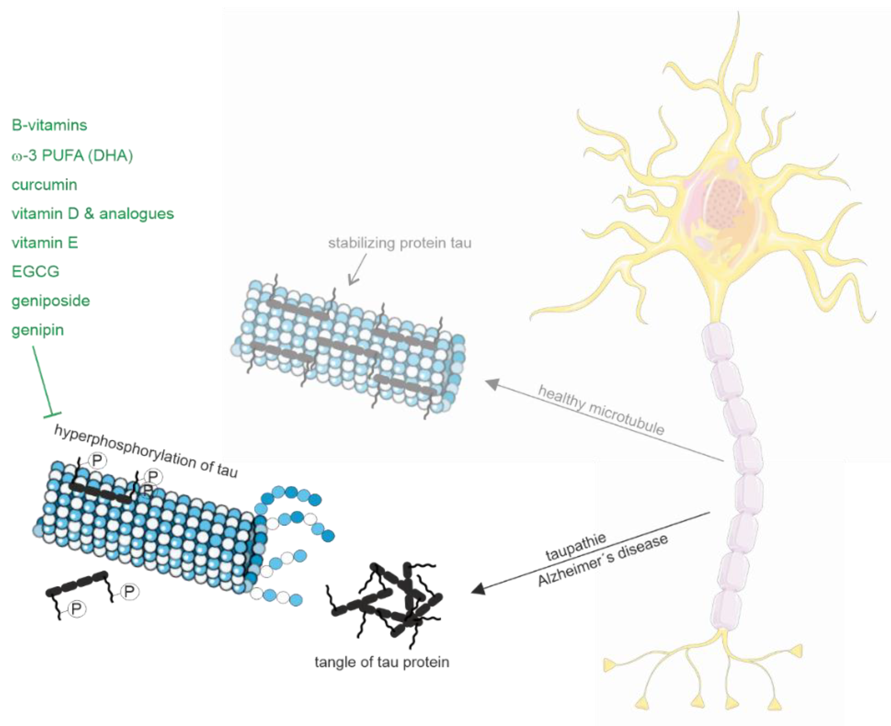
2.1.2. Tau Pathology

Besides the described protective properties of B-vitamins with respect to Aβ pathology, B-vitamins also improve Tau pathology through various molecular mechanisms (Figure 2). Homocysteine-induced changes in Tau hyperphosphorylation could be reduced by simultaneous supplementation of folate and vitamin B12 in rats [129]. In line, supplementation of folate and vitamin B12 alleviates hyperhomocysteinemia-induced Alzheimer-like pathologies, including Tau hyperphosphorylation, in the rat retina [130]. In this context, it has to be mentioned that in the retina, an outgrowth of the developing brain, tau hyperphosphorylation as well as Aβ accumulation are observed in early AD stages [178]. The homocysteine-induced Tau hyperphosphorylation in the hippocampi of rat brains could be ascribed to an inhibition of protein phosphatase 2A (PP2A) involved in Tau dephosphorylation [179]. Tau hyperphosphorylation and PP2A inhibition could be significantly antagonized by the simultaneous supplementation of folate and vitamin B12, indicating that B-vitamins elevate PP2A activity, leading to Tau dephosphorylation [129,179]. Furthermore, B-vitamins are discussed to inhibit several kinases involved in Tau phosphorylation, including glycogen synthase-3β (GSK-3β), cyclin-dependent kinase-5, C-jun N-terminal kinase, extracellular signal-regulated kinase and p38MAPK [129]. In addition, Tau polymerization in presence of vitamin B12 is inhibited by the direct binding of vitamin B12 to cysteine residues of Tau [180]. DHA has also been found to reduce Tau hyperphosphorylation by inhibition of kinases involved in Tau phosphorylation. DHA suppressed traumatic brain injury-induced tau hyperphosphorylation by inhibiting c-jun N-terminal kinases, improving neurological function [181]. The influence of PUFAs on c-jun N-terminal kinase could also be shown by the dietary treatment of 3xTg AD mice [104]. Dietary DHA treatment significantly reduced the steady state level of Tau protein after three months. After six months somatodendritic tau accumulation was significantly reduced in 3xTg AD mice, fed with a diet containing DHA alone or in combination with the n-6 PUFA docosapentaenoic acid (DPA). Mice fed the DHA-DPA diet revealed a reduction in phosphorylated c-Jun N-terminal kinase which correlated with reduced levels of early stage phosphor-tau epitopes. The n-3 PUFA DHA also inhibited c-Jun N-terminal kinase and phosphorylation of Tau in cultured hippocampal neurons and in 3xTg AD mice [182]. Treatment of 3xTg AD mice on a high-fat diet with fish oil or curcumin or a combination of both for 4 months reduced phosphorylated JNK and phosphorylated Tau. Furthermore, DHA-containing phosphatidylcholine showed a reduction in phosphorylated Tau in Aβ25-35-induced AD rats [183]. DHA has also been reported to inhibit GSK-3β phosphorylation and the phosphorylation of Tau proteins in APP/PS1 wildtype mice fed with DHA (400 mg/kg once daily for 2 months), thus inhibiting tau protein neurofibrillary tangle formation in the hippocampi of these mice [184]. Moreover, DHA treatment attenuated increased levels of hippocampal tau phosphorylation in rats fed with a high-fat diet, suggesting that DHA protects against the neurotoxic effects of phosphorylated Tau [185]. Notably, a recent study by Zussy et al. shows that the intranasal administration of nanovectorized DHA decreases the phosphorylation of Tau and restores cognitive functions in two complementary AD murine models, paving the way for the development of new approaches to prevent or treat AD [186].
Besides DHA, recent findings propose beneficial properties of vitamin D or vitamin D analogues with respect to tau pathology. Activation of the vitamin D receptor (VDR) by paricalcitol, a specific agonist of the VDR, reduced phosphorylation of Tau at Ser396 and Thr181 sites via inhibiting GSK-3β phosphorylation in APP/PS1 transgenic mice [187]. Maxacalcitol, an active vitamin D analogue, significantly decreased hyperphosphorylation of MAPK-38, ERK1/2 and tau proteins in experimental AD in rats [188]. In addition to the repressive effect of vitamin D on kinases involved in Tau phosphorylation subcutaneous injection of vitamin D (1,α25-dihydroxyvitamin D3) for 21 days in rats resulted in a 29% increase in PP2A activity in hippocampal tissue [189]. In line with the elevated PP2A activity, Tau phosphorylation in the hippocampus was reduced in aged rats after vitamin D administration, including pre-neurofibrillary tangle phospho-tau protein (pThr231), intraneuronal neurofibrillary tangle phospho-tau protein (pSer214), and extracellular neurofibrillary tangle phospho-tau protein (pSer404). 1,25(OH)2D3 also alleviates Aβ25-35-induced Tau hyperphosphorylation in SH-SY5Y cells [190]. Notably, a recent cross-sectional, explorative study investigating possible associations of vitamin D in CSF with biomarkers for AD, including tau protein and phosphorylated tau protein, revealed that higher levels of 25(OH) vitamin D were significantly associated with lower levels of tau protein as well as phosphorylated tau protein [191].
Vitamin E also exerts protective effects with respect to Tau pathology. Primary cultures of rat cortical neurons incubated with 5 µM β-amyloid peptide cause an oxidative-stress-induced activation of p38 MAPK, leading to tau hyperphosphorylation [192]. The Aβ-induced effects were prevented when neurons were co-incubated with Trolox, the water-soluble analog of vitamin E. Furthermore, high level of phosphorylated p38 MAPK in the hippocampus of APP/PS1 transgenic mice could be prevented by feeding mice with a diet supplemented with vitamin E [192]. Vice versa, dietary deficiency in vitamin E and folate under conditions of oxidative stress increased phospho-tau levels in mice expressing human apolipoprotein E4 (associated with increased risk of AD) [193]. A recent study investigated the effects of vitamin D and E on an insulin-resistant model induced in SK-N-SH neuronal cells, hypothesizing that treatment with vitamin D and E would reverse the effects of AD and improve insulin signaling [194]. Besides the improvement of the insulin signaling pathway upon vitamin D treatment, vitamin D significantly decreased GSK3β and Tau expression levels. Vitamin E alone as well as the combination of vitamin D and E also reduced GSK3β and Tau. In contrast to the proposed beneficial properties of DHA, vitamin B12, vitamin D and vitamin E, vitamin A (retinol) supplementation to human neuroblastoma cells elevated tau phosphorylation at Ser396 [195].
The polyphenol EGCG also exerts potential protective effects in respect to Tau pathology in addition to its protective effects regarding Aβ pathology. EGCG reduces sarkosyl-soluble phosphotau isoforms in TG2576 mice [196]. It is therefore discussed to decrease phospho-tau by direct binding and inhibition of heat shock protein 90 (HSP90), reported to be involved in the phosphorylation status of Tau [197]. Furthermore, Geniposide attenuated Tau hyperphosphorylation by reducing GSK-3 enzyme activity in streptozotocin-treated rats and mice [198,199]. Beside affecting GSK-3 geniposide has been reported to decrease Tau hyperphosphorylation and Aβ42 generation due to increased leptin signaling [200,201]. A recent study further demonstrates the potential beneficial effect of Genipin with respect to tau hyperphosphorylation and Tau fibril formation [143]. In this study, Genipin has been found to bind to Tau and to protect against heparin-induced Tau fibril formation. Additionally, Genipin downregulates the expression of the Tau-kinases CDK5 and GSK-3β in Tau-overexpressing cells.

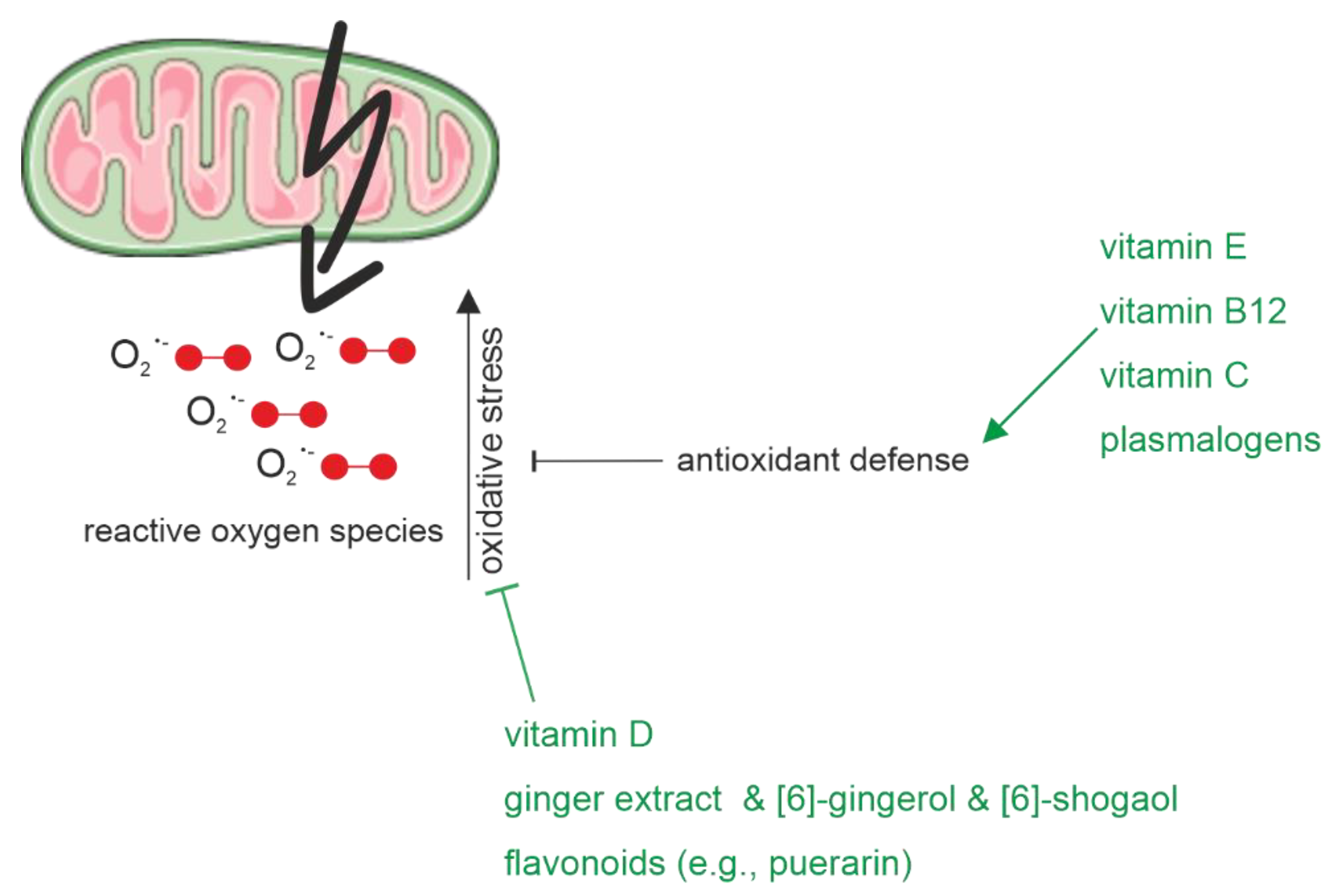
2.1.3. Oxidative Stress
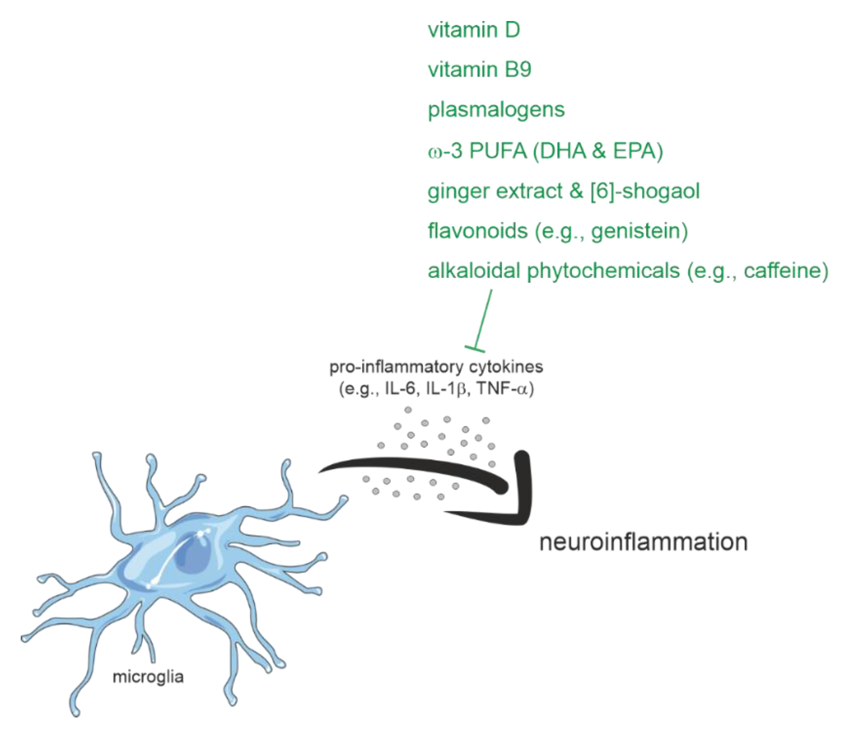
Oxidative stress, which can be defined as an imbalance between the formation of oxidant species and insufficient antioxidant defense [202], appears to be another hallmark of AD pathology. For example, a study analyzing human postmortem brain samples demonstrated that compared to non-demented control individuals, oxidized lipid and 4-hydroxy-nonenal (HNE) levels were significantly increased in brain samples from PwAD. This negatively affected Aβ levels [86]. Moreover, the levels of hydrogen peroxide, a major reactive oxygen species (ROS), were detected to be increased in the brains of PwAD compared to healthy controls and can be influenced by Aβ peptides [203,204,205]. It is discussed that the overproduction of ROS due to mitochondrial damage occurs earlier than the Aβ pathology or clinical symptoms [206]. Oligodendrocytes are the cells in the central nervous system that may contribute to oxidative stress due to their reduced glutathione levels compared to other brain cells [207]. Oligodendrocytes are the exclusive location for myelin-formation in the central nervous system. Impaired function of this cell type, e.g., due to oxidative stress, can lead to demyelination. This, in turn, reduces the action potential time of neurons and thereby worsens the cognitive decline in PwAD [208].
The before-mentioned mechanisms are representative reasons why antioxidants, which prevent and reduce free radical-mediated damage in neuronal cells, are important for the prevention and treatment of AD. Based on this, different pharmacological therapies are currently discussed [209]. In the following paragraph, some antioxidants from food sources and their potential impact on AD pathology due to oxidative stress are presented (Figure 3). One important antioxidant is vitamin E, which might also have beneficial effects on AD pathology due to its neuroprotective properties [210]. Vitamin E occurs in various forms in natural food (four tocopherols and four tocotrienols), of which α-tocopherol represents the most abundant and bioavailable antioxidant in humans [211]. Foods rich in vitamin E are for example vegetable oils, various nuts, seeds, or green leafy vegetables. On a molecular level, vitamin E can be classified as a lipophilic antioxidant that protects membranes from free radical-mediated oxidative damage [212]. The antioxidative properties of vitamin E are based on a hydroxyl group in its phenolic group on the chromanol ring, which can donate a hydrogen atom to neutralize free radicals, including ROS [213]. Besides α-tocopherol, also for α-tocotrienol, the most abundant form of the tocotrienol family, which is characterized by the unsaturated side chain, a reducing influence on the generation of ROS could be detected in human neuroblastoma cells [150]. Furthermore, in an animal study using a transgenic AD model, vitamin E was able to reduce lipid peroxidation [111]. Regarding human clinical or epidemiological studies there are only limited and inconsistent data available analyzing the role of vitamin E alone regarding oxidative stress [210]. Moreover, most of these kinds of studies used a combination of different nutrients or antioxidants, which will be discussed later.
A further fat-soluble vitamin with antioxidative properties is vitamin D. This secosteroid might mediate its beneficial influence on oxidative stress via transcriptional regulation involving the intracellular vitamin D receptor. It is reported that Vitamin D prevents oxidative stress-related oxidation of proteins and lipids as well as DNA damage by the facilitation of balanced mitochondrial activities [214]. On a cellular level, it was shown recently that the active form of vitamin D (1,25(OH)2 vitamin D3) can modulate Aβ-induced ROS by scavenging intracellular ROS [190]. Moreover, similar findings regarding the antioxidant potential of vitamin D were obtained in animal studies [215]. In line with this, increased oxidative stress was reported in an AD mouse model fed with a vitamin D-deficient diet for 13 weeks. Along with this, enzymes such as superoxide dismutase 1 (SOD1), glutathione peroxidase 4 (GPx4) or cystine/glutamate exchanger (xCT) were reported to be downregulated under these conditions [216].
In addition to fat-soluble vitamins, a positive influence on oxidative stress has also been reported for water-soluble vitamins. For example, vitamin B12 was recently shown to protect against the ROS-mediated oxidation of lipids in a cell model. Especially plasmalogens were protected from hydrogen peroxide-induced oxidative stress in the presence of vitamin B12. This was mediated by an increased expression of superoxide-dismutase (SOD) and catalase (CAT), two ROS-degrading enzymes. Furthermore, the transcription of alkylglycerone phosphate synthase (AGPS) and choline phosphotransferase 1 (CHPT1), two enzymes involved in the plasmalogen synthesis, was also elevated under oxidative stress conditions in the presence of vitamin B12 [217]. Besides vitamin B12, a recent study reported that vitamin C decreased oxidative stress and DNA damage caused by brain surgery (laparotomy) in an APP mouse model [218]. Vitamin C is known to have antioxidant effects [73]. On a structural level, this could be explained by its vulnerability to providing an electron for oxidizing radicals [219]. Moreover, Vitamin C has synergistic effects with vitamin E in the protection of low-density lipoprotein from oxidative damage [220]. In line with this, a study using a mouse model with a knockout of a neuronal vitamin C transporter to generate a vitamin C deficiency reported elevated oxidative stress in the brain cortex and reduced total glutathione in comparison to wild-type mice [221]. Moreover, supplementation of vitamin C (3.3 g/l) was able to prevent abnormal mitochondrial morphology found in vitamin C deficient 5x FAD mice [222]. Along with these findings in animal studies, a meta-analysis found a lowered relative risk (0.83 with 95% CI 0.72 to 0.94) for AD when vitamin C was consumed in the diet [223].
Regarding plasmalogens, it should be mentioned that this lipid class has an impact on the generation and caused damage to oxidative stress itself. This is due to characteristic moieties in their unique chemical structure: plasmalogens have a vinyl-ether bond at the sn-1 position of the glycerol-backbone, which makes them highly susceptible to oxidation. Furthermore, they have bound PUFAs at the sn-2 position frequently, which are also vulnerable to oxidative stress [224]. Analysis of cerebral cortex homogenates of rats under induced oxidative stress confirmed that plasmalogens are highly sensitive to oxidative stress. This was demonstrated, for example, by the 70% decrease in plasmalogens after 90 min of UV irradiation [225]. These and other findings suggest plasmalogens as antioxidant molecules, as the vinyl ether bond could be the first target for newly formed radicals.
Moreover, the beneficial effects of ginger, which has numerous medical properties, and its biologically active components including, among others, gingerols, shogaols, paradols, and zingerone, regarding AD were reported. It was reported in vitro, for example, that ginger extracts were able to prevent lipid peroxidation in rat brains [226]. In neuroblastoma cells, pretreatment with [227]-gingerol prevented the Aβ25-35-induced disruption of the mitochondrial membrane potential and effectively inhibited the accumulation of ROS by restoring endogenous antioxidant glutathione levels and upregulating the expression of antioxidative enzymes [228]. Moreover, also for [6]-shogaol it could be shown, that this component of ginger could recover an increased ROS production induced by H2O2 treatment in vitro [229].
Another nutritional factor with anti-oxidative properties is flavonoids. For example, for the natural isoflavone puerarin, an alleviation in oxidative stress was reported in an AD mouse model [230]. It seems to be unlikely that flavonoids represent direct ROS scavengers due to their low circulating concentrations in the brain [231]. More likely is the modulation of pathways, that include for example pro-survival signaling molecules such as Akt/protein kinase B, p38 mitogen-activated protein kinase or c-jun N-terminal kinase [232]. An additional mechanism of action was described in vitro and comprises the activation of transcription factors, such as Nrf2 or PPARγ [233].
Oxidative stress can, among others, lead to inflammation by the activation of microglia and astrocytes, which results in the release of pro-inflammatory cytokines. Inflammation, along with Aβ- and tau-pathology is a further hallmark of multifactorial AD and may also contribute to and exacerbate this neurodegenerative disorder. Due to this, the following paragraph is going to present the influence of nutritional components on inflammatory processes related to AD.

2.1.4. Inflammation

A further contributor to the development and exacerbation of AD is inflammation, which is mediated by astrocytes and microglia in the brain. Microglia are considered the major source of pro-inflammatory cytokines such as IL-6, IL-1β or TNF-α. These cytokines are important for the regulation and initiation of the inflammation process including the migration of leukocytes and immune cells.
In the study mentioned above, it was found in the mouse model that, due to vitamin D malnutrition, increased inflammatory stress in form of promoted glial activation and significantly increased secretion of inflammatory factors (IL-1β, IL-6, and TNFα) developed [216] (Figure 4). Another recent study, using vitamin D-deficient mouse brains for genomics analysis, reported a significantly increased expression of Casp4, a gene encoding caspase-4, which is part of the innate immune response [234]. Microglial caspase-4 expression is suggested to contribute to cognitive impairments in AD, such as hippocampal synaptic plasticity [235]. On a molecular level, it could be shown in human neuroblastoma cells that a supplementation of vitamin D and its analogues is able to tendentially decrease the levels of IL-1β [90]. Moreover, for acitretin, a derivate of the fat-soluble vitamin A, which is known to mediate anti-inflammatory properties [236], an immune stimulatory effect was shown in the 5xFAD mouse model and human CSF [237].
With respect to water-soluble vitamins, a randomized controlled trial reported decreased inflammation due to the supplementation of folic acid (vitamin B9). In a study including 120 people, a six-month treatment with 1.25 mg folic acid per day resulted in significantly lowered levels of TNF-α mRNA in participants of the intervention group compared to those of the control group. Based on these findings, the authors suggested inflammation as an essential player in the association between folic acid and AD [238].
Similar findings were obtained for plasmalogens on the cellular level recently. Pretreatment of human neuroblastoma cells with eicosapentaenoic acid-enriched ethanolamine plasmalogen in a neuroinflammation model, generated with help of conditioned medium from lipopolysaccharides-induced BV2 cells, resulted in the reversal of increased nitric oxide and TNF-α levels as well as of the reduced IL-10 levels [239]. Regarding lipids and fatty acids, a study found new evidence for the molecular mechanisms through which ω-3 fatty acids, EPA and DHA, may exert their known anti-inflammatory and neuroprotective properties [240]. A pre-treatment of human hippocampal progenitor cells with EPA or DHA prevented the decrease in neurogenesis and the increase in apoptosis, which both were induced by treatment with IL-1β, IL-6 and interferon-α (IFN-α) [241]. Moreover, a further study indicates another possible underlying mechanism since ω-3 PUFAs were found to attenuate the inflammation-induced hyperactivity of the immunoproteasomes in astrocytes [242]. In a rat model, the supplementation of EPA was able to normalize the IL-1β-induced elevation in TNF-α expression and thereby mediate beneficial effects regarding inflammatory processes [243].
Additionally, for some bioactive components of ginger beneficial effects regarding neuroinflammation were reported. For example, it could be shown that [6]-shogaol exerts anti-inflammatory influences by inhibiting the microglia-mediated production of proinflammatory cytokines (IL-1β, TNF-α) in vitro [244]. Similar findings were obtained in the brain of animal models of dementia, where treatment with [6]-shogaol was reported to play a role in inhibiting glial cell activation [245]. Moreover, ginger extract can inhibit Aβ-induced expression of proinflammatory genes, including TNF-α, COX-2, or IL-1β in a human monocytic cell line sharing properties with human microglial cells [246].
Regarding the numerous beneficial effects of flavonoids on neuroinflammation [247], it was for example recently reported, that genistein, an isoflavone abundant in soy, mediates its inhibitory effects on LPS-induced expression of TNF-α, IL-1β or IL-6 via the activation of a G protein-coupled estrogen receptor (GPER) [248]. Animal studies found the natural flavonoid eriodictyol, to regulate inflammatory mediators and cytokines via the NF-κb and MAPK pathways [249].
In the context of potential neuroprotective agents, phytochemicals should not be disregarded as they have, for example, beneficial effects on the neuroinflammatory cascade. With regard to AD pathology, alkaloidal phytochemicals, such as caffeine, berberine, huperzine A, galantamine, sophocarpidine, or nicotine, are of interest, among others, because of their neuroinflammatory properties, as recently reviewed [250]. Besides these nutritional approaches, the gut microbiota has become a new potential target to alleviate neuroinflammatory processes in AD, in recent years [251].

2.1.5. Multicomponent Nutritional Interventions

Regarding nutritional interventions, more and more evidence arises that combined dietary components show synergistic beneficial actions with respect to AD pathology. For example, it could be shown in an AD mouse model that the dietary vitamin E status influences the benefits of fish oil supplementation. With respect to oxidative stress, a dose-dependent attenuation was observed, expressed as modulatory effects on the antioxidant system [252]. A recent in vitro study found a preventive effect of the Fortasyn Connect multi-nutrient combination on reactive astrogliosis. This combination of DHA, EPA, uridine monophosphate, choline, phospholipids, folic acid, vitamins B12, B6, C, and E, and selenium, was able to prevent the reactive astrogliosis typical molecular and morphological changes that were induced by pro-inflammatory cytokines TNF-α and IFN-γ [253]. Moreover, in another study, various transcriptional patterns were detected in blood samples of participants having a diet supplemented with olives (olive oil), nuts (MUFA, PUFA, fibers, vitamin E), or fish (ω-3 fatty acids) according to the Mediterranean diet. Compared to controls, several genes were found to be differentially expressed and those were associated with inflammation, as for example IL-8, STK17B, or RGS1 [254]. In summary, the current literature suggests a combination of vitamins (vitamin D, vitamin B complex), fatty acids (the ω-3 fatty acids DHA and EPA), flavonoids (e.g., resveratrol), alkaloids (e.g., caffeine), and polyphenols (e.g., curcuminoids) as nutritional supplements for PwAD [255].
Based on the before presented findings regarding the beneficial potential of some nutritional components in the context of AD, clinical studies were performed, using the daily medical food Souvenaid (Nutricia Advanced Medical Nutrition), which contains a combination of DHA, EPA, phospholipids, choline, uridine monophosphate, folic acid, selenium, and the vitamins B12, B6, C, and E (Fortasyn Connect) [256]. For example, using APPswe/PS1DE9 transgenic mice, it has been demonstrated that diet can be considered a modifiable risk factor for prodromal and early AD. A 3-week intervention of a diet with Fortasyn was able to elevate markers of cholinergic synapses and to improve muscarinic neurotransmission in this transgenic AD mouse model [257]. Based on these promising findings, the first non-pharmacological intervention study, the LipiDiDiet trial, was performed. This double-blind and multicenter RCT was designed to analyze the influence of Fortasyn Connect on cognitive performance in prodromal PwAD. The readout was assessed using a neuropsychological test battery [258]. In 2021, the results of the 36-month intervention from 81 (36 control and 45 active) participants at the prodromal stage of AD from eleven study sites in Sweden, Finland, the Netherlands, and Germany were published. They reported significant reductions in the decline in cognition, memory, brain atrophy and AD progression. In summary, these findings indicate that the duration of the intervention as well as their starting point regarding the disease stage are crucial for their success [259].
Indications for the beneficial role of the combined intervention strategies regarding the progression of AD are also given by animal studies. For example, a recent study using a rat model, in which AD was induced by administering 70 mg/kg aluminum chloride via intraperitoneal injection for five weeks, reported that natural antioxidants enhance the effect of mental and physical activities. This was evident from the fact that the weekly expose to physical and mental activities combined with a treatment of 10 mg/kg EGCG (intraperitoneal injection), 400 mg/kg Vitamin C (per os), 100 mg/kg Vitamin E (per mouth) and 1 mg/kg selenium (per os) elevated the levels of total antioxidant capacity, superoxide dismutase (SOD), brain monoamines as well as the brain-derived neurotrophic factor (BDNF). Moreover, Aβ-, tau- or β-secretase levels were found to be reduced under these intervention conditions [260].

2.2. Physical (in)Activity and Alzheimer’s Disease

The extent, volume and intensity of physical (in) activity are closely related to health or illness, well-being and quality of life.
Physical inactivity has become one of the leading health risk factors globally, long underestimated, ranking among the frontrunners both in terms of attributable contribution to total deaths and DALY burden (DALY: disability-adjusted life years) [261]. Physical inactivity leads to a significant reduction in life expectancy and quality of life, as well as a marked increase in the likelihood of occurrence of many non-communicable diseases such as cardiovascular disease, type 2 diabetes mellitus and cancer [262,263]. Among the risk factors linked to dementia or AD, lack of physical activity plays a key role in increasing the prevalence of the disease [264].
In order to counteract the negative consequences of an inactive lifestyle, targeted promotion of physical activity is therefore of great importance for the physical health of PwAD, moreover, it has a positive effect on mental health and cognitive function [26]. Understanding the mechanisms behind the beneficial effects of physical activity and how physical activity can exert neuroprotective actions on the central nervous system are goals that have not yet been achieved. Rody et al. (2022) reiterate that the disease must be treated in the early stages, before symptoms appear, and that the combination of multiple healthy lifestyle factors can be a promising strategy. The same authors warn that physical activity in patients with cognitive decline or AD is a challenge and becomes more challenging when mobility problems are associated. In this case, the adherence and engagement of these individuals to long-term treatments can be, for example, maximized with virtual reality-based physical activity with exergames. However, more studies are needed to indicate the benefits of this type of treatment.
A meta-analysis by Aarsland et al. (2010), which included a total of 24 longitudinal studies involving 1378 people with vascular dementia, showed a significantly reduced risk of 0.62 (95% CI 0.42 to 0.92) for developing vascular dementia in physically active individuals [227].
Further studies have also found a preventive effect of regular physical activity in relation to the development of AD (summarized by Pedersen and Saltin, 2015 [265]). These findings are indirectly supported by a study by Nyberg et al. (2014), which found that low cardiovascular fitness was associated with an up to a 7-fold increased risk (HR 7.34, 95% CI 5.08 to 10.58) for early onset dementia. The greatest risks for early onset dementia and cognitive impairment were found in individuals with low cardiovascular fitness and low cognitive performance [266].
Several studies also investigated the effectiveness of regular physical activity in people with dementia or AD. A Cochrane review by Forbes et al. (2015) found evidence of the benefit of physical activity in improving cognitive performance in people with dementia [267]. The mean difference between the intervention and control groups was 0.43 (95% CI −0.05 to 0.92, p = 0.08). Positive significant effects of physical activity were found in relation to the performance of activities of daily living (ADL) in people with dementia. The predictable standardized mean difference between the control and intervention groups was 0.68 (95% CI 0.08 to 1.27, p-value 0.02). Furthermore, a reduction in burden was found for caregivers monitoring the participation of a family member with dementia in a physical activity program. The mean difference between the control and intervention groups was −15.30 (95% CI −24.73 to −5.87; p = 0.001).
A study by Kemoun et al. (2010) found an improvement in cognitive function in older people with dementia who participated in physical training for 15 weeks [268]. In contrast, participants in the control group experienced a decline in cognitive function. A similar result was seen regarding walking ability (walking speed and step length), which also improved in the intervention group, while it deteriorated in the control group. Positive effects of physical activity in relation to physical functions in older people with dementia were also found by Rolland et al. (2007) [269] and Steinberg et al. (2009) [270].
Furthermore, there is evidence that physical activity also has a positive effect on cognitive function in people with cognitive impairment but without dementia [271,272]. Erickson et al. (2011) also found a positive effect of physical activity on hippocampal volume [273].
Overall, it can be stated that there is good evidence for the effectiveness of the physical activity, especially regarding the prevention of dementia. The same applies to the effects of physical activity in people with dementia in relation to physical functions such as walking ability [265]. Physical activity might also have positive effects on the general function of PwAD [274].
Physical activity appears to have a direct positive impact on brain structures as well as indirectly reducing the risk of dementia via improved cardiometabolic functions. Another major advantage of physical activity is that virtually no risks or side effects have been identified and that individually adapted physical activities can be performed safely and with low risk [265,275].
The homeostasis of mitochondrial function appears to be linked to neural plasticity and the effects of physical activity. Sun et al. (2022) summarized the evidence in a review study where they point out that preventive physical activity for psychiatric and neurodegenerative disorders is efficient due to its effect on mitochondrial and neurogenic functions [276]. The effects of physical activity on neural and metabolic properties are not yet fully understood, but there is strong evidence that physical activity might decrease the inflammatory process, strength the neurogenesis and induce neuroprotective effects [276,277].
Against this background, physical activity also ranks highly in the World Health Organization (WHO) recommendations for dementia prevention [278] and is strongly recommended. Besides physical activity, there is only a strong recommendation for tobacco cessation. Conditional recommendations include a Mediterranean-like diet, interventions to treat alcohol use disorders, cognitive training, weight management, management of hypertension, management of diabetes and management of dyslipidemia (Table 1). Interdisciplinary cooperation is essential for progress in this field. As evidence, Chen et al. (2022) found that most publications that study physical activity and AD involve neuroscience, geriatrics, sports sciences, psychology, and rehabilitation [279].
In fact, several authors confirm that there is no sufficient evidence regarding the different types of physical activity, the different intensities, durations and frequencies, or the related mechanisms [26,279]. Nevertheless, different types of physical activities are recommended, including single strength and endurance training, but also multidomain training programs [280,281,282,283]. Endurance training might be particularly effective and produces greater effects than strength training. However, the optimal dose or specific dose–response relationships are still poorly understood and thus not well defined or derivable. Good randomized controlled trials should be conducted to define these parameters and assess the effects at different stages of the disease.
According to the current activity recommendations of the WHO (2020) for adults (aged 18–64 years) and for older adults (65 years and older), suggesting moderate-intensity aerobic physical activity for at least 150–300 min or 75–150 min of vigorous-intensity aerobic physical activity or a comparable combination of moderate- and vigorous-intensity activity throughout the week, would be necessary to bring substantial health benefits. Further, they recommend that muscle-strengthening activities should be practiced at moderate or greater intensity, including all major muscle groups on two or more days a week. In order to enhance functional capacity and prevent falls in older adults (65 years and older), it is recommended to incorporate in the lifestyle a diverse multicomponent physical activity on three or more days a week that emphasizes functional balance and strength training at moderate or greater intensity [284]. A recent review suggests that combined muscle strength, balance and motor function training can improve postural stability in older adults with AD and reduce cases of falls [285]. However, this type of training does not seem to be different for elderly people without AD. More studies should be carried out to identify whether these individuals have different physiological and biomechanical dysfunctions related to falls compared to healthy elderly people.
Although many steps have been taken in this field, there are still many open questions regarding physical activity and AD. Multidisciplinary work should be encouraged and treatment and prevention strategies should be designed considering the multidomain aspect of the disease.

2.3. Cognition-Oriented Treatments

COT are aimed at improving and/or maintaining cognitive processes of daily life functions to warrant participation as long as possible [7,22,286]. COT comprise three components, differing in the addressed focus of intervention: cognitive training, cognitive stimulation, and cognitive rehabilitation. While cognitive training (CT) is directed at restoring specific cognitive abilities such as memory, attention or problem solving through repeated practice of standardized tasks [7,25,286], cognitive stimulation (CST) can be regarded as a psychosocial intervention, mostly organized in group sessions [24,286,287]. Cognitive skills (language, memory, executive functions) are implicitly addressed in nonspecific tasks to trigger executive functions, language, and social interaction [7,24,287,288,289,290]. Cognitive rehabilitation (CR) is a person-centered approach, focusing on individual needs and goals to manage everyday activities and is mostly offered as an individual intervention. Characteristically, people with dementia are involved in goal setting and support for carers is offered [1,2,7,24,25,291,292]. The underlying mechanisms of effective COT are ascribed to the use of cognitive reserve, enabling people with AD to maintain functions despite evident disease pathology [2,293,294,295,296,297]. Results of an functional magnetic resonance imaging study confirmed that despite a significant reduction in grey matter volume CST led in comparison to usual treatment to an increase in resting-state default mode network connectivity in the posterior cingulate cortex and bilateral parietal cortices in PwAD [295].
Thus far, numerous reviews on the topic of COT with varying focus but promising results to maintain cognitive health in dementia were published [1,7,25,286,290,298]. In a recently issued overview of 46 systematic reviews on the effectiveness of COT in older adults on cognitive and non-cognitive outcomes, 23 reviews referred to MCI and the dementia population [25]. Overall, CT was associated with small mean effect size estimates on cognition in MCI (n = 5; Hedges’ g = 0.40) and dementia (n = 7; 0.38). No general statement could be made about the effectiveness of a computerized CT, compared to active or inactive controls, on cognitive function in adults with MCI [293]. In terms of psychosocial outcomes, there were heterogeneous results for MCI and no benefits for dementia. CST in dementia led to small mean effect size estimates on cognition (n = 5; 0.32) and on psychosocial outcomes (n = 4; 0.26). Compared to CT and CST, there are only few studies that focus on CR since achieving personalized daily living goals is difficult to assess in RCTs. Nevertheless, some trials exist. They demonstrated, for example, significant improvements in participants’ and informants’ ratings of attaining personally relevant everyday goals in early stage dementia [299] as well as reduced functional decline and delay in institutionalization [300]. Since there is an urgent need for approaches targeting daily functioning and its assessment, CR has recently gained increasing attention [2,299,301].
All in all, the included studies exhibited high heterogeneity in reported results and low to moderate methodological quality. Thus far, functional ability and caregiver outcomes have been poorly addressed [25].

2.3.1. Speech and Language Therapy within the Scope of Cognition-Oriented Treatments

Communication as a function of cognitive processes is already affected in the early stages of AD disease. In disease progression, reduced communication can form a major burden for PwAD and caregivers [302,303]. Therefore, it is astonishing that there is a lack of studies on communication-based interventions in dementia [297,304]. Language and/or communication functions are rarely assessed as specific outcome parameters [25,288,290,305,306,307] or rather represent higher cognitive functions as fluency tasks [306,308]. Table 2 provides an overview of cognitive language and communication difficulties and resources as AD progresses.

2.3.2. Effects on Language and Communication after Cognitive Stimulation

The systematic review by Lobbia et al. [290] showed moderate evidence for CST on language production and comprehension, communication skills and quality of life for people with mild to moderate AD, partly even with long-term-effects in studies published after this review [305,306]. These beneficial effects can possibly be attributed to the focus of CST on language activities and communicative interaction between group participants [290,309]. Even with moderate-severe AD communication-based group settings, independent of their format (e.g., CST, conversation therapy, reminiscence therapy [310,311] or treatments that combine social interaction with procedural tasks as described in the “breakfast club” [312] seem to maintain functional communication skills and improve language impairments [297].

2.3.3. Effects on Language and Communication after Cognitive Training and Cognitive Rehabilitation

Language therapy-specific research mostly includes studies of CT and CR approaches or a mix of both. More recently, a person-centered approach with individually determined goals has become prevalent in speech and language therapy to maximize communication success and quality of life [297,311,313]. Thus far, language therapy research is rarely considered in reviews dealing with COT, mostly due to small sample sizes and low evidence levels [24,286,290,304]. However, unlike RCTs, single-case research designs allow the evaluation of personalized daily living goals, which are a central component of CR [23,314]. A review of interventions focused on language and communication outcomes for people with AD [304] described, that lexical-semantic training approaches showed the greatest evidence for naming and word fluency tasks with mixed effects in terms of maintenance [315,316,317]. Furthermore, techniques such as spaced retrieval principles, memory-based approaches and errorless learning [304,318,319,320,321,322,323] are used to reduce the cognitive load in the lexical retrieval process. However, studies also indicate that especially in the early stage of AD errorless and trial-and-error tasks benefit comparably [2,299,324,325]. Beales et al. [326] see potential in strategy-based approaches, to achieve generalizations in lexical retrieval and connected speech. They achieved significant improvements in naming trained and untrained items in four participants with AD, directly after intervention and in a 6-week follow-up assessment. No change was found in communicative informativeness and efficiency. Nevertheless, the authors suggest that the chosen outcome parameters should be critically considered in order to successfully evaluate communication functions [326].

2.3.4. Dyadic Intervention Approaches and Communication Success

Studies emphasize communication partner engagement in dementia interventions as a potentially relevant factor in communication success and confidence [299,303,313,327,328]. Nguyen et al. (2019) demonstrated in a review and meta-analysis that caregiver support intervention groups, addressing educational and psychosocial needs, skills and communication training [299,320,329], achieved significant improvements in daily interactions in dementia care compared with control groups (95% CI 0.56 to 1.22; p < 0.001) [303]. Improved communication skills could even be maintained months after interventions (95% CI 0.67 to 1.05; p < 0.001). However, heterogeneity between studies was evident [303].

2.3.5. Non-Pharmacological Interventions and Functional Outcomes

To date, evidence regarding functional outcomes/ADL is sparse. According to a systematic review by Scott et al. [301] on non-pharmacological interventions to reduce the functional decline in PwAD living at home, only individually delivered interventions such as CR and tailored physical activity had an effect on functional abilities. These interventions ideally involve the PwAD and their caregivers, whereby structured guidance is critical to achieve personally relevant goals [299].
Table 2. Characteristics of cognitive language and communication difficulties in Alzheimer’s disease [330,331,332].
Language and CommunicationCognition
ImpairmentsResourcesImpairmentsResources
Mild StageReception
- Comprehension of abstract language/complex conversation
- Comprehension of simple sentences
- Reading comprehension
- Declarative/explicit memory
- Inconsistent problems with orientation
- Visuospatial skills
- Divided/selective attention
- Inconsistent problems with instrumental activities of daily living (IADL)
- Nondeclarative/sensory memory
- Awareness of language and memory lapses
- Sustained attention
- Concentration
Production
- Word retrieval for names, objects, locations
- Semantic paraphasias
- Irrelevant/vague comments
- Reduced content/error repairs in discourse
- Grammatical correct sentences
- Phonology/articulation
- Oral reading/writing
Moderate StageReception
- Comprehending complexinstructions/tasks
- Reading comprehension
- Reading comprehension for familiar words/phrases- Declarative memory
- Orientation
- Executive functions
- Attention in all domains
- Visuospatial skills
- Nondeclarative/sensory memory
Production
- Word retrieval
- Increase in circumlocutions/word repetition/paraphasias
- Disrupted conversation flow
- Decline in sentence length/ grammatical complexity/propositional content
- Increase in the use of pronouns/vague terms
- Lack of content in conversation
- Pragmatic abilities: maintain topics of conversation/knowledge of conversation perspectives/irrelevant content/inaccurate utterances
- Phonology
- Syntax
- Oral reading of simple texts
- Nonverbal conversation
Severe StageReception
- Auditory and reading comprehension
- Comprehension/interpretation of emotional state via facial expression/gestures/eye contact/prosody/voice tone- Memory
- Attention
- Fluctuated alertness
- Affective response to sensory stimuli/music
- Basic needs for attention/communication/touch present
Production
- Production of single words/short phrases
- Often inappropriate verbal/vocal production
- Repetitive vocal/physical behavior
- Mutism in the end stage
- Communication via facial expressions/gestures/eye contact

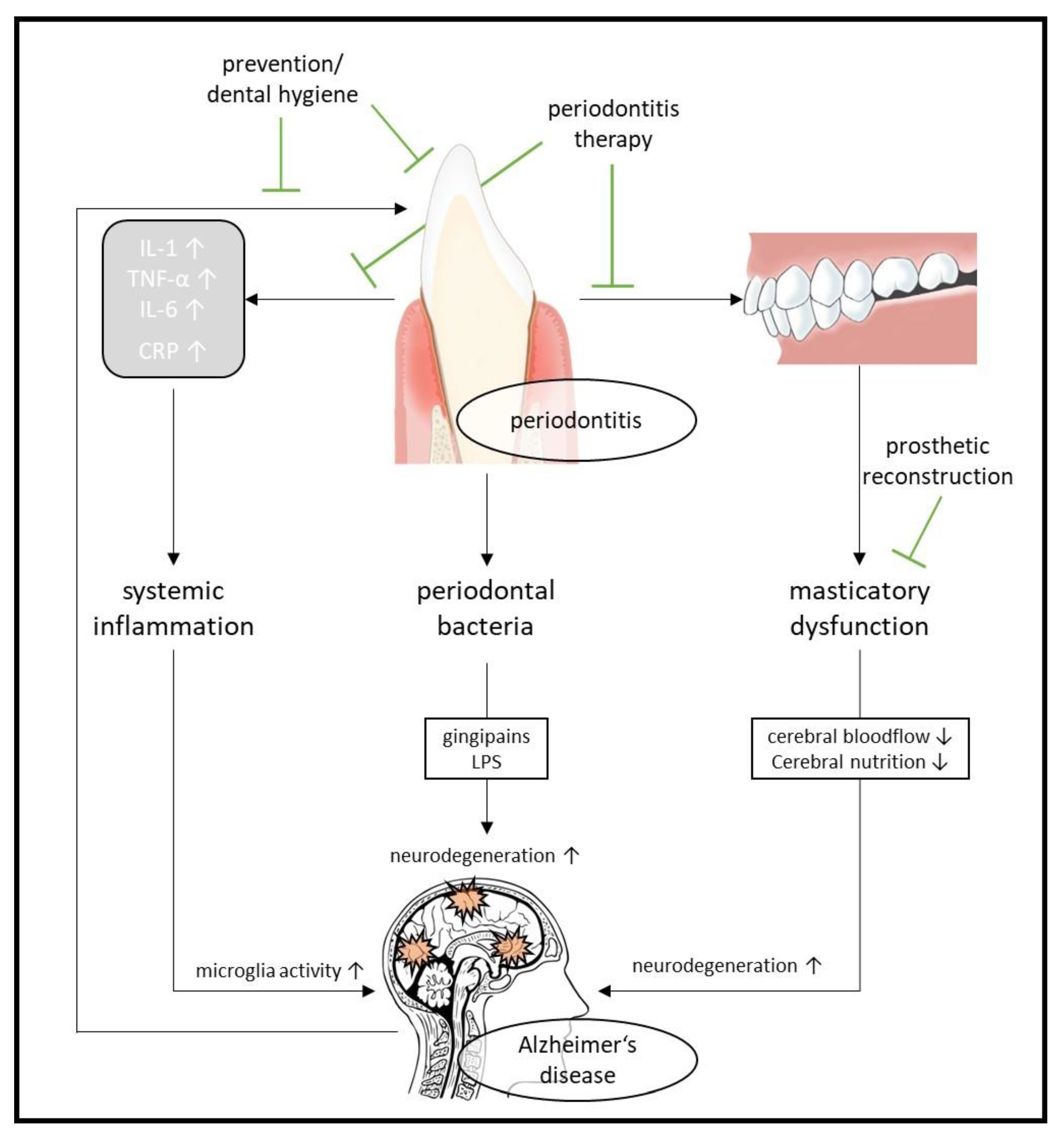
2.4. Oral Health and Alzheimer’s Disease

There is rising evidence for an association between intraoral inflammatory conditions and AD [17,18,19]. More than 50% of all people over the age of 35 worldwide are affected by periodontitis, and approximately 10–15% suffer from a severe form of the disease which can lead to the loss of dentition [333]. In particular, people over the age of 65 are at increased risk for periodontitis.
Periodontitis is a chronic, bacterial, host-dependent inflammation of tooth-supporting tissues that ultimately leads to loss of the periodontal attachment and, in continuum, to tooth loss through the activation of host proteinases [334,335]. While gingival inflammation that precedes periodontitis is generally triggered by the presence of biofilm, the development and progression of periodontal inflammation depend on the presence of oral dysbiosis [336]. Local degradation products and inflammation-induced milieu changes provide a selection advantage to specific periodontal pathogens such as the Gram-negative anaerobic species known as the “red complex” [337]. These pathogens include Porphyromonas gingivalis (P. gingivalis), Tannerella forsythia (T. forsysthia), and Treponema denticola (T. denticola). Additional risk factors, such as smoking, can exacerbate dysbiotic changes [338,339].
In this process, the dysbiotic biofilm causes not only a local inflammatory response but also a systemic inflammatory response, which is related to various noncommunicable diseases and can influence their development and progression [340].
Such a relationship is also discussed for dementia, especially AD, and the intraoral condition, which may be bidirectional: on the one hand, the reduction in memory capacity caused by AD is certainly causal for the decrease in personal oral hygiene ability and thus ultimately for the increase in the risk for the development of caries and periodontitis [341,342]. On the other hand, there is evidence that periodontitis itself and the associated microbial colonization and inflammatory response is associated with AD [18,19,343,344].
While the systematic reviews that included cross-sectional studies failed to assume a causal relationship between the presence of periodontal disease and the development of AD [344,345], a more recent systematic review that included only longitudinal data from observational studies came to the conclusion that causality was evident and that, considering the adjusted pooled risk ratio for dementia related to the periodontal disease of 1.38 (1.01 to 1.90), a fictive 75% reduction in the prevalence of severe periodontitis would have the potential to prevent far more than 1 million people worldwide from developing dementia such as AD [346].
Thus far, the underlying mechanisms in the involvement of periodontitis in AD have not been conclusively elucidated. Systemic inflammation, as described above, is one approach. Among others, increased IL-1, TNF-α, and IL-6 plasma levels play an important role, resulting in an increase in microglial activation [347]. The involvement of oral microorganisms and their metabolites is another frequently discussed aspect in the development and progression of dementia. The presence of the bacterium T. denticola associated with periodontitis was discovered in brain biopsies from deceased PwAD as early as 20 years ago [348]. The increased presence of antibodies against periodontal pathogens in the blood of persons suffering from dementia also suggested that these pathogens may play an important role in the etiopathogenesis of cognitive diseases [349,350,351]. The bacterium P. gingivalis plays a key role in this context because of its virulence factors [352]. This representative of the “red complex” was also recently shown to be increased in brain biopsies from PwAD [353]. P. gingivalis uses membrane lipopolysaccharides and special proteases, so-called gingipains, to bypass host defenses and degrade brain tissue. Gingipains in particular are thought to be highly relevant with regard to the initiation of the AD process and have therefore also led to the development of a new drug therapy approach—a gingipain inhibitor [353]. However, such a compound (COR388), which was later clinically tested in a phase II/III trial, failed to lead to therapeutic success and was discontinued—also due to side effects [354,355].
Even though the mechanism of involvement or even causality of periodontitis in AD has not yet been fully clarified, it is obvious to pay attention to early periodontitis diagnosis and its prevention or therapy in the attempt at AD prevention. Nevertheless, there are only few reliable clinical data available to date that deal with a possible improvement of AD through a therapy of periodontal diseases. Regarding the impact of dental and periodontal treatment on cognitive function before and after dental treatment, a longitudinal observational study has previously found that oral restoration of PwAD has the potential to improve cognitive function [356]. Another longitudinal study also demonstrated that regular oral home care had a positive effect on cognitive status [357]. For this purpose, current guidelines recommend brushing with a manual or electric toothbrush twice a day for at least two minutes, in combination with interdental care using interdental brushes [358]. In addition, the application of fluoridated toothpaste and mouth rinses based on Chlorhexamed or essential oils is effective [359]. As dementia progresses, this default can usually not be achieved by the persons themselves at some point, so professional support in-home care becomes necessary to maintain intraoral health as long as possible [360]. Recent data show that a dental nurse who professionally brushed the teeth of nursing home residents every 2 weeks after an initial professional dental cleaning in addition to daily care by relatives or caregivers could contribute to the reduction in intraoral inflammation and minimization of oral problems, such as root caries or tooth loss [361].
In summary, given the potential links between periodontal inflammation and AD, minimizing intraoral biofilm, and consequently reducing the inflammatory response is most likely to be beneficial with respect to the development and progression of dementia. For other chronic diseases, such as cardiovascular disease, this benefit has already been adequately demonstrated: For example, a recent systematic review with meta-analysis concluded that professional and home-based management of oral biofilm as a non-pharmacological intervention reduced systemic inflammation to an extent equivalent to that achieved by drug interventions to treat residual cardiovascular risk [340].
Thus, the goal should be to assess intraoral status soon after AD diagnosis and, if necessary, to remove any initial supra- and subgingival plaque by mechanical debridement with or without adjunctive use of antiseptics or antibiotics, followed by long-term individualized professional follow-up in combination with instructions on home oral hygiene for the person himself or a family or professional caregiver, optionally supported by a dental nurse to ensure the longest possible maintenance of oral health [362,363].
In addition to the possible mechanisms described above in the association of periodontitis and AD, tooth loss itself, which may result from periodontitis, also plays an important role as a risk factor for the development and progression of dementia (Figure 5). Reduced masticatory function due to tooth loss affects cerebral blood flow and nutrition [364] and led to a decrease in acetylcholine levels and the number of pyramidal cells in the hippocampus when studied in animals, which was associated with a disturbance in learning memory [365]. A recent systematic review, reporting on further animal studies, shows that the main mechanisms in the association of tooth loss and AD may be accelerated neurodegeneration caused by the loss of masticatory function and the accompanying decreased nerve stimulation, as well as a chronic systemic inflammatory stress state [366].
A further recent systematic review of case–control studies shows, in addition to aspects of the systemic effects of underlying diseases such as periodontitis, that tooth loss itself has an impact on the prognosis of dementia and is therefore a fundamental starting point in the planning of dental therapy for elderly persons: The goal should be, if possible, to maintain masticatory function through long-term functional tooth preservation, as this not only has a positive effect on cerebral blood flow and the consequent oxygenation of the brain but also allows for a favorable dementia-protective diet [367]. One possible therapy could be the provision of the missing support zones resulting from tooth loss by means of prosthetic replacement. It has been shown that the swallowing disorders and dysphagia associated with tooth loss could be improved by the use of prostheses, which had an effect not only on the nutritional status but also on the ability to perform ADL [368]. Further improvement of masticatory function was achieved by the additional support of such prostheses on implants placed in the jawbone, resulting in the need for intensive professional follow-up to protect the patients from possible future peri-implant infection [369]. Improvement of mastication by the provision of prosthesis and the therapy of oral complaints led to an increase in the social activity of patients [370]. The provision of dentures was also shown to be beneficial in terms of cognitive function [371].
The influence of individual oral processes and conditions on the oral health-related quality of life of people with AD has been demonstrated recently [372] as well as the long-known assumption that intraoral problems lead to pain and functional limitations resulting in a disease-promoting deterioration of physical, psychological, and social condition [373].
The ideal control of problems associated with tooth loss and oral diseases is the maintenance of oral health and the prevention of tooth loss long before the risk of developing dementia increases. It has already been successfully demonstrated that almost complete tooth preservation can be achieved through a lifelong, individualized and needs-oriented prevention program that begins in adolescence and includes regular check-ups, dental cleanings and instructions on oral hygiene provided by dental hygienists [374].
Although the current evidence suggests a bidirectional relationship between oral status and AD, there are so far only initial clinical studies on the effect of dental treatment of AD status. These are usually observational studies with small numbers of subjects, but they show promising initial results. Currently, initial intervention studies are in preparation, which already considers a first multidomain approach.

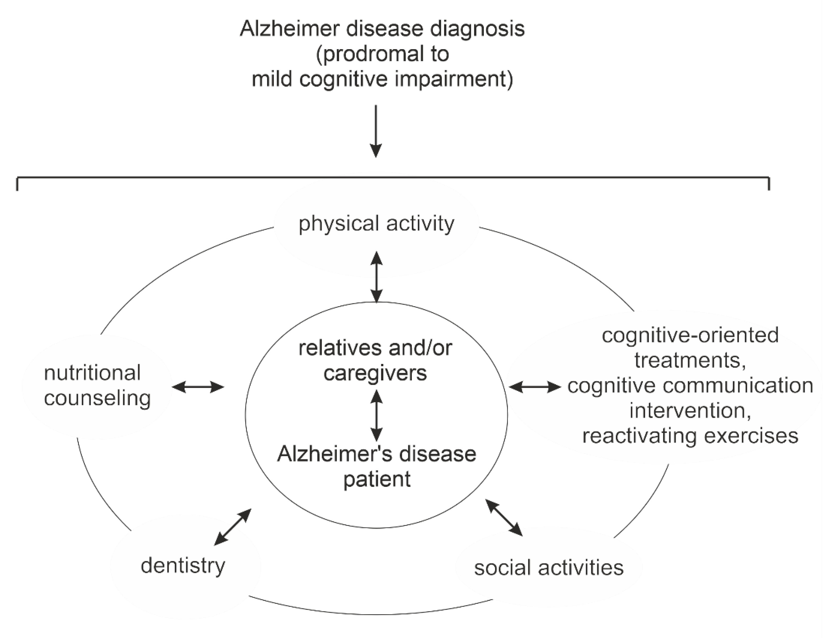
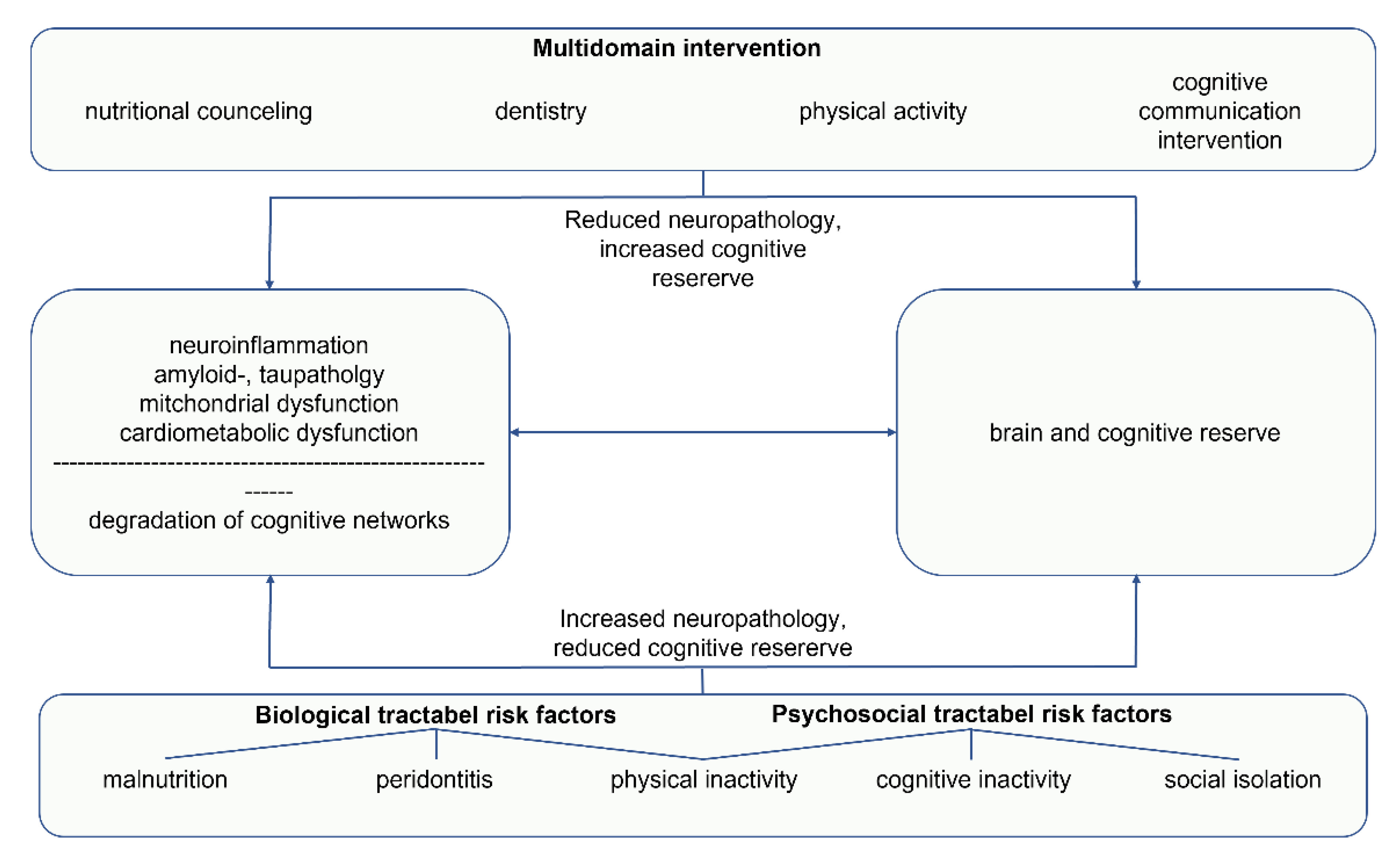
3. Multidomain Interventions

According to the BPS model [20], dementia is a complex condition, that involves interrelated tractable and fixed risk factors. Acting on several modifiable risk factors simultaneously seems to be particularly promising to influence the brain and cognitive reserve and maintain functioning [16,375,376]. Therefore, multidomain approaches, including psychosocial and educational support are of growing interest to delay the decline in dementia [16,314,377,378,379,380]. Figure 6 provides an overview of the interrelationships and interactions of individual interventions in AD.
Two recent reviews on multidomain interventions are of particular interest. The meta-analysis of Salzmann et al. (2022) on 28 studies directly compared multi- and single-domain interventions in MCI. The authors found greater pooled effect sizes in multidomain intervention groups for MCI compared to single-domain intervention groups in global cognition (SMD, 0.41; 95% CI 0.23 to 0.59; p < 0.001), executive function (SMD, 0.20; 95% CI 0.04 to 0.36; p = 0.01), memory (SMD, 0.29; 95% CI 0.14 to 0.45; p < 0.001) and verbal fluency (SMD, 0.30; 95% CI 0.12 to 0.49; p = 0.001) [379]. The systematic review of Chalfont et al. (2021) on multidomain interventions in dementia included 26 studies, of which 92% demonstrated improvement, maintenance, or delayed decline of cognitive functioning. Both reviews comprised, among other modes, cognitive, physical, and nutritional interventions, and educational support. Therefore, it is reasonable to combine these approaches, whereby cognition and physical training is up to date the most common combination [314,379,381,382]. A review of 17 studies found greater improvement in older adults with MCI on global cognition (SMD = 0.83, 95% CI 0.41 to 1.25; p = 0.0001) for combined cognitive and physical intervention compared with intervention in a single domain [381].
However, according to the review of Chalfont et al. (2020), to improve cognition, at least three modalities should be combined and one of them should have a cognitive approach. Moreover, the greatest effect sizes were identified in studies with individually tailored multidomain approaches [314]. Thus, interventions should be adapted to the individual’s needs and preferences to maintain motivation for active participation in therapy [314,383,384] and to reduce functional decline [301]. Caregiver involvement also seems to be crucial in multidomain interventions [314], especially when it comprises targeted caregiver components [385]. Astonishingly, the severity and length of intervention were not determinants of treatment in the review of Chalfont et al. (2020). However, continuous treatment could be crucial for maintaining functional abilities [386] or physical health [383] in PwAD. The multidomain activation therapy study Motor Stimulation, Activities of daily living, and Cognitive and Social Functioning (MAKS) led to stable results in ADL and cognition of persons with moderate dementia during a 12-month intervention period, whereas an increase in impairment for both measures was found in the control group. In a 10-month follow-up test scores of both groups deteriorated significantly, but scores in ADL remained significantly higher in the intervention group compared to the control group [386]. Prick et al. [383] considered too low therapy intensity crucial for missing therapy effects in mood, behavior and physical health after a multidomain intervention.
As indicated in the before mentioned studies, multidomain lifestyle intervention strategies could be beneficial regarding AD pathology and have therefore been the subject of different clinical trials [387]. A recent RCT with 67 participants over 60 years without any cognitive dysfunction reported improved cognitive functions after combined aerobic and memory training and the supplementation of the functional food soy peptide in form of a commercial drink for 90 days [388].
With reference to study participants, three large prevention trials were performed in the past years, namely preDIVA, MAPT, and FINGER. The preDIVA (Prevention of Dementia by Intensive Vascular Care) study was a cluster-randomized controlled trial in more than 3400 community-dwelling older individuals (aged 70–78 years), examining the effectiveness of a six-year multidomain vascular care intervention to prevent dementia. The intervention included the assessment of cardiovascular risk factors every four months and individually tailored lifestyle advice. No differences in the outcome cumulative incidence of dementia or incident cardiovascular disease between the intervention (more than 1890 participants) and control group (1636 participants), which obtained usual care, was found. One possible reason for the obtained results is seen in the high standards of usual care and a modest baseline cardiovascular risk. However, importantly, as a secondary outcome in the intervention group, a significantly reduced risk of non-AD dementia could be detected [389].
The Multidomain Alzheimer Preventive Trial (MAPT) included more than 1500 participants and was conducted over three years. In a randomized, placebo-controlled design the intervention took place in more than 40 group sessions with the content of nutrition, COT, physical activity as well as three preventive consultations. As a specific compound, some of the non-demented community-dwelling participants received ω-3 PUFAs. No significant differences were found in cognition between the intervention groups. However, in comparison to the placebo group, there was a significantly less cognitive decline in the multidomain + PUFA group [390].
The randomized controlled Finnish Geriatric Intervention Study to Prevent Cognitive Impairment and Disability (FINGER) ran for two years and included more than 1200 at-risk elderly people. Interventions were conducted in group and individual sessions comprising diet, COT, physical exercise, and vascular risk monitoring. The control group received usual health care. Results revealed a significant between-group difference in the change in cognition [378] and were not modified by parameters such as cognition, socioeconomic status, cardiovascular factors, or sociodemographic status [391]. Thus, the findings underline the potential benefit of FINGER in a large elderly population to maintain or improve cognitive functioning [378,391]. Based on these results, the current step is to expand the FINGER-type trials to a World-Wide FINGERS (WW-FINGERS) network, facilitating not only international collaborations but also the opportunity to elaborate globally implementable and effective preventive approaches [392,393].
Multidomain lifestyle interventions are a current research topic. Among others, the Multimodal Preventive Trial for Alzheimer’s Disease (MIND-ADmini) also examines the potential effects of multidomain interventions in individuals with prodromal AD. Up to date more than 90 participants were randomized and enrolled for the multidomain lifestyle intervention alone or in combination with medical food (Fortasyn Connect) in comparison to regular health advice/care in the control group [394].
The ongoing randomized controlled Japan-Multimodal Intervention Trial (J-MINT) uses original outcome parameters as cognitive changes at 18-month follow-up, changes in ADL, frailty status, blood markers, dementia-related blood bio-makers, and neuroimaging to verify whether the multidomain intervention could prevent the progression of cognitive decline among older adults with MCI [395]. The intervention consists of the management of vascular risk factors, group-based physical activity and self-monitoring of physical activity, nutritional counseling, and COT.
The first approach of interdisciplinary cooperation of dental hygiene and physiotherapy can be found in the planned study of Jockusch et al. (2022) aiming to activate the masticatory function to improve cognition in AD [396].
Comparable to single-domain interventions, communication-based interventions and outcomes have rarely been considered in multidomain approaches so far. A combination of stimulative walking activity and conversation did not result in a significant decrease in nonredundant information in connected speech compared to a structured conversation-only group [384], among others probably because of limited mobility in the combined group, which may have led to an overload of cognitive capacity [384]. The Language-Enriched Exercise Plus Socialization (LEEPS) Program combined language stimulation with physical activity in older adults with AD and related disorders, leading to stability in cognition, mood, and physical performance over 11 months and in a small group even over 20 months. However, there was a high dropout rate, mainly due to entry into long-term care, worsening health or death [377]. While language abilities were not specifically tested in the study of La Rue et al. (2015), Arkin and Mahendra (2001) used communication-based outcome measures in a similar approach. Although experimental and control groups did not differ in the Arizona Battery for Communication Disorders of Dementia (ABCD) [397], the specific analysis showed a significantly more differentiated use of nouns in the experimental group. However, results were based on case series [398].

4. Discussion

A large-scale meta-analysis and systematic review of more than 150 RCTs and more than 240 observational prospective studies identified evidence-based modifiable factors for the prevention of AD [399]. The Lancet Commission also recommends addressing risk factors that favor the development of dementia across the lifespan, as this can affect cognitive reserve and delay neuropathological developments [16,314]. Physical activity and cognitive activities stimulate cerebral blood flow and the production of neurotrophic factors that influence hippocampal neuroplasticity [314,382,400]. In particular, physical activity and dietary interventions are known to address similar mechanisms involved in AD. For example, a reduction in oxidative stress or effects on mitochondria, in general, are known to be involved in both disciplines [401]. In this context, the effect of PGC-1α, a mitochondrial super-regulator, has been reported to be affected by AD and might be an interesting target for further studies [402]. Moreover, dietary interventions influence cellular energy metabolism and can therefore have an impact on synaptic plasticity [314], a parameter, which highly correlates with cognitive reserve and cognitive activity in general. Synaptic plasticity is further influenced by different lipids [403]. E.g., plasmalogens or phosphatidyl-choline in combination with choline/UMP-choline or DHA have been shown or are at least discussed to increase several synaptic markers such as synaptophysin or PSD95 [404,405]. Interventions in the prodromal and early stages of AD may correct the deficiency of key nutritional elements that could otherwise lead to the loss of synapses and neurons [256].
Obviously, both physical activity and dietary interventions elevate cerebral blood flow, another important factor necessary for cognitive activity. These are only a few examples that combine different approaches to adjust the dysregulated metabolic homeostasis in AD synergistically from different angles, resulting in an elevated beneficial potential with respect to AD compared to approaches based on a single intervention. A further important aspect is the multifactorial component of AD: as reviewed above, AD can be caused by several different molecular mechanisms. Apparently, addressing only one mechanism has been proven to be not sufficient to deal with AD up to now, making a multidisciplinary approach, addressing these different risk factors or molecular mechanisms involved in AD, even more attractive.
In line with this argumentation, multidomain interventions have already been shown to be effective in stimulating modifiable risk factors in parallel to delay the progression of dementia [314,379,382], even with existing structural changes in the brain [295,400,406]. To improve cognition, multidomain interventions should comprise at least three modalities, one of which is a cognitive approach [314]. Up to now, cognition and physical training is the most common combination of the multidomain approaches [314,379,381,382] whereby nutritional interventions and educational support play an important role in supporting and counseling PwAD and their families throughout the course of the disease [378,385,407,408]. The success of psychoeducational interventions depends on clear communication of theoretical contents and the active involvement of carers in practice sequences to implement new skills [303,385]. Support of oral hygiene and health, prevention and diagnosis of periodontal disease are promising approaches in dementia care that have not yet been addressed in multidomain interventions but should receive more attention in the future [363,367,371,409]. In this context, diabetes mellitus, recently identified as an additional risk factor for the development of AD [410], should also be mentioned, even though the underlying mechanisms have not yet been clarified [411]. It is known that there is a bidirectional relationship between the presence of intraoral inflammation, especially periodontitis, and diabetes mellitus [412]. Thus, an improvement of the intraoral condition also fulfills the previously formulated need to implement a diabetes mellitus-protective lifestyle for AD prevention [399].
Person-centered approaches are a characteristic feature of CR [23,292,299] and similarly, multidomain interventions with individualized contents achieve the greatest effect sizes [314].
Regarding nutritional interventions, more and more evidence arises that combined dietary components show synergistic beneficial actions with respect to AD pathology [255,256]. Multidomain interventions that target preventive, or early stage disease show potential for delaying the onset of dementia [259,378,389,390]. Dietary modifications combined with aerobic physical activity also led to cognitive improvements in participants with elevated blood pressure [413]. Since high blood pressure is also a risk factor for the onset and progression of AD, combined nutritional approaches with specific physical activity are promising for AD.
In many studies, cognition is the primary outcome used to test the effectiveness of the intervention. Unfortunately, most of the chosen assessments or outcomes have little relation to the demands of daily living [314,326], but rather measure higher cognitive functions [308]. This is all the more astonishing because impairments in communication already occur in the early phase of the disease. Future work should therefore consider communication skills as an outcome measure. Corresponding parameters would be, for example, the proportion of topic-related utterances and empty utterances or the global coherence in the discourse of people with dementia [288,305].
Although there is a growing interest in the effects of dental therapy on the occurrence and progression of AD, dentistry still has a minor role in the multidisciplinary context. Despite interrelated biochemical processes in intraoral inflammation and AD [349,355], there are few clinical studies investigating the effectiveness of oral treatment approaches in AD. Difficulties in recruitment include ethical issues, the health status of people with disabilities, lack of compliance, required involvement of relatives, treatment limitations, and economic issues [351,396,414]. However, oral treatment approaches are very promising to be integrated into a multidomain approach. In particular, approaches to nutrition, oral hygiene, mobility and masticatory function can be optimally coordinated and adapted to individual needs, which overall contributes to an improvement in the quality of life of PwAD. Sessions can take place in groups as well as individually and thus promote social activities, as is also recommended in this context [16].
Future research should address the feasibility of multidomain approaches in clinical practice. To establish this approach, orientation sheets can be developed and evaluated. A proposal for an interdisciplinary approach to nutrition therapy/counseling is outlined below (Table 3).

5. Conclusions

Pleiotropic mechanisms are known to result in AD. Although these molecular mechanisms are well-known and can be addressed by pharmaceutical and non-pharmaceutical approaches, including nutrition, physical activity or increasing the cognitive reserve, beneficial effects for PwAD are not sufficient to cope with the disease. However, treatments based on multicomponent or even multidisciplinary approaches revealed more pronounced benefits. Unfortunately, the number of studies and the different included disciplines are limited. For example, oral health, which obviously interferes with nutritional status but also many other factors reviewed above, is mostly neglected. Therefore, we suggest a tight interlink between different treatment strategies resulting in an interdisciplinary approach, covering in particular nutritional counseling, supervised physical training, oral health, and cognitive-oriented communication training to maintain quality of life in AD (Figure 7). Importantly, all of these single approaches are already available now and the required interlink between these approaches seems to be feasible and associated with only a moderate additional effort. As an example, a simple route card for PwAD or nutritional counselors, caregivers, or relatives addressing these interlinks is suggested, helping to individualize and adopt treatment. Due to the synergistic action of these interventions, a significantly greater benefit for the PwAD might be expected. Even before the onset of AD, the approaches listed could potentially be considered preventive approaches. The awareness of the presented correlations and the possible preventive effectiveness should also be discussed with patients who do not yet suffer from AD. Although the reviewed literature is promising, further studies addressing these interdisciplinary aims are needed to prove effectiveness.

Author Contributions

Writing—original draft preparation, I.A., K.D., T.R., A.A.L., M.T., J.P.B., T.T., H.S.G., M.O.W.G. All authors have read and agreed to the published version of the manuscript.

Funding

This research received no external funding.

Data Availability Statement

Not applicable.

Acknowledgments

The authors thank Servier Medical Art since parts of the figure were drawn by using pictures from Servier Medical Art. Servier Medical Art by Servier is licensed under a Creative Commons Attribution 3.0 Unported License (https://creativecommons.org/licenses/by/3.0/ (accessed on 1 September 2020)).

Conflicts of Interest

The authors declare no conflict of interest.

References

  1. Clare, L.; Teale, J.C.; Toms, G.; Kudlicka, A.; Evans, I.; Abrahams, S.; Goldstein, L.H.; Hindle, J.V.; Ho, A.K.; Jahanshahi, M.; et al. Cognitive rehabilitation, self-management, psychotherapeutic and caregiver support interventions in progressive neurodegenerative conditions: A scoping review. NeuroRehabilitation 2018, 43, 443–471. [Google Scholar] [CrossRef] [PubMed]
  2. Kudlicka, A.; Martyr, A.; Bahar-Fuchs, A.; Woods, B.; Clare, L. Cognitive rehabilitation for people with mild to moderate dementia. Cochrane Database Syst. Rev. 2019, 28, 707. [Google Scholar] [CrossRef]
  3. Selkoe, D.J. Cell biology of protein misfolding: The examples of Alzheimer’s and parkinson’s diseases. Nat. Cell Biol. 2004, 6, 1054–1061. [Google Scholar] [CrossRef] [PubMed]
  4. Dubois, B.; Hampel, H.; Feldman, H.H.; Scheltens, P.; Aisen, P.; Andrieu, S.; Bakardjian, H.; Benali, H.; Bertram, L.; Blennow, K.; et al. Preclinical Alzheimer’s disease: Definition, natural history, and diagnostic criteria. Alzheimer’s Dement. 2016, 12, 292–323. [Google Scholar] [CrossRef]
  5. Kumar, A.; Sidhu, J.; Goyal, A.; Tsao, J.W. Alzheimer disease. In Statpearls; StatPearls Publishing: Treasure Island, FL, USA, 2022. [Google Scholar]
  6. Breijyeh, Z.; Karaman, R. Comprehensive review on Alzheimer’s disease: Causes and treatment. Molecules 2020, 25, 5789. [Google Scholar] [CrossRef]
  7. Bahar-Fuchs, A.; Martyr, A.; Goh, A.M.; Sabates, J.; Clare, L. Cognitive training for people with mild to moderate dementia. Cochrane Database Syst. Rev. 2019, 3, CD013069. [Google Scholar] [CrossRef]
  8. Wu, J.; Ma, Y.; Ren, Z. Rehabilitative effects of virtual reality technology for mild cognitive impairment: A systematic review with meta-analysis. Front. Psychol. 2020, 11, 1811. [Google Scholar] [CrossRef]
  9. Mueller, K.D.; Hermann, B.; Mecollari, J.; Turkstra, L.S. Connected speech and language in mild cognitive impairment and Alzheimer’s disease: A review of picture description tasks. J. Clin. Exp. Neuropsychol. 2018, 40, 917–939. [Google Scholar] [CrossRef]
  10. Wattmo, C.; Minthon, L.; Wallin, A.K. Mild versus moderate stages of Alzheimer’s disease: Three-year outcomes in a routine clinical setting of cholinesterase inhibitor therapy. Alzheimer’s. Res. 2016, 8, 7. [Google Scholar] [CrossRef] [PubMed] [Green Version]
  11. Eldufani, J.; Blaise, G. The role of acetylcholinesterase inhibitors such as neostigmine and rivastigmine on chronic pain and cognitive function in aging: A review of recent clinical applications. Alzheimer’s Dement. 2019, 5, 175–183. [Google Scholar] [CrossRef]
  12. Singh, R.; Sadiq, N.M. Cholinesterase inhibitors. In Statpearls; StatPearls Publishing: Treasure Island, FL, USA, 2022. [Google Scholar]
  13. Chen, J.H. Microscopic features for the identification of chinese drugs in ranunculaceus. Zhong Yao Tong Bao 1985, 10, 15–17. [Google Scholar]
  14. Khoury, R.; Grysman, N.; Gold, J.; Patel, K.; Grossberg, G.T. The role of 5 ht6-receptor antagonists in Alzheimer’s disease: An update. Expert Opin. Investig. Drugs 2018, 27, 523–533. [Google Scholar] [CrossRef] [PubMed]
  15. Wang, R.; Reddy, P.H. Role of glutamate and nmda receptors in Alzheimer’s disease. J. Alzheimer’s Dis. 2017, 57, 1041–1048. [Google Scholar] [CrossRef] [Green Version]
  16. Livingston, G.; Huntley, J.; Sommerlad, A.; Ames, D.; Ballard, C.; Banerjee, S.; Brayne, C.; Burns, A.; Cohen-Mansfield, J.; Cooper, C.; et al. Dementia prevention, intervention, and care: 2020 report of the lancet commission. Lancet 2020, 396, 413–446. [Google Scholar] [CrossRef]
  17. Dioguardi, M.; Crincoli, V.; Laino, L.; Alovisi, M.; Sovereto, D.; Mastrangelo, F.; Lo Russo, L.; Lo Muzio, L. The role of periodontitis and periodontal bacteria in the onset and progression of Alzheimer’s disease: A systematic review. J. Clin. Med. 2020, 9, 495. [Google Scholar] [CrossRef] [PubMed] [Green Version]
  18. Kaliamoorthy, S.; Nagarajan, M.; Sethuraman, V.; Jayavel, K.; Lakshmanan, V.; Palla, S. Association of Alzheimer’s disease and periodontitis—A systematic review and meta-analysis of evidence from observational studies. Med. Pharm. Rep. 2022, 95, 144–151. [Google Scholar] [CrossRef]
  19. Parra-Torres, V.; Melgar-Rodríguez, S.; Muñoz-Manríquez, C.; Sanhueza, B.; Cafferata, E.A.; Paula-Lima, A.C.; Díaz-Zúñiga, J. Periodontal bacteria in the brain-implication for Alzheimer’s disease: A systematic review. Oral Dis. 2021. [Google Scholar] [CrossRef] [PubMed]
  20. Spector, A.; Orrell, M. Using a biopsychosocial model of dementia as a tool to guide clinical practice. Int. Psychogeriatr. 2010, 22, 957–965. [Google Scholar] [CrossRef] [PubMed]
  21. Revolta, C.; Orrell, M.; Spector, A. The biopsychosocial (bps) model of dementia as a tool for clinical practice. A pilot study. Int. Psychogeriatr. 2016, 28, 1079–1089. [Google Scholar] [CrossRef]
  22. Clare, L.; Linden, D.E.J.; Woods, R.T.; Whitaker, R.; Evans, S.J.; Parkinson, C.H.; van Paasschen, J.; Nelis, S.M.; Hoare, Z.; Yuen, K.S.L.; et al. Goal-oriented cognitive rehabilitation for people with early-stage Alzheimer disease: A single-blind randomized controlled trial of clinical efficacy. Am. J. Geriatr. Psychiatry Off. J. Am. Assoc. Geriatr. Psychiatry 2010, 18, 928–939. [Google Scholar] [CrossRef]
  23. Clare, L.; Kudlicka, A.; Oyebode, J.R.; Jones, R.W.; Bayer, A.; Leroi, I.; Kopelman, M.; James, I.A.; Culverwell, A.; Pool, J.; et al. Goal-oriented cognitive rehabilitation for early-stage Alzheimer’s and related dementias: The great rct. Health Technol. Assess. 2019, 23, 1–242. [Google Scholar] [CrossRef] [PubMed]
  24. Ninot, G. Psychosocial interventions and dementia. Pract. Guide Fond. Médéric Alzheimer 2021, 1–100. [Google Scholar] [CrossRef]
  25. Gavelin, H.M.; Lampit, A.; Hallock, H.; Sabatés, J.; Bahar-Fuchs, A. Cognition-oriented treatments for older adults: A systematic overview of systematic reviews. Neuropsychol. Rev. 2020, 30, 167–193. [Google Scholar] [CrossRef] [Green Version]
  26. Rody, T.; De Amorim, J.A.; De Felice, F.G. The emerging neuroprotective roles of exerkines in Alzheimer’s disease. Front. Aging Neurosci. 2022, 14, 965190. [Google Scholar] [CrossRef] [PubMed]
  27. Yusufov, M.; Weyandt, L.L.; Piryatinsky, I. Alzheimer’s disease and diet: A systematic review. Int. J. Neurosci. 2017, 127, 161–175. [Google Scholar] [CrossRef] [PubMed]
  28. O’Brien, R.J.; Wong, P.C. Amyloid precursor protein processing and Alzheimer’s disease. Annu. Rev. Neurosci. 2011, 34, 185–204. [Google Scholar] [CrossRef] [Green Version]
  29. Lee, S.J.; Nam, E.; Lee, H.J.; Savelieff, M.G.; Lim, M.H. Towards an understanding of amyloid-beta oligomers: Characterization, toxicity mechanisms, and inhibitors. Chem. Soc. Rev. 2017, 46, 310–323. [Google Scholar] [CrossRef] [PubMed]
  30. Dyrks, T.; Weidemann, A.; Multhaup, G.; Salbaum, J.M.; Lemaire, H.G.; Kang, J.; Muller-Hill, B.; Masters, C.L.; Beyreuther, K. Identification, transmembrane orientation and biogenesis of the amyloid a4 precursor of Alzheimer’s disease. EMBO J. 1988, 7, 949–957. [Google Scholar] [CrossRef] [PubMed]
  31. Wasco, W.; Bupp, K.; Magendantz, M.; Gusella, J.F.; Tanzi, R.E.; Solomon, F. Identification of a mouse brain cdna that encodes a protein related to the Alzheimer disease-associated amyloid beta protein precursor. Proc. Natl. Acad. Sci. USA 1992, 89, 10758–10762. [Google Scholar] [CrossRef] [Green Version]
  32. Muller, U.C.; Deller, T.; Korte, M. Not just amyloid: Physiological functions of the amyloid precursor protein family. Nat. Rev. Neurosci. 2017, 18, 281–298. [Google Scholar] [CrossRef]
  33. Roberts, S.B.; Ripellino, J.A.; Ingalls, K.M.; Robakis, N.K.; Felsenstein, K.M. Non-amyloidogenic cleavage of the beta-amyloid precursor protein by an integral membrane metalloendopeptidase. J. Biol. Chem. 1994, 269, 3111–3116. [Google Scholar] [CrossRef]
  34. Edwards, D.R.; Handsley, M.M.; Pennington, C.J. The adam metalloproteinases. Mol. Asp. Med. 2008, 29, 258–289. [Google Scholar] [CrossRef] [PubMed]
  35. Dulin, F.; Leveille, F.; Ortega, J.B.; Mornon, J.P.; Buisson, A.; Callebaut, I.; Colloc’h, N. P3 peptide, a truncated form of a beta devoid of synaptotoxic effect, does not assemble into soluble oligomers. FEBS Lett. 2008, 582, 1865–1870. [Google Scholar] [CrossRef] [PubMed]
  36. Furukawa, K.; Sopher, B.L.; Rydel, R.E.; Begley, J.G.; Pham, D.G.; Martin, G.M.; Fox, M.; Mattson, M.P. Increased activity-regulating and neuroprotective efficacy of alpha-secretase-derived secreted amyloid precursor protein conferred by a c-terminal heparin-binding domain. J. Neurochem. 1996, 67, 1882–1896. [Google Scholar] [CrossRef]
  37. Meziane, H.; Dodart, J.C.; Mathis, C.; Little, S.; Clemens, J.; Paul, S.M.; Ungerer, A. Memory-enhancing effects of secreted forms of the beta-amyloid precursor protein in normal and amnestic mice. Proc. Natl. Acad. Sci. USA 1998, 95, 12683–12688. [Google Scholar] [CrossRef] [Green Version]
  38. Hampel, H.; Vassar, R.; De Strooper, B.; Hardy, J.; Willem, M.; Singh, N.; Zhou, J.; Yan, R.; Vanmechelen, E.; De Vos, A.; et al. The beta-secretase bace1 in Alzheimer’s disease. Biol. Psychiatry 2021, 89, 745–756. [Google Scholar] [CrossRef] [PubMed]
  39. Vassar, R.; Bennett, B.D.; Babu-Khan, S.; Kahn, S.; Mendiaz, E.A.; Denis, P.; Teplow, D.B.; Ross, S.; Amarante, P.; Loeloff, R.; et al. Beta-secretase cleavage of Alzheimer’s amyloid precursor protein by the transmembrane aspartic protease bace. Science 1999, 286, 735–741. [Google Scholar] [CrossRef] [Green Version]
  40. Wang, M.; Jing, T.; Wang, X.; Yao, D. Beta-secretase/bace1 promotes app endocytosis and processing in the endosomes and on cell membrane. Neurosci. Lett. 2018, 685, 63–67. [Google Scholar] [CrossRef]
  41. Gouras, G.K.; Xu, H.; Jovanovic, J.N.; Buxbaum, J.D.; Wang, R.; Greengard, P.; Relkin, N.R.; Gandy, S. Generation and regulation of beta-amyloid peptide variants by neurons. J. Neurochem. 1998, 71, 1920–1925. [Google Scholar] [CrossRef]
  42. Wang, R.; Sweeney, D.; Gandy, S.E.; Sisodia, S.S. The profile of soluble amyloid beta protein in cultured cell media. Detection and quantification of amyloid beta protein and variants by immunoprecipitation-mass spectrometry. J. Biol. Chem. 1996, 271, 31894–31902. [Google Scholar] [CrossRef] [Green Version]
  43. Schieb, H.; Kratzin, H.; Jahn, O.; Mobius, W.; Rabe, S.; Staufenbiel, M.; Wiltfang, J.; Klafki, H.W. Beta-amyloid peptide variants in brains and cerebrospinal fluid from amyloid precursor protein (app) transgenic mice: Comparison with human Alzheimer amyloid. J. Biol. Chem. 2011, 286, 33747–33758. [Google Scholar] [CrossRef]
  44. Haass, C. Take five--bace and the gamma-secretase quartet conduct Alzheimer’s amyloid beta-peptide generation. EMBO J. 2004, 23, 483–488. [Google Scholar] [CrossRef] [PubMed]
  45. Wolfe, M.S.; Xia, W.; Ostaszewski, B.L.; Diehl, T.S.; Kimberly, W.T.; Selkoe, D.J. Two transmembrane aspartates in presenilin-1 required for presenilin endoproteolysis and gamma-secretase activity. Nature 1999, 398, 513–517. [Google Scholar] [CrossRef] [PubMed]
  46. Riddell, D.R.; Christie, G.; Hussain, I.; Dingwall, C. Compartmentalization of beta-secretase (asp2) into low-buoyant density, noncaveolar lipid rafts. Curr. Biol. 2001, 11, 1288–1293. [Google Scholar] [CrossRef] [Green Version]
  47. Vetrivel, K.S.; Cheng, H.; Kim, S.H.; Chen, Y.; Barnes, N.Y.; Parent, A.T.; Sisodia, S.S.; Thinakaran, G. Spatial segregation of gamma-secretase and substrates in distinct membrane domains. J. Biol. Chem. 2005, 280, 25892–25900. [Google Scholar] [CrossRef] [Green Version]
  48. Grundke-Iqbal, I.; Iqbal, K.; Tung, Y.C.; Quinlan, M.; Wisniewski, H.M.; Binder, L.I. Abnormal phosphorylation of the microtubule-associated protein tau (tau) in Alzheimer cytoskeletal pathology. Proc. Natl. Acad. Sci. USA 1986, 83, 4913–4917. [Google Scholar] [CrossRef] [PubMed] [Green Version]
  49. Grundke-Iqbal, I.; Iqbal, K.; Quinlan, M.; Tung, Y.C.; Zaidi, M.S.; Wisniewski, H.M. Microtubule-associated protein tau. A component of Alzheimer paired helical filaments. J. Biol. Chem. 1986, 261, 6084–6089. [Google Scholar] [CrossRef]
  50. Goedert, M.; Spillantini, M.G.; Cairns, N.J.; Crowther, R.A. Tau proteins of Alzheimer paired helical filaments: Abnormal phosphorylation of all six brain isoforms. Neuron 1992, 8, 159–168. [Google Scholar] [CrossRef]
  51. Binder, L.I.; Frankfurter, A.; Rebhun, L.I. The distribution of tau in the mammalian central nervous system. J. Cell Biol. 1985, 101, 1371–1378. [Google Scholar] [CrossRef] [Green Version]
  52. Butner, K.A.; Kirschner, M.W. Tau protein binds to microtubules through a flexible array of distributed weak sites. J. Cell Biol. 1991, 115, 717–730. [Google Scholar] [CrossRef] [Green Version]
  53. Lindwall, G.; Cole, R.D. Phosphorylation affects the ability of tau protein to promote microtubule assembly. J. Biol. Chem. 1984, 259, 5301–5305. [Google Scholar] [CrossRef]
  54. Alonso, A.C.; Zaidi, T.; Grundke-Iqbal, I.; Iqbal, K. Role of abnormally phosphorylated tau in the breakdown of microtubules in Alzheimer disease. Proc. Natl. Acad. Sci. USA 1994, 91, 5562–5566. [Google Scholar] [CrossRef] [Green Version]
  55. Martin, L.; Latypova, X.; Wilson, C.M.; Magnaudeix, A.; Perrin, M.L.; Yardin, C.; Terro, F. Tau protein kinases: Involvement in Alzheimer’s disease. Ageing Res. Rev. 2013, 12, 289–309. [Google Scholar] [CrossRef]
  56. Liu, F.; Grundke-Iqbal, I.; Iqbal, K.; Gong, C.X. Contributions of protein phosphatases pp1, pp2a, pp2b and pp5 to the regulation of tau phosphorylation. Eur. J. Neurosci. 2005, 22, 1942–1950. [Google Scholar] [CrossRef]
  57. Kins, S.; Kurosinski, P.; Nitsch, R.M.; Gotz, J. Activation of the erk and jnk signaling pathways caused by neuron-specific inhibition of pp2a in transgenic mice. Am. J. Pathol. 2003, 163, 833–843. [Google Scholar] [CrossRef] [Green Version]
  58. Vogelsberg-Ragaglia, V.; Schuck, T.; Trojanowski, J.Q.; Lee, V.M. Pp2a mrna expression is quantitatively decreased in Alzheimer’s disease hippocampus. Exp. Neurol. 2001, 168, 402–412. [Google Scholar] [CrossRef]
  59. Kinney, J.W.; Bemiller, S.M.; Murtishaw, A.S.; Leisgang, A.M.; Salazar, A.M.; Lamb, B.T. Inflammation as a central mechanism in Alzheimer’s disease. Alzheimer’s Dement. 2018, 4, 575–590. [Google Scholar] [CrossRef] [PubMed]
  60. Chakrabarty, P.; Jansen-West, K.; Beccard, A.; Ceballos-Diaz, C.; Levites, Y.; Verbeeck, C.; Zubair, A.C.; Dickson, D.; Golde, T.E.; Das, P. Massive gliosis induced by interleukin-6 suppresses abeta deposition in vivo: Evidence against inflammation as a driving force for amyloid deposition. FASEB J. 2010, 24, 548–559. [Google Scholar] [CrossRef] [PubMed] [Green Version]
  61. Shaftel, S.S.; Kyrkanides, S.; Olschowka, J.A.; Miller, J.N.; Johnson, R.E.; O’Banion, M.K. Sustained hippocampal il-1 beta overexpression mediates chronic neuroinflammation and ameliorates Alzheimer plaque pathology. J. Clin. Investig. 2007, 117, 1595–1604. [Google Scholar] [CrossRef] [PubMed] [Green Version]
  62. Grammas, P. Neurovascular dysfunction, inflammation and endothelial activation: Implications for the pathogenesis of Alzheimer’s disease. J. Neuroinflammation 2011, 8, 26. [Google Scholar] [CrossRef] [Green Version]
  63. Meraz-Rios, M.A.; Toral-Rios, D.; Franco-Bocanegra, D.; Villeda-Hernandez, J.; Campos-Pena, V. Inflammatory process in Alzheimer’s disease. Front. Integr. Neurosci. 2013, 7, 59. [Google Scholar] [CrossRef] [PubMed]
  64. Goldgaber, D.; Harris, H.W.; Hla, T.; Maciag, T.; Donnelly, R.J.; Jacobsen, J.S.; Vitek, M.P.; Gajdusek, D.C. Interleukin 1 regulates synthesis of amyloid beta-protein precursor mrna in human endothelial cells. Proc. Natl. Acad. Sci. USA 1989, 86, 7606–7610. [Google Scholar] [CrossRef] [PubMed] [Green Version]
  65. Quintanilla, R.A.; Orellana, D.I.; Gonzalez-Billault, C.; Maccioni, R.B. Interleukin-6 induces Alzheimer-type phosphorylation of tau protein by deregulating the cdk5/p35 pathway. Exp. Cell Res. 2004, 295, 245–257. [Google Scholar] [CrossRef] [PubMed]
  66. Nunomura, A.; Perry, G.; Aliev, G.; Hirai, K.; Takeda, A.; Balraj, E.K.; Jones, P.K.; Ghanbari, H.; Wataya, T.; Shimohama, S.; et al. Oxidative damage is the earliest event in Alzheimer disease. J. Neuropathol. Exp. Neurol. 2001, 60, 759–767. [Google Scholar] [CrossRef] [PubMed] [Green Version]
  67. Sultana, R.; Perluigi, M.; Butterfield, D.A. Redox proteomics identification of oxidatively modified proteins in Alzheimer’s disease brain and in vivo and in vitro models of ad centered around abeta(1-42). J. Chromatogr. B Anal. Technol. Biomed. Life Sci. 2006, 833, 3–11. [Google Scholar] [CrossRef] [PubMed]
  68. Pratico, D.; Clark, C.M.; Liun, F.; Rokach, J.; Lee, V.Y.; Trojanowski, J.Q. Increase of brain oxidative stress in mild cognitive impairment: A possible predictor of Alzheimer disease. Arch. Neurol. 2002, 59, 972–976. [Google Scholar] [CrossRef] [PubMed] [Green Version]
  69. Keller, J.N.; Schmitt, F.A.; Scheff, S.W.; Ding, Q.; Chen, Q.; Butterfield, D.A.; Markesbery, W.R. Evidence of increased oxidative damage in subjects with mild cognitive impairment. Neurology 2005, 64, 1152–1156. [Google Scholar] [CrossRef] [PubMed]
  70. Gibson, G.E.; Sheu, K.F.; Blass, J.P. Abnormalities of mitochondrial enzymes in Alzheimer disease. J. Neural Transm. 1998, 105, 855–870. [Google Scholar] [CrossRef]
  71. Perez Ortiz, J.M.; Swerdlow, R.H. Mitochondrial dysfunction in Alzheimer’s disease: Role in pathogenesis and novel therapeutic opportunities. Br. J. Pharm. 2019, 176, 3489–3507. [Google Scholar] [CrossRef]
  72. Bartochowski, Z.; Conway, J.; Wallach, Y.; Chakkamparambil, B.; Alakkassery, S.; Grossberg, G.T. Dietary interventions to prevent or delay Alzheimer’s disease: What the evidence shows. Curr. Nutr. Rep. 2020, 9, 210–225. [Google Scholar] [CrossRef]
  73. Monacelli, F.; Acquarone, E.; Giannotti, C.; Borghi, R.; Nencioni, A. Vitamin c, aging and Alzheimer’s disease. Nutrients 2017, 9, 670. [Google Scholar] [CrossRef] [PubMed]
  74. Roman, G.C.; Jackson, R.E.; Gadhia, R.; Roman, A.N.; Reis, J. Mediterranean diet: The role of long-chain omega-3 fatty acids in fish; polyphenols in fruits, vegetables, cereals, coffee, tea, cacao and wine; probiotics and vitamins in prevention of stroke, age-related cognitive decline, and Alzheimer disease. Rev. Neurol. 2019, 175, 724–741. [Google Scholar] [CrossRef] [PubMed]
  75. McGrattan, A.M.; McGuinness, B.; McKinley, M.C.; Kee, F.; Passmore, P.; Woodside, J.V.; McEvoy, C.T. Diet and inflammation in cognitive ageing and Alzheimer’s disease. Curr. Nutr. Rep. 2019, 8, 53–65. [Google Scholar] [CrossRef] [PubMed] [Green Version]
  76. Sastry, P.S. Lipids of nervous tissue: Composition and metabolism. Prog. Lipid Res. 1985, 24, 69–176. [Google Scholar] [CrossRef]
  77. Naudi, A.; Cabre, R.; Jove, M.; Ayala, V.; Gonzalo, H.; Portero-Otin, M.; Ferrer, I.; Pamplona, R. Lipidomics of human brain aging and Alzheimer’s disease pathology. Int. Rev. Neurobiol. 2015, 122, 133–189. [Google Scholar]
  78. Pararasa, C.; Ikwuobe, J.; Shigdar, S.; Boukouvalas, A.; Nabney, I.T.; Brown, J.E.; Devitt, A.; Bailey, C.J.; Bennett, S.J.; Griffiths, H.R. Age-associated changes in long-chain fatty acid profile during healthy aging promote pro-inflammatory monocyte polarization via ppargamma. Aging Cell 2016, 15, 128–139. [Google Scholar] [CrossRef]
  79. Schonfeld, P.; Reiser, G. Why does brain metabolism not favor burning of fatty acids to provide energy? Reflections on disadvantages of the use of free fatty acids as fuel for brain. J. Cereb. Blood Flow Metab. 2013, 33, 1493–1499. [Google Scholar] [CrossRef] [Green Version]
  80. Grimm, M.O.; Mett, J.; Hartmann, T. The impact of vitamin e and other fat-soluble vitamins on Alzheimer s disease. Int. J. Mol. Sci. 2016, 17, 1785. [Google Scholar] [CrossRef] [Green Version]
  81. Lauer, A.A.; Grimm, H.S.; Apel, B.; Golobrodska, N.; Kruse, L.; Ratanski, E.; Schulten, N.; Schwarze, L.; Slawik, T.; Sperlich, S.; et al. Mechanistic link between vitamin b12 and Alzheimer’s disease. Biomolecules 2022, 12, 129. [Google Scholar] [CrossRef]
  82. Naudi, A.; Cabre, R.; Dominguez-Gonzalez, M.; Ayala, V.; Jove, M.; Mota-Martorell, N.; Pinol-Ripoll, G.; Gil-Villar, M.P.; Rue, M.; Portero-Otin, M.; et al. Region-specific vulnerability to lipid peroxidation and evidence of neuronal mechanisms for polyunsaturated fatty acid biosynthesis in the healthy adult human central nervous system. Biochim. Biophys. Acta Mol. Cell Biol. Lipids 2017, 1862, 485–495. [Google Scholar] [CrossRef]
  83. Sahlin, C.; Pettersson, F.E.; Nilsson, L.N.; Lannfelt, L.; Johansson, A.S. Docosahexaenoic acid stimulates non-amyloidogenic app processing resulting in reduced abeta levels in cellular models of Alzheimer’s disease. Eur. J. Neurosci. 2007, 26, 882–889. [Google Scholar] [CrossRef] [PubMed]
  84. Grimm, M.O.; Kuchenbecker, J.; Grosgen, S.; Burg, V.K.; Hundsdorfer, B.; Rothhaar, T.L.; Friess, P.; de Wilde, M.C.; Broersen, L.M.; Penke, B.; et al. Docosahexaenoic acid reduces amyloid beta production via multiple pleiotropic mechanisms. J. Biol. Chem. 2011, 286, 14028–14039. [Google Scholar] [CrossRef] [PubMed]
  85. Eckert, G.P.; Chang, S.; Eckmann, J.; Copanaki, E.; Hagl, S.; Hener, U.; Muller, W.E.; Kogel, D. Liposome-incorporated dha increases neuronal survival by enhancing non-amyloidogenic app processing. Biochim. Biophys. Acta 2011, 1808, 236–243. [Google Scholar] [CrossRef] [PubMed] [Green Version]
  86. Grimm, M.O.; Haupenthal, V.J.; Mett, J.; Stahlmann, C.P.; Blumel, T.; Mylonas, N.T.; Endres, K.; Grimm, H.S.; Hartmann, T. Oxidized docosahexaenoic acid species and lipid peroxidation products increase amyloidogenic amyloid precursor protein processing. Neurodegener. Dis. 2016, 16, 44–54. [Google Scholar] [CrossRef]
  87. Yang, X.; Sheng, W.; Sun, G.Y.; Lee, J.C. Effects of fatty acid unsaturation numbers on membrane fluidity and alpha-secretase-dependent amyloid precursor protein processing. Neurochem. Int. 2011, 58, 321–329. [Google Scholar] [CrossRef] [Green Version]
  88. Amtul, Z.; Westaway, D.; Cechetto, D.F.; Rozmahel, R.F. Oleic acid ameliorates amyloidosis in cellular and mouse models of Alzheimer’s disease. Brain Pathol. 2011, 21, 321–329. [Google Scholar] [CrossRef] [PubMed]
  89. Grimm, M.O.; Haupenthal, V.J.; Rothhaar, T.L.; Zimmer, V.C.; Grosgen, S.; Hundsdorfer, B.; Lehmann, J.; Grimm, H.S.; Hartmann, T. Effect of different phospholipids on alpha-secretase activity in the non-amyloidogenic pathway of Alzheimer’s disease. Int. J. Mol. Sci. 2013, 14, 5879–5898. [Google Scholar] [CrossRef] [Green Version]
  90. Grimm, M.O.W.; Thiel, A.; Lauer, A.A.; Winkler, J.; Lehmann, J.; Regner, L.; Nelke, C.; Janitschke, D.; Benoist, C.; Streidenberger, O.; et al. Vitamin d and its analogues decrease amyloid-beta (abeta) formation and increase abeta-degradation. Int. J. Mol. Sci. 2017, 18, 2764. [Google Scholar] [CrossRef] [Green Version]
  91. Emken, E.A. Nutrition and biochemistry of trans and positional fatty acid isomers in hydrogenated oils. Annu. Rev. Nutr. 1984, 4, 339–376. [Google Scholar] [CrossRef]
  92. Grimm, M.O.; Rothhaar, T.L.; Grosgen, S.; Burg, V.K.; Hundsdorfer, B.; Haupenthal, V.J.; Friess, P.; Kins, S.; Grimm, H.S.; Hartmann, T. Trans fatty acids enhance amyloidogenic processing of the Alzheimer amyloid precursor protein (app). J. Nutr. Biochem. 2012, 23, 1214–1223. [Google Scholar] [CrossRef]
  93. Phivilay, A.; Julien, C.; Tremblay, C.; Berthiaume, L.; Julien, P.; Giguere, Y.; Calon, F. High dietary consumption of trans fatty acids decreases brain docosahexaenoic acid but does not alter amyloid-beta and tau pathologies in the 3xTg-ad model of Alzheimer’s disease. Neuroscience 2009, 159, 296–307. [Google Scholar] [CrossRef] [PubMed]
  94. Obregon, D.F.; Rezai-Zadeh, K.; Bai, Y.; Sun, N.; Hou, H.; Ehrhart, J.; Zeng, J.; Mori, T.; Arendash, G.W.; Shytle, D.; et al. Adam10 activation is required for green tea (-)-epigallocatechin-3-gallate-induced alpha-secretase cleavage of amyloid precursor protein. J. Biol. Chem. 2006, 281, 16419–16427. [Google Scholar] [CrossRef] [PubMed]
  95. Rezai-Zadeh, K.; Shytle, D.; Sun, N.; Mori, T.; Hou, H.; Jeanniton, D.; Ehrhart, J.; Townsend, K.; Zeng, J.; Morgan, D.; et al. Green tea epigallocatechin-3-gallate (egcg) modulates amyloid precursor protein cleavage and reduces cerebral amyloidosis in Alzheimer transgenic mice. J. Neurosci. 2005, 25, 8807–8814. [Google Scholar] [CrossRef] [PubMed] [Green Version]
  96. Levites, Y.; Amit, T.; Mandel, S.; Youdim, M.B. Neuroprotection and neurorescue against abeta toxicity and pkc-dependent release of nonamyloidogenic soluble precursor protein by green tea polyphenol (-)-epigallocatechin-3-gallate. FASEB J. 2003, 17, 952–954. [Google Scholar] [CrossRef] [PubMed]
  97. Ettcheto, M.; Cano, A.; Manzine, P.R.; Busquets, O.; Verdaguer, E.; Castro-Torres, R.D.; Garcia, M.L.; Beas-Zarate, C.; Olloquequi, J.; Auladell, C.; et al. Epigallocatechin-3-gallate (egcg) improves cognitive deficits aggravated by an obesogenic diet through modulation of unfolded protein response in appswe/ps1de9 mice. Mol. Neurobiol. 2020, 57, 1814–1827. [Google Scholar] [CrossRef]
  98. Wang, Z.; Huang, X.; Zhao, P.; Zhao, L.; Wang, Z.Y. Catalpol inhibits amyloid-beta generation through promoting alpha-cleavage of app in swedish mutant app overexpressed n2a cells. Front. Aging Neurosci. 2018, 10, 66. [Google Scholar] [CrossRef] [PubMed]
  99. Janitschke, D.; Nelke, C.; Lauer, A.A.; Regner, L.; Winkler, J.; Thiel, A.; Grimm, H.S.; Hartmann, T.; Grimm, M.O.W. Effect of caffeine and other methylxanthines on abeta-homeostasis in sh-sy5y cells. Biomolecules 2019, 9, 689. [Google Scholar] [CrossRef] [Green Version]
  100. Hooijmans, C.R.; Van der Zee, C.E.; Dederen, P.J.; Brouwer, K.M.; Reijmer, Y.D.; van Groen, T.; Broersen, L.M.; Lutjohann, D.; Heerschap, A.; Kiliaan, A.J. Dha and cholesterol containing diets influence Alzheimer-like pathology, cognition and cerebral vasculature in appswe/ps1de9 mice. Neurobiol. Dis. 2009, 33, 482–498. [Google Scholar] [CrossRef]
  101. Hashimoto, M.; Hossain, S.; Agdul, H.; Shido, O. Docosahexaenoic acid-induced amelioration on impairment of memory learning in amyloid beta-infused rats relates to the decreases of amyloid beta and cholesterol levels in detergent-insoluble membrane fractions. Biochim. Biophys. Acta 2005, 1738, 91–98. [Google Scholar] [CrossRef]
  102. Stillwell, W.; Shaikh, S.R.; Zerouga, M.; Siddiqui, R.; Wassall, S.R. Docosahexaenoic acid affects cell signaling by altering lipid rafts. Reprod. Nutr. Dev. 2005, 45, 559–579. [Google Scholar] [CrossRef] [Green Version]
  103. Cole, G.M.; Frautschy, S.A. Docosahexaenoic acid protects from amyloid and dendritic pathology in an Alzheimer’s disease mouse model. Nutr. Health 2006, 18, 249–259. [Google Scholar] [CrossRef] [PubMed]
  104. Green, K.N.; Martinez-Coria, H.; Khashwji, H.; Hall, E.B.; Yurko-Mauro, K.A.; Ellis, L.; LaFerla, F.M. Dietary docosahexaenoic acid and docosapentaenoic acid ameliorate amyloid-beta and tau pathology via a mechanism involving presenilin 1 levels. J. Neurosci. 2007, 27, 4385–4395. [Google Scholar] [CrossRef] [PubMed]
  105. Lim, G.P.; Calon, F.; Morihara, T.; Yang, F.; Teter, B.; Ubeda, O.; Salem, N., Jr.; Frautschy, S.A.; Cole, G.M. A diet enriched with the omega-3 fatty acid docosahexaenoic acid reduces amyloid burden in an aged Alzheimer mouse model. J. Neurosci. 2005, 25, 3032–3040. [Google Scholar] [CrossRef] [PubMed] [Green Version]
  106. Perez, S.E.; Berg, B.M.; Moore, K.A.; He, B.; Counts, S.E.; Fritz, J.J.; Hu, Y.S.; Lazarov, O.; Lah, J.J.; Mufson, E.J. Dha diet reduces ad pathology in young appswe/ps1 delta e9 transgenic mice: Possible gender effects. J. Neurosci. Res. 2010, 88, 1026–1040. [Google Scholar] [PubMed] [Green Version]
  107. Amtul, Z.; Uhrig, M.; Wang, L.; Rozmahel, R.F.; Beyreuther, K. Detrimental effects of arachidonic acid and its metabolites in cellular and mouse models of Alzheimer’s disease: Structural insight. Neurobiol. Aging 2012, 33, 831.e21–831.e31. [Google Scholar] [CrossRef]
  108. Liu, Y.; Yang, L.; Conde-Knape, K.; Beher, D.; Shearman, M.S.; Shachter, N.S. Fatty acids increase presenilin-1 levels and [gamma]-secretase activity in pswt-1 cells. J. Lipid Res. 2004, 45, 2368–2376. [Google Scholar] [CrossRef] [Green Version]
  109. Jiang, Q. Natural forms of vitamin e: Metabolism, antioxidant, and anti-inflammatory activities and their role in disease prevention and therapy. Free Radic. Biol. Med. 2014, 72, 76–90. [Google Scholar] [CrossRef] [Green Version]
  110. Browne, D.; McGuinness, B.; Woodside, J.V.; McKay, G.J. Vitamin e and Alzheimer’s disease: What do we know so far? Clin. Interv. Aging 2019, 14, 1303–1317. [Google Scholar] [CrossRef] [Green Version]
  111. Sung, S.; Yao, Y.; Uryu, K.; Yang, H.; Lee, V.M.; Trojanowski, J.Q.; Pratico, D. Early vitamin e supplementation in young but not aged mice reduces abeta levels and amyloid deposition in a transgenic model of Alzheimer’s disease. FASEB J. 2004, 18, 323–325. [Google Scholar] [CrossRef]
  112. Wang, S.W.; Yang, S.G.; Liu, W.; Zhang, Y.X.; Xu, P.X.; Wang, T.; Ling, T.J.; Liu, R.T. Alpha-tocopherol quinine ameliorates spatial memory deficits by reducing beta-amyloid oligomers, neuroinflammation and oxidative stress in transgenic mice with Alzheimer’s disease. Behav. Brain Res. 2016, 296, 109–117. [Google Scholar] [CrossRef]
  113. Pilleron, S.; Desport, J.C.; Jesus, P.; Mbelesso, P.; Ndamba-Bandzouzi, B.; Dartigues, J.F.; Clement, J.P.; Preux, P.M.; Guerchet, M. Diet, alcohol consumption and cognitive disorders in central africa: A study from the epidemca program. J. Nutr. Health Aging 2015, 19, 657–667. [Google Scholar] [CrossRef] [PubMed]
  114. Pedrini, S.; Chatterjee, P.; Hone, E.; Martins, R.N. High-density lipoprotein-related cholesterol metabolism in Alzheimer’s disease. J. Neurochem. 2021, 159, 343–377. [Google Scholar] [CrossRef] [PubMed]
  115. Jeong, W.; Lee, H.; Cho, S.; Seo, J. Apoe4-induced cholesterol dysregulation and its brain cell type-specific implications in the pathogenesis of Alzheimer’s disease. Mol. Cells 2019, 42, 739–746. [Google Scholar] [PubMed]
  116. Loera-Valencia, R.; Goikolea, J.; Parrado-Fernandez, C.; Merino-Serrais, P.; Maioli, S. Alterations in cholesterol metabolism as a risk factor for developing Alzheimer’s disease: Potential novel targets for treatment. J. Steroid. Biochem. Mol. Biol. 2019, 190, 104–114. [Google Scholar] [CrossRef] [PubMed]
  117. Wang, H.; Kulas, J.A.; Wang, C.; Holtzman, D.M.; Ferris, H.A.; Hansen, S.B. Regulation of beta-amyloid production in neurons by astrocyte-derived cholesterol. Proc. Natl. Acad. Sci. USA 2021, 118, e2102191118. [Google Scholar] [CrossRef]
  118. Fassbender, K.; Simons, M.; Bergmann, C.; Stroick, M.; Lutjohann, D.; Keller, P.; Runz, H.; Kuhl, S.; Bertsch, T.; von Bergmann, K.; et al. Simvastatin strongly reduces levels of Alzheimer’s disease beta -amyloid peptides abeta 42 and abeta 40 in vitro and in vivo. Proc. Natl. Acad. Sci. USA 2001, 98, 5856–5861. [Google Scholar] [CrossRef] [Green Version]
  119. Grimm, M.O.; Grimm, H.S.; Tomic, I.; Beyreuther, K.; Hartmann, T.; Bergmann, C. Independent inhibition of Alzheimer disease beta- and gamma-secretase cleavage by lowered cholesterol levels. J. Biol. Chem. 2008, 283, 11302–11311. [Google Scholar] [CrossRef] [Green Version]
  120. Kao, Y.C.; Ho, P.C.; Tu, Y.K.; Jou, I.M.; Tsai, K.J. Lipids and Alzheimer’s disease. Int. J. Mol. Sci. 2020, 21, 1505. [Google Scholar] [CrossRef]
  121. Briones, T.L.; Darwish, H. Vitamin d mitigates age-related cognitive decline through the modulation of pro-inflammatory state and decrease in amyloid burden. J. Neuroinflammation 2012, 9, 244. [Google Scholar] [CrossRef] [Green Version]
  122. Grimm, M.O.; Lehmann, J.; Mett, J.; Zimmer, V.C.; Grosgen, S.; Stahlmann, C.P.; Hundsdorfer, B.; Haupenthal, V.J.; Rothhaar, T.L.; Herr, C.; et al. Impact of vitamin d on amyloid precursor protein processing and amyloid-beta peptide degradation in Alzheimer’s disease. Neurodegener. Dis. 2014, 13, 75–81. [Google Scholar] [CrossRef]
  123. Wang, L.; Hara, K.; Van Baaren, J.M.; Price, J.C.; Beecham, G.W.; Gallins, P.J.; Whitehead, P.L.; Wang, G.; Lu, C.; Slifer, M.A.; et al. Vitamin d receptor and Alzheimer’s disease: A genetic and functional study. Neurobiol. Aging 2012, 33, 1844.e1841–1849. [Google Scholar] [CrossRef]
  124. Yu, J.; Gattoni-Celli, M.; Zhu, H.; Bhat, N.R.; Sambamurti, K.; Gattoni-Celli, S.; Kindy, M.S. Vitamin d3-enriched diet correlates with a decrease of amyloid plaques in the brain of abetapp transgenic mice. J. Alzheimer’s Dis. 2011, 25, 295–307. [Google Scholar] [CrossRef]
  125. Lai, R.H.; Hsu, C.C.; Yu, B.H.; Lo, Y.R.; Hsu, Y.Y.; Chen, M.H.; Juang, J.L. Vitamin d supplementation worsens Alzheimer’s progression: Animal model and human cohort studies. Aging Cell 2022, 21, e13670. [Google Scholar] [CrossRef] [PubMed]
  126. Zhuo, J.M.; Pratico, D. Acceleration of brain amyloidosis in an Alzheimer’s disease mouse model by a folate, vitamin b6 and b12-deficient diet. Exp. Gerontol. 2010, 45, 195–201. [Google Scholar] [CrossRef] [PubMed] [Green Version]
  127. Fuso, A.; Nicolia, V.; Cavallaro, R.A.; Ricceri, L.; D’Anselmi, F.; Coluccia, P.; Calamandrei, G.; Scarpa, S. B-vitamin deprivation induces hyperhomocysteinemia and brain s-adenosylhomocysteine, depletes brain s-adenosylmethionine, and enhances ps1 and bace expression and amyloid-beta deposition in mice. Mol. Cell. Neurosci. 2008, 37, 731–746. [Google Scholar] [CrossRef]
  128. Fuso, A.; Nicolia, V.; Pasqualato, A.; Fiorenza, M.T.; Cavallaro, R.A.; Scarpa, S. Changes in presenilin 1 gene methylation pattern in diet-induced b vitamin deficiency. Neurobiol. Aging 2011, 32, 187–199. [Google Scholar] [CrossRef]
  129. Wei, W.; Liu, Y.H.; Zhang, C.E.; Wang, Q.; Wei, Z.; Mousseau, D.D.; Wang, J.Z.; Tian, Q.; Liu, G.P. Folate/vitamin-b12 prevents chronic hyperhomocysteinemia-induced tau hyperphosphorylation and memory deficits in aged rats. J. Alzheimer’s Dis. 2011, 27, 639–650. [Google Scholar] [CrossRef] [PubMed] [Green Version]
  130. Guo, J.; Ni, S.; Li, Q.; Wang, J.Z.; Yang, Y. Folate/vitamin b alleviates hyperhomocysteinemia-induced Alzheimer-like pathologies in rat retina. Neurosci. Bull. 2019, 35, 325–335. [Google Scholar] [CrossRef]
  131. Adaikalakoteswari, A.; Finer, S.; Voyias, P.D.; McCarthy, C.M.; Vatish, M.; Moore, J.; Smart-Halajko, M.; Bawazeer, N.; Al-Daghri, N.M.; McTernan, P.G.; et al. Vitamin b12 insufficiency induces cholesterol biosynthesis by limiting s-adenosylmethionine and modulating the methylation of srebf1 and ldlr genes. Clin. Epigenetics 2015, 7, 14. [Google Scholar] [CrossRef] [Green Version]
  132. Perla-Kajan, J.; Wloczkowska, O.; Ziola-Frankowska, A.; Frankowski, M.; Smith, A.D.; de Jager, C.A.; Refsum, H.; Jakubowski, H. Paraoxonase 1, b vitamins supplementation, and mild cognitive impairment. J. Alzheimer’s Dis. 2021, 81, 1211–1229. [Google Scholar] [CrossRef]
  133. Zhang, C.; Luo, J.; Yuan, C.; Ding, D. Vitamin b12, b6, or folate and cognitive function in community-dwelling older adults: A systematic review and meta-analysis. J. Alzheimer’s Dis. 2020, 77, 781–794. [Google Scholar] [CrossRef] [PubMed]
  134. Li, S.; Guo, Y.; Men, J.; Fu, H.; Xu, T. The preventive efficacy of vitamin b supplements on the cognitive decline of elderly adults: A systematic review and meta-analysis. BMC Geriatr. 2021, 21, 367. [Google Scholar] [CrossRef] [PubMed]
  135. Ma, F.; Zhou, X.; Li, Q.; Zhao, J.; Song, A.; An, P.; Du, Y.; Xu, W.; Huang, G. Effects of folic acid and vitamin b12, alone and in combination on cognitive function and inflammatory factors in the elderly with mild cognitive impairment: A single-blind experimental design. Curr. Alzheimer Res. 2019, 16, 622–632. [Google Scholar] [CrossRef] [PubMed]
  136. Jeon, S.Y.; Bae, K.; Seong, Y.H.; Song, K.S. Green tea catechins as a bace1 (beta-secretase) inhibitor. Bioorg. Med. Chem. Lett. 2003, 13, 3905–3908. [Google Scholar] [CrossRef] [PubMed]
  137. Marambaud, P.; Zhao, H.; Davies, P. Resveratrol promotes clearance of Alzheimer’s disease amyloid-beta peptides. J. Biol. Chem. 2005, 280, 37377–37382. [Google Scholar] [CrossRef] [Green Version]
  138. Choi, C.W.; Choi, Y.H.; Cha, M.R.; Kim, Y.S.; Yon, G.H.; Hong, K.S.; Park, W.K.; Kim, Y.H.; Ryu, S.Y. In vitro bace-1 inhibitory activity of resveratrol oligomers from the seed extract of paeonia lactiflora. Planta Med. 2011, 77, 374–376. [Google Scholar] [CrossRef]
  139. Koukoulitsa, C.; Villalonga-Barber, C.; Csonka, R.; Alexi, X.; Leonis, G.; Dellis, D.; Hamelink, E.; Belda, O.; Steele, B.R.; Micha-Screttas, M.; et al. Biological and computational evaluation of resveratrol inhibitors against Alzheimer’s disease. J. Enzym. Inhib. Med. Chem. 2016, 31, 67–77. [Google Scholar] [CrossRef] [Green Version]
  140. Chen, Y.; Shi, G.W.; Liang, Z.M.; Sheng, S.Y.; Shi, Y.S.; Peng, L.; Wang, Y.P.; Wang, F.; Zhang, X.M. Resveratrol improves cognition and decreases amyloid plaque formation in tg6799 mice. Mol. Med. Rep. 2019, 19, 3783–3790. [Google Scholar] [CrossRef]
  141. Zhao, H.F.; Li, N.; Wang, Q.; Cheng, X.J.; Li, X.M.; Liu, T.T. Resveratrol decreases the insoluble abeta1-42 level in hippocampus and protects the integrity of the blood-brain barrier in ad rats. Neuroscience 2015, 310, 641–649. [Google Scholar] [CrossRef]
  142. Youn, K.; Park, J.H.; Lee, S.; Lee, S.; Lee, J.; Yun, E.Y.; Jeong, W.S.; Jun, M. Bace1 inhibition by genistein: Biological evaluation, kinetic analysis, and molecular docking simulation. J. Med. Food 2018, 21, 416–420. [Google Scholar] [CrossRef] [PubMed]
  143. Li, M.; Cai, N.; Gu, L.; Yao, L.; Bi, D.; Fang, W.; Lin, Z.; Wu, Y.; Xu, H.; Li, H.; et al. Genipin attenuates tau phosphorylation and abeta levels in cellular models of Alzheimer’s disease. Mol. Neurobiol. 2021, 58, 4134–4144. [Google Scholar] [CrossRef]
  144. Talia, S.; Benarous, K.; Lamrani, M.; Yousfi, M. Lepidine b from lepidium sativum seeds as multi-functional anti- Alzheimer’s disease agent: In vitro and in silico studies. Curr. Comput. Aided Drug Des. 2021, 17, 360–377. [Google Scholar] [CrossRef]
  145. Kurochkin, I.V.; Guarnera, E.; Berezovsky, I.N. Insulin-degrading enzyme in the fight against Alzheimer’s disease. Trends Pharm. Sci. 2018, 39, 49–58. [Google Scholar] [CrossRef]
  146. Nalivaeva, N.N.; Zhuravin, I.A.; Turner, A.J. Neprilysin expression and functions in development, ageing and disease. Mech. Ageing Dev. 2020, 192, 111363. [Google Scholar] [CrossRef]
  147. Grimm, M.O.; Mett, J.; Stahlmann, C.P.; Haupenthal, V.J.; Blumel, T.; Stotzel, H.; Grimm, H.S.; Hartmann, T. Eicosapentaenoic acid and docosahexaenoic acid increase the degradation of amyloid-beta by affecting insulin-degrading enzyme. Biochem. Cell Biol. 2016, 94, 534–542. [Google Scholar] [CrossRef]
  148. Mett, J.; Lauer, A.A.; Janitschke, D.; Griebsch, L.V.; Theiss, E.L.; Grimm, H.S.; Koivisto, H.; Tanila, H.; Hartmann, T.; Grimm, M.O.W. Medium-chain length fatty acids enhance abeta degradation by affecting insulin-degrading enzyme. Cells 2021, 10, 2941. [Google Scholar] [CrossRef] [PubMed]
  149. Grimm, M.O.; Stahlmann, C.P.; Mett, J.; Haupenthal, V.J.; Zimmer, V.C.; Lehmann, J.; Hundsdorfer, B.; Endres, K.; Grimm, H.S.; Hartmann, T. Vitamin e: Curse or benefit in Alzheimer’s disease? A systematic investigation of the impact of alpha-, gamma- and delta-tocopherol on ass generation and degradation in neuroblastoma cells. J. Nutr. Health Aging 2015, 19, 646–656. [Google Scholar] [CrossRef]
  150. Grimm, M.O.; Regner, L.; Mett, J.; Stahlmann, C.P.; Schorr, P.; Nelke, C.; Streidenberger, O.; Stoetzel, H.; Winkler, J.; Zaidan, S.R.; et al. Tocotrienol affects oxidative stress, cholesterol homeostasis and the amyloidogenic pathway in neuroblastoma cells: Consequences for Alzheimer’s disease. Int. J. Mol. Sci. 2016, 17, 1809. [Google Scholar] [CrossRef] [Green Version]
  151. Liu, J.; Zhang, Y.; Deng, X.; Yin, F. Geniposide decreases the level of abeta1-42 in the hippocampus of streptozotocin-induced diabetic rats. Acta Biochim. Biophys. Sin. 2013, 45, 787–791. [Google Scholar] [CrossRef] [PubMed] [Green Version]
  152. Huang, J.Z.; Wu, J.; Xiang, S.; Sheng, S.; Jiang, Y.; Yang, Z.; Hua, F. Catalpol preserves neural function and attenuates the pathology of Alzheimer’s disease in mice. Mol. Med. Rep. 2016, 13, 491–496. [Google Scholar] [CrossRef] [PubMed] [Green Version]
  153. Liu, C.; Chen, K.; Lu, Y.; Fang, Z.; Yu, G. Catalpol provides a protective effect on fibrillary abeta1-42 -induced barrier disruption in an in vitro model of the blood-brain barrier. Phytother. Res. 2018, 32, 1047–1055. [Google Scholar] [CrossRef] [PubMed]
  154. El Shatshat, A.; Pham, A.T.; Rao, P.P.N. Interactions of polyunsaturated fatty acids with amyloid peptides abeta40 and abeta42. Arch. Biochem. Biophys. 2019, 663, 34–43. [Google Scholar] [CrossRef] [PubMed]
  155. Johansson, A.S.; Garlind, A.; Berglind-Dehlin, F.; Karlsson, G.; Edwards, K.; Gellerfors, P.; Ekholm-Pettersson, F.; Palmblad, J.; Lannfelt, L. Docosahexaenoic acid stabilizes soluble amyloid-beta protofibrils and sustains amyloid-beta-induced neurotoxicity in vitro. FEBS J. 2007, 274, 990–1000. [Google Scholar] [CrossRef] [PubMed]
  156. Alam, P.; Siddiqi, M.K.; Chaturvedi, S.K.; Zaman, M.; Khan, R.H. Vitamin b12 offers neuronal cell protection by inhibiting abeta-42 amyloid fibrillation. Int. J. Biol. Macromol. 2017, 99, 477–482. [Google Scholar] [CrossRef]
  157. Andrade, S.; Loureiro, J.A.; Pereira, M.C. Vitamin b12 inhibits abeta fibrillation and disaggregates preformed fibrils in the presence of synthetic neuronal membranes. ACS Chem. Neurosci. 2021, 12, 2491–2502. [Google Scholar] [CrossRef] [PubMed]
  158. Ono, K.; Yamada, M. Vitamin a and Alzheimer’s disease. Geriatr. Gerontol. Int. 2012, 12, 180–188. [Google Scholar] [CrossRef]
  159. Ono, K.; Yoshiike, Y.; Takashima, A.; Hasegawa, K.; Naiki, H.; Yamada, M. Vitamin a exhibits potent antiamyloidogenic and fibril-destabilizing effects in vitro. Exp. Neurol. 2004, 189, 380–392. [Google Scholar] [CrossRef]
  160. Takasaki, J.; Ono, K.; Yoshiike, Y.; Hirohata, M.; Ikeda, T.; Morinaga, A.; Takashima, A.; Yamada, M. Vitamin a has anti-oligomerization effects on amyloid-beta in vitro. J. Alzheimer’s Dis. 2011, 27, 271–280. [Google Scholar] [CrossRef] [Green Version]
  161. Ding, Y.; Qiao, A.; Wang, Z.; Goodwin, J.S.; Lee, E.S.; Block, M.L.; Allsbrook, M.; McDonald, M.P.; Fan, G.H. Retinoic acid attenuates beta-amyloid deposition and rescues memory deficits in an Alzheimer’s disease transgenic mouse model. J. Neurosci. 2008, 28, 11622–11634. [Google Scholar] [CrossRef] [PubMed] [Green Version]
  162. Joshi, P.; Chia, S.; Yan, X.; Perni, M.; Habchi, J.; Vendruscolo, M. Vitamin a and vitamin e metabolites comodulate amyloid-b aggregation. bioRxiv 2021. [Google Scholar] [CrossRef]
  163. Ringman, J.M.; Frautschy, S.A.; Cole, G.M.; Masterman, D.L.; Cummings, J.L. A potential role of the curry spice curcumin in Alzheimer’s disease. Curr. Alzheimer Res. 2005, 2, 131–136. [Google Scholar] [CrossRef] [PubMed] [Green Version]
  164. Ono, K.; Hasegawa, K.; Naiki, H.; Yamada, M. Curcumin has potent anti-amyloidogenic effects for Alzheimer’s beta-amyloid fibrils in vitro. J. Neurosci. Res. 2004, 75, 742–750. [Google Scholar] [CrossRef] [PubMed]
  165. Yang, F.; Lim, G.P.; Begum, A.N.; Ubeda, O.J.; Simmons, M.R.; Ambegaokar, S.S.; Chen, P.P.; Kayed, R.; Glabe, C.G.; Frautschy, S.A.; et al. Curcumin inhibits formation of amyloid beta oligomers and fibrils, binds plaques, and reduces amyloid in vivo. J. Biol. Chem. 2005, 280, 5892–5901. [Google Scholar] [CrossRef] [PubMed] [Green Version]
  166. Chainoglou, E.; Hadjipavlou-Litina, D. Curcumin in health and diseases: Alzheimer’s disease and curcumin analogues, derivatives, and hybrids. Int. J. Mol. Sci. 2020, 21, 1975. [Google Scholar] [CrossRef] [PubMed] [Green Version]
  167. Fu, Z.; Aucoin, D.; Ahmed, M.; Ziliox, M.; Van Nostrand, W.E.; Smith, S.O. Capping of abeta42 oligomers by small molecule inhibitors. Biochemistry 2014, 53, 7893–7903. [Google Scholar] [CrossRef] [PubMed] [Green Version]
  168. Mithu, V.S.; Sarkar, B.; Bhowmik, D.; Das, A.K.; Chandrakesan, M.; Maiti, S.; Madhu, P.K. Curcumin alters the salt bridge-containing turn region in amyloid beta(1-42) aggregates. J. Biol. Chem. 2014, 289, 11122–11131. [Google Scholar] [CrossRef] [PubMed] [Green Version]
  169. Doytchinova, I.; Atanasova, M.; Salamanova, E.; Ivanov, S.; Dimitrov, I. Curcumin inhibits the primary nucleation of amyloid-beta peptide: A molecular dynamics study. Biomolecules 2020, 10, 1323. [Google Scholar] [CrossRef]
  170. Garcia-Alloza, M.; Borrelli, L.A.; Rozkalne, A.; Hyman, B.T.; Bacskai, B.J. Curcumin labels amyloid pathology in vivo, disrupts existing plaques, and partially restores distorted neurites in an Alzheimer mouse model. J. Neurochem. 2007, 102, 1095–1104. [Google Scholar] [CrossRef]
  171. Ahmed, R.; VanSchouwen, B.; Jafari, N.; Ni, X.; Ortega, J.; Melacini, G. Molecular mechanism for the (-)-epigallocatechin gallate-induced toxic to nontoxic remodeling of abeta oligomers. J. Am. Chem. Soc. 2017, 139, 13720–13734. [Google Scholar] [CrossRef]
  172. Ehrnhoefer, D.E.; Bieschke, J.; Boeddrich, A.; Herbst, M.; Masino, L.; Lurz, R.; Engemann, S.; Pastore, A.; Wanker, E.E. Egcg redirects amyloidogenic polypeptides into unstructured, off-pathway oligomers. Nat. Struct. Mol. Biol. 2008, 15, 558–566. [Google Scholar] [CrossRef]
  173. Zhan, C.; Chen, Y.; Tang, Y.; Wei, G. Green tea extracts egcg and egc display distinct mechanisms in disrupting abeta42 protofibril. ACS Chem. Neurosci. 2020, 11, 1841–1851. [Google Scholar] [CrossRef] [PubMed]
  174. Li, F.; Zhan, C.; Dong, X.; Wei, G. Molecular mechanisms of resveratrol and egcg in the inhibition of abeta42 aggregation and disruption of abeta42 protofibril: Similarities and differences. Phys. Chem. Chem. Phys. 2021, 23, 18843–18854. [Google Scholar] [CrossRef]
  175. Zhang, S.; Zhu, Q.; Chen, J.Y.; OuYang, D.; Lu, J.H. The pharmacological activity of epigallocatechin-3-gallate (egcg) on Alzheimer’s disease animal model: A systematic review. Phytomedicine 2020, 79, 153316. [Google Scholar] [CrossRef] [PubMed]
  176. Hirohata, M.; Ono, K.; Takasaki, J.; Takahashi, R.; Ikeda, T.; Morinaga, A.; Yamada, M. Anti-amyloidogenic effects of soybean isoflavones in vitro: Fluorescence spectroscopy demonstrating direct binding to abeta monomers, oligomers and fibrils. Biochim. Biophys. Acta 2012, 1822, 1316–1324. [Google Scholar] [CrossRef] [PubMed] [Green Version]
  177. Luo, Y.; Smith, J.V.; Paramasivam, V.; Burdick, A.; Curry, K.J.; Buford, J.P.; Khan, I.; Netzer, W.J.; Xu, H.; Butko, P. Inhibition of amyloid-beta aggregation and caspase-3 activation by the ginkgo biloba extract egb761. Proc. Natl. Acad. Sci. USA 2002, 99, 12197–12202. [Google Scholar] [CrossRef] [PubMed] [Green Version]
  178. Feng, R.; Li, L.; Yu, H.; Liu, M.; Zhao, W. Melanopsin retinal ganglion cell loss and circadian dysfunction in Alzheimer’s disease (review). Mol. Med. Rep. 2016, 13, 3397–3400. [Google Scholar] [CrossRef] [Green Version]
  179. Zhang, C.E.; Tian, Q.; Wei, W.; Peng, J.H.; Liu, G.P.; Zhou, X.W.; Wang, Q.; Wang, D.W.; Wang, J.Z. Homocysteine induces tau phosphorylation by inactivating protein phosphatase 2a in rat hippocampus. Neurobiol. Aging 2008, 29, 1654–1665. [Google Scholar] [CrossRef]
  180. Rafiee, S.; Asadollahi, K.; Riazi, G.; Ahmadian, S.; Saboury, A.A. Vitamin b12 inhibits tau fibrillization via binding to cysteine residues of tau. ACS Chem. Neurosci. 2017, 8, 2676–2682. [Google Scholar] [CrossRef]
  181. Zhu, W.; Zhao, L.; Li, T.; Xu, H.; Ding, Y.; Cui, G. Docosahexaenoic acid ameliorates traumatic brain injury involving jnk-mediated tau phosphorylation signaling. Neurosci. Res. 2020, 157, 44–50. [Google Scholar] [CrossRef]
  182. Ma, Q.L.; Yang, F.; Rosario, E.R.; Ubeda, O.J.; Beech, W.; Gant, D.J.; Chen, P.P.; Hudspeth, B.; Chen, C.; Zhao, Y.; et al. Beta-amyloid oligomers induce phosphorylation of tau and inactivation of insulin receptor substrate via c-jun n-terminal kinase signaling: Suppression by omega-3 fatty acids and curcumin. J. Neurosci. 2009, 29, 9078–9089. [Google Scholar] [CrossRef] [Green Version]
  183. Qu, M.H.; Yang, X.; Wang, Y.; Tang, Q.; Han, H.; Wang, J.; Wang, G.D.; Xue, C.; Gao, Z. Docosahexaenoic acid-phosphatidylcholine improves cognitive deficits in an abeta23-35-induced Alzheimer’s disease rat model. Curr. Top. Med. Chem. 2016, 16, 558–564. [Google Scholar] [CrossRef]
  184. Bie, N.; Li, J.; Li, C.; Lian, R.; Qin, L.; Wang, C. Protective effect and mechanism of docosahexaenoic acid on the cognitive function in female app/ps1 mice. Food Funct. 2021, 12, 11435–11448. [Google Scholar] [CrossRef]
  185. Xu, J.; Ni, B.; Ma, C.; Rong, S.; Gao, H.; Zhang, L.; Xiang, X.; Huang, Q.; Deng, Q.; Huang, F. Docosahexaenoic acid enhances hippocampal insulin sensitivity to promote cognitive function of aged rats on a high-fat diet. J. Adv. Res. 2022. [Google Scholar] [CrossRef]
  186. Zussy, C.; John, R.; Urgin, T.; Otaegui, L.; Vigor, C.; Acar, N.; Canet, G.; Vitalis, M.; Morin, F.; Planel, E.; et al. Intranasal administration of nanovectorized docosahexaenoic acid (dha) improves cognitive function in two complementary mouse models of Alzheimer’s disease. Antioxidants 2022, 11, 838. [Google Scholar] [CrossRef]
  187. Wu, T.Y.; Zhao, L.X.; Zhang, Y.H.; Fan, Y.G. Activation of vitamin d receptor inhibits tau phosphorylation is associated with reduction of iron accumulation in app/ps1 transgenic mice. Neurochem. Int. 2022, 153, 105260. [Google Scholar] [CrossRef]
  188. Saad El-Din, S.; Rashed, L.; Medhat, E.; Emad Aboulhoda, B.; Desoky Badawy, A.; Mohammed ShamsEldeen, A.; Abdelgwad, M. Active form of vitamin d analogue mitigates neurodegenerative changes in Alzheimer’s disease in rats by targeting keap1/nrf2 and mapk-38p/erk signaling pathways. Steroids 2020, 156, 108586. [Google Scholar] [CrossRef] [PubMed]
  189. Briones, T.L.; Darwish, H. Decrease in age-related tau hyperphosphorylation and cognitive improvement following vitamin d supplementation are associated with modulation of brain energy metabolism and redox state. Neuroscience 2014, 262, 143–155. [Google Scholar] [CrossRef] [PubMed] [Green Version]
  190. Lin, C.I.; Chang, Y.C.; Kao, N.J.; Lee, W.J.; Cross, T.W.; Lin, S.H. 1,25(oh)2d3 alleviates abeta(25-35)-induced tau hyperphosphorylation, excessive reactive oxygen species, and apoptosis through interplay with glial cell line-derived neurotrophic factor signaling in sh-sy5y cells. Int. J. Mol. Sci. 2020, 21, 4215. [Google Scholar] [CrossRef] [PubMed]
  191. Soares, J.Z.; Valeur, J.; Saltyte Benth, J.; Knapskog, A.B.; Selbaek, G.; Bogdanovic, N.; Pettersen, R. Associations between intrathecal levels of vitamin d, cytokines, and core biomarkers of Alzheimer’s disease: A cross-sectional study. J. Alzheimer’s Dis. 2022, 89, 825–834. [Google Scholar] [CrossRef] [PubMed]
  192. Giraldo, E.; Lloret, A.; Fuchsberger, T.; Vina, J. Abeta and tau toxicities in Alzheimer’s are linked via oxidative stress-induced p38 activation: Protective role of vitamin e. Redox. Biol. 2014, 2, 873–877. [Google Scholar] [CrossRef] [Green Version]
  193. Chan, A.; Rogers, E.; Shea, T.B. Dietary deficiency in folate and vitamin e under conditions of oxidative stress increases phospho-tau levels: Potentiation by apoe4 and alleviation by s-adenosylmethionine. J. Alzheimer’s Dis. 2009, 17, 483–487. [Google Scholar] [CrossRef] [PubMed]
  194. Zaulkffali, A.S.; Md Razip, N.N.; Syed Alwi, S.S.; Abd Jalil, A.; Abd Mutalib, M.S.; Gopalsamy, B.; Chang, S.K.; Zainal, Z.; Ibrahim, N.N.; Zakaria, Z.A.; et al. Vitamins d and e stimulate the pi3k-akt signalling pathway in insulin-resistant sk-n-sh neuronal cells. Nutrients 2019, 11, 2525. [Google Scholar] [CrossRef]
  195. Kunzler, A.; Kolling, E.A.; da Silva, J.D., Jr.; Gasparotto, J.; de Bittencourt Pasquali, M.A.; Moreira, J.C.F.; Gelain, D.P. Retinol (vitamin a) increases alpha-synuclein, beta-amyloid peptide, tau phosphorylation and rage content in human sh-sy5y neuronal cell line. Neurochem. Res. 2017, 42, 2788–2797. [Google Scholar] [CrossRef] [PubMed]
  196. Rezai-Zadeh, K.; Arendash, G.W.; Hou, H.; Fernandez, F.; Jensen, M.; Runfeldt, M.; Shytle, R.D.; Tan, J. Green tea epigallocatechin-3-gallate (egcg) reduces beta-amyloid mediated cognitive impairment and modulates tau pathology in Alzheimer transgenic mice. Brain Res. 2008, 1214, 177–187. [Google Scholar] [CrossRef] [PubMed]
  197. Yin, Z.; Henry, E.C.; Gasiewicz, T.A. (-)-epigallocatechin-3-gallate is a novel hsp90 inhibitor. Biochemistry 2009, 48, 336–345. [Google Scholar] [CrossRef] [PubMed] [Green Version]
  198. Gao, Y.; Tan, L.; Yu, J.T.; Tan, L. Tau in Alzheimer’s disease: Mechanisms and therapeutic strategies. Curr. Alzheimer Res. 2018, 15, 283–300. [Google Scholar] [CrossRef]
  199. Zhang, Y.; Yin, F.; Liu, J.; Liu, Z.; Guo, L.; Xia, Z.; Zidichouski, J. Geniposide attenuates insulin-deficiency-induced acceleration of beta-amyloidosis in an app/ps1 transgenic model of Alzheimer’s disease. Neurochem. Int. 2015, 89, 7–16. [Google Scholar] [CrossRef]
  200. Liu, J.; Liu, Z.; Zhang, Y.; Yin, F. Leptin signaling plays a critical role in the geniposide-induced decrease of tau phosphorylation. Acta Biochim. Biophys. Sin. 2015, 47, 1018–1022. [Google Scholar] [CrossRef] [PubMed] [Green Version]
  201. Liu, Z.; Zhang, Y.; Liu, J.; Yin, F. Geniposide attenuates the level of abeta1-42 via enhancing leptin signaling in cellular and app/ps1 transgenic mice. Arch. Pharm. Res. 2017, 40, 571–578. [Google Scholar] [CrossRef] [PubMed]
  202. Sies, H. Findings in redox biology: From h2o2 to oxidative stress. J. Biol. Chem. 2020, 295, 13458–13473. [Google Scholar] [CrossRef]
  203. Yang, J.; Yang, J.; Liang, S.H.; Xu, Y.; Moore, A.; Ran, C. Imaging hydrogen peroxide in Alzheimer’s disease via cascade signal amplification. Sci. Rep. 2016, 6, 35613. [Google Scholar] [CrossRef] [Green Version]
  204. Milton, N.G. Role of hydrogen peroxide in the aetiology of Alzheimer’s disease: Implications for treatment. Drugs Aging 2004, 21, 81–100. [Google Scholar] [CrossRef]
  205. Kaminsky, Y.G.; Kosenko, E.A. Effects of amyloid-beta peptides on hydrogen peroxide-metabolizing enzymes in rat brain in vivo. Free Radic. Res. 2008, 42, 564–573. [Google Scholar] [CrossRef]
  206. Tonnies, E.; Trushina, E. Oxidative stress, synaptic dysfunction, and Alzheimer’s disease. J. Alzheimer’s Dis. 2017, 57, 1105–1121. [Google Scholar] [CrossRef] [Green Version]
  207. Sinyor, B.; Mineo, J.; Ochner, C. Alzheimer’s disease, inflammation, and the role of antioxidants. J. Alzheimer’s Dis. Rep. 2020, 4, 175–183. [Google Scholar] [CrossRef]
  208. Nasrabady, S.E.; Rizvi, B.; Goldman, J.E.; Brickman, A.M. White matter changes in Alzheimer’s disease: A focus on myelin and oligodendrocytes. Acta Neuropathol. Commun. 2018, 6, 22. [Google Scholar] [CrossRef] [Green Version]
  209. Aborode, A.T.; Pustake, M.; Awuah, W.A.; Alwerdani, M.; Shah, P.; Yarlagadda, R.; Ahmad, S.; Silva Correia, I.F.; Chandra, A.; Nansubuga, E.P.; et al. Targeting oxidative stress mechanisms to treat Alzheimer’s and parkinson’s disease: A critical review. Oxid. Med. Cell. Longev. 2022, 2022, 7934442. [Google Scholar] [CrossRef]
  210. Lloret, A.; Esteve, D.; Monllor, P.; Cervera-Ferri, A.; Lloret, A. The effectiveness of vitamin e treatment in Alzheimer’s disease. Int. J. Mol. Sci. 2019, 20, 879. [Google Scholar] [CrossRef] [Green Version]
  211. Joshi, Y.B.; Pratico, D. Vitamin e in aging, dementia, and Alzheimer’s disease. Biofactors 2012, 38, 90–97. [Google Scholar] [CrossRef]
  212. Wolf, G. The discovery of the antioxidant function of vitamin e: The contribution of henry a. Mattill. J. Nutr. 2005, 135, 363–366. [Google Scholar] [CrossRef] [Green Version]
  213. Wang, X.; Quinn, P.J. Vitamin e and its function in membranes. Prog. Lipid Res. 1999, 38, 309–336. [Google Scholar] [CrossRef]
  214. Wimalawansa, S.J. Vitamin d deficiency: Effects on oxidative stress, epigenetics, gene regulation, and aging. Biology 2019, 8, 30. [Google Scholar] [CrossRef]
  215. Ali, A.; Shah, S.A.; Zaman, N.; Uddin, M.N.; Khan, W.; Ali, A.; Riaz, M.; Kamil, A. Vitamin d exerts neuroprotection via sirt1/nrf-2/ nf-kb signaling pathways against d-galactose-induced memory impairment in adult mice. Neurochem. Int. 2021, 142, 104893. [Google Scholar] [CrossRef] [PubMed]
  216. Fan, Y.G.; Pang, Z.Q.; Wu, T.Y.; Zhang, Y.H.; Xuan, W.Q.; Wang, Z.; Yu, X.; Li, Y.C.; Guo, C.; Wang, Z.Y. Vitamin d deficiency exacerbates Alzheimer-like pathologies by reducing antioxidant capacity. Free Radic. Biol. Med. 2020, 161, 139–149. [Google Scholar] [CrossRef] [PubMed]
  217. Theiss, E.L.; Griebsch, L.V.; Lauer, A.A.; Janitschke, D.; Erhardt, V.K.J.; Haas, E.C.; Kuppler, K.N.; Radermacher, J.; Walzer, O.; Portius, D.; et al. Vitamin b12 attenuates changes in phospholipid levels related to oxidative stress in sh-sy5y cells. Cells 2022, 11, 2574. [Google Scholar] [CrossRef] [PubMed]
  218. Rong, Y.; Mi, X.; Ni, C.; Liu, T.; Yang, N.; Hong, J.; Li, Y.; Li, Z.; Han, D.; Guo, X. Protective effect of vitamin c on DNA damage in surgery-induced cognitive dysfunction in app/ps1 mice. Neurosci. Lett. 2022, 784, 136740. [Google Scholar] [CrossRef] [PubMed]
  219. Buettner, G.R. The pecking order of free radicals and antioxidants: Lipid peroxidation, alpha-tocopherol, and ascorbate. Arch. Biochem. Biophys. 1993, 300, 535–543. [Google Scholar] [CrossRef] [PubMed]
  220. Han, Q.Q.; Shen, T.T.; Wang, F.; Wu, P.F.; Chen, J.G. Preventive and therapeutic potential of vitamin c in mental disorders. Curr. Med. Sci. 2018, 38, 1–10. [Google Scholar] [CrossRef]
  221. Dixit, S.; Bernardo, A.; Walker, J.M.; Kennard, J.A.; Kim, G.Y.; Kessler, E.S.; Harrison, F.E. Vitamin c deficiency in the brain impairs cognition, increases amyloid accumulation and deposition, and oxidative stress in app/psen1 and normally aging mice. ACS Chem. Neurosci. 2015, 6, 570–581. [Google Scholar] [CrossRef] [Green Version]
  222. Kook, S.Y.; Lee, K.M.; Kim, Y.; Cha, M.Y.; Kang, S.; Baik, S.H.; Lee, H.; Park, R.; Mook-Jung, I. High-dose of vitamin c supplementation reduces amyloid plaque burden and ameliorates pathological changes in the brain of 5xfad mice. Cell Death Dis. 2014, 5, e1083. [Google Scholar] [CrossRef] [Green Version]
  223. Li, F.J.; Shen, L.; Ji, H.F. Dietary intakes of vitamin e, vitamin c, and beta-carotene and risk of Alzheimer’s disease: A meta-analysis. J. Alzheimer’s Dis. 2012, 31, 253–258. [Google Scholar] [CrossRef] [PubMed]
  224. Udagawa, J.; Hino, K. Plasmalogen in the brain: Effects on cognitive functions and behaviors attributable to its properties. Brain Res. Bull. 2022, 188, 197–202. [Google Scholar] [CrossRef] [PubMed]
  225. Stadelmann-Ingrand, S.; Favreliere, S.; Fauconneau, B.; Mauco, G.; Tallineau, C. Plasmalogen degradation by oxidative stress: Production and disappearance of specific fatty aldehydes and fatty alpha-hydroxyaldehydes. Free Radic. Biol. Med. 2001, 31, 1263–1271. [Google Scholar] [CrossRef]
  226. Oboh, G.; Ademiluyi, A.O.; Akinyemi, A.J. Inhibition of acetylcholinesterase activities and some pro-oxidant induced lipid peroxidation in rat brain by two varieties of ginger (zingiber officinale). Exp. Toxicol. Pathol. 2012, 64, 315–319. [Google Scholar] [CrossRef] [PubMed]
  227. Lee, C.; Park, G.H.; Kim, C.Y.; Jang, J.H. [6]-gingerol attenuates beta-amyloid-induced oxidative cell death via fortifying cellular antioxidant defense system. Food Chem. Toxicol. 2011, 49, 1261–1269. [Google Scholar] [CrossRef]
  228. Shim, S.; Kwon, J. Effects of [6]-shogaol on cholinergic signaling in ht22 cells following neuronal damage induced by hydrogen peroxide. Food Chem. Toxicol. 2012, 50, 1454–1459. [Google Scholar] [CrossRef] [PubMed]
  229. Zhou, Y.; Xie, N.; Li, L.; Zou, Y.; Zhang, X.; Dong, M. Puerarin alleviates cognitive impairment and oxidative stress in app/ps1 transgenic mice. Int. J. Neuropsychopharmacol. 2014, 17, 635–644. [Google Scholar] [CrossRef] [Green Version]
  230. Hole, K.L.; Williams, R.J. Flavonoids as an intervention for Alzheimer’s disease: Progress and hurdles towards defining a mechanism of action. Brain Plast. 2021, 6, 167–192. [Google Scholar] [CrossRef] [PubMed]
  231. Hwang, S.L.; Yen, G.C. Modulation of akt, jnk, and p38 activation is involved in citrus flavonoid-mediated cytoprotection of pc12 cells challenged by hydrogen peroxide. J. Agric. Food Chem. 2009, 57, 2576–2582. [Google Scholar] [CrossRef]
  232. Pallauf, K.; Duckstein, N.; Hasler, M.; Klotz, L.O.; Rimbach, G. Flavonoids as putative inducers of the transcription factors nrf2, foxo, and ppargamma. Oxid. Med. Cell. Longev. 2017, 2017, 4397340. [Google Scholar] [CrossRef] [Green Version]
  233. Grimm, M.O.W.; Lauer, A.A.; Grosgen, S.; Thiel, A.; Lehmann, J.; Winkler, J.; Janitschke, D.; Herr, C.; Beisswenger, C.; Bals, R.; et al. Profiling of Alzheimer’s disease related genes in mild to moderate vitamin d hypovitaminosis. J. Nutr. Biochem. 2019, 67, 123–137. [Google Scholar] [CrossRef] [PubMed]
  234. Kajiwara, Y.; McKenzie, A.; Dorr, N.; Gama Sosa, M.A.; Elder, G.; Schmeidler, J.; Dickstein, D.L.; Bozdagi, O.; Zhang, B.; Buxbaum, J.D. The human-specific casp4 gene product contributes to Alzheimer-related synaptic and behavioural deficits. Hum. Mol. Genet. 2016, 25, 4315–4327. [Google Scholar] [CrossRef]
  235. Endres, K.; Fahrenholz, F.; Lotz, J.; Hiemke, C.; Teipel, S.; Lieb, K.; Tuscher, O.; Fellgiebel, A. Increased csf apps-alpha levels in patients with Alzheimer disease treated with acitretin. Neurology 2014, 83, 1930–1935. [Google Scholar] [CrossRef]
  236. Dos Santos Guilherme, M.; Stoye, N.M.; Rose-John, S.; Garbers, C.; Fellgiebel, A.; Endres, K. The synthetic retinoid acitretin increases il-6 in the central nervous system of Alzheimer disease model mice and human patients. Front. Aging Neurosci. 2019, 11, 182. [Google Scholar] [CrossRef] [PubMed] [Green Version]
  237. Chen, H.; Liu, S.; Ji, L.; Wu, T.; Ji, Y.; Zhou, Y.; Zheng, M.; Zhang, M.; Xu, W.; Huang, G. Folic acid supplementation mitigates Alzheimer’s disease by reducing inflammation: A randomized controlled trial. Mediat. Inflamm. 2016, 2016, 5912146. [Google Scholar] [CrossRef] [Green Version]
  238. Yang, T.X.; Zhu, Y.F.; Wang, C.C.; Yang, J.Y.; Xue, C.H.; Huang, Q.R.; Wang, Y.M.; Zhang, T.T. Epa-enriched plasmalogen attenuates the cytotoxic effects of lps-stimulated microglia on the sh-sy5y neuronal cell line. Brain Res. Bull. 2022, 186, 143–152. [Google Scholar] [CrossRef]
  239. Parolini, C. Marine n-3 polyunsaturated fatty acids: Efficacy on inflammatory-based disorders. Life Sci. 2020, 263, 118591. [Google Scholar] [CrossRef]
  240. Borsini, A.; Nicolaou, A.; Camacho-Munoz, D.; Kendall, A.C.; Di Benedetto, M.G.; Giacobbe, J.; Su, K.P.; Pariante, C.M. Omega-3 polyunsaturated fatty acids protect against inflammation through production of lox and cyp450 lipid mediators: Relevance for major depression and for human hippocampal neurogenesis. Mol. Psychiatry 2021, 26, 6773–6788. [Google Scholar] [CrossRef]
  241. Zgorzynska, E.; Dziedzic, B.; Markiewicz, M.; Walczewska, A. Omega-3 pufas suppress il-1beta-induced hyperactivity of immunoproteasomes in astrocytes. Int. J. Mol. Sci. 2021, 22, 5410. [Google Scholar] [CrossRef]
  242. Dong, Y.; Xu, M.; Kalueff, A.V.; Song, C. Dietary eicosapentaenoic acid normalizes hippocampal omega-3 and 6 polyunsaturated fatty acid profile, attenuates glial activation and regulates bdnf function in a rodent model of neuroinflammation induced by central interleukin-1beta administration. Eur. J. Nutr. 2018, 57, 1781–1791. [Google Scholar] [CrossRef]
  243. Ha, S.K.; Moon, E.; Ju, M.S.; Kim, D.H.; Ryu, J.H.; Oh, M.S.; Kim, S.Y. 6-shogaol, a ginger product, modulates neuroinflammation: A new approach to neuroprotection. Neuropharmacology 2012, 63, 211–223. [Google Scholar] [CrossRef]
  244. Moon, M.; Kim, H.G.; Choi, J.G.; Oh, H.; Lee, P.K.; Ha, S.K.; Kim, S.Y.; Park, Y.; Huh, Y.; Oh, M.S. 6-shogaol, an active constituent of ginger, attenuates neuroinflammation and cognitive deficits in animal models of dementia. Biochem. Biophys. Res. Commun. 2014, 449, 8–13. [Google Scholar] [CrossRef]
  245. Grzanna, R.; Phan, P.; Polotsky, A.; Lindmark, L.; Frondoza, C.G. Ginger extract inhibits beta-amyloid peptide-induced cytokine and chemokine expression in cultured thp-1 monocytes. J. Altern. Complement. Med. 2004, 10, 1009–1013. [Google Scholar] [CrossRef] [PubMed]
  246. Spagnuolo, C.; Moccia, S.; Russo, G.L. Anti-inflammatory effects of flavonoids in neurodegenerative disorders. Eur. J. Med. Chem. 2018, 153, 105–115. [Google Scholar] [CrossRef] [PubMed]
  247. Du, Z.R.; Feng, X.Q.; Li, N.; Qu, J.X.; Feng, L.; Chen, L.; Chen, W.F. G protein-coupled estrogen receptor is involved in the anti-inflammatory effects of genistein in microglia. Phytomedicine 2018, 43, 11–20. [Google Scholar] [CrossRef] [PubMed]
  248. He, P.; Yan, S.; Zheng, J.; Gao, Y.; Zhang, S.; Liu, Z.; Liu, X.; Xiao, C. Eriodictyol attenuates lps-induced neuroinflammation, amyloidogenesis, and cognitive impairments via the inhibition of nf-kappab in male c57bl/6j mice and bv2 microglial cells. J. Agric. Food Chem. 2018, 66, 10205–10214. [Google Scholar] [CrossRef]
  249. Uddin, M.S.; Kabir, M.T.; Al Mamun, A.; Behl, T.; Mansouri, R.A.; Aloqbi, A.A.; Perveen, A.; Hafeez, A.; Ashraf, G.M. Exploring potential of alkaloidal phytochemicals targeting neuroinflammatory signaling of Alzheimer’s disease. Curr. Pharm. Des. 2021, 27, 357–366. [Google Scholar] [CrossRef]
  250. Thu Thuy Nguyen, V.; Endres, K. Targeting gut microbiota to alleviate neuroinflammation in Alzheimer’s disease. Adv. Drug Deliv. Rev. 2022, 188, 114418. [Google Scholar] [CrossRef] [PubMed]
  251. Dong, S.; Huang, X.; Zhen, J.; Van Halm-Lutterodt, N.; Wang, J.; Zhou, C.; Yuan, L. Dietary vitamin e status dictates oxidative stress outcomes by modulating effects of fish oil supplementation in Alzheimer disease model appswe/ps1de9 mice. Mol. Neurobiol. 2018, 55, 9204–9219. [Google Scholar] [CrossRef]
  252. Badia-Soteras, A.; de Vries, J.; Dykstra, W.; Broersen, L.M.; Verkuyl, J.M.; Smit, A.B.; Verheijen, M.H.G. High-throughput analysis of astrocyte cultures shows prevention of reactive astrogliosis by the multi-nutrient combination fortasyn connect. Cells 2022, 11, 1428. [Google Scholar] [CrossRef] [PubMed]
  253. Bottero, V.; Potashkin, J.A. A comparison of gene expression changes in the blood of individuals consuming diets supplemented with olives, nuts or long-chain omega-3 fatty acids. Nutrients 2020, 12, 3765. [Google Scholar] [CrossRef] [PubMed]
  254. Fiala, M.; Kooij, G.; Wagner, K.; Hammock, B.; Pellegrini, M. Modulation of innate immunity of patients with Alzheimer’s disease by omega-3 fatty acids. FASEB J. 2017, 31, 3229–3239. [Google Scholar] [CrossRef] [PubMed]
  255. van Wijk, N.; Broersen, L.M.; de Wilde, M.C.; Hageman, R.J.; Groenendijk, M.; Sijben, J.W.; Kamphuis, P.J. Targeting synaptic dysfunction in Alzheimer’s disease by administering a specific nutrient combination. J. Alzheimer’s Dis. 2014, 38, 459–479. [Google Scholar] [CrossRef] [PubMed] [Green Version]
  256. Janickova, H.; Rudajev, V.; Dolejsi, E.; Koivisto, H.; Jakubik, J.; Tanila, H.; El-Fakahany, E.E.; Dolezal, V. Lipid-based diets improve muscarinic neurotransmission in the hippocampus of transgenic appswe/ps1de9 mice. Curr. Alzheimer Res. 2015, 12, 923–931. [Google Scholar] [CrossRef] [PubMed]
  257. Rasmussen, J. The lipididiet trial: What does it add to the current evidence for fortasyn connect in early Alzheimer’s disease? Clin. Interv. Aging 2019, 14, 1481–1492. [Google Scholar] [CrossRef] [PubMed] [Green Version]
  258. Soininen, H.; Solomon, A.; Visser, P.J.; Hendrix, S.B.; Blennow, K.; Kivipelto, M.; Hartmann, T.; LipiDiDiet clinical study, g. 36-month lipididiet multinutrient clinical trial in prodromal Alzheimer’s disease. Alzheimer’s Dement. 2021, 17, 29–40. [Google Scholar] [CrossRef]
  259. Ali, A.A.; Abd El-Fattah, A.I.; Abu-Elfotuh, K.; Elariny, H.A. Natural antioxidants enhance the power of physical and mental activities versus risk factors inducing progression of Alzheimer’s disease in rats. Int. Immunopharmacol. 2021, 96, 107729. [Google Scholar] [CrossRef]
  260. World Health Organization. The European Health Report 2005: Public Health Action for Healthier Children and Populations; World Health Organization, Regional Office for Europe: Geneva, Switzerland, 2005.
  261. Finger, J.D.; Mensink, G.B.M.; Lange, C.; Manz, K. Gesundheitsfördernde körperliche aktivität in der freizeit bei erwachsenen in deutschland. J. Health Monit. 2017, 2, 37–44. [Google Scholar]
  262. Lee, I.M.; Shiroma, E.J.; Lobelo, F.; Puska, P.; Blair, S.N.; Katzmarzyk, P.T.; Lancet Physical Activity Series Working, G. Effect of physical inactivity on major non-communicable diseases worldwide: An analysis of burden of disease and life expectancy. Lancet 2012, 380, 219–229. [Google Scholar] [CrossRef] [Green Version]
  263. Luck, T.; Riedel-Heller, S.G. Prevention of Alzheimer’s dementia in germany: A projection of the possible potential of reducing selected risk factors. Nervenarzt 2016, 87, 1194–1200. [Google Scholar] [CrossRef]
  264. Aarsland, D.; Sardahaee, F.S.; Anderssen, S.; Ballard, C.; Alzheimer’s Society Systematic Review, g. Is physical activity a potential preventive factor for vascular dementia? A systematic review. Aging Ment. Health 2010, 14, 386–395. [Google Scholar] [CrossRef] [PubMed]
  265. Pedersen, B.K.; Saltin, B. Exercise as medicine—Evidence for prescribing exercise as therapy in 26 different chronic diseases. Scand. J. Med. Sci. Sports 2015, 25 (Suppl. S3), 1–72. [Google Scholar] [PubMed] [Green Version]
  266. Nyberg, J.; Aberg, M.A.; Schioler, L.; Nilsson, M.; Wallin, A.; Toren, K.; Kuhn, H.G. Cardiovascular and cognitive fitness at age 18 and risk of early-onset dementia. Brain 2014, 137, 1514–1523. [Google Scholar] [CrossRef] [PubMed] [Green Version]
  267. Forbes, D.; Forbes, S.C.; Blake, C.M.; Thiessen, E.J.; Forbes, S. Exercise programs for people with dementia. Cochrane Database Syst. Rev. 2015, 4, CD006489. [Google Scholar] [CrossRef] [Green Version]
  268. Kemoun, G.; Thibaud, M.; Roumagne, N.; Carette, P.; Albinet, C.; Toussaint, L.; Paccalin, M.; Dugue, B. Effects of a physical training programme on cognitive function and walking efficiency in elderly persons with dementia. Dement. Geriatr. Cogn. Disord. 2010, 29, 109–114. [Google Scholar] [CrossRef]
  269. Rolland, Y.; Pillard, F.; Klapouszczak, A.; Reynish, E.; Thomas, D.; Andrieu, S.; Riviere, D.; Vellas, B. Exercise program for nursing home residents with Alzheimer’s disease: A 1-year randomized, controlled trial. J. Am. Geriatr. Soc. 2007, 55, 158–165. [Google Scholar] [CrossRef]
  270. Steinberg, M.; Leoutsakos, J.M.; Podewils, L.J.; Lyketsos, C.G. Evaluation of a home-based exercise program in the treatment of Alzheimer’s disease: The maximizing independence in dementia (mind) study. Int. J. Geriatr. Psychiatry 2009, 24, 680–685. [Google Scholar] [CrossRef] [Green Version]
  271. Baker, L.D.; Frank, L.L.; Foster-Schubert, K.; Green, P.S.; Wilkinson, C.W.; McTiernan, A.; Plymate, S.R.; Fishel, M.A.; Watson, G.S.; Cholerton, B.A.; et al. Effects of aerobic exercise on mild cognitive impairment: A controlled trial. Arch. Neurol. 2010, 67, 71–79. [Google Scholar] [CrossRef] [Green Version]
  272. Lautenschlager, N.T.; Cox, K.L.; Flicker, L.; Foster, J.K.; van Bockxmeer, F.M.; Xiao, J.; Greenop, K.R.; Almeida, O.P. Effect of physical activity on cognitive function in older adults at risk for Alzheimer disease: A randomized trial. JAMA 2008, 300, 1027–1037. [Google Scholar] [CrossRef] [Green Version]
  273. Erickson, K.I.; Voss, M.W.; Prakash, R.S.; Basak, C.; Szabo, A.; Chaddock, L.; Kim, J.S.; Heo, S.; Alves, H.; White, S.M.; et al. Exercise training increases size of hippocampus and improves memory. Proc. Natl. Acad. Sci. USA 2011, 108, 3017–3022. [Google Scholar] [CrossRef] [Green Version]
  274. Yu, F.; Swartwood, R.M. Feasibility and perception of the impact from aerobic exercise in older adults with Alzheimer’s disease. Am. J. Alzheimer’s Dis. Other Demen. 2012, 27, 397–405. [Google Scholar] [CrossRef] [PubMed]
  275. Freberg, E.; Taglialatela, G. Exercise as a potential therapeutic strategy to target the clinical link between depression and Alzheimer’s disease: A narrative review. J. Alzheimer’s Dis. 2022, 89, 759–767. [Google Scholar] [CrossRef]
  276. Sun, L.; Liu, T.; Liu, J.; Gao, C.; Zhang, X. Physical exercise and mitochondrial function: New therapeutic interventions for psychiatric and neurodegenerative disorders. Front. Neurol. 2022, 13, 929781. [Google Scholar] [CrossRef] [PubMed]
  277. de Almeida, E.J.R.; Ibrahim, H.J.; Chitolina Schetinger, M.R.; de Andrade, C.M.; Cardoso, A.M. Modulation of inflammatory mediators and microglial activation through physical exercise in Alzheimer’s and parkinson’s diseases. Neurochem. Res. 2022, 47, 3221–3240. [Google Scholar] [CrossRef] [PubMed]
  278. World Health Organization. Risk reduction of cognitive decline and dementia. In WHO Guidelines; World Health Organization: Geneva, Switzerland, 2019. [Google Scholar]
  279. Chen, B.; Fu, Y.; Song, G.; Zhong, W.; Guo, J. Research trends and hotspots of exercise for Alzheimer’s disease: A bibliometric analysis. Front. Aging Neurosci. 2022, 14, 984705. [Google Scholar] [CrossRef]
  280. Barha, C.K.; Davis, J.C.; Falck, R.S.; Nagamatsu, L.S.; Liu-Ambrose, T. Sex differences in exercise efficacy to improve cognition: A systematic review and meta-analysis of randomized controlled trials in older humans. Front. Neuroendocr. 2017, 46, 71–85. [Google Scholar] [CrossRef]
  281. de Souto Barreto, P.; Demougeot, L.; Vellas, B.; Rolland, Y. Exercise training for preventing dementia, mild cognitive impairment, and clinically meaningful cognitive decline: A systematic review and meta-analysis. J. Gerontol. A Biol. Sci. Med. Sci. 2018, 73, 1504–1511. [Google Scholar] [CrossRef]
  282. Northey, J.M.; Cherbuin, N.; Pumpa, K.L.; Smee, D.J.; Rattray, B. Exercise interventions for cognitive function in adults older than 50: A systematic review with meta-analysis. Br. J. Sports Med. 2018, 52, 154–160. [Google Scholar] [CrossRef] [Green Version]
  283. Song, D.; Yu, D.S.F.; Li, P.W.C.; Lei, Y. The effectiveness of physical exercise on cognitive and psychological outcomes in individuals with mild cognitive impairment: A systematic review and meta-analysis. Int. J. Nurs. Stud. 2018, 79, 155–164. [Google Scholar] [CrossRef]
  284. World Health Organization. Guidelines on Physical Activity and Sedentary Behaviour; World Health Organization: Geneva, Switzerland, 2020.
  285. Adzhar, M.A.; Manlapaz, D.; Singh, D.K.A.; Mesbah, N. Exercise to improve postural stability in older adults with Alzheimer’s disease: A systematic review of randomized control trials. Int. J. Environ. Res. Public Health 2022, 19, 10350. [Google Scholar] [CrossRef]
  286. Bahar-Fuchs, A.; Clare, L.; Woods, B. Cognitive training and cognitive rehabilitation for mild to moderate Alzheimer’s disease and vascular dementia. Cochrane Database Syst. Rev. 2013, 6, CD003260. [Google Scholar] [CrossRef] [PubMed] [Green Version]
  287. Bertrand, É.; Spector, A. Cognitive stimulation therapy. In Practical Guide Fondation Médéric Alzheimer; Fondation Médéric Alzheimer: Paris, France, 2021; pp. 40–47. [Google Scholar]
  288. Capotosto, E.; Belacchi, C.; Gardini, S.; Faggian, S.; Piras, F.; Mantoan, V.; Salvalaio, E.; Pradelli, S.; Borella, E. Cognitive stimulation therapy in the italian context: Its efficacy in cognitive and non-cognitive measures in older adults with dementia. Int. J. Geriatr. Psychiatry 2017, 32, 331–340. [Google Scholar] [CrossRef] [PubMed]
  289. Carbone, E.; Piras, F.; Pastore, M.; Borella, E. The role of individual characteristics in predicting short- and long-term cognitive and psychological benefits of cognitive stimulation therapy for mild-to-moderate dementia. Front. Aging Neurosci. 2021, 13, 811127. [Google Scholar] [CrossRef] [PubMed]
  290. Lobbia, A.; Carbone, E.; Faggian, S.; Gardini, S.; Piras, F.; Spector, A.; Borella, E. The efficacy of cognitive stimulation therapy (cst) for people with mild-to-moderate dementia. Eur. Psychol. 2019, 24, 257–277. [Google Scholar] [CrossRef] [Green Version]
  291. Clare, L. Rehabilitation for people living with dementia: A practical framework of positive support. PLoS Med. 2017, 14, e1002245. [Google Scholar] [CrossRef] [PubMed] [Green Version]
  292. Kudlicka, A. Cognitive rehabilitationn. In Practical Guide Fondation Médéric Alzheimer; Fondation Médéric Alzheimer: Paris, France, 2021; pp. 32–39. [Google Scholar]
  293. Gates, N.J.; Vernooij, R.W.; Di Nisio, M.; Karim, S.; March, E.; Martínez, G.; Rutjes, A.W. Computerised cognitive training for preventing dementia in people with mild cognitive impairment. Cochrane Database Syst. Rev. 2019, 3, CD012279. [Google Scholar] [CrossRef]
  294. Hafdi, M.; Hoevenaar-Blom, M.P.; Richard, E. Multi-domain interventions for the prevention of dementia and cognitive decline. Cochrane Database Syst. Rev. 2021, 11, CD013572. [Google Scholar] [CrossRef]
  295. Liu, T.; Spector, A.; Mograbi, D.C.; Cheung, G.; Wong, G.H.Y. Changes in default mode network connectivity in resting-state fmri in people with mild dementia receiving cognitive stimulation therapy. Brain Sci. 2021, 11, 1137. [Google Scholar] [CrossRef]
  296. Stern, Y. Cognitive reserve in ageing and Alzheimer’s disease. Lancet Neurol. 2012, 11, 1006–1012. [Google Scholar] [CrossRef] [Green Version]
  297. Swan, K.; Hopper, M.; Wenke, R.; Jackson, C.; Till, T.; Conway, E. Speech-language pathologist interventions for communication in moderate-severe dementia: A systematic review. Am. J. Speech-Lang. Pathol. 2018, 27, 836–852. [Google Scholar] [CrossRef]
  298. Gates, N.J.; Rutjes, A.W.; Di Nisio, M.; Karim, S.; Chong, L.-Y.; March, E.; Martínez, G.; Vernooij, R.W. Computerised cognitive training for 12 or more weeks for maintaining cognitive function in cognitively healthy people in late life. Cochrane Database Syst. Rev. 2020, 2, CD012277. [Google Scholar] [CrossRef] [PubMed]
  299. Clare, L.; Kudlicka, A.; Oyebode, J.R.; Jones, R.W.; Bayer, A.; Leroi, I.; Kopelman, M.; James, I.A.; Culverwell, A.; Pool, J.; et al. Individual goal-oriented cognitive rehabilitation to improve everyday functioning for people with early-stage dementia: A multicentre randomised controlled trial (the great trial). Int. J. Geriatr. Psychiatry 2019, 34, 709–721. [Google Scholar] [CrossRef] [PubMed] [Green Version]
  300. Amieva, H.; Robert, P.H.; Grandoulier, A.-S.; Meillon, C.; Rotrou, J.d.; Andrieu, S.; Berr, C.; Desgranges, B.; Dubois, B.; Girtanner, C.; et al. Group and individual cognitive therapies in Alzheimer’s disease: The etna3 randomized trial. Int. Psychogeriatr. 2016, 28, 707–717. [Google Scholar] [CrossRef] [PubMed]
  301. Scott, I.; Cooper, C.; Leverton, M.; Burton, A.; Beresford-Dent, J.; Rockwood, K.; Butler, L.; Rapaport, P. Effects of nonpharmacological interventions on functioning of people living with dementia at home: A systematic review of randomised controlled trials. Int. J. Geriatr. Psychiatry 2019, 34, 1386–1402. [Google Scholar] [CrossRef] [PubMed]
  302. Collins, R.; Hunt, A.; Quinn, C.; Martyr, A.; Pentecost, C.; Clare, L. Methods and approaches for enhancing communication with people with moderate-to-severe dementia that can facilitate their inclusion in research and service evaluation: Findings from the ideal programme. Dementia 2022, 21, 1135–1153. [Google Scholar] [CrossRef]
  303. Nguyen, H.; Terry, D.; Phan, H.; Vickers, J.; McInerney, F. Communication training and its effects on carer and care-receiver outcomes in dementia settings: A systematic review. J. Clin. Nurs. 2019, 28, 1050–1069. [Google Scholar] [CrossRef]
  304. Morello, A.N.d.C.; Lima, T.M.; Brandão, L. Language and communication non-pharmacological interventions in patients with Alzheimer’s disease: A systematic review. Communication intervention in Alzheimer. Dement. Neuropsychol. 2017, 11, 227–241. [Google Scholar] [CrossRef] [Green Version]
  305. Carbone, E.; Gardini, S.; Pastore, M.; Piras, F.; Vincenzi, M.; Borella, E. Cognitive stimulation therapy for older adults with mild-to-moderate dementia in italy: Effects on cognitive functioning, and on emotional and neuropsychiatric symptoms. J. Gerontol. Ser. B Psychol. Sci. Soc. Sci. 2021, 76, 1700–1710. [Google Scholar] [CrossRef]
  306. Juárez-Cedillo, T.; Gutiérrez-Gutiérrez, L.; Sánchez-Hurtado, L.A.; Martínez-Rodríguez, N.; Juarez-Cedillo, E. Randomized controlled trial of multi-component cognitive stimulation therapy (sadem) in community-dwelling demented adults. J. Alzheimer’s Dis. JAD 2020, 78, 1033–1045. [Google Scholar] [CrossRef]
  307. Spector, A.; Thorgrimsen, L.; Woods, B.; Royan, L.; Davies, S.; Butterworth, M.; Orrell, M. Efficacy of an evidence-based cognitive stimulation therapy programme for people with dementia: Randomised controlled trial. Br. J. Psychiatry J. Ment. Sci. 2003, 183, 248–254. [Google Scholar] [CrossRef]
  308. Henderson, S.K.; Peterson, K.A.; Patterson, K.; Lambon Ralph, M.A.; Rowe, J.B. Verbal fluency tests assess global cognitive status but have limited diagnostic differentiation: Evidence from a large-scale examination of six neurodegenerative diseases. medRxiv, 2022; preprint. [Google Scholar]
  309. Spector, A.; Orrell, M.; Woods, B. Cognitive stimulation therapy (cst): Effects on different areas of cognitive function for people with dementia. Int. J. Geriatr. Psychiatry 2010, 25, 1253–1258. [Google Scholar] [CrossRef]
  310. Saragih, I.D.; Tonapa, S.I.; Yao, C.-T.; Saragih, I.S.; Lee, B.-O. Effects of reminiscence therapy in people with dementia: A systematic review and meta-analysis. J. Psychiatr. Ment. Health Nurs. 2022, 29, 883–903. [Google Scholar] [CrossRef]
  311. Woods, B.; O’Philbin, L.; Farrell, E.M.; Spector, A.E.; Orrell, M. Reminiscence therapy for dementia. Cochrane Database Syst. Rev. 2018, 3, CD001120. [Google Scholar] [CrossRef] [PubMed]
  312. Pietro, M.J.S.; Boczko, F. The breakfast club: Results of a study examining the effectiveness of a multi-modality group communication treatment. Am. J. Alzheimer’s Dis. 1998, 13, 146–158. [Google Scholar] [CrossRef]
  313. Rogalski, E.J.; Khayum, B. A life participation approach to primary progressive aphasia intervention. Semin. Speech Lang. 2018, 39, 284–296. [Google Scholar] [PubMed]
  314. Chalfont, G.; Milligan, C.; Simpson, J. A mixed methods systematic review of multimodal non-pharmacological interventions to improve cognition for people with dementia. Dementia 2020, 19, 1086–1130. [Google Scholar] [CrossRef] [Green Version]
  315. Flanagan, K.J.; Copland, D.A.; van Hees, S.; Byrne, G.J.; Angwin, A.J. Semantic feature training for the treatment of anomia in Alzheimer disease: A preliminary investigation. Cogn. Behav. Neurol. Off. J. Soc. Behav. Cogn. Neurol. 2016, 29, 32–43. [Google Scholar] [CrossRef] [PubMed]
  316. Flurie, M.; Ungrady, M.; Reilly, J. Evaluating a maintenance-based treatment approach to preventing lexical dropout in progressive anomia. J. Speech Lang. Hear. Res. JSLHR 2020, 63, 4082–4095. [Google Scholar] [CrossRef]
  317. Jelcic, N.; Cagnin, A.; Meneghello, F.; Turolla, A.; Ermani, M.; Dam, M. Effects of lexical-semantic treatment on memory in early Alzheimer disease: An observer-blinded randomized controlled trial. Neurorehabilit. Neural Repair 2012, 26, 949–956. [Google Scholar] [CrossRef]
  318. Beales, A.; Whitworth, A.; Cartwright, J. A review of lexical retrieval intervention in primary progressive aphasia and Alzheimer’s disease: Mechanisms of change, generalisation, and cognition. Aphasiology 2018, 32, 1360–1387. [Google Scholar] [CrossRef]
  319. Bourgeois, M.S. Memory and Communication Aids for People with Dementia; Health Professions Press: Baltimore, MD, USA, 2014; p. 150. [Google Scholar]
  320. Bourgeois, M.S. Caregiving for persons with dementia: Evidence-based resources for slps. Top. Lang. Disord. 2019, 39, 89–103. [Google Scholar] [CrossRef]
  321. Bourgeois, M.S.; Camp, C.J.; Antenucci, V.; Fox, K. Voicemychoice&#8482;: Facilitating understanding of preferences of residents with dementia. Adv. Aging Res. 2016, 05, 131–141. [Google Scholar]
  322. Clare, L.; Wilson, B.A.; Carter, G.; Breen, K.; Gosses, A.; Hodges, J.R. Intervening with everyday memory problems in dementia of Alzheimer type: An errorless learning approach. J. Clin. Exp. Neuropsychol. 2000, 22, 132–146. [Google Scholar] [CrossRef]
  323. Haslam, C.; Hodder, K.I.; Yates, P.J. Errorless learning and spaced retrieval: How do these methods fare in healthy and clinical populations? J. Clin. Exp. Neuropsychol. 2011, 33, 432–447. [Google Scholar] [CrossRef] [PubMed]
  324. Clare, L.; Jones, R.S.P. Errorless learning in the rehabilitation of memory impairment: A critical review. Neuropsychol. Rev. 2008, 18, 1–23. [Google Scholar] [CrossRef] [PubMed]
  325. Voigt-Radloff, S.; Werd, M.M.E.d.; Leonhart, R.; Boelen, D.H.E.; Olde Rikkert, M.G.M.; Fliessbach, K.; Klöppel, S.; Heimbach, B.; Fellgiebel, A.; Dodel, R.; et al. Structured relearning of activities of daily living in dementia: The randomized controlled redali-dem trial on errorless learning. Alzheimer’s Res. Ther. 2017, 9, 22. [Google Scholar] [CrossRef] [PubMed] [Green Version]
  326. Beales, A.; Whitworth, A.; Cartwright, J.; Panegyres, P.K.; Kane, R.T. Making the right connections: Maximizing lexical generalization in lexical impairments in primary progressive aphasia and Alzheimer’s disease. Am. J. Speech-Lang. Pathol. 2021, 30, 697–712. [Google Scholar] [CrossRef]
  327. Jokel, R.; Meltzer, J.; D.R., J.; D.M., L.; J.C., J.; A.N., E.; D.T., C. Group intervention for individuals with primary progressive aphasia and their spouses: Who comes first? J. Commun. Disord. 2017, 66, 51–64. [Google Scholar] [CrossRef]
  328. Rogalski, E.; Roberts, A.; Salley, E.; Saxon, M.; Fought, A.; Esparza, M.; Blaze, E.; Coventry, C.; Mesulam, M.-M.; Weintraub, S.; et al. Communication partner engagement: A relevant factor for functional outcomes in speech-language therapy for aphasic dementia. J. Gerontol. Ser. B Psychol. Sci. Soc. Sci. 2022, 77, 1017–1025. [Google Scholar] [CrossRef]
  329. Eggenberger, E.; Heimerl, K.; Bennett, M.I. Communication skills training in dementia care: A systematic review of effectiveness, training content, and didactic methods in different care settings. Int. Psychogeriatr. 2013, 25, 345–358. [Google Scholar] [CrossRef] [PubMed]
  330. Bourgeois, M.S.; Hickey, E.M. Dementia: From Diagnosis to Management—A Functional Approach; Taylor & Francis: New York, NY, USA, 2009. [Google Scholar]
  331. Volkmer, A. Assessment and Therapy for Language and Cognitive Communication Difficulties in Dementia and Other Progressive Diseases; J. R. Press: Guildford, UK, 2013. [Google Scholar]
  332. Knels, C.; Grün, F.; Schuster, P. Sprache und Ernährung bei Demenz; Georg Thieme Verlag: Stuttgart, Germany, 2018. [Google Scholar]
  333. Wu, L.; Zhang, S.-Q.; Zhao, L.; Ren, Z.-H.; Hu, C.-Y. Global, regional, and national burden of periodontitis from 1990 to 2019: Results from the global burden of disease study 2019. J. Periodontol. 2022, 93, 1445–1454. [Google Scholar] [CrossRef] [PubMed]
  334. Papapanou, P.N.; Sanz, M.; Buduneli, N.; Dietrich, T.; Feres, M.; Fine, D.H.; Flemmig, T.F.; Garcia, R.; Giannobile, W.V.; Graziani, F.; et al. Periodontitis: Consensus report of workgroup 2 of the 2017 world workshop on the classification of periodontal and peri-implant diseases and conditions. J. Periodontol. 2018, 89 (Suppl. S1), S173–S182. [Google Scholar] [CrossRef] [PubMed] [Green Version]
  335. Page, R.C.; Offenbacher, S.; Schroeder, H.E.; Seymour, G.J.; Kornman, K.S. Advances in the pathogenesis of periodontitis: Summary of developments, clinical implications and future directions. Periodontology 2000 1997, 14, 216–248. [Google Scholar] [CrossRef] [PubMed]
  336. Slots, J. Periodontitis: Facts, fallacies and the future. Periodontology 2000 2017, 75, 7–23. [Google Scholar] [CrossRef]
  337. Socransky, S.S.; Haffajee, A.D. Periodontal microbial ecology. Periodontology 2000 2005, 38, 135–187. [Google Scholar] [CrossRef]
  338. Camelo-Castillo, A.J.; Mira, A.; Pico, A.; Nibali, L.; Henderson, B.; Donos, N.; Tomás, I. Subgingival microbiota in health compared to periodontitis and the influence of smoking. Front. Microbiol. 2015, 6, 119. [Google Scholar] [CrossRef] [Green Version]
  339. Haffajee, A.D.; Socransky, S.S. Relationship of cigarette smoking to the subgingival microbiota. J. Clin. Periodontol. 2001, 28, 377–388. [Google Scholar] [CrossRef]
  340. Luthra, S.; Orlandi, M.; Hussain, S.B.; Leira, Y.; Botelho, J.; Machado, V.; Mendes, J.J.; Marletta, D.; Harden, S.; D’Aiuto, F. Treatment of periodontitis and c-reactive protein: A systematic review and meta-analysis of randomized clinical trials. J. Clin. Periodontol. 2022. [Google Scholar] [CrossRef]
  341. Yamaguchi, S.; Horigome, Y.; Endo, K.; Komagata, M.; Komai, S.; Komaki, K.; Miyata, H.; Sugano, K.; Ito, S.; Itabashi, S.; et al. Caregiver-reported dementia as a predictor of oral health among patients receiving home-visit dental treatment: A retrospective cohort study. Clin. Exp. Dent. Res. 2021, 7, 49–55. [Google Scholar] [CrossRef]
  342. Lopez-Jornet, P.; Zamora Lavella, C.; Pons-Fuster Lopez, E.; Tvarijonaviciute, A. Oral health status in older people with dementia: A case-control study. J. Clin. Med. 2021, 10, 477. [Google Scholar] [CrossRef] [PubMed]
  343. Leira, Y.; Domínguez, C.; Seoane, J.; Seoane-Romero, J.; Pías-Peleteiro, J.M.; Takkouche, B.; Blanco, J.; Aldrey, J.M. Is periodontal disease associated with Alzheimer’s disease? A systematic review with meta-analysis. Neuroepidemiology 2017, 48, 21–31. [Google Scholar] [CrossRef] [PubMed]
  344. Leira, Y.; Seoane, J.; Blanco, M.; Rodríguez-Yáñez, M.; Takkouche, B.; Blanco, J.; Castillo, J. Association between periodontitis and ischemic stroke: A systematic review and meta-analysis. Eur. J. Epidemiol. 2017, 32, 43–53. [Google Scholar] [CrossRef]
  345. Kapellas, K. The association between periodontal disease and dementia: A systematic review and meta-analysis. Dent. Oral Biol. Craniofacial Res. 2019. [Google Scholar] [CrossRef]
  346. Nadim, R.; Tang, J.; Dilmohamed, A.; Yuan, S.; Wu, C.; Bakre, A.T.; Partridge, M.; Ni, J.; Copeland, J.R.; Anstey, K.J.; et al. Influence of periodontal disease on risk of dementia: A systematic literature review and a meta-analysis. Eur. J. Epidemiol. 2020, 35, 821–833. [Google Scholar] [CrossRef] [PubMed]
  347. Rogers, J. The inflammatory response in Alzheimer’s disease. J. Periodontol. 2008, 79, 1535–1543. [Google Scholar] [CrossRef] [PubMed]
  348. Riviere, G.R.; Riviere, K.H.; Smith, K.S. Molecular and immunological evidence of oral treponema in the human brain and their association with Alzheimer’s disease. Oral Microbiol. Immunol. 2002, 17, 113–118. [Google Scholar] [CrossRef]
  349. Kamer, A.R.; Craig, R.G.; Pirraglia, E.; Dasanayake, A.P.; Norman, R.G.; Boylan, R.J.; Nehorayoff, A.; Glodzik, L.; Brys, M.; de Leon, M.J. Tnf-alpha and antibodies to periodontal bacteria discriminate between Alzheimer’s disease patients and normal subjects. J. Neuroimmunol. 2009, 216, 92–97. [Google Scholar] [CrossRef] [Green Version]
  350. Sparks Stein, P.; Steffen, M.J.; Smith, C.; Jicha, G.; Ebersole, J.L.; Abner, E.; Dawson, D. Serum antibodies to periodontal pathogens are a risk factor for Alzheimer’s disease. Alzheimer’s Dement. 2012, 8, 196–203. [Google Scholar] [CrossRef] [Green Version]
  351. Noble, J.M.; Scarmeas, N.; Celenti, R.S.; Elkind, M.S.; Wright, C.B.; Schupf, N.; Papapanou, P.N. Serum igg antibody levels to periodontal microbiota are associated with incident Alzheimer disease. PLoS ONE 2014, 9, e114959. [Google Scholar] [CrossRef] [Green Version]
  352. Singhrao, S.K.; Olsen, I. Assessing the role of porphyromonas gingivalis in periodontitis to determine a causative relationship with Alzheimer’s disease. J. Oral Microbiol. 2019, 11, 1563405. [Google Scholar] [CrossRef] [Green Version]
  353. Dominy, S.S.; Lynch, C.; Ermini, F.; Benedyk, M.; Marczyk, A.; Konradi, A.; Nguyen, M.; Haditsch, U.; Raha, D.; Griffin, C.; et al. Porphyromonas gingivalis in Alzheimer’s disease brains: Evidence for disease causation and treatment with small-molecule inhibitors. Sci. Adv. 2019, 5, eaau3333. [Google Scholar] [CrossRef] [Green Version]
  354. Cortexyme Inc. Gain Trial: Phase 2/3 Study of Cor388 in Subjects with Alzheimer’s Disease. Available online: https://clinicaltrials.gov/ct2/show/NCT03823404 (accessed on 11 September 2022).
  355. Jungbauer, G.; Stähli, A.; Zhu, X.; Auber Alberi, L.; Sculean, A.; Eick, S. Periodontal microorganisms and Alzheimer disease—A causative relationship? Periodontology 2000 2022, 89, 59–82. [Google Scholar] [CrossRef]
  356. Rolim, T.d.S.; Fabri, G.M.C.; Nitrini, R.; Anghinah, R.; Teixeira, M.J.; Siqueira, J.T.T.d.; Cesari, J.A.F.; Siqueira, S.R.D.T.d. Evaluation of patients with Alzheimer’s disease before and after dental treatment. Arq. Neuro-Psiquiatr. 2014, 72, 919–924. [Google Scholar] [CrossRef] [Green Version]
  357. Paganini-Hill, A.; White, S.C.; Atchison, K.A. Dentition, dental health habits, and dementia: The leisure world cohort study. J. Am. Geriatr. Soc. 2012, 60, 1556–1563. [Google Scholar] [CrossRef]
  358. Graetz, C.; Fawzy El-Sayed, K.; Sälzer, S.; Dörfer, C.E. Häusliches mechanisches biofilmmanagement in der prävention und therapie der gingivitis: Awmf-registernummer 083-022. 2018. Available online: https://register.awmf.org/de/leitlinien/detail/083-022 (accessed on 1 November 2022).
  359. Auschill, T.; Sälzer, S.; Arweiler, N. Häusliches chemisches biofilmmanagement in der prävention und therapie der gingivitis: Awmf-registernummer: 083-016. 2018. Available online: https://register.awmf.org/de/leitlinien/detail/083-016 (accessed on 1 November 2022).
  360. Zenthöfer, A.; Baumgart, D.; Cabrera, T.; Rammelsberg, P.; Schröder, J.; Corcodel, N.; Hassel, A.J. Poor dental hygiene and periodontal health in nursing home residents with dementia: An observational study. Odontology 2017, 105, 208–213. [Google Scholar] [CrossRef] [PubMed]
  361. Barbe, A.G.; Kottmann, H.E.; Derman, S.H.M.; Noack, M.J. Efficacy of regular professional brushing by a dental nurse for 3 months in nursing home residents-a randomized, controlled clinical trial. Int. J. Dent. Hyg. 2019, 17, 327–335. [Google Scholar] [CrossRef] [PubMed] [Green Version]
  362. Sanz, M.; Herrera, D.; Kebschull, M.; Chapple, I.; Jepsen, S.; Beglundh, T.; Sculean, A.; Tonetti, M.S. Treatment of stage i–iii periodontitis-the efp s3 level clinical practice guideline. J. Clin. Periodontol. 2020, 47 (Suppl. 22), 4–60. [Google Scholar] [CrossRef]
  363. Desta, N.T. Pathophysiological association between periodontal disease and Alzheimer’s disease: Importance of periodontal health in the elderly. J. Oral Biosci. 2021, 63, 351–359. [Google Scholar] [CrossRef]
  364. Li, J.; Xu, H.; Pan, W.; Wu, B. Association between tooth loss and cognitive decline: A 13-year longitudinal study of chinese older adults. PLoS ONE 2017, 12, e0171404. [Google Scholar] [CrossRef] [PubMed]
  365. Makiura, T.; Ikeda, Y.; Hirai, T.; Terasawa, H.; Hamaue, N.; Minami, M. Influence of diet and occlusal support on learning memory in rats behavioral and biochemical studies. Res. Commun. Mol. Pathol. Pharmacol. 2000, 107, 269–277. [Google Scholar] [PubMed]
  366. Wang, X.; Hu, J.; Jiang, Q. Tooth loss-associated mechanisms that negatively affect cognitive function: A systematic review of animal experiments based on occlusal support loss and cognitive impairment. Front. Neurosci. 2022, 16, 811335. [Google Scholar] [CrossRef] [PubMed]
  367. Dioguardi, M.; Di Gioia, G.; Caloro, G.A.; Capocasale, G.; Zhurakivska, K.; Troiano, G.; Lo Russo, L.; Lo Muzio, L. The association between tooth loss and Alzheimer’s disease: A systematic review with meta-analysis of case control studies. Dent. J. 2019, 7, 49. [Google Scholar] [CrossRef] [Green Version]
  368. Furuta, M.; Komiya-Nonaka, M.; Akifusa, S.; Shimazaki, Y.; Adachi, M.; Kinoshita, T.; Kikutani, T.; Yamashita, Y. Interrelationship of oral health status, swallowing function, nutritional status, and cognitive ability with activities of daily living in japanese elderly people receiving home care services due to physical disabilities. Community Dent. Oral Epidemiol. 2013, 41, 173–181. [Google Scholar] [CrossRef] [PubMed]
  369. Faggion, C.M. Critical appraisal of evidence supporting the placement of dental implants in patients with neurodegenerative diseases. Gerodontology 2016, 33, 2–10. [Google Scholar] [CrossRef] [PubMed]
  370. Park, H.N.; Yoon, J.Y. Relationship between oral health and social activity among community-dwelling older adults in korea: Focusing on the mediating effect of depressive symptoms. Geriatr. Gerontol. Int. 2022, 22, 766–772. [Google Scholar] [CrossRef] [PubMed]
  371. Ahmed, S.E.; Mohan, J.; Kalaignan, P.; Kandasamy, S.; Raju, R.; Champakesan, B. Influence of dental prostheses on cognitive functioning in elderly population: A systematic review. J. Pharm. Bioallied Sci. 2021, 13, S788–S794. [Google Scholar] [CrossRef] [PubMed]
  372. Ming, Y.; Hsu, S.-W.; Yen, Y.-Y.; Lan, S.-J. Association of oral health-related quality of life and Alzheimer disease: A systematic review. J. Prosthet. Dent. 2020, 124, 168–175. [Google Scholar] [CrossRef] [Green Version]
  373. Locker, D.; Slade, G. Association between clinical and subjective indicators of oral health status in an older adult population. Gerodontology 1994, 11, 108–114. [Google Scholar] [CrossRef]
  374. Axelsson, P.; Nyström, B.; Lindhe, J. The long-term effect of a plaque control program on tooth mortality, caries and periodontal disease in adults. Results after 30 years of maintenance. J. Clin. Periodontol. 2004, 31, 749–757. [Google Scholar] [CrossRef]
  375. Shigihara, Y.; Hoshi, H.; Shinada, K.; Okada, T.; Kamada, H. Non-pharmacological treatment changes brain activity in patients with dementia. Sci. Rep. 2020, 10, 6744. [Google Scholar] [CrossRef] [Green Version]
  376. Zucchella, C.; Sinforiani, E.; Tamburin, S.; Federico, A.; Mantovani, E.; Bernini, S.; Casale, R.; Bartolo, M. The multidisciplinary approach to Alzheimer’s disease and dementia. A narrative review of non-pharmacological treatment. Front. Neurol. 2018, 9, 1058. [Google Scholar] [CrossRef] [PubMed] [Green Version]
  377. La Rue, A.; Felten, K.; Turkstra, L. Intervention of multi-modal activities for older adults with dementia translation to rural communities. Am. J. Alzheimer’s Dis. Other Dement. 2015, 30, 468–477. [Google Scholar] [CrossRef]
  378. Ngandu, T.; Lehtisalo, J.; Solomon, A.; Levälahti, E.; Ahtiluoto, S.; Antikainen, R.; Bäckman, L.; Hänninen, T.; Jula, A.; Laatikainen, T.; et al. A 2 year multidomain intervention of diet, exercise, cognitive training, and vascular risk monitoring versus control to prevent cognitive decline in at-risk elderly people (finger): A randomised controlled trial. Lancet 2015, 385, 2255–2263. [Google Scholar] [CrossRef]
  379. Salzman, T.; Sarquis-Adamson, Y.; Son, S.; Montero-Odasso, M.; Fraser, S. Associations of multidomain interventions with improvements in cognition in mild cognitive impairment: A systematic review and meta-analysis. JAMA Netw. Open 2022, 5, e226744. [Google Scholar] [CrossRef] [PubMed]
  380. Sikkes, S.A.M.; Tang, Y.; Jutten, R.J.; Wesselman, L.M.P.; Turkstra, L.S.; Brodaty, H.; Clare, L.; Cassidy-Eagle, E.; Cox, K.L.; Chételat, G.; et al. Toward a theory-based specification of non-pharmacological treatments in aging and dementia: Focused reviews and methodological recommendations. Alzheimer’s Dement. J. Alzheimer’s Assoc. 2021, 17, 255–270. [Google Scholar] [CrossRef]
  381. Han, K.; Tang, Z.; Bai, Z.; Su, W.; Zhang, H. Effects of combined cognitive and physical intervention on enhancing cognition in older adults with and without mild cognitive impairment: A systematic review and meta-analysis. Front. Aging Neurosci. 2022, 14, 878025. [Google Scholar] [CrossRef] [PubMed]
  382. Sharew, N.T. The effect of multimodal non-pharmacological interventions on cognitive function improvement for people with dementia: A systematic review. Front. Public Health 2022, 10, 894930. [Google Scholar] [CrossRef] [PubMed]
  383. Prick, A.-E.; Lange, J.d.; Scherder, E.; Twisk, J.; Pot, A.M. The effects of a multicomponent dyadic intervention on the mood, behavior, and physical health of people with dementia: A randomized controlled trial. Clin. Interv. Aging 2016, 11, 383–395. [Google Scholar] [CrossRef] [Green Version]
  384. Tappen, R.M.; Williams, C.L.; Barry, C.; Disesa, D. Conversation intervention with Alzheimer’s patients: Increasing the relevance of communication. Clin. Gerontol. 2002, 24, 63–75. [Google Scholar] [CrossRef] [Green Version]
  385. Walter, E.; Pinquart, M. How effective are dementia caregiver interventions? An updated comprehensive meta-analysis. Gerontologist 2020, 60, 609–619. [Google Scholar] [CrossRef] [PubMed]
  386. Luttenberger, K.; Hofner, B.; Graessel, E. Are the effects of a non-drug multimodal activation therapy of dementia sustainable? Follow-up study 10 months after completion of a randomised controlled trial. BMC Neurol. 2012, 12, 151. [Google Scholar] [CrossRef] [PubMed] [Green Version]
  387. Kivipelto, M.; Mangialasche, F.; Ngandu, T. Lifestyle interventions to prevent cognitive impairment, dementia and Alzheimer disease. Nat. Rev. Neurol. 2018, 14, 653–666. [Google Scholar] [CrossRef] [PubMed]
  388. Imaoka, M.; Nakao, H.; Nakamura, M.; Tazaki, F.; Maebuchi, M.; Ibuki, M.; Takeda, M. Effect of multicomponent exercise and nutrition support on the cognitive function of older adults: A randomized controlled trial. Clin. Interv. Aging 2019, 14, 2145–2153. [Google Scholar] [CrossRef] [PubMed] [Green Version]
  389. Moll van Charante, E.P.; Richard, E.; Eurelings, L.S.; van Dalen, J.W.; Ligthart, S.A.; van Bussel, E.F.; Hoevenaar-Blom, M.P.; Vermeulen, M.; van Gool, W.A. Effectiveness of a 6-year multidomain vascular care intervention to prevent dementia (prediva): A cluster-randomised controlled trial. Lancet 2016, 388, 797–805. [Google Scholar] [CrossRef]
  390. Andrieu, S.; Guyonnet, S.; Coley, N.; Cantet, C.; Bonnefoy, M.; Bordes, S.; Bories, L.; Cufi, M.N.; Dantoine, T.; Dartigues, J.F.; et al. Effect of long-term omega 3 polyunsaturated fatty acid supplementation with or without multidomain intervention on cognitive function in elderly adults with memory complaints (mapt): A randomised, placebo-controlled trial. Lancet Neurol. 2017, 16, 377–389. [Google Scholar] [CrossRef]
  391. Rosenberg, A.; Ngandu, T.; Rusanen, M.; Antikainen, R.; Backman, L.; Havulinna, S.; Hanninen, T.; Laatikainen, T.; Lehtisalo, J.; Levalahti, E.; et al. Multidomain lifestyle intervention benefits a large elderly population at risk for cognitive decline and dementia regardless of baseline characteristics: The finger trial. Alzheimer’s Dement. 2018, 14, 263–270. [Google Scholar] [CrossRef]
  392. Rosenberg, A.; Mangialasche, F.; Ngandu, T.; Solomon, A.; Kivipelto, M. Multidomain interventions to prevent cognitive impairment, Alzheimer’s disease, and dementia: From finger to world-wide fingers. J. Prev. Alzheimer’s Dis. 2020, 7, 29–36. [Google Scholar] [CrossRef] [PubMed]
  393. Kivipelto, M.; Mangialasche, F.; Snyder, H.M.; Allegri, R.; Andrieu, S.; Arai, H.; Baker, L.; Belleville, S.; Brodaty, H.; Brucki, S.M.; et al. World-wide fingers network: A global approach to risk reduction and prevention of dementia. Alzheimer’s Dement. 2020, 16, 1078–1094. [Google Scholar] [CrossRef] [PubMed]
  394. Sindi, S.; Thunborg, C.; Rosenberg, A.; Andersen, P.; Andrieu, S.; Broersen, L.M.; Coley, N.; Couderc, C.; Duval, C.Z.; Faxen-Irving, G.; et al. Multimodal preventive trial for Alzheimer’s disease: Mind-admini pilot trial study design and progress. J. Prev. Alzheimer’s Dis. 2022, 9, 30–39. [Google Scholar] [CrossRef]
  395. Sugimoto, T.; Sakurai, T.; Akatsu, H.; Doi, T.; Fujiwara, Y.; Hirakawa, A.; Kinoshita, F.; Kuzuya, M.; Lee, S.; Matsuo, K.; et al. The japan-multimodal intervention trial for prevention of dementia (j-mint): The study protocol for an 18-month, multicenter, randomized, controlled trial. J. Prev. Alzheimer’s Dis. 2021, 8, 465–476. [Google Scholar] [CrossRef] [PubMed]
  396. Jockusch, J.; Wiedemeier, D.; Nitschke, I. The orbid (oral health, bite force and dementia) pilot study: A study protocol for new approaches to masticatory muscle training and efficient recruitment for longitudinal studies in people with dementia. Int. J. Environ. Res. Public Health 2022, 19, 3700. [Google Scholar] [CrossRef] [PubMed]
  397. Bayles, K.A.; Tomoeda, C.K.; Dharmaperwira-Prins, R. Abcd: Arizona Battery for Communication Disorders of Dementia; Canyonlands Publishing: Bellemont, AZ, USA, 1993. [Google Scholar]
  398. Arkin, S.; Mahendra, N. Discourse analysis of Alzheimer’s patients before and after intervention: Methodology and outcomes. Aphasiology 2001, 15, 533–569. [Google Scholar] [CrossRef]
  399. Yu, J.T.; Xu, W.; Tan, C.C.; Andrieu, S.; Suckling, J.; Evangelou, E.; Pan, A.; Zhang, C.; Jia, J.; Feng, L.; et al. Evidence-based prevention of Alzheimer’s disease: Systematic review and meta-analysis of 243 observational prospective studies and 153 randomised controlled trials. J. Neurol. Neurosurg. Psychiatry 2020, 91, 1201–1209. [Google Scholar] [CrossRef] [PubMed]
  400. Stephen, R.; Ngandu, T.; Liu, Y.; Peltonen, M.; Antikainen, R.; Kemppainen, N.; Laatikainen, T.; Lötjönen, J.; Rinne, J.; Strandberg, T.; et al. Change in caide dementia risk score and neuroimaging biomarkers during a 2-year multidomain lifestyle randomized controlled trial: Results of a post-hoc subgroup analysis. J. Gerontol. Ser. A 2021, 76, 1407–1414. [Google Scholar] [CrossRef]
  401. Cheng, A.; Hou, Y.; Mattson, M.P. Mitochondria and neuroplasticity. ASN Neuro 2010, 2, e00045. [Google Scholar] [CrossRef] [PubMed]
  402. Robinson, A.; Grosgen, S.; Mett, J.; Zimmer, V.C.; Haupenthal, V.J.; Hundsdorfer, B.; Stahlmann, C.P.; Slobodskoy, Y.; Muller, U.C.; Hartmann, T.; et al. Upregulation of pgc-1alpha expression by Alzheimer’s disease-associated pathway: Presenilin 1/amyloid precursor protein (app)/intracellular domain of app. Aging Cell 2014, 13, 263–272. [Google Scholar] [CrossRef] [PubMed] [Green Version]
  403. Chianese, R.; Coccurello, R.; Viggiano, A.; Scafuro, M.; Fiore, M.; Coppola, G.; Operto, F.F.; Fasano, S.; Laye, S.; Pierantoni, R.; et al. Impact of dietary fats on brain functions. Curr. Neuropharmacol. 2018, 16, 1059–1085. [Google Scholar] [CrossRef]
  404. Wurtman, R.J.; Ulus, I.H.; Cansev, M.; Watkins, C.J.; Wang, L.; Marzloff, G. Synaptic proteins and phospholipids are increased in gerbil brain by administering uridine plus docosahexaenoic acid orally. Brain Res. 2006, 1088, 83–92. [Google Scholar] [CrossRef]
  405. Wurtman, R.J.; Cansev, M.; Sakamoto, T.; Ulus, I.H. Use of phosphatide precursors to promote synaptogenesis. Annu. Rev. Nutr. 2009, 29, 59–87. [Google Scholar] [CrossRef]
  406. Stephen, R.; Liu, Y.; Ngandu, T.; Antikainen, R.; Hulkkonen, J.; Koikkalainen, J.; Kemppainen, N.; Lötjönen, J.; Levälahti, E.; Parkkola, R.; et al. Brain volumes and cortical thickness on mri in the finnish geriatric intervention study to prevent cognitive impairment and disability (finger). Alzheimer’s Res. Ther. 2019, 11, 53. [Google Scholar] [CrossRef] [PubMed] [Green Version]
  407. Vicente de Sousa, O.; Soares Guerra, R.; Sousa, A.S.; Pais Henriques, B.; Pereira Monteiro, A.; Amaral, T.F. Impact of nutritional supplementation and a psychomotor program on patients with Alzheimer’s disease. Am. J. Alzheimer’s Dis. Other Dement. 2017, 32, 329–341. [Google Scholar]
  408. He, J.; Wang, J.; Zhong, H.; Guan, C. The effectiveness of multi-component interventions on the positive and negative aspects of well-being among informal caregivers of people with dementia: A systematic review and meta-analysis. Int. J. Environ. Res. Public Health 2022, 19, 6973. [Google Scholar]
  409. Harding, A.; Robinson, S.; Crean, S.; Singhrao, S.K. Can better management of periodontal disease delay the onset and progression of Alzheimer’s disease? J. Alzheimer’s Dis. 2017, 58, 337–348. [Google Scholar] [CrossRef] [Green Version]
  410. Athanasaki, A.; Melanis, K.; Tsantzali, I.; Stefanou, M.I.; Ntymenou, S.; Paraskevas, S.G.; Kalamatianos, T.; Boutati, E.; Lambadiari, V.; Voumvourakis, K.I.; et al. Type 2 diabetes mellitus as a risk factor for Alzheimer’s disease: Review and meta-analysis. Biomedicines 2022, 10, 778. [Google Scholar] [CrossRef] [PubMed]
  411. Janoutova, J.; Machaczka, O.; Zatloukalova, A.; Janout, V. Is Alzheimer’s disease a type 3 diabetes? A review. Cent. Eur. J. Public Health 2022, 30, 139–143. [Google Scholar] [CrossRef] [PubMed]
  412. Stohr, J.; Barbaresko, J.; Neuenschwander, M.; Schlesinger, S. Bidirectional association between periodontal disease and diabetes mellitus: A systematic review and meta-analysis of cohort studies. Sci. Rep. 2021, 11, 13686. [Google Scholar] [CrossRef]
  413. Smith, P.J.; Blumenthal, J.A.; Babyak, M.A.; Craighead, L.; Welsh-Bohmer, K.A.; Browndyke, J.N.; Strauman, T.A.; Sherwood, A. Effects of the dietary approaches to stop hypertension diet, exercise, and caloric restriction on neurocognition in overweight adults with high blood pressure. Hypertension 2010, 55, 1331–1338. [Google Scholar]
  414. Nitschke, I.; Hahnel, S. Zahnmedizinische versorgung älterer menschen: Chancen und herausforderungen. Bundesgesundheitsblatt Gesundh. Gesundh. 2021, 64, 802–811. [Google Scholar] [CrossRef]
Figure 1. Influence of nutritional components on Aβ pathology. PUFA: polyunsaturated fatty acid. DHA: docosahexaenoic acid. EPA: eicosapentaenoic acid. AA: arachidonic acid. MCFA: medium-chain saturated fatty acids. OA: oleic acid. DA: decanoic acid. LA: lauric acid. EGCG: epigallocatechin gallate.
Biomedicines 10 02922 g001
Figure 2. Influence of nutritional components on tau pathology. PUFA: polyunsaturated fatty acid. DHA: docosahexaenoic acid.
Biomedicines 10 02922 g002
Figure 3. Influence of nutritional components on oxidative stress.
Biomedicines 10 02922 g003
Figure 4. Influence of nutritional components on inflammation. PUFA: polyunsaturated fatty acid. DHA: docosahexaenoic acid. EPA: eicosapentaenoic acid.
Biomedicines 10 02922 g004
Figure 5. Schematic (simplified) diagram of currently discussed relationships between oral status and Alzheimer’s disease and possible dental interventions.
Biomedicines 10 02922 g005
Figure 6. Schematic overview of the interrelationships and interactions of individual interventions in Alzheimer’s disease.
Biomedicines 10 02922 g006
Figure 7. Association between biological and psychosocial tractable risk factors, intervention, neuropathology, and cognitive reserve.
Biomedicines 10 02922 g007
Table 1. WHO recommendations for risk reduction in cognitive decline and dementia (source: own illustration according to World Health Organization, 2019).
WHO Recommendations
Strong recommendation- Physical activity (aerobic, resistance training or multicomponent physical activity)
- Tobacco cessation
Conditional recommendation- Mediterranean-like diet
- Interventions to treat alcohol use disorders
- Cognitive training
- Weight management
- Management of hypertension
- Management of diabetes
- Management of dyslipidaemia
Table 3. Nutritional Intervention: A suggested 6-step program for nutritional counselors.
6-Step Program
StepInvolved DisciplinesContent Clarification and Coordination
1Oral health/dentist- Potential risk factors that interfere with food intake?
- Dental status sufficient that food intake is not perceived as unpleasant?
- Xerostomia, dysphagia present?
- Problems present in the mechanical comminution of food?
- Current antibiotic treatment due to periodontitis etc.?
2Speech and language therapy- Clarification of existing dysphagia/dysphagia intervention
- Language/reading comprehension present?
- What to consider when communicating?
- Participation in social/communication groups?
3Sport science and physiotherapy- Age corrected grip strength test to estimate malnutrition available?
- WHO recommended sports load possible? (How many times/duration per week)
- Sport practice preferably under sunlight?
- Participation in regular sport activity groups?
- Short individual sport program for home available?
4Medical performance- Signs of malnutrition present based on blood test (albumin, vitamin D, selene, vitamin B12)?
- Any antibiotic treatments? (e.g., antibiotics and calcium; vitamin K and anticoagulant drugs)
- Any medication that interferes with drug-degrading enzymes? (such as Cyp)
- Any redocumentations that interfere with vitamin uptake or uptake of essential fatty acid? (proton-pump inhibitors)
5Relatives/professional caregivers- Any food intolerances or personal preferences such as vegetarian diets?
- Regular food intake helps to structure the daily routine
- Feeding or food intake should be done in an upright position and not lying down to avoid aspiration pneumonias
- Utilize food intake, cooking classes, cooking in general to prevent social isolation
- Use (of liked) foods to create sensory stimuli
- Participation in social groups? E.g., regular meetings for a joint breakfast?
6Nutritional advice- Adjust caloric intake to achieve an age-appropriate normal weight
- Avoid unintentional weight loss
- When dental health affects food intake, additive use of liquid food might be necessary
- Explain the Mediterranean diet: rich in antioxidants, vitamins, PUFA, polyphenols, phytochemicals, and vegetables in general
- Explain potential beneficial use of medium chain fatty acids (coconut oil)
- Supplementation of vitamin B12, vitamin D
- Consider LipiDiDiet-based supplementation
- Ensure sufficient intake of omega 3 fatty acids such as DHA (e.g., two times/week fatty sea fish)
- Avoid trans fatty acid and highly processed food in general
- In case of antibiotic treatment: make sure that the microbiome is built up afterwards through nutrition (increase fiber intake, probiotics if necessary)
- Consider further phytochemicals as supplementation such as EGCG (or green tea extract), Ginseng, Gingko, resveratrol, etc.
- Moderate coffee consumption
Publisher’s Note: MDPI stays neutral with regard to jurisdictional claims in published maps and institutional affiliations.

