Review

Hidradenitis Suppurativa in Patients with HIV: A Scoping Review

by
Laura Macca
1,†,
Vittoria Moscatt
2,†,
Manuela Ceccarelli
2,
Ylenia Ingrasciotta
3,
Giuseppe Nunnari
4,* and
Claudio Guarneri
5,*
1
Department of Clinical and Experimental Medicine, Section of Dermatology, University of Messina, Italy C/O A.O.U.P. “Gaetano Martino”, via Consolare Valeria 1, 98125 Messina, Italy
2
Unit of Infectious Diseases, Department of Clinical and Experimental Medicine, University of Catania, Italy C/O ARNAS “Garibaldi”, “Nesima” Hospital, Via Palermo 636, 95122 Catania, Italy
3
Department of Diagnostics and Public Health, University of Verona, 37134 Verona, Italy
4
Unit of Infectious Diseases, Department of Clinical and Experimental Medicine, University of Messina, Italy C/O A.O.U.P. “Gaetano Martino”, via Consolare Valeria 1, 98124 Messina, Italy
5
Department of Biomedical and Dental Sciences and Morphofunctional Imaging, Section of Dermatology, University of Messina, Italy C/O A.O.U.P. “Gaetano Martino”, via Consolare Valeria 1, 98125 Messina, Italy
*
Authors to whom correspondence should be addressed.
These authors contributed equally to this work.
Biomedicines 2022, 10(11), 2761; https://doi.org/10.3390/biomedicines10112761
Submission received: 30 September 2022 / Revised: 24 October 2022 / Accepted: 27 October 2022 / Published: 31 October 2022

Abstract

:
Hidradenitis suppurativa (HS) is a chronic, debilitating skin disease of the apocrine glands. Bibliographic search revealed few studies concerning the association between HS and human immunodeficiency virus (HIV). To assess this link, we performed a systematic review of the current knowledge through a careful analysis of the relevant and authoritative medical literature in the field. Results showed that people with HIV are particularly susceptible to developing HS with the characteristic involvement of atypical sites, such as face or thighs, due to HIV-related immunosuppression. Based on the pathogenesis of both conditions and according to our review, we suggest that HIV screening should be routinely performed in suspected cases while monitoring and integrated approach in management are mandatory in the management of HIV-positive patients with HS.

1. Introduction

Hidradenitis suppurativa (HS) is an inflammatory follicular skin disease characterized by chronic, painful, deep-seated, inflamed lesions at the apocrine gland-bearing areas of the body (e.g., axillary, inguinal and anogenital regions) [1,2,3]. With a reported prevalence of up to 4%, HS is more than twice as common in young women compared to young men [1]. Although the pathogenic mechanism has still not been fully understood, it is believed that follicular hyperkeratosis causes plugging and dilation, resulting in hair follicle rupture [4,5]. This event is followed by a significant inflammatory response, with the recruitment of self-perpetuating inflammatory mediators [4,5].
Patients with HS are at high risk of infections because of the skin-barrier alterations coming from erosions and suppurating lesions, treatment with immunosuppressants, also for topical use, and comorbidities such as diabetes [6]. According with these potential hazards, serious infections may occur, including those due to the human immunodeficiency virus (HIV), that significantly influences the clinical course and presentation of such chronic skin conditions [7]. HIV is an RNA virus grouped to the family of Retroviridae, associated with slow onset of immunosuppressive diseases and neurologic disorders [8]. HIV tropism for CD4-expressing T-cells and myeloid cells is the major determinant in the pathogenesis of the disease. Indeed, an uninhibited viral replication causes a progressive reduction of CD4 T-cells count and, consequently, an insufficient ability to activate and control immune response [9]. The viral transmission requires direct exposition to infected blood or secretions and cutaneous/mucosal damage. A wide variety of behavioral and biologic risk factors were associated with the risk of transmission, including the frequency and amount of different sexual partners, the condoms use (or non-use), the use of hormonal contraceptives, immunologic status, the presence or absence of advanced HIV infection (AIDS), circumcision, and sexually transmitted diseases [10,11,12,13,14,15,16]. The acute burst of viremia and wide dissemination of virus in primary HIV infection seems to be associated with HIV acute syndrome [17]. As the natural history of the infection continues, acute infection is followed by a period of viral latency, normally lasting 3–10 years, after that the collapse of the immune system begins. Acquired Immunodeficiency Syndrome (AIDS) is defined as the last stage of HIV infection (stage C) and usually develops 8–10 years after primary infection in the absence of treatment. AIDS may be manifested in several different ways, including lymphadenopathy and fever, opportunistic infections, malignancies, and AIDS-related dementia [18]. The causative agents of the secondary infections are usually opportunistic organism such as P. Jirovecii, atypical mycobacteria, T. Gondii, Candida, CMV and other organisms that do not ordinary cause disease in the absence of a compromised immune system. However, secondary infections can also be caused by common bacterial and mycobacterial pathogens [19,20,21]. With the advent of highly active combination antiretroviral therapy in the mid-1990s and routine use of antimicrobial prophylaxis, the rates of AIDS defining opportunistic illnesses among HIV-infected adults and children have declined dramatically in the industrialized countries [22,23]. Nonetheless, opportunistic illnesses remain a leading cause of hospitalization and death among HIV-infected persons. HIV patients are more easily subject to cancers, rheumatological and skin disorders than the seronegative population. Moreover, the clinical course of this diseases is often associated with high morbidity and mortality [22,23].
To assess the association between HIV and HS, we performed a systematic review of the current literature on the topic and discussed the results.

2. Materials and Methods
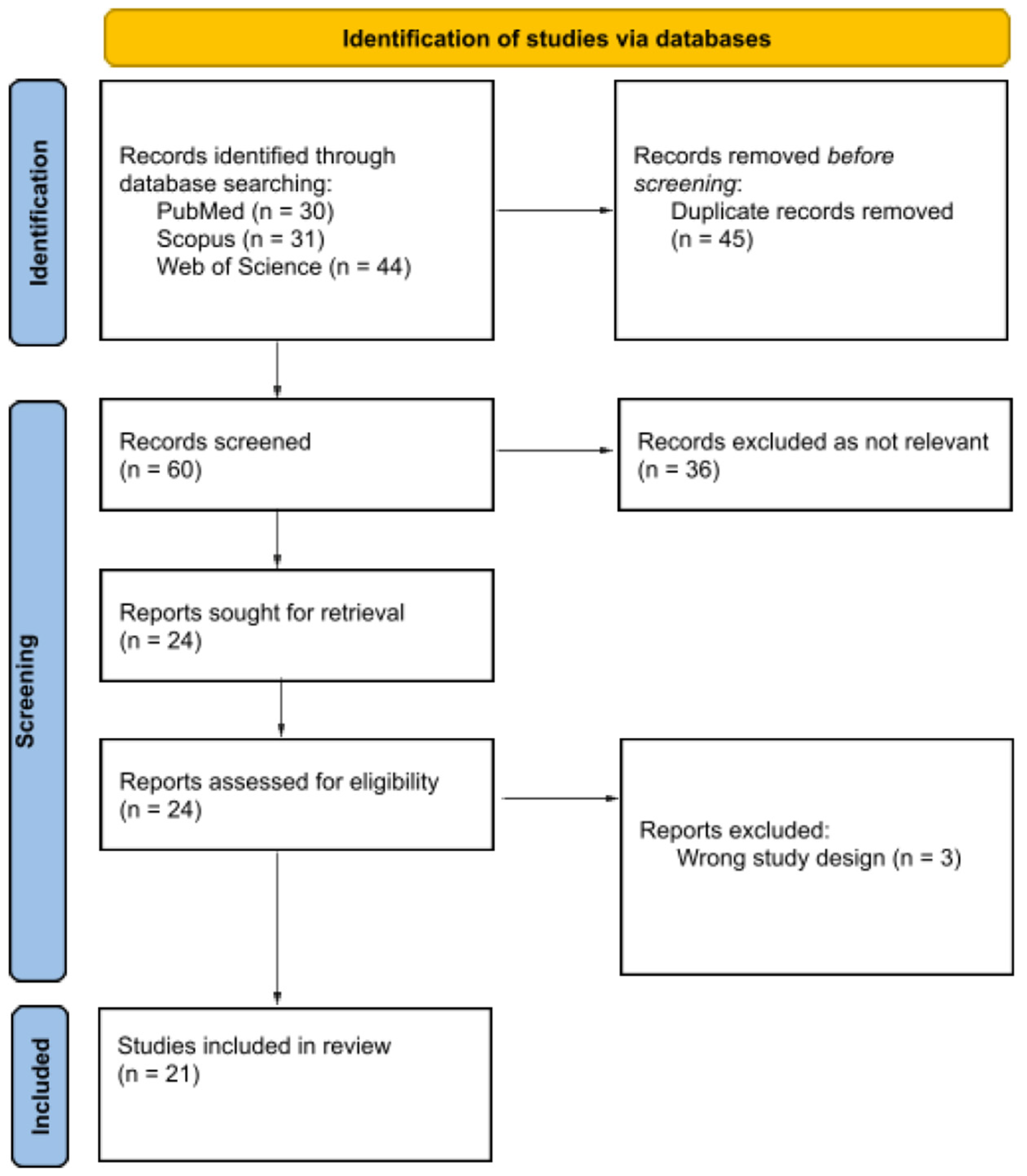
With the purpose of reviewing the pertinent literature, we checked the PubMed (https://ncbi.nlm.nih.gov/PubMed, accessed on 14 April 2022), Scopus and Web of Science databases using the search string “Hidradenitis suppurativa” [All Fields] AND “HIV” [All Fields] OR “AIDS” [All Fields], without time limits. Using the PRISMA flowchart for the drafting of our systematic literature, we read the abstracts of each article whose title suggested the association between hidradenitis suppurativa and HIV. The entire article was read only if the abstract met our inclusion criteria: English language, research papers, and studies on human population only. Papers identified as irrelevant to the topic in question as well as performed on animals or cells were excluded.

3. Results

As of 14 April 2022, a PubMed search of “Hidradenitis suppurativa” AND “HIV” OR “AIDS” yielded 105 articles of which 45 were removed being duplicate records, 36 were not considered because they were not relevant to the outcome of interest, not written in English, not performed on human populations only and three were excluded because they were not research papers (Figure 1). The other 21 concerning the association between HS and HIV were included in our systematic review. Nine case reports out of these twenty-one papers were studied and reviewed in their epidemiological, clinical and treatment/outcomes features (Table 1).

4. Discussion

4.1. Epidemiology

Patients with HS have multiple potential risk factors that can increase the infection rate, including epidermal disruption, immune dysregulation, and topical or oral immunosuppressive treatments [32,33]. The study conducted by Lee et al., examined the associations of HS with serious acute infections (e.g., bacterial, fungal, or viral), chronic infections (e.g., HIV, hepatitis B) and antibiotic-resistant infections [32,34,35]. The authors showed a higher prevalence of cutaneous, extracutaneous, and systemic infections in adults and children with HS than in patients with psoriasis and atopic dermatitis, which lead to increased mortality and costs of care [32,36]. Regarding the relationship between HIV and the increased rates and severity of chronic inflammatory skin disorders, such as HS, to date it remains poorly understood. Being an immunodeficiency syndrome, HIV could promote the onset and progression of HS owing to immune dysregulation [37]. Bui et al. evaluated in their study a population of patients affected by HS and HIV, assessing the rates of HS misdiagnosis in the HIV + community [37]. Data from 63 adults revealed that the onset of HS in patients with HS and HIV was later than in those living with HS only (2nd–3rd decade of life). HIV diagnosis was made before HS onset in 63.8% of subjects [37]. A predisposing effect of HIV infection in the development of HS has been suggested considering the late age of HS onset on one side and the high rate of HS onset after initial HIV diagnosis on the other [37]. Indeed, Deng et al. found a six-fold higher rate of HS diagnosis in patients with HIV compared with those without HIV [38]. However, further studies are needed to establish the exact prevalence and incidence, being both conditions largely misdiagnosed or not regarded by patients, especially in the elderly population.

4.2. Pathogenesis

The “primum movens” in the pathogenesis of HS is follicular hyperkeratosis that leads to rupture of the hair follicle, and subsequent inflammation of apocrine glands [1]. The presence of retained hair tracts in pseudocysts and fistulas, as highlighted at ultrasound, confirms that hair follicle is the main actor in the pathogenesis and suggests laser epilation as first approach in mild cases [39,40]. Furthermore, the presence of superinfections, although not responsible of the HS, may aggravate its clinical course [41]. However, several cytokines seem to be particularly relevant in the HS immunopathogenesis [1]. Among these, tumor necrosis factor-alpha (TNF-α) and interleukin (IL)-17 represent the key cytokines, in fact their increase correlates with disease severity [4,38]. Most notably, TNF-α levels in injured tissues of patients with HS are even higher than those found in patients with psoriasis. TNF-α leads to an increase of the T helper (Th) 17/regulatory T cells ratio, which results in an augmented production of IL-17 [4]. This last induces the expression of other cytokines such as IL-1β, IL-6, and TNF-α through a mechanism involving the nod-like receptor protein 3 (NLRP3) inflammasome [4]. Moreover, TNF-alpha might predispose to insulin resistance acting on adipocytes and muscle cells to induce insulin signaling defects and suppressing the secretion of adiponectin from adipocytes [4]. Additionally, the relationship between smoking and HS is thought to involve TNF-α. Nicotine induces infundibular epithelial hyperplasia causing follicular occlusion and rupture and, at the same time, stimulates macrophages to produce IL-1β and TNF-α and increases expression of matrix metalloproteases (MMPs). Nicotine excretion in sweat also induces TNF-α release by keratinocytes and Th17 cells [4]. There are still other cytokines involved in the pathogenesis of HS: among these it is worth mentioning IL-12 and IL-23 that play a role in the establishment of chronic inflammation [4]. IL-23 is also involved in the development and maintenance of Th17 cells [4]. Even though the precise mechanism that leads to HIV and HS association has not been clearly elucidated, it is generally agreed that HIV may activate the immune system in a persistent manner, due to the increased stimulation caused by the virion and other viral gene products [38]. Thus, during HIV infection, it is the overactivation of T cells themself that may result in an accelerated depletion of these cells through higher rates of apoptosis [38]. HIV is associated with an increased release of proinflammatory cytokines such as TNF-α, IL-6 and IL-18, this phenomenon is thought to facilitate the development of HS by promoting the expression of Th17 cells and IL-17 [38,42,43].

4.3. Clinical Manifestations

It is interesting to underline how atypical presentations of dermatosis and unusual associations between dermatitis characterized by extremely different pathophysiological mechanisms could suggest retroviral infection [6,24,27,28,29]. The involvement of atypical sites, such as face or thighs, must be a “wake-up call”, because it could be due to HIV-related immunosuppression, as noticed by Dhadke et al. in a middle-aged male with poor health status [28] and by Rankin et al. in a man who presented HS exclusively on his face [6]. Even the association between psoriasis and HS should alert the dermatologist to rule out or confirm a possible HIV infection, as described by Bouaddi et al. in a 45-year-old man [29]. Marfatia et al. reported a case of a 35-year-old HIV-positive man with recurrent nodular skin lesions with foul smelling discharge over face, gluteal region, thighs, and axilla. Once again, this case emphasizes how HIV-induced immunological changes alter the clinical course of chronic skin conditions as HS leading to atypical manifestations and therapeutic challenges [24], as also noticed by Manglani et al. [27]. The author highlighted a rare presentation of HS in a 13-year-old pre-pubertal HIV-infected male child who did not respond to common modalities of treatment (oral antibiotics, oral steroids, and local antibiotic wash) and had to be treated with highly active antiretroviral therapy (HAART) (zidovudine, lamivudine, and nevirapine) with no recurrences observed during follow-up visits [27]. This suggests that initiating HAART for HS should be considered as a therapeutic option in HIV-positive patients who do not respond to traditional treatments of HS [27]. HIV infection is able to considerably impair the clinical course and presentation of chronic skin diseases like HS and certain their complications, such as secondary bacterial infections [7]. Given that the majority of data related to the safety of immunosuppressants in seropositive patients comes from clinical cases, physicians should be updated about possible interactions and adverse events associated with the use of biological drugs in HS (e.g., adalimumab) in patients on HAART [44]. In addition, dermatologists should keep in mind cutaneous tuberculosis or other cutaneous mycobacterial infections on their differential diagnosis, since they may mimic the presentation of HS in HIV patients [7]. A routine full body skin examination and a multidisciplinary approach with other healthcare providers, (e.g., patient’s primary care physician and infectious disease provider), is extremely important to improve the quality patient care.

4.4. Histopathology

In addition to studying the cytokine landscape of HS, understanding the histopathologic alterations in this complicated disease is also of some influence. The central role of follicular occlusion in the development and progression of HS corresponds to the typical histological feature, with hyperkeratosis and hyperplasia of the follicular epithelium [45,46,47,48]. Interestingly, Boar et al. characterized the inflammatory infiltrate of HS through immunohistochemical stains (IHC), reporting a high percentage of T-cells subset in the lymphocytic cell population [49]. Like psoriasis, also HS is therefore an inflammatory skin disease associated with the activation of T-cell in which the treatments that decrease the T-cell count improve it [1,36]. Specifically, the lymphocytic infiltrate is represented by cells positive for CD3, CD4, CD8, CD68 and CD79 with a CD4+/CD8+ ratio of 2.1:1 [48,49]. From the other side, HIV infection is marked by a gradual depletion in CD4+ T-cell number and an augmented CD8+ T-cell number with an inverted CD4+/CD8+ cells ratio [36,50,51].
It could seem paradoxical that severe skin manifestations are more frequent in HIV-patients than the general population [37]. However, it is common opinion that the immune dysregulation caused by both pathologies could represent the right interpretation [36,37,51].
An interesting data was the discovery of atypical CD30-positive lymphoid cells described in common cutaneous non-neoplastic disorders including HS [52]. This finding was observed by Cepeda et al. in both HIV negative and positive patients, with a particularly prominent increase in CD30 positive cells in these latter [52]. In the setting of HIV infection, this could be referred to their upregulation [52,53]. Potential differential diagnostic problems with CD30-positive lymphoproliferative disorders could arise given that the presence of these atypical CD30+ lymphoid cells does not allow to formulate alone a diagnosis of malignancy, being they a part of the common reactive infiltrates. [52,54]. Therefore, close collaboration between oncological, dermatological, and infectious disease specialists is sometimes mandatory for a correct management of HS [29,31].

4.5. Therapy

The complexity of the clinical and therapeutic management of HS is burdened by a high rate of clinical failures and recurrences; more so in people living with HIV [24,26]. Depending on the severity of the disease, its extension and patients’ comorbidities, HS treatment includes topical or systemic antibiotics (such as doxycycline, clindamycin, or erythromycin), corticosteroids, isotretinoin, antiandrogens and immunosuppressors [6,24,25,26,27,28,29,30,31,55,56,57]. Severe cases of HS may also require surgical removal of the lesions, thus causing difficult to accept cosmetic alterations [25]. The combination of oral clindamycin and rifampin for 10-12 weeks has been reported as potentially beneficial in the treatment of stage 2 of HS [58]. Nevertheless, the administration of rifampin is challenging in patients receiving antiretroviral therapy. Rifampin is a potent cytochrome P450 inducer (CYP450 1A2, 2B6, 2C8/9, 2C19 and 3A4, weak inducer of CYP450 2D6). It also interacts with different transporters, inducing P-glycoprotein (PGP) and UDP-glucuronosyltransferase enzymes (UGT) and inhibiting Organic Anion Transporter (OAT) and Organic Anion Transporting Polypeptide (OATP) [59]. Unfortunately, many antiretroviral drugs share at least one of the abovementioned metabolisms and their co-administration with rifampin can compromise HIV treatment [60].
Retinoids, such as acitretin, are one of the oldest drugs in the management of HS. Their use on HIV patients should be careful as it may easily cause an increase in triglycerides and transaminases [26]. Retinoid-caused hypertriglyceridemia may also be responsible for an increase of cardiovascular risk, thus leading to different cardiovascular manifestations including myocardial infarction [26].
HS in HIV is usually more aggressive and often refractory to isotretinoin. However, Rankin et al. described the case of a 31-year-old male HIV patient with HS affecting his right jawline, who responded well to isotretinoin and intralesional triamcinolone [6].
Recently, biologic therapies have been increasingly used as treatment solutions for patients with moderate-to-severe disease. TNF-alpha inhibitors, including IgG monoclonal antibodies (e.g., adalimumab and infliximab), are the most prescribed category [57]. As TNF-alpha is a proinflammatory cytokine, its blockage ultimately leads to both reduction of inflammation and increase of susceptibility to infection [42]. However, other targets have been studied and other molecules have been used as possible treatment for HS such as: anti-IL-12/IL-23 (ustekinumab), IL-1 alpha inhibitors (anakinra and bermekimab), IL-17 inhibitors (secukinumab) and anti-IL-23 (guselkumab). They have shown to be effective in HS management even if they are not yet approved by the United States Food and Drug Administration (FDA) nor by the European Medicines Agency (EMA) [57].
As mentioned above, HIV infection can alter the clinical course of HS as seropositive patients often present with more severe cutaneous manifestations and are unresponsive to standard therapies [7].
Although biologics have already been reported as available therapeutic options for moderate to severe psoriasis [61,62] as they do not interfere with ART response, their use in HIV patients with HS is still controversial, as there is not enough scientific evidence due to the absence of randomized placebo-controlled trials. Moreover, the risk of infectious complications related to biologic therapy in already immunosuppressed patients has limited its use considerably [30,56].
Along this line, J.D. Claytor et al. decided to discontinue infliximab and methotrexate combination therapy in a 55-year-old woman with HS when an HIV test performed during the treatment came back positive. However, the immunosuppressive therapy has not been started again after she reached a good viral control and HS progressed aggressively [31].
Early in vitro studies demonstrated the involvement of TNF-alpha in HIV replication and clinical manifestations. TNF-alpha stimulates HIV expression through nuclear factor kappa beta (NFkB) pathway activation and is also a selective activator of lymphocyte T-cells. Furthermore, TNF-alpha production is high in HIV patients undergoing antiretroviral treatment and tends to increase as the infection progresses [55].
Several studies have shown a possible role of TNF-alpha in Acquired Immune Deficiency Syndrome (AIDS) manifestations such as fever, fatigue, cachexia, aphthous ulcers and dementia [56]. Thus, TNF-alpha blockade may play a role in HIV treatment. On this note, anti-TNFalpha therapy has already been administered to treat immune reconstitution inflammatory syndrome in HIV patients who failed steroid treatment [56].
Different case reports demonstrated the safety and efficacy of anti-TNF-alpha monoclonal antibodies in the treatment of HS/HIV patients, as shown in Table 1.
Even though the impact of anti-TNF-alpha on viral load and CD4+ cell count is one of the main concerns about monoclonal antibodies administration in HIV patients, only one case report describes a possible contribution of infliximab to a reduced CD4+ count and increased viral load [25].
Alecsandru at al. reported a case of a 47-year-old HIV male patient who never needed antiretroviral therapy with persistent cutaneous lesions [25]. His CD4+ T-lymphocytes were 546 cells/μL, CD8+ lymphocytes were 795 cells/μL and CD4+/CD8+ 0.6. HIV-1 viral load was 24,870 copies/mL. Tuberculin test came out negative. However, antituberculosis prophylaxis was started two months before infliximab administration. The patients soon reported an improvement of the symptomatology. After 10 months of treatment, the CD4+ cell count decreased and viral load increased, antiretroviral therapy was then initiated. No adverse event has been reported and the clinical response persisted during the maintenance therapy [25].
On the contrary, Husein-ElAhmed et al. and A. Molina-Leyva et al. described the cases of two HIV patients with refractory HS treated with anti-TNF-alpha [26,30]. Both patients were already on antiretroviral treatment and the viral load was undetectable. Husein-ElAhmed et al. administered infliximab with little improvement of skin lesions. After two weeks a recurrence was observed, and neodymium-yttrium-aluminium-garnet laser therapy was performed with partial response. Molina-Leyva et al. prescribed adalimumab treatment with a complete and maintained response at follow-ups. No impact on CD4+ or viral load was detected at follow-ups in both cases [26,30].

5. Conclusions

HS in HIV positive patients often presents a more aggressive course and is refractory to standard therapy compared to HIV negative patients [56]. Biological therapies have begun to be prescribed to reach disease remission in these patients. However, the efficacy and the risk related to its or other molecules administration in people living with HIV needs to be studied further.
Even if the incidence of adverse effect is low in the reviewed cases, new randomized placebo-controlled trials are still needed to be able to compare safety and efficacy of biologic therapies between seronegative and seropositive patients.
As only adalimumab is approved by FDA and EMA for HS treatment, new studies are also necessary to optimize treatment and to find new targets, thus broadening the therapeutic choices.
In conclusion, given the chance of opportunistic infection and other serious adverse effects, HIV screening should be routinely performed before biological therapy administration.
Immunosuppressive therapy could be used cautiously in patients on antiretroviral therapy with stable CD4 count and undetectable viral load at baseline, and patients should be closely monitored.
Evidence and recommendations for physicians about the management of HS in HIV positive patients are summarized in Table 2.

Author Contributions

Conceptualization, L.M. and C.G.; methodology, M.C. and V.M.; software, Y.I.; validation, L.M., G.N. and C.G.; formal analysis, L.M. and M.C.; investigation, L.M. and V.M.; resources, L.M. and Y.I.; data curation, M.C.; writing—original draft preparation, L.M. and V.M.; writing—review and editing, M.C. and C.G.; visualization, M.C.; supervision, G.N.; project administration, L.M.; funding acquisition, C.G. All authors have read and agreed to the published version of the manuscript.

Funding

This research received no external funding.

Institutional Review Board Statement

Not applicable.

Informed Consent Statement

Not applicable.

Data Availability Statement

Not applicable.

Conflicts of Interest

C.G. has received consultation fees and/or grants for research projects, advisory panels and giving educational lectures from Wyeth-Pfizer, Abbott Immunology-Abbvie, Janssen-Cilag, Novartis, LEO-Pharma, LEO-Pharma Denmark, Ely-Lilly, Celgene, Merck-Serono, Sanofi-Aventis, Amgen and Almirall.

References

  1. Li Pomi, F.; Macca, L.; Motolese, A.; Ingrasciotta, Y.; Berretta, M.; Guarneri, C. Neoplastic Implications in Patients Suffering from Hidradenitis Suppurativa under Systemic Treatments. Biomedicines 2021, 9, 1594. [Google Scholar] [CrossRef] [PubMed]
  2. Elkin, K.; Daveluy, S.; Avanaki, K. (Mohammad) Hidradenitis Suppurativa: Current Understanding, Diagnostic and Surgical Challenges, and Developments in Ultrasound Application. Ski. Res. Technol. 2020, 26, 11–19. [Google Scholar] [CrossRef] [PubMed] [Green Version]
  3. Campanati, A.; Orciani, M.; Sorgentoni, G.; Consales, V.; Offidani, A.; di Primio, R. Pathogenetic Characteristics of Mesenchymal Stem Cells in Hidradenitis Suppurativa. JAMA Dermatol. 2018, 154, 1184. [Google Scholar] [CrossRef] [PubMed]
  4. Goldburg, S.R.; Strober, B.E.; Payette, M.J. Hidradenitis Suppurativa. J. Am. Acad. Dermatol. 2020, 82, 1045–1058. [Google Scholar] [CrossRef] [PubMed]
  5. Zouboulis, C.C.; Benhadou, F.; Byrd, A.S.; Chandran, N.S.; Giamarellos-Bourboulis, E.J.; Fabbrocini, G.; Frew, J.W.; Fujita, H.; González-López, M.A.; Guillem, P.; et al. What Causes Hidradenitis Suppurativa ?—15 Years After. Exp. Dermatol. 2020, 29, 1154–1170. [Google Scholar] [CrossRef]
  6. Rankin, B.D.; Haber, R.M. Case Report of Hidradenitis Suppurativa Localized to the Face in an HIV Patient. SAGE Open Med. Case Rep. 2021, 9, 2050313X2110579. [Google Scholar] [CrossRef]
  7. Gomez, J.; Barnes, L.A.; Yost, J.M.; Gordon, J.; Ginsberg, B.A.; Aleshin, M. Hidradenitis Suppurativa in Sexual and Gender Minorities: A Review and Considerations for Providers. J. Am. Acad. Dermatol 2022, in press. [CrossRef]
  8. Keele, B.F.; van Heuverswyn, F.; Li, Y.; Bailes, E.; Takehisa, J.; Santiago, M.L.; Bibollet-Ruche, F.; Chen, Y.; Wain, L.V.; Liegeois, F.; et al. Chimpanzee Reservoirs of Pandemic and Nonpandemic HIV-1. Science 2006, 313, 523–526. [Google Scholar] [CrossRef] [Green Version]
  9. Eggena, M.P.; Barugahare, B.; Jones, N.; Okello, M.; Mutalya, S.; Kityo, C.; Mugyenyi, P.; Cao, H. Depletion of Regulatory T Cells in HIV Infection Is Associated with Immune Activation. J. Immunol. 2005, 174, 4407–4414. [Google Scholar] [CrossRef] [Green Version]
  10. Epstein, H.; Morris, M. Concurrent Partnerships and HIV: An Inconvenient Truth. J. Int. AIDS Soc. 2011, 14, 13. [Google Scholar] [CrossRef]
  11. Hughes, J.P.; Baeten, J.M.; Lingappa, J.R.; Magaret, A.S.; Wald, A.; de Bruyn, G.; Kiarie, J.; Inambao, M.; Kilembe, W.; Farquhar, C.; et al. Determinants of Per-Coital-Act HIV-1 Infectivity Among African HIV-1–Serodiscordant Couples. J. Infect. Dis. 2012, 205, 358–365. [Google Scholar] [CrossRef] [PubMed] [Green Version]
  12. Tanser, F.; Bärnighausen, T.; Hund, L.; Garnett, G.P.; McGrath, N.; Newell, M.-L. Effect of Concurrent Sexual Partnerships on Rate of New HIV Infections in a High-Prevalence, Rural South African Population: A Cohort Study. Lancet 2011, 378, 247–255. [Google Scholar] [CrossRef] [Green Version]
  13. Polis, C.B.; Curtis, K.M. Use of Hormonal Contraceptives and HIV Acquisition in Women: A Systematic Review of the Epidemiological Evidence. Lancet Infect. Dis. 2013, 13, 797–808. [Google Scholar] [CrossRef]
  14. Polis, C.B.; Curtis, K.M.; Hannaford, P.C.; Phillips, S.J.; Chipato, T.; Kiarie, J.N.; Westreich, D.J.; Steyn, P.S. An Updated Systematic Review of Epidemiological Evidence on Hormonal Contraceptive Methods and HIV Acquisition in Women. AIDS 2016, 30, 2665–2683. [Google Scholar] [CrossRef] [PubMed] [Green Version]
  15. Butler, A.R.; Smith, J.A.; Polis, C.B.; Gregson, S.; Stanton, D.; Hallett, T.B. Modelling the Global Competing Risks of a Potential Interaction between Injectable Hormonal Contraception and HIV Risk. AIDS 2013, 27, 105–113. [Google Scholar] [CrossRef] [Green Version]
  16. Weiss, H.A.; Quigley, M.A.; Hayes, R.J. Male Circumcision and Risk of HIV Infection in Sub-Saharan Africa: A Systematic Review and Meta-Analysis. AIDS 2000, 14, 2361–2370. [Google Scholar] [CrossRef]
  17. Cohen, M.S.; Gay, C.L.; Busch, M.P.; Hecht, F.M. The Detection of Acute HIV Infection. J. Infect. Dis. 2010, 202, S270–S277. [Google Scholar] [CrossRef] [Green Version]
  18. Zetola, N.M.; Pilcher, C.D. Diagnosis and Management of Acute HIV Infection. Infect. Dis. Clin. N. Am. 2007, 21, 19–48. [Google Scholar] [CrossRef]
  19. Buchacz, K.; Baker, R.K.; Palella, F.J.; Chmiel, J.S.; Lichtenstein, K.A.; Novak, R.M.; Wood, K.C.; Brooks, J.T. AIDS-Defining Opportunistic Illnesses in US Patients, 1994–2007: A Cohort Study. AIDS 2010, 24, 1549–1559. [Google Scholar] [CrossRef]
  20. Bonnet, F.; Lewden, C.; May, T.; Heripret, L.; Jougla, E.; Bevilacqua, S.; Costagliola, D.; Salmon, D.; Chêne, G.; Morlat, P.; et al. Opportunistic Infections as Causes of Death in HIV-Infected Patients in the HAART Era in France. Scand. J. Infect. Dis. 2005, 37, 482–487. [Google Scholar] [CrossRef]
  21. Palella, F.J.; Delaney, K.M.; Moorman, A.C.; Loveless, M.O.; Fuhrer, J.; Satten, G.A.; Aschman, D.J.; Holmberg, S.D. Declining Morbidity and Mortality among Patients with Advanced Human Immunodeficiency Virus Infection. N. Engl. J. Med. 1998, 338, 853–860. [Google Scholar] [CrossRef] [PubMed]
  22. Kaplan, J.E.; Hanson, D.; Dworkin, M.S.; Frederick, T.; Bertolli, J.; Lindegren, M.L.; Holmberg, S.; Jones, J.L. Epidemiology of Human Immunodeficiency Virus–Associated Opportunistic Infections in the United States in the Era of Highly Active Antiretroviral Therapy. Clin. Infect. Dis. 2000, 30, S5–S14. [Google Scholar] [CrossRef] [PubMed] [Green Version]
  23. Long, J.L.; Engels, E.A.; Moore, R.D.; Gebo, K.A. Incidence and Outcomes of Malignancy in the HAART Era in an Urban Cohort of HIV-Infected Individuals. AIDS 2008, 22, 489–496. [Google Scholar] [CrossRef] [PubMed]
  24. Marfatia, Y.; Singhal, P.; Khambhati, R. Hidradenitis Suppurativa in AIDS. Indian J. Sex Transm. Dis. AIDS 2010, 31, 45. [Google Scholar] [CrossRef] [PubMed] [Green Version]
  25. Alecsandru, D.; Padilla, B.; Izquierdo, J.A.A.; Fernández-Cruz, E.; Sánchez-Ramón, S. Severe Refractory Hidradenitis Suppurativa in an HIV-Positive Patient Successfully Treated With Infliximab. Arch. Dermatol. 2010, 146, 1343. [Google Scholar] [CrossRef] [PubMed]
  26. Husein-ElAhmed, H.; Fernandez-Pugnaire, M.-A.; Ruiz-Carrascosa, J.-C. Severe Hidradenitis Suppurative in an HIV-Positive Male: Use of Multiple Treatment Modalities, Including Tumor Necrosis Factor Blockade. AIDS Patient Care STDS 2011, 25, 507–508. [Google Scholar] [CrossRef] [PubMed]
  27. Manglani, M.; Prabhu, G.; Phiske, M.; Laddha, P. Hidradenitis Suppurativa in a HIV-Infected Child. J. Postgrad. Med. 2012, 58, 207. [Google Scholar] [CrossRef]
  28. Dhadke, S.V.; Korade, M.B.; Sangle, S.A.; Dhadke, V.N. Hidradenitis Suppurativa Complicating Epithelial Malignancy in Immunocompromised Patient. J. Assoc. Physicians India 2016, 64, 90–92. [Google Scholar] [PubMed]
  29. Bouaddi, M.; Hassam, B. Hidradenite Suppurative et Psoriasis: Anguille Sous Roche. Pan Afr. Med. J. 2017, 27, 200. [Google Scholar] [CrossRef]
  30. Molina-Leyva, A.; Badiola, J. Severe Refractory Hidradenitis Suppurativa Successfully Treated with Adalimumab in an HIV-Positive/Hepatitis C Virus-Positive Patient. AIDS 2018, 32, 2436–2438. [Google Scholar] [CrossRef]
  31. Claytor, J.D.; Viramontes, O.; Conner, S.; Wen, K.W.; Beck, K.; Chin-Hong, P.V.; Henrich, T.J.; Peluso, M.J. TNF-α Inhibition in the Setting of Undiagnosed HIV Infection: A Call for Enhanced Screening Guidelines. AIDS 2021, 35, 2163–2168. [Google Scholar] [CrossRef] [PubMed]
  32. Lee, H.H.; Patel, K.R.; Singam, V.; Rastogi, S.; Silverberg, J.I. Associations of Cutaneous and Extracutaneous Infections with Hidradenitis Suppurativa in U.S. Children and Adults. Br. J. Dermatol. 2020, 182, 327–334. [Google Scholar] [CrossRef] [PubMed]
  33. Kurayev, A.; Ashkar, H.; Saraiya, A.; Gottlieb, A.B. Hidradenitis Suppurativa: Review of the Pathogenesis and Treatment. J. Drugs Dermatol. 2016, 15, 1017–1022. [Google Scholar]
  34. Guet-Revillet, H.; Coignard-Biehler, H.; Jais, J.-P.; Quesne, G.; Frapy, E.; Poirée, S.; le Guern, A.-S.; le Flèche-Matéos, A.; Hovnanian, A.; Consigny, P.-H.; et al. Bacterial Pathogens Associated with Hidradenitis Suppurativa, France. Emerg. Infect. Dis. 2014, 20, 1990–1998. [Google Scholar] [CrossRef] [PubMed]
  35. Sartorius, K.; Killasli, H.; Oprica, C.; Sullivan, A.; Lapins, J. Bacteriology of Hidradenitis Suppurativa Exacerbations and Deep Tissue Cultures Obtained during Carbon Dioxide Laser Treatment. Br. J. Dermatol. 2012, 166, 879–883. [Google Scholar] [CrossRef]
  36. Motolese, A.; Ceccarelli, M.; Macca, L.; Li Pomi, F.; Ingrasciotta, Y.; Nunnari, G.; Guarneri, C. Novel Therapeutic Approaches to Psoriasis and Risk of Infectious Disease. Biomedicines 2022, 10, 228. [Google Scholar] [CrossRef]
  37. Bui, H.; Skiba, P.N.; Breskin, A.; Sayed, C. Hidradenitis Suppurativa among Patients with HIV: Investigating Patient and Disease Characteristics and Misdiagnosis. Br. J. Dermatol. 2021, 185, 1070–1072. [Google Scholar] [CrossRef]
  38. Deng, P.H.; Wang, C.J.; Armstrong, A.W. An Association between Hidradenitis Suppurativa and HIV. Br. J. Dermatol. 2020, 182, 490–491. [Google Scholar] [CrossRef]
  39. Wortsman, X.; Wortsman, J. Ultrasound Detection of Retained Hair Tracts in Hidradenitis Suppurativa. Dermatol. Surg. 2015, 41, 867–869. [Google Scholar] [CrossRef]
  40. Nazzaro, G.; Zerboni, R.; Passoni, E.; Barbareschi, M.; Marzano, A.V.; Muratori, S.; Veraldi, S. High-frequency Ultrasound in Hidradenitis Suppurativa as Rationale for Permanent Hair Laser Removal. Ski. Res. Technol. 2019, 25, 587–588. [Google Scholar] [CrossRef]
  41. Benzecry, V.; Grancini, A.; Guanziroli, E.; Nazzaro, G.; Barbareschi, M.; Marzano, A.V.; Muratori, S.; Veraldi, S. Hidradenitis Suppurativa/Acne Inversa: A Prospective Bacteriological Study and Review of the Literature. G. Ital. Di Dermatol. E Venereol. 2020, 155, 459–463. [Google Scholar] [CrossRef] [PubMed]
  42. Ceccarelli, M.; Venanzi Rullo, E.; Vaccaro, M.; Facciolà, A.; d’Aleo, F.; Paolucci, I.A.; Cannavò, S.P.; Cacopardo, B.; Pinzone, M.R.; Pellicanò, G.F.; et al. HIV-associated Psoriasis: Epidemiology, Pathogenesis, and Management. Dermatol. Ther. 2019, 32, e12806. [Google Scholar] [CrossRef] [PubMed]
  43. Facciolà, A.; Venanzi Rullo, E.; Ceccarelli, M.; D’Andrea, F.; Coco, M.; Micali, C.; Cacopardo, B.; Marino, A.; Cannavò, S.P.; di Rosa, M.; et al. Malignant Melanoma in HIV: Epidemiology, Pathogenesis, and Management. Dermatol. Ther. 2020, 33, e13180. [Google Scholar] [CrossRef] [PubMed]
  44. Venanzi Rullo, E.; Maimone, M.G.; Fiorica, F.; Ceccarelli, M.; Guarneri, C.; Berretta, M.; Nunnari, G. Non-Melanoma Skin Cancer in People Living With HIV: From Epidemiology to Clinical Management. Front. Oncol. 2021, 11, 689789. [Google Scholar] [CrossRef]
  45. Von Laffert, M.; Stadie, V.; Wohlrab, J.; Marsch, W.C. Hidradenitis Suppurativa/Acne Inversa: Bilocated Epithelial Hyperplasia with Very Different Sequelae. Br. J. Dermatol. 2011, 164, 367–371. [Google Scholar] [CrossRef]
  46. Jemec, G.B.E.; Thomsen, B.M.; Hansen, U. The Homogeneity of Hidradenitis Suppurativa Lesions. APMIS 1997, 105, 378–383. [Google Scholar] [CrossRef]
  47. ATTANOOS, R.L.; APPLETON, M.A.C.; HUGHES, L.E.; ANSELL, I.D.; DOUGLAS-JONES, A.G.; WILLIAMS, G.T. Granulomatous Hidradenitis Suppurativa and Cutaneous Crohn’s Disease. Histopathology 1993, 23, 111–115. [Google Scholar] [CrossRef] [PubMed]
  48. Smith, S.D.B.; Okoye, G.A.; Sokumbi, O. Histopathology of Hidradenitis Suppurativa: A Systematic Review. Dermatopathology 2022, 9, 251–257. [Google Scholar] [CrossRef]
  49. Boer, J.; Weltevreden, E.F. Hidradenitis Suppurativa or Acne Inversa. A Clinicopathological Study of Early Lesions. Br. J. Dermatol. 1996, 135, 721–725. [Google Scholar] [CrossRef]
  50. De Simone, C.; Perino, F.; Caldarola, G.; D’Agostino, M.; Peris, K. Treatment of Psoriasis with Etanercept in Immunocompromised Patients: Two Case Reports. J. Int. Med. Res. 2016, 44, 67–71. [Google Scholar] [CrossRef] [Green Version]
  51. Morar, N.; Willis-Owen, S.A.; Maurer, T.; Bunker, C.B. HIV-Associated Psoriasis: Pathogenesis, Clinical Features, and Management. Lancet Infect. Dis. 2010, 10, 470–478. [Google Scholar] [CrossRef]
  52. Cepeda, L.T.; Pieretti, M.; Chapman, S.F.; Horenstein, M.G. CD30-Positive Atypical Lymphoid Cells in Common Non-Neoplastic Cutaneous Infiltrates Rich in Neutrophils and Eosinophils. Am. J. Surg. Pathol. 2003, 27, 912–918. [Google Scholar] [CrossRef] [PubMed]
  53. Keane, N.M.; Price, P.; Lee, S.; Stone, S.F.; French, M.A. An Evaluation of Serum Soluble CD30 Levels and Serum CD26 (DPPIV) Enzyme Activity as Markers of Type 2 and Type 1 Cytokines in HIV Patients Receiving Highly Active Antiretroviral Therapy. Clin. Exp. Immunol. 2008, 126, 111–116. [Google Scholar] [CrossRef] [PubMed]
  54. Diaz-Cascajo, C. Strong Immunoexpression of the Monoclonal Antibody CD-30 in Lymphocytic Infiltrates of the Skin Not By Itself Evidence for Diagnosing Malignant Lymphoma. Am. J. Dermatopathol. 2001, 23, 79–80. [Google Scholar] [CrossRef]
  55. Gallitano, S.M.; McDermott, L.; Brar, K.; Lowenstein, E. Use of Tumor Necrosis Factor (TNF) Inhibitors in Patients with HIV/AIDS. J. Am. Acad. Dermatol. 2016, 74, 974–980. [Google Scholar] [CrossRef]
  56. Akarsu, S.; Avcı, C. Effects of Biologic Therapies for Cutaneous Inflammatory Diseases in HIV-Infected Individuals: Reliable or Not? J. Basic Clin. Health Sci. 2018, 2, 61–67. [Google Scholar] [CrossRef]
  57. Rosales Santillan, M.; Morss, P.C.; Porter, M.L.; Kimball, A.B. Biologic Therapies for the Treatment of Hidradenitis Suppurativa. Expert Opin. Biol. Ther. 2020, 20, 621–633. [Google Scholar] [CrossRef]
  58. Saunte, D.M.L.; Jemec, G.B.E. Hidradenitis Suppurativa. JAMA 2017, 318, 2019. [Google Scholar] [CrossRef]
  59. Chen, J.; Raymond, K. Roles of Rifampicin in Drug-Drug Interactions: Underlying Molecular Mechanisms Involving the Nuclear Pregnane X Receptor. Ann. Clin. Microbiol. Antimicrob. 2006, 5, 3. [Google Scholar] [CrossRef] [Green Version]
  60. Dooley, K.E.; Flexner, C.; Andrade, A.S. Drug Interactions Involving Combination Antiretroviral Therapy and Other Anti-Infective Agents: Repercussions for Resource-Limited Countries. J. Infect. Dis. 2008, 198, 948–961. [Google Scholar] [CrossRef] [Green Version]
  61. Bardazzi, F.; Magnano, M.; Campanati, A.; Loconsole, F.; Carpentieri, A.; Potenza, C.; Bernardini, N.; Lernia, V.; Carrera, C.; Raone, B.; et al. Biologic Therapies in HIV-Infected Patients with Psoriasis: An Italian Experience. Acta Derm. Venereol. 2017, 97, 989–990. [Google Scholar] [CrossRef] [PubMed]
  62. Campanati, A.; Ganzetti, G.; Giuliodori, K.; Molinelli, E.; Offidani, A. Biologic Therapy in Psoriasis: Safety Profile. Curr. Drug Saf. 2016, 11, 4–11. [Google Scholar] [CrossRef] [PubMed]
Figure 1. Flow diagram of studies assessed in systematic review. Diagram shows several steps of studies selection in systematic review, also describing exclusion criteria and final number of included articles for each step.
Biomedicines 10 02761 g001
Table 1. Reported cases of hidradenitis suppurativa in individuals with HIV.
Authors, Reference Number and Year Type of Study Sex/Age (Year) Duration Location SeverityTreatmentOutcome
Marfatia Y et al. [24], 2010Case reportM/352 yearsFace, thighs, gluteal region, and axillaNot reportedDoxycicline, ibuprofen, dapsonePartial response with recurrence
Alecsandru D et al. [25], 2010Case reportM/47>1 yearAxillary, gluteal, perianal, thoracic and abdominalSevereInfliximab, clindamycinRemission
Husein-ElAhmed H et al. [26], 2011Case reportM/4710 weeksInguino-scrotal perineum and gluteal areasNot reportedInfliximabPartial response with recurrence
Mangiani M et al. [27], 2012Case reportM/131 monthsRight axillaNot reportedAntiretroviral therapy (zidovudine, lamivudine, nevirapine)Remission
Dhadke S et al. [28], 2016Case reportM/“Middle-age”10 daysRight cheek, neck, chest, shoulder, and axillaNot reportedLocal antibiotic washes, amoxicillin and clavulanic acid, prednisoloneExitus
Bouaddi M et al. [29], 2017Case reportM/456 monthsArmpitsMildNot reportedNot reported
Molina-Leyva A et al. [30], 2018Case reportW/3916 weeksLeft and right axillaSevereAdalimumabRemission
Claytor J et al. [31], 2021Case reportF/55“several months”Axillary and groinNot reportedInfliximab and methotrexate (discontinued after HIV diagnosis)Uncontrolled after infliximab and methotrexate discontinuation
Rankin B et al. [6], 2021Case reportM/314 monthsRight jawlineModerateDoxycycline, Isotretinoin, intralesional triamcinolone acetonideRemission
Table 2. Pearls for physicians: evidence and recommendations.
Pearls for Physicians
EvidenceRecommendations
Patients with HIV may develop recalcitrant forms of inflammatory comorbiditiesA multidisciplinary approach is mandatory in case of suspect cutaneous manifestations
Patients with HS have multiple potential risk factors that can increase their infection rateAs HS and HIV are largely misdiagnosed or not regarded by patients, particular attention should be paid to anamnesis and screening
HIV could promote the onset and progression of HS owing to immune dysregulationPhysicians may aware of late forms of HS
Atypical clinical presentations (and the association between physiopathologically different dermatoses) could be subtended by retroviral infectionThe involvement of atypical sites should be considered as a ’wake-up call’
Clinical and therapeutic management of HS patients is characterized by a high rate of failures and recurrences, more so with HIVManagement and treatment should be as tailored as possible
First-line treatment options may be ineffective in HIV&HS patients or contraindicated because of concomitant medicationsDifferent treatments, including surgery, must be considered, especially in most severe cases
Clinical experience seems to demonstrate that TNF-alpha inhibitors and newer biologics interfering with IL-17 and IL-23 can provide improvement in patients with HIVBiotechnological treatments should be considered for treatment in motivated HIV&HS patients, who are monitored for CD4+ count
Publisher’s Note: MDPI stays neutral with regard to jurisdictional claims in published maps and institutional affiliations.

