Article

The Transcriptional Regulator Prdm1 Is Essential for the Early Development of the Sensory Whisker Follicle and Is Linked to the Beta-Catenin First Dermal Signal

1
Laboratory of Stem Cell Dynamics, School of Life Sciences, Ecole Polytechnique Fédérale Lausanne, 1015 Lausanne, Switzerland
2
Department of Oncology and Hemato-Oncology, University of Milan, Via Santa Sofia 9, 20122 Milan, Italy
3
Department of Experimental Oncology, European Institute of Oncology IRCCS, Via Adamello 16, 20139 Milan, Italy
4
Laboratory of Developmental Genomics, School of Life Sciences, Ecole Polytechnique Fédérale Lausanne, 1015 Lausanne, Switzerland
5
Department of Genetic Medicine and Development, Faculty of Medicine, University of Geneva Medical School, 1211 Geneva, Switzerland
6
BioInformatics Competence Center, UNIL-EPFL, 1015 Lausanne, Switzerland
7
Histology Core Facility, Ecole Polytechnique Fédérale Lausanne, 1015 Lausanne, Switzerland
8
Institut de Génétique et de Biologie Moléculaire et Cellulaire, 67404 Illkirch, France
9
Centre National de la Recherche Scientifique (CNRS), UMR7104, 67404 Illkirch, France
10
Institut National de la Santé et de la Recherche Médicale (INSERM), U1258, 67404 Illkirch, France
11
Université de Strasbourg, 67404 Illkirch, France
12
Division of Cell Matrix Biology and Regenerative Medicine, University of Manchester, Manchester M139PL, UK
13
Division of Neuroscience, IRCCS San Raffaele Hospital, 20132 Milan, Italy
14
Centre Hospitalier Universitaire Vaudois, 1011 Lausanne, Switzerland
15
Duke-NUS Graduate Medical School, Singapore 169857, Singapore
16
Department of Plastic, Reconstructive and Aesthetic Surgery, Singapore General Hospital, Singapore 169608, Singapore
17
A*STAR Skin Research Labs, Singapore 138648, Singapore
*
Author to whom correspondence should be addressed.
Biomedicines 2022, 10(10), 2647; https://doi.org/10.3390/biomedicines10102647
Submission received: 10 September 2022 / Revised: 6 October 2022 / Accepted: 12 October 2022 / Published: 20 October 2022
(This article belongs to the Section Molecular Genetics and Genetic Diseases)

Abstract

:
Prdm1 mutant mice are one of the rare mutant strains that do not develop whisker hair follicles while still displaying a pelage. Here, we show that Prdm1 is expressed at the earliest stage of whisker development in clusters of mesenchymal cells before placode formation. Its conditional knockout in the murine soma leads to the loss of expression of Bmp2, Shh, Bmp4, Krt17, Edar, and Gli1, though leaving the β-catenin-driven first dermal signal intact. Furthermore, we show that Prdm1 expressing cells not only act as a signaling center but also as a multipotent progenitor population contributing to the several lineages of the adult whisker. We confirm by genetic ablation experiments that the absence of macro vibrissae reverberates on the organization of nerve wiring in the mystacial pads and leads to the reorganization of the barrel cortex. We demonstrate that Lef1 acts upstream of Prdm1 and identify a primate-specific deletion of a Lef1 enhancer named Leaf. This loss may have been significant in the evolutionary process, leading to the progressive defunctionalization and disappearance of vibrissae in primates.

1. Introduction

The whisker follicles (or vibrissae—from the Latin vibrio) are complex, self-renewing sensory micro-organs loaded with multipotent epithelial stem cells localized on the snout [1,2]. These stem cells permit the regular replacement of the whisker (a terminally differentiated hair) and, consequently, allow for the continuity of tactile mechanical sensing [3,4,5].
Conserved throughout evolution, whiskers underwent a reduction throughout the primate adaptive radiation [6] and disappeared completely in the human lineage; yet vestiges of whisker capsular skeletal muscles remain in the human upper lip [7]. These specialized hair follicles are bigger both in length and width compared to those of the pelage and are enveloped by vascular sinuses conferring rigidity to the hair shaft. Fibers of striated muscle have an insertion on the capsula and encompass the vascular sinuses. While macro vibrissae are motile and used for distance detecting/object locating, micro vibrissae are immotile and used for object identification.
The processing of whisker-acquired information occurs in the barrel cortex, where each whisker is represented by a discrete and well-defined cytoarchitectonic structure referred to as the barrel [8]. The barrel map occupies a large area of the rodent brain, it is in large part genetically specified and forms early on during development [9]. As the whisker pattern is established earlier and independently from innervation, the hypothesis that whiskers impose their own pattern onto the somatosensory cortex in the homeomorphic fashion has arisen [10].
Prdm1 (also known as Blimp1) is a zinc-finger transcriptional repressor [11] that was shown to be a master regulator controlling terminal differentiation of B-lymphocytes [12,13,14]; it also governs T-cell homeostasis [15] and primordial germ cell specification [16], stem cell maintenance in the sebaceous gland [17], and skin differentiation [18,19]. Prdm1 was also shown to play a crucial role during whisker development in mice [20].
Sox2Cre driven Prdm1 conditional knockout mice are one of the very rare transgenic animals entirely lacking whisker (vibrissae) follicles, while pelage hair follicles develop physiologically [20]. The loss of this gene impairs whisker development, but the exact stage at which the development is halted has not yet been identified. Furthermore, it is still not known which type of mystacial vibrissae (macro- and/or micro-) are impacted in these mutant mice.
Moreover, Robertson et al. demonstrated that Prdm1 positive mesenchymal cells give rise to the mature dermal papilla (DP) and expand to form a mesenchymal layer immediately surrounding the hair follicles [20]. However, what this mesenchymal layer gives rise to in the adult whisker is not yet known.
In addition, the reverberation of whisker development halt onto the organization barrel cortex was not investigated yet. The re-organization of the somatosensory cortex is a phenomenon of great evolutionary importance, given the expansion of the brain areas dedicated to the processing of the sensory organs.
In summary, despite its clear role in whisker development, relatively little is known about the regulation of Prdm1 expression during whisker development. Investigating this subject is of fundamental importance as the loss of regulatory elements in genes might explain the morphogenetic changes that occurred throughout the primate adaptive radiation and that led to the reduction in snout size and vibrissae while hands and eyes of diurnal primates took over as sensory organs.

2. Materials and Methods

2.1. Mouse Strains

OF1 and C57/B6J2 mice were obtained from Charles River Breeding Laboratories. Prdm1MEGFP transgenic mice were kindly provided by Mitinori Saitou; Lef1tm1 Rug mice were provided by Rudolf Grosschedl and Werner Held. Sox2Cre, Sox2CreERT2, Prdm1Cre, Wnt1Cre, Prdm1 CA, and ROSAYFP mice were obtained from the Jackson Laboratory (Bar Harbor, United States) (Table 1). The strains carrying the Cre recombinase and ROSAYFP were kept in heterozygosity, while the conditional knockout was performed in homozygosity. Genotyping was conducted using the primers listed in Table 2. All animals were maintained in a 12 h light cycle providing food and water ad libitum. Mice were sacrificed by intra-peritoneal (i.p.) injection of pentobarbital. Experiments were conducted in accordance with the EU Directive (86/609/EEC) for the care and use of laboratory animals and that of the Swiss Confederation. Mating of adult female and male mice was carried out overnight. Time-pregnant mice were sacrificed by injection of pentobarbital and uteri with embryos were removed by dissection.

2.2. In Situ Hybridization

RNA-FISH was performed using 8-μm paraffin sections. Digoxigenin-labeled probes for specific transcripts were prepared by PCR with primers designed using published sequences. The mRNA expression patterns were visualized by immunoreactivity with anti-digoxigenin horseradish peroxidase-conjugated Fab-fragments (Roche, Basel, Switzerland), according to the manufacturer’s instructions. The amplification was carried out using the TSA Plus Cyanine 3/5 System (Perkin Elmer, Waltham, United States). Gli1 and Wnt10b ISH were performed with the ACD RNAscope (Newark, United States).

2.3. In Vivo Lineage Tracing

For the lineage tracing experiment, Sox2CreERT2 crossed with ROSAYFP pregnant females were induced at embryonic day E12 and E12.5 with 2 mg tamoxifen and 1 mg of progesterone (Sigma-Aldrich, St. Louis, United States) by intraperitoneal injection. The transgenic animals were retrieved at E17 and perinatally and then processed for histology and immunostaining.

2.4. Proliferation Experiments

Pregnant female mice carrying Prdm1MEGFP embryos were injected with 200 µL of EdU (30 mg/ mL) and analyzed 2 h after the first injection. Embryos were retrieved, genotyped under the UV lamp, and processed for histology and immunostaining.

2.5. Histology and Immunostaining

All samples were removed and fixed overnight in 4% paraformaldehyde at 4 °C. Tissues were washed three times in PBS for 5 min and incubated overnight in 30% sucrose in PBS at 4 °C; eventually, they were then embedded in OCT and kept at −80  °C. Sections of 10 µm thickness were cut using a CM3050S Leica cryostat (Leica Microsystems, Wetzlar, Germany).
Sections were incubated in blocking buffer (1% BSA, 0.3% Triton in PBS) for 1 hour at room temperature. Primary antibodies (listed in Table 3) were incubated overnight at 4 °C. Sections were rinsed three times in PBS and incubated with appropriate secondary antibodies diluted to 1:1000 and DAPI in blocking buffer for 1 h at room temperature. Sections were again washed three times with PBS. The primary antibodies used are listed in the following table. The following secondary antibodies were used: anti-mouse, anti-rabbit, anti-rat, anti-goat, conjugated to Alexa Fluor 488, 568 and 647 (Molecular Probes, Eugene, United States). Nuclei were stained in DAPI solution (1:2000) and slides were mounted in DAKO fluorescent mounting medium. As for the AP reaction, SIGMAFAST™ Fast Red TR was used and visualized by confocal microscopy (Leica) at 568 nm.

2.6. Imaging

Fluorescence microscopy images were captured under the LSM 780 confocal microscope (Carl Zeiss, Jena, Germany); transmission microscopy images were acquired with either the Olympus Ax70 or the Zeiss Axioscope 2 Plus. x. Adobe Photoshop software was used for image processing.

2.7. RT–PCR

Total RNA was isolated from the embryonic whisker pad using RNAeasy Mini Kit (Qiagen, Hilden, Germany) according to the manufacturer’s instructions. Total RNA was extracted, and 250 ng of each sample were reverse transcribed using the Superscript III enzyme and random primers (Life Technologies, Carlsbad, United States).

2.8. Quantitative PCR

For qPCR, 1 µL of cDNA was amplified with the Taqman Universal Mastermix II (Life Technologies) in a 10 µL total reaction volume; 5 µL of the Mastermix, 1.5 µL of CDNA, and 3.5µL of assay mix were included in the reaction. The primers were bought from Applied Biosciences. The TaqMan assays were performed using a 79,000 HT Fast Real-Time system (AB). For data analysis, the mouse Eef1alpha, β-actin, Gapdh, and Tbp housekeeping genes were used as internal controls. Gene expression profiling was achieved using the Comparative CT method (DDCT) of relative quantification [21] using the SDS 2.4 software (Applied Biosystems, Waltham, United States).

2.9. Interspecies Sequence Comparison

The comparison of the conserved noncoding elements and deletions in mouse–rat, mouse–guinea pig, mouse–squirrel, mouse–rabbit, mouse–human, mouse–chimp, mouse–gorilla, mouse–orangutan, mouse–rhesus, mouse–dolphin, mouse–cow, mouse–cat, mouse–dog, mouse–horse, mouse–elephant was performed using the Vertebrate Multiz Alignment and Conservation Track in the UCSC genome browser, using a window size of 2 kb. HiC data on topological associated domains (TADs) from ES cells were obtained from http://3dgenome.fsm.northwestern.edu/view.php (accessed on 11 April 2020) [22,23].

2.10. 4C-Seq

For each sample, we harvested at least 1 × 10⁷ cells and obtained 7–10 μg of output double-digested, double-ligated DNA. We collected 120 embryonic whisker pads, both at E12.5 and E13. NlaIII and DpnII were used as primary and secondary cutters, respectively; ligation was performed by using the Concentrated T4 DNA ligase from Promega. Primer sets for Lef1 promoter and Leaf enhancer are described in Table 4.
PCRs were multiplexed and sequenced with Illumina HiSeq2000. Then, 4C-seq reads were processed through the HTS station according to previously described procedures [24,25] and visualized with gFeatBrowser (http://www.gfeatbrowser.com, accessed on 12 January 2022). Briefly, raw reads were demultiplexed and aligned to the mouse mm10 reference genome (GRCm38). The regions directly surrounding the viewpoints, chr3: 131′104′979–131′112′546 for the Lef1 promoter viewpoint and chr3: 131′016′310–131′022′769 for the Leaf viewpoint, were excluded from the analysis. Normalization was performed by dividing the fragment scores by the mean of fragments scores falling into a region defined as +/−1 Mb around the center of the bait coordinates. Smoothed signals were obtained by applying a running mean algorithm with a window size of eleven fragments. The profile corrected data were generated by applying an approach similar to the one described in [26] using a fit with a slope −1 in a log-log scale [27]. The 4C-Seq data are deposited in GEO under the accession number GSE193356 (Table 5).

2.11. Statistical Analysis

To compare the contacts between the Lef1 promoter and Leaf, in the E12.5 whisker pad and the adult kidney, we considered the normalized and profile corrected fragments from region chr3:131′008′663–131′026′430 for Leaf viewpoint, and those from region chr3:131106987–131227057 for Lef1 promoter. Statistical differences were assessed by applying an unpaired non-parametric two-tailed Mann–Whitney test after having excluded the normality of the data with a D’Agostino and Pearson omnibus normality test. Differences were considered significant if the p-value, p, was less than 0.05 and shown in figures as * (* p < 0.05, ** p < 0.01, *** p < 0.001, **** p ≤ 0.001).
For Leaf, fragments of region Chr3:131008663–131026430 (mm10) were analyzed whereas, for the Lef1 promoter and coding sequence, the fragments of region Chr3:131106987-131227057 were analyzed. The normality of the data was excluded by the D’Agostino and Pearson omnibus normality test. Statistical differences were assessed by applying an unpaired non-parametric two-tailed Mann–Whitney test. Differences were regarded as significant if p < 0.05, and significances are shown in figures as * (* p < 0.05, ** p < 0.01, *** p < 0.001, **** p ≤ 0.001). Analyses were conducted using GraphPad Prism version 8.0.

2.12. CUT&Tag

CUT&Tag was performed on wild-type E12 whisker pads dissociated to a single cell suspension according to the v1.5 Epycypher Protocol (https://www.epicypher.com/resources/protocols/cutana-cut-and-tag-protocol/, accessed on 12 January 2022). Rabbit anti-H3K4Me1 (Abcam, Ab8895) and IgG (Cell Signaling, 2729S) were used at 0.5 ug per sample. Samples were sequenced at a depth of 10Mio each with the Illumina NovaSeq 6000 system (51 bp paired-end runs).
The peak calling for the CUT&Tag experiments was performed per each replicate with SEACR v1.3 [28] using the norm and stringent parameters. Due to the poor distribution of fragment lengths observed for IgG replicate 1 and the subsequent low reproduction rates (see Supplementary Figure S6), we rather called the peaks without IgG by setting the top fraction of peaks considered to 1%, 2%, 3%, 4%, and 5% to assess the overall behavior. The peaks represented in the figures are the ones obtained from the top 1%. CUT&Tag data are deposited in GEO under the accession number GSE192851 (Table 5).

3. Results

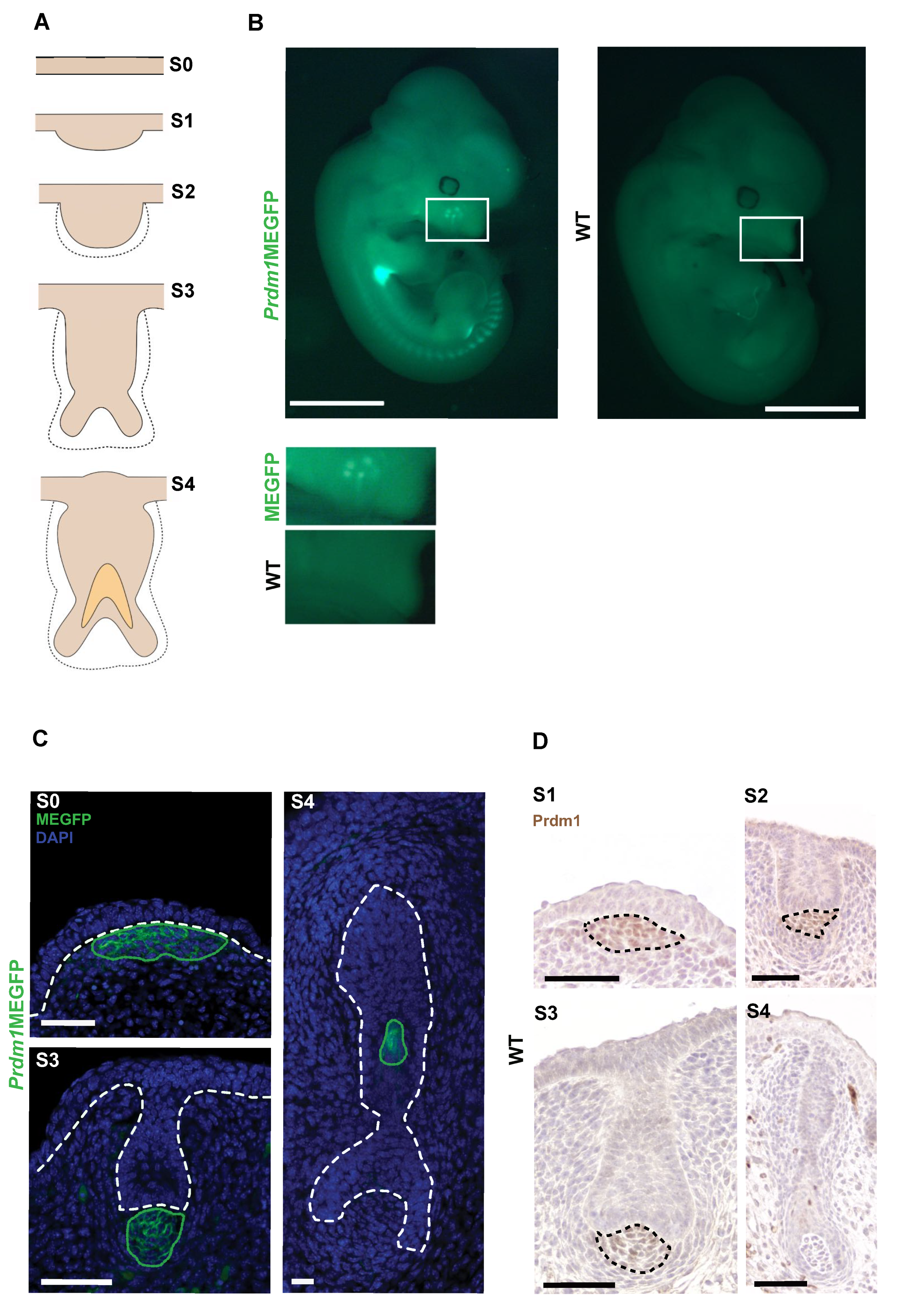
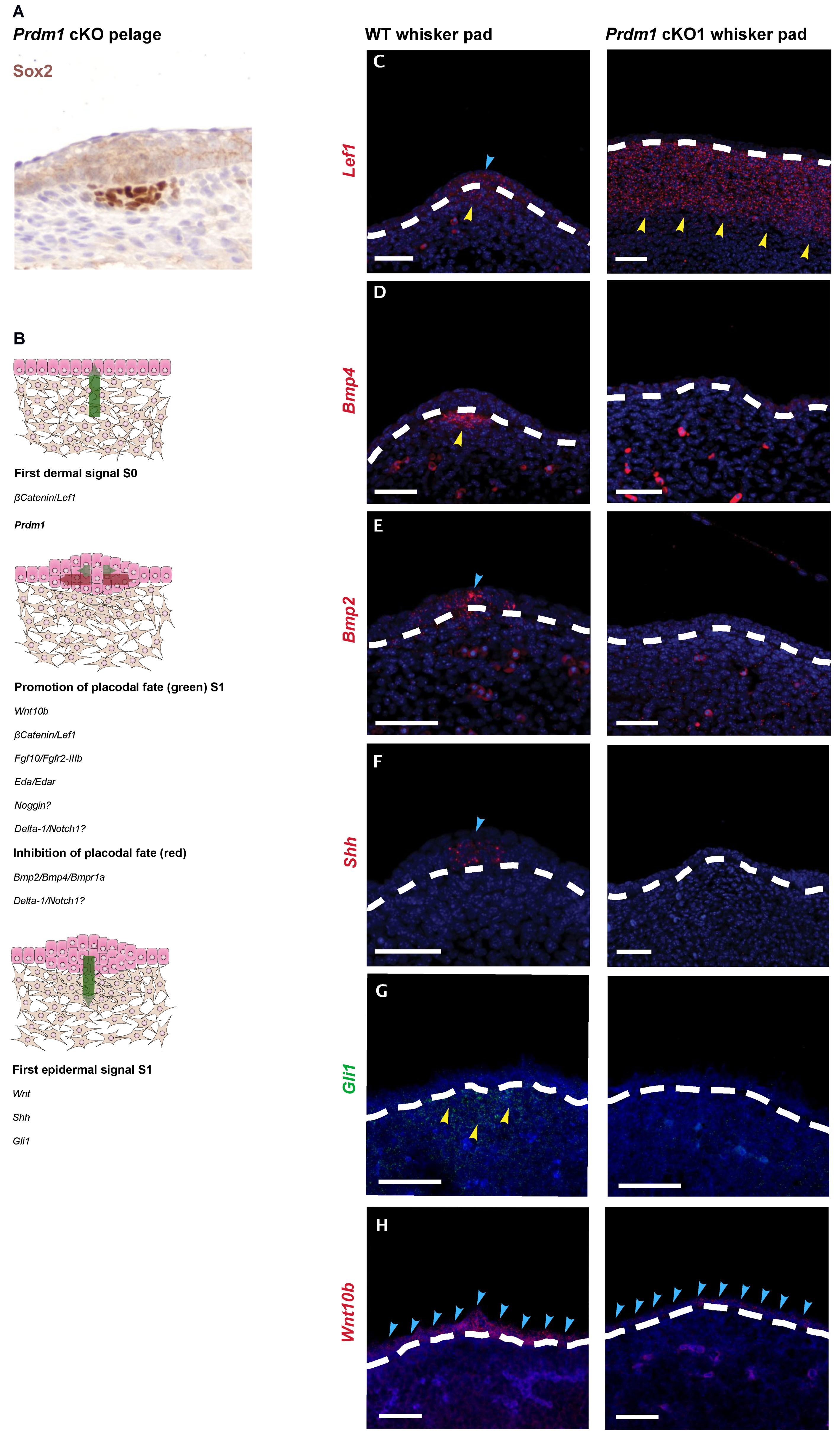
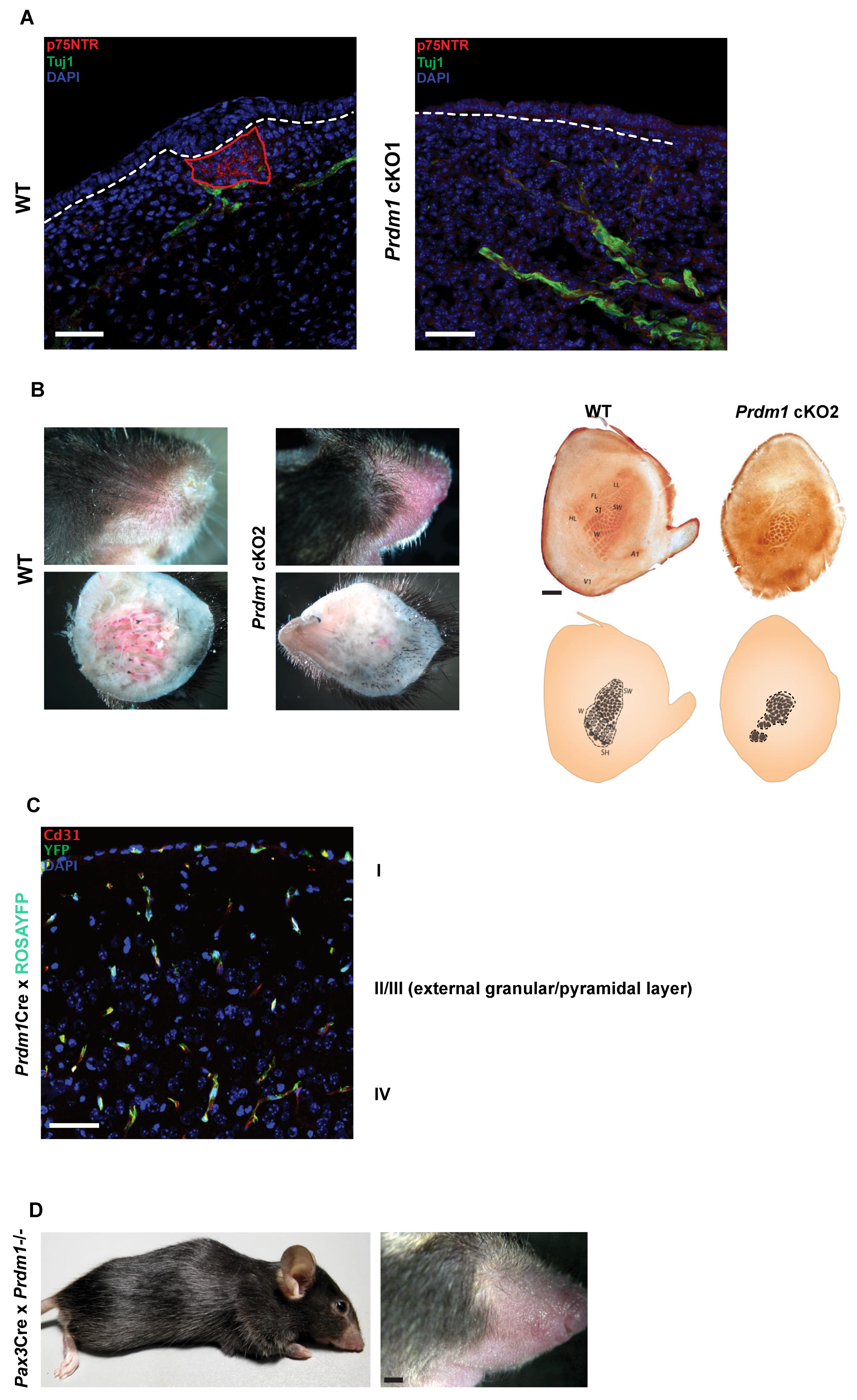
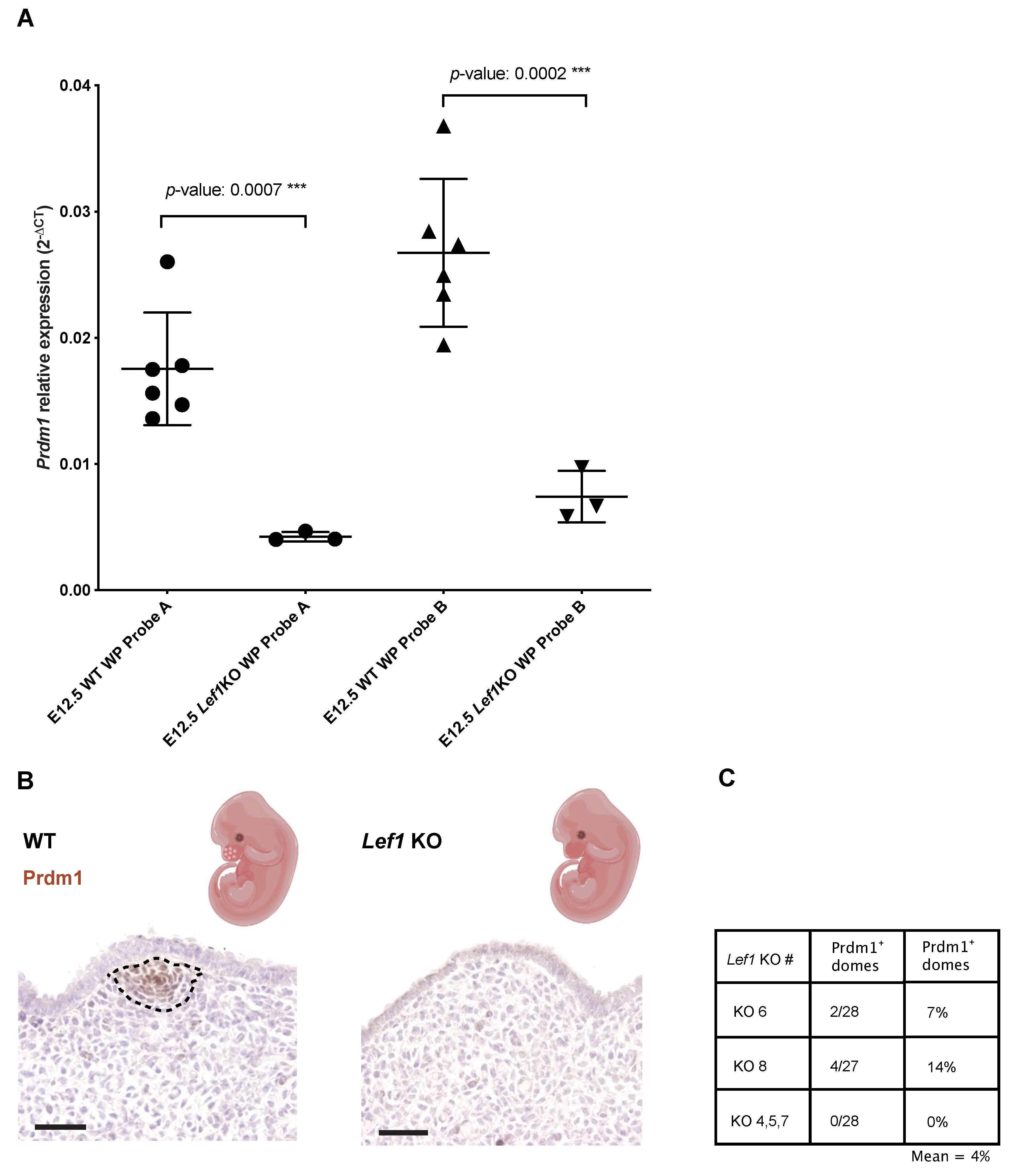
3.1. Prdm1 Is an Essential Gene for Whisker Follicle Development

We localized Prdm1 expression by anti-GFP immunofluorescence in Prdm1mEGFP reporter [17] embryonic whisker pads during the first stages of whisker development (Figure 1A,B).
The expression of mEGFP in heterozygous embryos can be detected in a specific cluster of mesenchymal cells underlying the monolayer of embryonic epidermis (referred to as stage 0 of whisker development, [29]) that will later form the whisker epidermal placode. It continues to be expressed in this compartment until stage 4, when its expression is turned on in the inner root sheath (IRS) of the follicle. At stage 5, mEGFP expression disappears in the DP (Figure 1C). Those results are validated by Prdm1 immunohistochemistry (IHC) in wild-type whisker pads (Figure 1D).
On the other hand, Prdm1 expression in the dermal condensate of both head and back pelage hair follicles starts at embryonic day E14.5 but is transient as it disappears at E16.5 (Figure S1).
To position Prdm1 in the molecular cascade leading to whisker formation, we generated Prdm1 knockout embryos. It was previously shown that the constitutional knockout of Prdm1 leads to severe impairment of the placenta resulting in early embryonic lethality [16] Therefore, we used a Sox2Cre deleter strain to bypass the placental developmental halt [20,30]. By combining Sox2Cre with a floxed Prdm1 allele [14], we were able to obtain viable E17.5 Prdm1 homozygous conditional knockout embryos (referred to as cKO1 in this study). We started by analyzing E12.5 and E13.5 cKO1 embryos, the relevant time points being when whisker follicle formation begins. cKO1 embryos recapitulate previous findings (Figure S2A) [20] and, microscopically, neither whisker placode can be detected, nor the dermal condensate formation occurs (Figure S2A). On the other hand, the development of pelage hair follicles is not impaired at E15.5 (Figure 2A), as observed in [20].
To understand where Prdm1 stands in the molecular cascade leading to whisker formation (Figure 2B), we looked with fluorescent in situ hybridization (FISH) at the expression of the genes involved in the first molecular steps of whisker follicle morphogenesis, both in the epithelial and mesenchymal compartment. β-catenin is involved in establishing the first dermal signal [31]; consistent with this, Lef1 is expressed throughout the mesenchyme of the mouse vibrissa pad prior to vibrissa follicle development at E11, and initiation of vibrissa follicle development is dependent on its expression [32,33]; at E12.5, its expression is confined to the epithelial placode and underlying mesenchyme. In E12.5 homozygous cKO1 mice, Lef1 is expressed homogeneously in the mesenchyme of the whisker pad, thus, indicating that the first signal is present, even though there is no further development, which would require the physiological restriction of Lef1 expression in the whisker placode and underlying mesenchyme (Figure 2C).
In Prdm1 cKO1 mice, no placode formation can be observed both macroscopically and microscopically (Figure 2 and Figure S2A). In the wild-type whisker pad, there is a patterned upregulation of molecules involved in the promotion and inhibition of placodal fate. This includes Bmp4 (Figure 2D) in the pre-follicle mesenchyme, Bmp2 (Figure 2E) in the epithelial compartment, and Shh (Figure 2F) secreted by the placodal cells. However, those are clearly absent in Prdm1 cKO1 mice (Figure 2D–F). Gli1 is not upregulated in the pre-follicular mesenchyme (Figure 2G). We found that Wnt10b is diffusely expressed in the monolayer of epidermal cells in the sites of whisker formation in cKO1 and does not show a marked upregulation in placodes (as in the wild-type whisker pads) (Figure 2H). Furthermore, RT-qPCR on whisker pads at E12.5 shows a statistically significant decrease in Edar, involved in the promotion of placode, and Keratin 17, whose expression arises within the single-layered, undifferentiated ectoderm of E10.5 mouse embryos and giving rise, in the ensuing 48 h, to the epidermal placodes [34] (Figure S2B). Altogether, these data show that in the absence of Prdm1, the gene cascade involved in placode formation is not activated, despite the presence of the initial Lef1 expression in the whisker placode mesenchyme.
Figure 2. Prdm1 conditional knockout locks the developing whisker pad at the level of the first dermal signal leaving pelage hair follicle formation unperturbed. (A) Sox2 immunohistochemistry on E15.5 head skin of Prdm1 cKO1 embryos. Pelage hair follicle placodes are present as well as the mesenchymal condensate. (B) Expression of early whisker developmental genes in the Prdm1 cKO1 mouse. Molecular mechanisms underlying the early steps of whisker development. The dermis delivers a β-catenin-based homogenous first signal to the overlying epidermis in order to initiate placode formation. The placode in turns both sustains their growth and inhibits the formation of other placodes in the surrounding epidermis. The promotion of the placodal fate is sustained by several molecules including Wnt10b, βcatenin/Lef1, Fgf10/Fgfr2-IIIb, Eda/Edar, Noggin, Delta-1/Notch1, whereas the inhibition is based upon Bmp2/Bmp4/Bmpr1a and Delta-1/Notch1. Thus, the placode conveys a first epithelial signal, leading to the clustering of the mesenchymal cells underneath into the dermal condensate. This process mainly relays on Wnt and Shh signaling. “Image adapted with permission from [35]. Copyright 2002, Elsevier.” Fluorescent ISH on E12.5 wild-type embryos (S1 of whisker development) reveals that Lef1 expression is confined to the epithelial placode and underlying mesenchyme (C); Bmp4 the underlying mesenchyme I (D); Bmp2 marks the epithelial placode (E); Shh is expressed by the placode and induces the condensation of the mesenchyme (F); Wnt10b expression is restricted in placodes (G); Gli1 is upregulated in the pre-follicular mesenchyme. In E12.5 cKO1 embryos, Lef1 expression is homogenous throughout the mesenchyme; Bmp2, Bmp4, Shh, and Gli1 are no longer detectable, while Wnt10b fails to be upregulated in placode areas; whisker follicles cannot, thus, reach stage 1 of whisker development. The dashed lines indicate the dermo–epidermal junction. Hybridization is marked with arrows (the yellow arrows) indicate the hybridization of the probes in the placode, the yellow ones in the mesenchyme); (H) Sox2 IHC on E15 head skin of Prdm1 cKO1 mouse reveals that the development of head hair follicles is unperturbed by the deletion of Prdm1. Scale bar: 50 μm.
Biomedicines 10 02647 g002

3.2. The Whisker Inducing Mesenchyme Contributes to Several Lineages of the Adult Whisker

To investigate the proliferative activity of Prdm1 expressing cells, we administered a short pulse (2 h) of nucleotide analog ethynyldeoxyuridine (EdU) to Prdm1MEGFP pregnant mice carrying E12.5 embryos. We could observe that Prdm1 expressing cells can be classified into two subpopulations (Figure 3A): quiescent—contiguous to the embryonic epithelium—and proliferative at its periphery (asterisks in Figure 3A).
We then aimed at identifying the progeny of Prdm1 expressing cells in the whisker through lineage tracing by crossing the Prdm1Cre transgenic strain [17] with ROSAYFP mice [36] and collecting litters at different time points during embryonic development (Figure S3).
At E12.5, YFP-expressing cells are located in the whisker mesenchymal condensate recapitulating the GFP expression previously reported in the Prdm1MEGFP embryonic whisker pad. However, at E13.5, the population of YFP-positive cells expands and encompasses the area of mesenchyme surrounding the whisker hair germ. Prdm1 can be detected in this area (except for the precursors of the DP) in whisker pad sections of both Prdm1MEGFP and WT mice at E12.5 and E13.5 (Figure 3B).
Given that Prdm1Cre is a transgenic strain that does not allow the spatiotemporal tracing of Prdm1 expressing cells and that Prdm1 is expressed in different territories, a Prdm1Cre lineage analysis does not allow the precise analysis of one given population. As we observed that Sox2 is co-expressed with Prdm1 at E12.5 in the whisker mesenchymal condensate (Figure 3B), we consequently crossed Sox2CreERT2 [37] with ROSAYFP mice and injected tamoxifen in pregnant females at E12.5. Double transgenic litters were analyzed at E14.5, E17.5, and post-natal days (P) 1–3. Forty-eight hours after tamoxifen injection, YFP-positive cells were detected in the dermal condensate of the whisker hair germs (Figure 3C). As the epidermal downward growth proceeds, the mesenchymal YFP cells progressively encapsulate it (Figure 3C). When the whisker follicle reaches its final anatomical configuration, YFP is expressed by the whole DP, dermal sheath (DS), and abundantly by cells residing in the vascular sinuses (Figure 3D). More precisely, the latter are enmeshed with CD31 positive endothelial cells and display a perithelial position: both the latter and the expression of markers such as Ng2, Pdgfrβ, and Tnap indicate that they are pericytes (Figure 3D).

3.3. Prdm1 Genetic Ablation Leads to the Reorganization of the Rodent Barrel Cortex

To study the impact of whisker loss on the nervous system, we looked at their innervation in the developing whisker pad of cKO1 fetuses. In the wild-type whisker pad, the afferent branches of the infraorbital nerve (labeled by p75NTR) encapsulate the whisker mesenchymal condensate without penetrating it; on the contrary, they terminate as free nerve endings in the KO counterpart (Figure 4A).
To evaluate the consequences of the impaired whisker innervation on the nervous system, we have adopted the Wnt1Cre as a deleter strain [38]. Wnt1 is expressed throughout the dorsal neural tube, thus, the Cre-Lox recombination occurs in all neural crest derivatives. This includes the whisker pad mesenchyme and, in particular, the mesenchymal compartment surrounding the developing whisker follicle. Wnt1Cre-driven Prdm1 conditional KO mice (referred to as the cKO2) are viable and lack almost all the macro vibrissae, except for the 1–3 distal ones of the first row, as observed both macroscopically and microscopically (Figure S4A–E). Macroscopically, pelage hair follicle seems to be absent only on the snout, even though microscopically they are present in the histological sections (Figure S4F,G).
We retrieved the brains of both WT and cKO2 mice at postnatal day (P) 21 to account for the developmental maturation of the somatosensory system and sectioned their flattened somatosensory cortex tangentially to visualize the organization of the barrel cortex (Figure 4B). The cytochrome oxidase staining revealed that cKO2 barrel cortex undergoes a major rearrangement; the residual macro vibrissae are represented by enlarged barrels; the barrels corresponding to the micro vibrissae are, however, still present, even though their pattern is highly disorganized (Figure 4C). The deletion of macro vibrissae obtained with the Wnt1Cre deleter strain was confirmed using another delete strain active in the neural crest, the Pax3Cre mice (Figure 4D) [39].
To exclude the expression of Prdm1 in the developing barrel cortex—and thus that the barrel cortex phenotype can be ascribed to the loss of Prdm1 in the nervous system—we crossed Prdm1Cre with ROSAYFP mice and looked at YFP expression in the cerebral cortex (Figure 4D). YFP is expressed only by endothelial cells and not by the thalamocortical axons or by layer four cortical neurons, thus, excluding the aforementioned possibility.

3.4. Prdm1 Knockouts Lead to Disrupted Trigeminal Nerve Wiring and Major Reorganization of the Barrel Cortex

We reasoned that β-catenin/Lef1 might act upstream of Prdm1 during whisker follicle development. To prove this, we obtained Lef1 constitutional KO embryos by crossing heterozygous Lef1tm1Rug mice [30] and investigated Prdm1 expression in the E12.5 whisker pad both at the mRNA and protein level.
At a molecular level, the quantity of Prdm1 transcript in the Lef1 KO embryos is lower compared to the wild-type counterpart, as demonstrated by RT-qPCR (Figure 5A). We sectioned the E12.5 whisker pads from Lef1 KO and WT mice and localized Prdm1 by IHC, focusing on the mesenchymal cells located under the characteristic surface elevations of the whisker pad that constitute the sites of whisker placode induction—hereafter referred to as domes. We analyzed the transverse sections of the embryonic whisker pad through (a) the primitive nasal cavity, (b) the vomeronasal organ, and (c) the tongue of five Lef1 KO embryos. Out of five Lef1 KOs, three domes did not express Prdm1. As for the remaining two embryos, we could observe Prdm1 expression in 2/28 and 4/27 domes (Figure 5B,C).

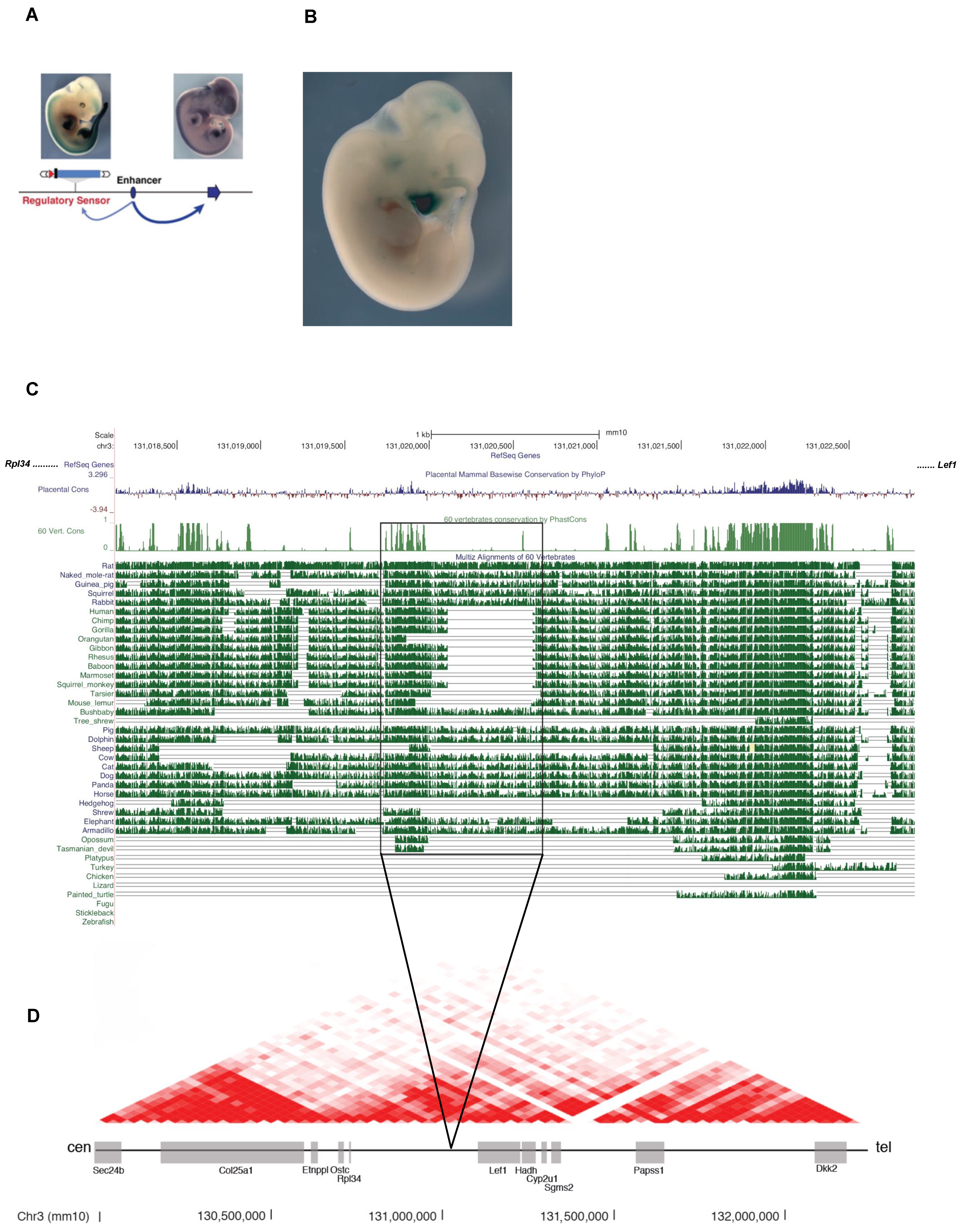
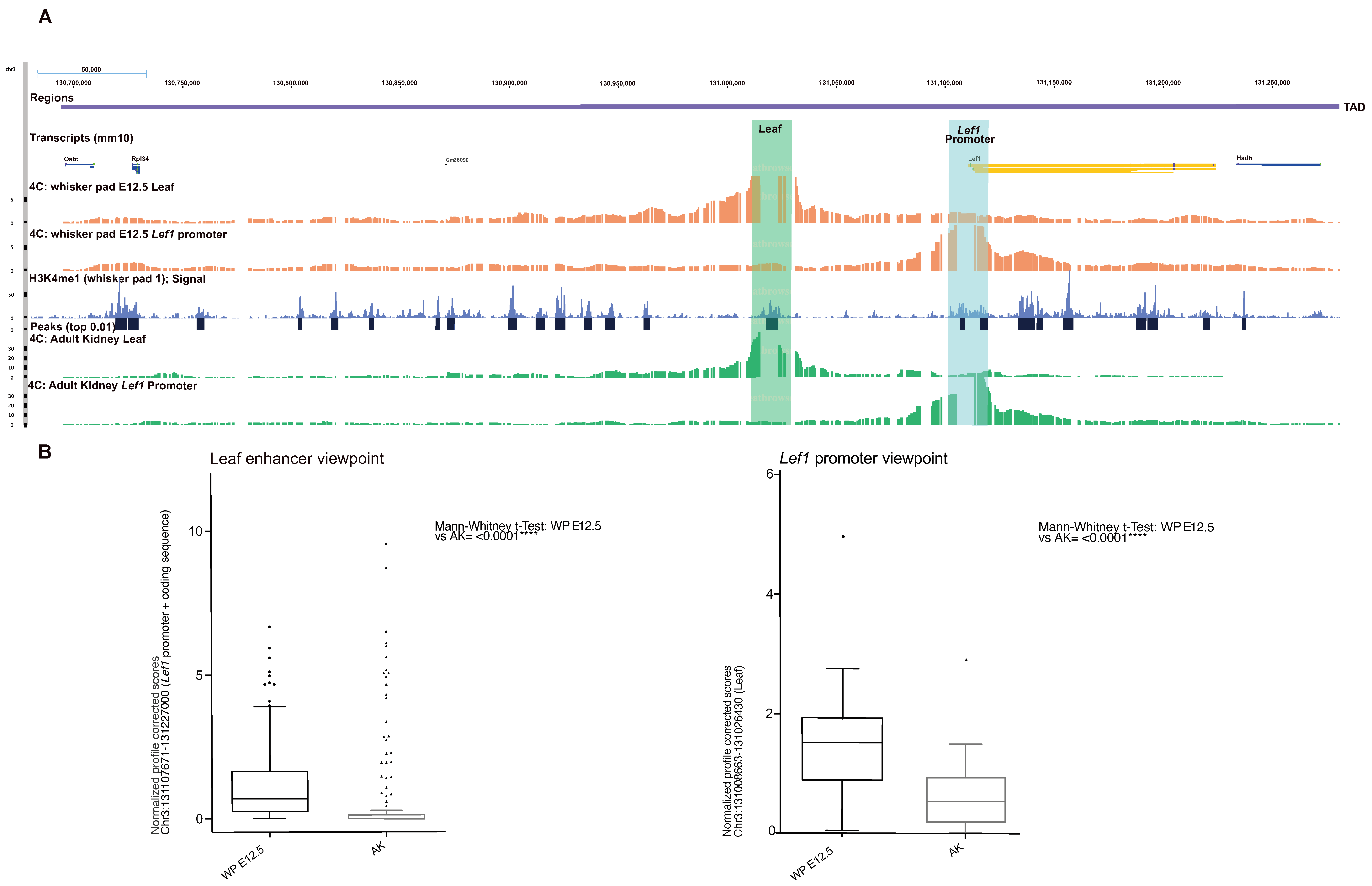
3.5. Identification of Regulatory Elements Lost during Evolution in Whisker Morphogenesis

To understand if the de-regulation of Prdm1 and/or Lef1 might help to explain vibrissae reduction and their eventual loss in humans, we looked at their regulatory regions. As for Lef1, a transposable enhancer trap mapping to a particular locus (chr3:130,927,182–130,927,529 in mm10 assembly) suggests that its regulatory region is located on the centromeric side of the gene in the adjacent gene desert (TRACER LacZ expression database, SB line name 183038-emb20) [40]. The transgene expression can be observed at E11.5 in the brain, mammary glands, whisker pad, and the tip of tail, tissues/organs where Lef1 is physiologically expressed during development (Figure 6A).
We next compared the multispecies alignment of animals with and without whiskers in the aforementioned locus, searching for putative regulatory elements. Conservation scored by PhastCons indicated the presence of an 878bp element conserved throughout the two categories of species (chr3:131,019,746–131,020,624), which, in turn, contains a 521 bp sub-region specifically absent in animals deprived of functional whiskers (human, chimp, gorilla, gibbon, rhesus, baboon, and squirrel monkey) mapping chr3:131,020,103–131,020,624 (mm10 assembly) (Figure 6C). Altogether, this DNA element that we called Leaf (chr3: 131,019,746–131,020,624) is located in the same topological associating domain (TAD) where Lef1 resides (Figure 6D) [22,23].
Figure 6. The Lef1 regulatory region lies centromeric to its promoter and contains a primate-specific deletion named Leaf. (A) The TRACER database pinpoints that the regulatory region of Lef1 resides centromeric to its promoter and that is active in E11.5 whisker pads. (B) A transposable enhancer trap mapping to a particular locus adjacent to Lef1 (chr3:130,927,182–130,927,529) shows that this region is active in the whole E11.5 whisker pad (revealed by LacZ staining), coherently with the homogeneous expression of the first dermal signal. (C) Multispecies alignment shows a primate-specific deletion of a sequence adjacent to a conserved region. Species analyzed include animals with developed whiskers (i.e., rat, cat, dog, rabbit, guinea pig, squirrel, horse, naked mole rat, pig), primates (human, chimp, gorilla, gibbon, rhesus, baboon, squirrel monkey, orangutan, marmoset, tarsier) and the human lineages. (D) HiC profile centered over Lef1 in murine ES cells [22] generated with [23] Cen: centromeric. Tel: telomeric.
Biomedicines 10 02647 g006
We performed 4C-Seq to observe if Leaf can contact the promoter of Lef1. The analysis was conducted on whole micro-dissected whisker pads at E12.5 using primers positioned in the promoter of Lef1. The adult kidney was used as the negative control as Lef1 is not expressed in this tissue (Figure 7A,B and Figure S5A–C) [41].
We found that Lef1 promoter scores contact mainly in cis, within an area of around 700 kb surrounding the Lef1 locus, corresponding to its TAD. More specifically, the centromeric region contains most of the peaks of interaction, whereas telomeric contacts occur chiefly with the coding sequence of Lef1, extending until the end of the coding sequence of the neighboring gene Hadh.
In the E12.5 whisker pad, the promoter of Lef1 contacts several centromeric regions, among which is the one containing the primate-specific deletion (Leaf). We quantified the number of contacts from the Lef1 promoter to Leaf in the E12.5 whisker pad and compared them to the adult kidney, tissue in which Lef1 is expressed at low levels (Figure 7B). We found high statistical significance between the scores mapping Leaf in the E12.5 whisker pad compared to the adult kidney in both normalized and profile corrected datasets (****, Mann–Whitney test, p = 0.0008 in normalized and p < 0.0001 in PC).
In the reciprocal experiment, we scored the contacts from the murine Leaf enhancer with the promoter and coding sequence of Lef1. Similarly, we showed a high statistical significance between the scores mapping Lef1 in the E12.5 whisker pad compared to the adult kidney (****, Mann–Whitney test, p < 0.0001 in normalized and p < 0.0001 in PC), suggesting productive long-range interactions between Leaf and Lef1 in the whisker pad, confirming our observations from the specular viewpoint.
To validate Leaf as an enhancer, we then performed CUT&Tag [42] on E12 wild-type whisker pads to map the presence of monomethylation of histone 3 lysine 4 (H3K4me1)—a post-translational modification enriched at active and poised enhancers. We observed a clear enrichment of H3K4me1 at the Leaf locus (Figure 7A). This result was reproducible across three wild-type whisker pads analyzed (Figures S5A and S6), which, when taken together with our previous results, clearly demonstrates Leaf as an enhancer that contacts the Lef1 promoter (and vice versa).

3.6. Expression of an Ar Regulatory Enhancer Specifically Lost in Humans in the Early Steps of Whisker Development

It was recently demonstrated that a specific enhancer of the androgen receptor gene (Ar) is active in the murine whisker mesenchyme during development [43]. The authors proposed that the loss of this enhancer is associated with the loss of both sensory vibrissae and penile spines in the human lineage [43,44]. Notably, the castration or genetic deletion of the Ar in mice results in reduced growth of whisker follicles in mice without their full disappearance.
To understand if the Ar is involved in the early steps of whisker development together with Prdm1 and Lef1, we performed both IHC and RT-qPCR on embryonic whisker pads at E12.5. The Ar IHC shows no signal either in the whisker placodes or in the mesenchyme underneath it at E12.5 and the RT-qPCR confirmed the absence of expression in the micro-dissected whisker pads at E12.5 and E13.5 (Figure S5B,C), thus, excluding the hypothesis of its early involvement in whisker formation.

4. Discussion

Bringing to light the molecular mechanisms underlying vibrissae development and their evolutionary reduction—until their complete disappearance in humans—is key to understanding how the modifications in molecular circuitries led to their unique features specific to different organisms.
While previous work has shown that Prdm1 and Lef1 have an indispensable role in whisker formation in rodents [20,31], their precise positioning in the morphogenic molecular cascade has remained elusive. The mesenchymal structures constituting the adult whiskers are of neural crest origin [45]. In line with this, Prdm1 expressing cells give rise to the dermal papilla, while the cells failing to incorporate into it migrate to surround the shaft of the forming follicle, giving rise to an adult progeny that has not yet been described [20]. Each adult whisker is innervated by branches of the trigeminal nerve and is represented in a one-to-one fashion in the barrel cortex. A synergy between the barrel cortex genetic programs and the physical presence of whisker follicles on the snout determines the development and somatotopic map of the barrel cortex, a puzzle to be completed with the ablation of whisker developmental genes. Previously, the deletion of a whisker-specific enhancer from the human androgen receptor in humans was described as a distinctive and characteristic feature of the human lineage [43,44]. Given that the Ar is not expressed during the early stages of whisker development and that Ar knockout mice still develop vibrissae [46], surely other regulatory elements—and most probably the ones of genes involved in the early phases of morphogenesis—are involved in the evolutionary loss of whisker follicles.
Our experiments clearly indicate that Prdm1 operates at the level of the first dermal signal during whisker formation and that in its absence, the development of pelage hair follicles is not perturbed during the early stages of development [47]. Intriguingly, Prdm1 is no longer expressed during human pelage hair follicle development [48], indicating that its upstream genes no longer induce its expression in hair follicles. Lef1 (and thus the β-catenin-based first dermal signal) is indispensable to induce Prdm1 expression, though the mechanism by which this occurs needs further investigation. We envision the cascade leading to whisker formation as a circuit where genes are activated in a specific sequence to start its formation and sustain its growth. As for the first dermal signal, Lef1 and subsequently Prdm1 represent the first genes of the circuit that trigger the successive phases (i.e., the formation of the placode first and then of the first epidermal signal).
We show that the population of cells expressing Prdm1/Sox2 in the embryonic whisker mesenchyme gives rise to several lineages of the adult whisker, including the DP and DS, thus, representing a population of multipotent progenitors. This result explains the common inductive properties of the DP and DS, as reported by others [49]. Additionally, we show that it gives rise to pericytes residing in the whiskers’ vascular sinuses.
We demonstrate that in the absence of Prdm1 expression, the afferent branches of the infraorbital nerve do not organize into plexi surrounding the mesenchymal condensates but terminate as free nerve endings. Axonal guidance is most probably absent because of the lack of nerve guidance molecules secreted by Prdm1 positive cells. The Wnt1Cre driven Prdm1 KO (cKO2) mice lack almost all macro vibrissae, although they retain micro vibrissae; we can, thus, describe Prdm1 as strictly necessary for macro vibrissae development. Those conditional knockout mice have major rearrangements in the barrel cortex also affecting the representation of micro vibrissae with relevant evolutionary consequences.
King and Wilson [50] postulated that “regulatory mutations account for the major biological differences between chimp and human”. It was already elegantly demonstrated how enhancers regulate craniofacial morphology [51], highlighting the prominent role of enhancers in morphological evolution. The multispecies deletion we identified in the regulatory region of Lef1 that is specific to several primates (including human, chimp, gorilla, gibbon, rhesus, and baboon) is flanking a well-conserved region among many mammals, altogether forming a regulatory element named Leaf. Using 4C-Seq analyses and H3K4me1 CUT&Tag, we have shown that Leaf is an enhancer in E12 embryonic whisker pads.
While great apes have lost macro vibrissae, although retaining the micro vibrissae on lips (a phenotype reminiscent of our Prdm1 cKO2 mice), as well as cheeks and eyebrows, humans are the only known mammals to have lost both. In the model we envisage, whisker loss was a multi-step process that started during the divergence of the species and that relied on the loss of tissue-specific regulatory elements of several genes involved in whisker formation. More specifically, the loss of Leaf and other putative enhancers might have contributed to the downregulation of the expression of genes fundamental for whisker development in the snout of primates, thus, contributing to whisker loss together with other mechanisms that have yet to be identified.

Supplementary Materials

The following supporting information can be downloaded at: https://www.mdpi.com/article/10.3390/biomedicines10102647/s1, Figure S1, (A) Prdm1 IHC on developing head/back pelage hair follicles. Prdm1 expression in pelage hair follicle development is transient, lasts until hair germ formation (S2), and is independent of the embryonic origin of the mesenchyme. Dashed circles envelop the areas where Prdm1 is expressed. Scale bar: 50 μm. (B) WT E12.5 embryo compared to Prdm1 cKO1. cKO1 embryos display no whisker placodes macroscopically. Figure S2, (A) Expression of Sox2 in wild-type and Prdm1 cKO1 whisker pad at E13.5. No dermal condensate can be detected in cKO1 embryos. Scale bar: 500 μm (B) Edar and Krt17 expression evaluated by RT-qPCR on E13 whisker pads of wild-type and cKO1 mice. Figure S3, Prdm1Cre lineage tracing in developing whisker pads. Prdm1Cre was crossed with a ROSAYFP strain and developing whisker pads were analyzed at E12.5 and E13. YFP-positive cells encompass the developing whisker. Scale bar: 50 μm. Figure S4, Wnt1Cre driven homozygous Prdm1 knockout mice (cKO2) lack almost all the macro vibrissae except for the 1–3 distal ones of the first row, as observed both macroscopically and microscopically (C,D,E) compared to wild-type animals (A,B). The barrel cortices of the correspondent animals were stained with cytochrome oxidase and revealed a major rearrangement. Residual macro vibrissae are represented by enlarged barrels; the barrels representing micro vibrissae are still present, though highly disorganized. cKO2 whisker pads display unperturbed pelage hair follicle formation at P21 (G) compared to wild-type animals (F). Figure S5, (A) Additional H3K4me1 profiles across all three CUT&Tag replicates. The green box indicates the genomic region where Leaf resides. (B) Ar IHC shows no expression in the E12.5 whisker pad compared to the dorsal root ganglia (positive control). (C)Validation of the lack of Ar expression in E12.5 whisker pad by RT-qPCR. Figure S6, Quality controls of CUT&Tag H3K4me1 in E12 whisker pads. A. Sequencing depth B. Alignment Rate C. Unique Fragments D. Fragment Length E. Correlation plot between the H3k4me1 samples and the IgG controls. F. Number of significant H3K4me1 peaks with or without IgG control G. Fraction of Reads in Peaks in the different sets of peaks H. Peak reproducibility rate. Figure S6, (A) Additional H3K4me1 profiles across all three CUT&Tag replicates. The green box indicates the genomic region where Leaf resides. (B) AR IHC shows no expression in the E12.5 whisker pad compared to the dorsal root ganglia (positive control). (C)Validation of the lack of Ar expression in E12.5 whisker pad by RT-qPCR.

Author Contributions

P.G.M., conceptualization of the study, experimental work, manuscript writing; F.D. (Fabrice Darbellay), conceptualization and experimental work on the evolutionary part of the paper, manuscript writing; M.L., bioinformatic analyses; A.Y.C., CUT&Tag H3K4Me1 experiments; B.M., J.C., F.D. (Frederic Droux), and M.S., experimental work; G.-F.M., A.H. and J.S.-D., histology, immunohistochemistry, and ACD RNAScope ISH; S.D.V., original experiments on GFP expression in the whisker pad Pax3Cre Prdm1 knockout mice; Y.B., conceptualization of the study; G.T. evaluation of the CUT&Tag data; P.G.M.,Y.B. and G.C., supervision of the study. All authors have read and agreed to the published version of the manuscript.

Funding

Y.B. was supported by SNF grants FNS 31003A-156812 and by the Lee Seng Teik and Lee Hoo Leng Distinguished Professorship in Plastic Surgery and Regenerative Medicine at Duke-NUS Medical School. P.G.M. was funded by an “Assegno di Ricerca tipo A” from the University of Milan. The work was partly funded by MRC grant MRC MR/S015116/1 to G.C. A.Y.C. was funded by Fondazione Regionale per la Ricerca Biomedica, project number 2729068.

Institutional Review Board Statement

The animal study protocol was approved by the Canton de Vaud (or Ethics Committee) (protocol codes 2472 and 2472.1).

Informed Consent Statement

Not applicable.

Data Availability Statement

All bioinformatic data are available at GEO (NCBI) under the accession codes GSE193356 and GSE192851.

Acknowledgments

We are grateful to Michiko Kanemitsu for kindly providing us with the results for Figure 4 and Figure S4. We thank Mitinori Saitou and Azim Surani for providing the Prdm1mEGFP mice, Rudolf Grosscheldl and Werner Held for providing the Lef1tm1Rug mice, Irene Pizzitola for breeding and mating Lef1tm1Rug mice, Denis Duboule for support, discussions, and suggestions on the evolutionary part of the paper, the EPFL CPG (Emilie Gesina, Gisele Ferrand) and the animal house of Epalinges (Francis Derouet, Lisa Arlandi) for mice handling, Orbicia Riccio, Elisabeth Joye and Severine Urfer for sharing probes for in situ hybridization, Olga de Sousa Silva and Lai Quiewen for technical help.

Conflicts of Interest

The authors declare no conflict of interest.

References

  1. Oshima, H.; Rochat, A.; Kedzia, C.; Kobayashi, K.; Barrandon, Y. Morphogenesis and renewal of hair follicles from adult multipotent stem cells. Cell 2001, 104, 233–245. [Google Scholar] [CrossRef] [Green Version]
  2. Claudinot, S.; Nicolas, M.; Oshima, H.; Rochat, A.; Barrandon, Y. Long-term renewal of hair follicles from clonogenic multipotent stem cells. Proc. Natl. Acad. Sci. USA 2005, 102, 14677–14682. [Google Scholar] [CrossRef] [PubMed] [Green Version]
  3. Petersen, C.C.H. The functional organization of the barrel cortex. Neuron 2007, 56, 339–355. [Google Scholar] [CrossRef] [PubMed] [Green Version]
  4. Brecht, M. Barrel cortex and whisker-mediated behaviors. Curr. Opin. Neurobiol. 2007, 17, 408–416. [Google Scholar] [CrossRef] [PubMed]
  5. Diamond, M.E.; von Heimendahl, M.; Knutsen, P.M.; Kleinfeld, D.; Ahissar, E. “Where” and “what” in the whisker sensorimotor system. Nat. Rev. Neurosci. 2008, 9, 601–612. [Google Scholar] [CrossRef] [PubMed]
  6. Van Horn, R.N. Vibrissae structure in the rhesus monkey. Folia Primatol. 1970, 13, 241–285. [Google Scholar] [CrossRef] [PubMed]
  7. Tamatsu, Y.; Tsukahara, K.; Hotta, M.; Shimada, K. Vestiges of vibrissal capsular muscles exist in the human upper lip. Clin. Anat. 2007, 20, 628–631. [Google Scholar] [CrossRef]
  8. Woolsey, T.A.; Van der Loos, H. The structural organization of layer IV in the somatosensory region (SI) of mouse cerebral cortex. The description of a cortical field composed of discrete cytoarchitectonic units. Brain Res. 1970, 17, 205–242. [Google Scholar] [CrossRef]
  9. Erzurumlu, R.S.; Gaspar, P. Development and critical period plasticity of the barrel cortex. Eur. J. Neurosci. 2012, 35, 1540–1553. [Google Scholar] [CrossRef] [PubMed] [Green Version]
  10. Andrés, F.L.; Van der Loos, H. From sensory periphery to cortex: The architecture of the barrelfield as modified by various early manipulations of the mouse whiskerpad. Anat. Embryol. 1985, 172, 11–20. [Google Scholar] [CrossRef] [PubMed]
  11. Keller, A.D.; Maniatis, T. Identification and characterization of a novel repressor of beta-interferon gene expression. Genes Dev. 1991, 5, 868–879. [Google Scholar] [CrossRef] [PubMed] [Green Version]
  12. Angelin-Duclos, C.; Cattoretti, G.; Lin, K.I.; Calame, K. Commitment of B lymphocytes to a plasma cell fate is associated with Blimp-1 expression in vivo. J. Immunol. 2000, 165, 5462–5471. [Google Scholar] [CrossRef] [PubMed] [Green Version]
  13. Shaffer, A.L.; Lin, K.I.; Kuo, T.C.; Yu, X.; Hurt, E.M.; Rosenwald, A.; Giltnane, J.M.; Yang, L.; Zhao, H.; Calame, K.; et al. Blimp-1 orchestrates plasma cell differentiation by extinguishing the mature B cell gene expression program. Immunity 2002, 17, 51–62. [Google Scholar] [CrossRef] [Green Version]
  14. Shapiro-Shelef, M.; Lin, K.-I.; McHeyzer-Williams, L.J.; Liao, J.; McHeyzer-Williams, M.G.; Calame, K. Blimp-1 is required for the formation of immunoglobulin secreting plasma cells and pre-plasma memory B cells. Immunity 2003, 19, 607–620. [Google Scholar] [CrossRef] [Green Version]
  15. Martins, G.A.; Cimmino, L.; Shapiro-Shelef, M.; Szabolcs, M.; Herron, A.; Magnusdottir, E.; Calame, K. Transcriptional repressor Blimp-1 regulates T cell homeostasis and function. Nat. Immunol. 2006, 7, 457–465. [Google Scholar] [CrossRef] [PubMed]
  16. Vincent, S.D.; Dunn, N.R.; Sciammas, R.; Shapiro-Shalef, M.; Davis, M.M.; Calame, K.; Bikoff, E.K.; Robertson, E.J. The zinc finger transcriptional repressor Blimp1/Prdm1 is dispensable for early axis formation but is required for specification of primordial germ cells in the mouse. Development 2005, 132, 1315–1325. [Google Scholar] [CrossRef] [PubMed] [Green Version]
  17. Ohinata, Y.; Payer, B.; O’Carroll, D.; Ancelin, K.; Ono, Y.; Sano, M.; Barton, S.C.; Obukhanych, T.; Nussenzweig, M.; Tarakhovsky, A.; et al. Blimp1 is a critical determinant of the germ cell lineage in mice. Nature 2005, 436, 207–213. [Google Scholar] [CrossRef]
  18. Magnúsdóttir, E.; Kalachikov, S.; Mizukoshi, K.; Savitsky, D.; Ishida-Yamamoto, A.; Panteleyev, A.A.; Calame, K. Epidermal terminal differentiation depends on B lymphocyte-induced maturation protein-1. Proc. Natl. Acad. Sci. USA 2007, 104, 14988–14993. [Google Scholar] [CrossRef] [Green Version]
  19. Horsley, V.; O’Carroll, D.; Tooze, R.; Ohinata, Y.; Saitou, M.; Obukhanych, T.; Nussenzweig, M.; Tarakhovsky, A.; Fuchs, E. Blimp1 defines a progenitor population that governs cellular input to the sebaceous gland. Cell 2006, 126, 597–609. [Google Scholar] [CrossRef] [Green Version]
  20. Robertson, E.J.; Charatsi, I.; Joyner, C.J.; Koonce, C.H.; Morgan, M.; Islam, A.; Paterson, C.; Lejsek, E.; Arnold, S.J.; Kallies, A.; et al. Blimp1 regulates development of the posterior forelimb, caudal pharyngeal arches, heart and sensory vibrissae in mice. Development 2007, 134, 4335–4345. [Google Scholar] [CrossRef]
  21. Livak, K.J.; Schmittgen, T.D. Analysis of relative gene expression data using real-time quantitative PCR and the 2(-Delta Delta C(T)) Method. Methods 2001, 25, 402–408. [Google Scholar] [CrossRef] [PubMed]
  22. Dixon, J.R.; Selvaraj, S.; Yue, F.; Kim, A.; Li, Y.; Shen, Y.; Hu, M.; Liu, J.S.; Ren, B. Topological domains in mammalian genomes identified by analysis of chromatin interactions. Nature 2012, 485, 376–380. [Google Scholar] [CrossRef] [PubMed] [Green Version]
  23. Wang, Y.; Song, F.; Zhang, B.; Zhang, L.; Xu, J.; Kuang, D.; Li, D.; Choudhary, M.N.K.; Li, Y.; Hu, M.; et al. The 3D Genome Browser: A web-based browser for visualizing 3D genome organization and long-range chromatin interactions. Genome Biol. 2018, 19, 1–12. [Google Scholar] [CrossRef] [PubMed] [Green Version]
  24. David, F.P.A.; Delafontaine, J.; Carat, S.; Ross, F.J.; Lefebvre, G.; Jarosz, Y.; Sinclair, L.; Noordermeer, D.; Rougemont, J.; Leleu, M. HTSstation: A web application and open-access libraries for high-throughput sequencing data analysis. PLoS ONE 2014, 9, e85879. [Google Scholar] [CrossRef]
  25. Noordermeer, D.; Leleu, M.; Splinter, E.; Rougemont, J.; De Laat, W.; Duboule, D. The dynamic architecture of Hox gene clusters. Science 2011, 334, 222–225. [Google Scholar] [CrossRef] [Green Version]
  26. Tolhuis, B.; Blom, M.; Kerkhoven, R.M.; Pagie, L.; Teunissen, H.; Nieuwland, M.; Simonis, M.; de Laat, W.; van Lohuizen, M.; van Steensel, B. Interactions among Polycomb domains are guided by chromosome architecture. PLoS Genet. 2011, 7, e1001343. [Google Scholar] [CrossRef] [Green Version]
  27. Lieberman-Aiden, E.; van Berkum, N.L.; Williams, L.; Imakaev, M.; Ragoczy, T.; Telling, A.; Amit, I.; Lajoie, B.R.; Sabo, P.J.; Dorschner, M.O.; et al. Comprehensive mapping of long-range interactions reveals folding principles of the human genome. Science 2009, 326, 289–293. [Google Scholar] [CrossRef] [Green Version]
  28. Meers, M.P.; Tenenbaum, D.; Henikoff, S. Peak calling by Sparse Enrichment Analysis for CUTnRUN chromatin profiling. Epigenet. Chromatin. 2019, 12, 42. [Google Scholar] [CrossRef] [Green Version]
  29. Hardy, M.H. The secret life of the hair follicle. Trends Genet. 1992, 8, 55–61. [Google Scholar] [CrossRef]
  30. Hayashi, S.; Tenzen, T.; McMahon, A.P. Maternal inheritance of Cre activity in a Sox2Cre deleter strain. Genesis 2003, 37, 51–53. [Google Scholar] [CrossRef]
  31. Noramly, S.; Freeman, A.; Morgan, B.A. beta-catenin signaling can initiate feather bud development. Development 1999, 126, 3509–3521. [Google Scholar] [CrossRef] [PubMed]
  32. Van Genderen, C.; Okamura, R.M.; Fariñas, I.; Quo, R.G.; Parslow, T.G.; Bruhn, L.; Grosschedl, R. Development of several organs that require inductive epithelial-mesenchymal interactions is impaired in LEF-1-deficient mice. Genes Dev. 1994, 8, 2691–2703. [Google Scholar] [CrossRef] [PubMed] [Green Version]
  33. Kratochwil, K.; Dull, M.; Farinas, I.; Galceran, J.; Grosschedl, R. Lef1 expression is activated by BMP-4 and regulates inductive tissue interactions in tooth and hair development. Genes Dev. 1996, 10, 1382–1394. [Google Scholar] [CrossRef] [PubMed] [Green Version]
  34. McGowan, K.M.; Coulombe, P.A. Onset of keratin 17 expression coincides with the definition of major epithelial lineages during skin development. J. Cell Biol. 1998, 143, 469–486. [Google Scholar] [CrossRef] [PubMed] [Green Version]
  35. Millar, S.E. Molecular mechanisms regulating hair follicle development. J. Investig. Dermatol. 2002, 118, 216–225. [Google Scholar] [CrossRef] [PubMed]
  36. Madisen, L.; Zwingman, T.A.; Sunkin, S.M.; Oh, S.W.; Zariwala, H.A.; Gu, H.; Ng, L.L.; Palmiter, R.D.; Hawrylycz, M.J.; Jones, A.R.; et al. A robust and high-throughput Cre reporting and characterization system for the whole mouse brain. Nat. Neurosci. 2010, 13, 133–140. [Google Scholar] [CrossRef] [PubMed] [Green Version]
  37. Arnold, K.; Sarkar, A.; Yram, M.A.; Polo, J.M.; Bronson, R.; Sengupta, S.; Seandel, M.; Geijsen, N.; Hochedlinger, K. Sox2(+) adult stem and progenitor cells are important for tissue regeneration and survival of mice. Cell Stem. Cell 2011, 9, 317–329. [Google Scholar] [CrossRef] [Green Version]
  38. Danielian, P.S.; Muccino, D.; Rowitch, D.H.; Michael, S.K.; McMahon, A.P. Modification of gene activity in mouse embryos in utero by a tamoxifen-inducible form of Cre recombinase. Curr. Biol. 1998, 8, 1323–1326. [Google Scholar] [CrossRef] [Green Version]
  39. Lang, D.; Lu, M.M.; Huang, L.; Engleka, K.A.; Zhang, M.; Chu, E.Y.; Lipner, S.; Skoultchi, A.; Millar, S.E.; Epstein, J.A. Pax3 functions at a nodal point in melanocyte stem cell differentiation. Nature 2005, 433, 884–887. [Google Scholar] [CrossRef]
  40. Chen, C.-K.; Symmons, O.; Uslu, V.V.; Tsujimura, T.; Ruf, S.; Smedley, D.; Spitz, F. TRACER: A resource to study the regulatory architecture of the mouse genome. BMC Genom. 2013, 14, 215. [Google Scholar] [CrossRef]
  41. Darbellay, F.; Necsulea, A. Comparative Transcriptomics Analyses across Species, Organs, and Developmental Stages Reveal Functionally Constrained lncRNAs. Mol. Biol. Evol. 2020, 37, 240–259. [Google Scholar] [CrossRef] [PubMed] [Green Version]
  42. Henikoff, S.; Henikoff, J.G.; Kaya-Okur, H.S.; Ahmad, K. Efficient chromatin accessibility mapping in situ by nucleosome-tethered tagmentation. eLife 2020, 9, e63274. [Google Scholar] [CrossRef] [PubMed]
  43. McLean, C.Y.; Reno, P.L.; Pollen, A.A.; Bassan, A.I.; Capellini, T.D.; Guenther, C.; Indjeian, V.B.; Lim, X.; Menke, D.B.; Schaar, B.T.; et al. Human-specific loss of regulatory DNA and the evolution of human-specific traits. Nature 2011, 471, 216–219. [Google Scholar] [CrossRef] [PubMed] [Green Version]
  44. Reno, P.L.; McLean, C.Y.; Hines, J.E.; Capellini, T.D.; Bejerano, G.; Kingsley, D.M. A penile spine/vibrissa enhancer sequence is missing in modern and extinct humans but is retained in multiple primates with penile spines and sensory vibrissae. PLoS ONE 2013, 8, e84258. [Google Scholar] [CrossRef] [PubMed] [Green Version]
  45. Wong, C.E.; Paratore, C.; Dours-Zimmermann, M.T.; Rochat, A.; Pietri, T.; Suter, U.; Zimmermann, D.R.; Dufour, S.; Thiery, J.P.; Meijer, D.; et al. Neural crest-derived cells with stem cell features can be traced back to multiple lineages in the adult skin. J. Cell Biol. 2006, 175, 1005–1015. [Google Scholar] [CrossRef] [PubMed]
  46. Yeh, S.; Tsai, M.-Y.; Xu, Q.; Mu, X.-M.; Lardy, H.; Huang, K.-E.; Lin, H.; Yeh, S.-D.; Altuwaijri, S.; Zhou, X.; et al. Generation and characterization of androgen receptor knockout (ARKO) mice: An in vivo model for the study of androgen functions in selective tissues. Proc. Natl. Acad. Sci. USA 2002, 99, 13498–13503. [Google Scholar] [CrossRef] [Green Version]
  47. Telerman, S.B.; Rognoni, E.; Sequeira, I.; Pisco, A.O.; Lichtenberger, B.M.; Culley, O.J.; Viswanathan, P.; Driskell, R.R.; Watt, F.M. Dermal Blimp1 Acts Downstream of Epidermal TGFβ and Wnt/β-Catenin to Regulate Hair Follicle Formation and Growth. J. Investig. Dermatol. 2017, 137, 2270–2281. [Google Scholar] [CrossRef] [PubMed] [Green Version]
  48. Sellheyer, K.; Krahl, D. Blimp-1: A marker of terminal differentiation but not of sebocytic progenitor cells. J. Cutan. Pathol. 2010, 37, 362–370. [Google Scholar] [CrossRef] [PubMed]
  49. Oliver, R.F. Whisker growth after removal of the dermal papilla and lengths of follicle in the hooded rat. J. Embryol. Exp. Morphol. 1966, 15, 331–347. [Google Scholar] [CrossRef] [PubMed]
  50. Jahoda, C.A.; Oliver, R.F. Histological studies of the effects of wounding vibrissa follicles in the hooded rat. J. Embryol. Exp. Morphol. 1984, 83, 95–108. [Google Scholar] [CrossRef] [PubMed]
  51. Attanasio, C.; Nord, A.S.; Zhu, Y.; Blow, M.J.; Li, Z.; Liberton, D.K.; Morrison, H.; Plajzer-Frick, I.; Holt, A.; Hosseini, R.; et al. Fine tuning of craniofacial morphology by distant-acting enhancers. Science 2013, 342, 1241006. [Google Scholar] [CrossRef] [PubMed]
Figure 1. Prdm1 is expressed in the early mesenchymal condensate during whisker development. (A) Schematic representation of the whisker follicle developmental stages (S1–S4). Whisker development starts around embryonic day E12.5 (day 0 of whisker development) when an early dermal condensate appears in the whisker pad below the embryonic epidermis, which will, in turn, thicken to form a placode in stage 1. Subsequently, an epidermal down growth (stage 2) and a dermal papilla (stage 3) are formed. A hollow cone (stage 4) develops by the hardening of cells belonging to the hair matrix, thus, giving rise to the inner root sheath. Image elaborated on (Hardy 1992). (B) Prdm1MEGFP versus wild-type (wt) embryo at E12.5. The fluorescent signal is observed in the developing whisker pad, forelimb, hindlimb, and somites. The whisker pads are highlighted in the white boxes. Right below full embryo pictures is the magnification of the transgenic and wild-type whisker pads. (D) Prdm1 IHC on developing whisker pad (E12.5 to E15.5). Left panel, Prdm1 is expressed in the mesenchymal compartment from stage 1 (S1) to stage 3 (S3) of whisker development. Black dashed circles envelop the areas where Prdm1 is expressed. (C) GFP immunofluorescence on Prdm1MEGFP whisker pads. On the top left, Prdm1 expression can be detected before placode formation (S0). The GFP expression in the dermal fibroblasts at stage 3 (S3) and in the IRS at stage 4 (S4) confirms that the reporter mouse recapitulates the endogenous pattern of expression of Prdm1. The white dashed lines indicate the epidermal–dermal junction in S0–S3 and demarcate the follicle from the surrounding mesenchyme in S4. The green dotted lines indicate the areas of GFP expression in Prdm1MEGFP mice. Scale bar: 50 μm.
Biomedicines 10 02647 g001
Figure 3. Prdm1/Sox2 cells in early whisker mesenchymal condensate contribute to several lineages of the adult whisker. (A)The vast majority of GFP-positive cells do not incorporate I contrarily to the peripheral ones at E12.5. The yellow dashed area indicates the separation between the two populations (asterisk indicates Prdm1+ peripheral cycling cells). (B) The Sox2 immunofluorescence on Prdm1MEGFP whisker pads reveals that Sox2 marks a subpopulation of Prdm1 positive cells. Note that Sox2 is also expressed in the putative oligodendrocytes surrounding the nerve endings surrounding the whisker pre-mesenchymal condensate. Cre expression was induced upon tamoxifen injection in E12.5 Sox2CreERT2 crossed with ROSAYFP embryos and examined for YFP expression either in the early stages (E13, E14) or at the completion of development (E17, P3). (C) Note that YFP is first expressed in a cluster of mesenchymal cells right underneath the hair germ; when the latter becomes the hair peg, the YFP cluster envelops it into a mesenchymal cup. (D) Analysis at later time points (E17-P3) reveals the extensive contribution of YFP+ cells to several lineages of the whisker follicle including the DP (starred) and the DS (asterisks). Several YFP+ cells in close contact with the endothelial ones (expressing Cd31) can be observed inside the vascular sinuses; those cells express markers of pericytes such as Tnap, Ng2, and Pdgfrβ (white triangles indicate areas of co-expression). White dashed lines (AC) indicate the epidermal–dermal junction. Green dotted lines indicate clusters of GFP positive cells (A,B); yellow dotted lines indicate the progeny of Sox2 positive cells (YFP positive, C). Scale bars: 50 μm, 10 μm (Ng2), 20 μm (Tnap).
Biomedicines 10 02647 g003
Figure 4. Prdm1 knockouts lead to disrupted trigeminal nerve wiring and a major reorganization of the barrel cortex. (A)Tuj1 immunofluorescence on Prdm1 cKO whisker pad at E13.5 reveals the innervation process in the early stages of whisker development. Note that the nerve fibers encapsulate the mesenchymal condensate in the wild-type, whereas they act as free nerve endings in cKO1 fetuses. The red dotted line indicates the area of dermal condensate expressing p75NTR, while the white one indicates the separation between the epidermis and the dermis. Scale bar: 50 μm. (B) On the left, closeup of whisker pads of WT and Wnt1Cre driven Prdm1 KO (cKO2) mice. On the right, cytochrome oxidase staining on the barrel cortex of Wnt1Cre driven Prdm1 KO (cKO2) mice. Note the absence of the vast majority of barrels representing the macro vibrissae and the rearrangement of the ones representing the micro vibrissae. (C) Cd31 (red) and GFP immunofluorescence on the somatosensory cortex of Prdm1Cre crossed with ROSAYFP mice. Note that the axons and neurons of layer four of the cortex have never expressed Prdm1 and that YFP+ positive cells are of endothelial origin. Scale bar: 50 μm. (D) Pax3Cre driven Prdm1 KO (cKO2) whisker pads recapitulate the phenotype of cKO2. Scale bar: 1 mm.
Biomedicines 10 02647 g004
Figure 5. Lef1 acts upstream of Prdm1 during the whisker developmental program. (A) Quantification of Prdm1 by RT-qPCR in both wild-type and Lef1 KO E12.5 whisker pads (each dot represents a replicate) indicates a severe decrease in Prdm1 expression in Lef1 KO mice compared to the WT counterpart (p-value: 0.0007 ***). (B) Prdm1 IHC on WT and Lef1 KO whisker pads (E12.5). Note the absence of expression of Prdm1 in the ectodermal elevation preconfiguring sites of whisker induction in the Lef1 KO embryos (KO 4, 5, 7). (C) Quantification of Prdm1 expressing domes in Lef1 KO whisker pad. Lef1 KO # indicates the number assigned to the KO embryo analyzed. ***: p-value ≤ 0.001.
Biomedicines 10 02647 g005
Figure 7. Lef1 promoter contacts Leaf (and vice-versa) in early whisker development. (A) 4C-Seq on E12.5 whisker pad and adult kidney (negative control). Normalized contacts in the E12.5 whisker pad (orange) or the adult kidney (green), from the Lef1 promoter (1st track) and Leaf (2nd track), in the TAD region where Leaf resides. The H3K4me1 profile on E12 whisker pads is represented together with the significant peaks (blue). The green box indicates the genomic region where Leaf resides. (B) Boxplots of normalized and profile corrected scores from/to Lef1 promoter to/from Leaf region in E12.5 whisker pad and adult kidney. ****: p-value ≤ 0.0001.
Biomedicines 10 02647 g007
Table 1. Mouse strains used in the study.
Strain NameProvider
B6.Cg-Tg(Prdm1-cre)1Masu/JThe Jackson Laboratory
B6.129-Prdm1tm1Clme/JThe Jackson Laboratory
Prdm1MEGFPMitinori Saitou, MTA
B6.Cg-Gt(ROSA)26Sortm3(CAG-EYFP)Hze/JThe Jackson Laboratory
Tg(Sox2-cre)1AmcThe Jackson Laboratory
B6;129S-Sox2tm1(cre/ERT2)Hoch/JThe Jackson Laboratory
STOCK Tg(Wnt1-cre)11Rth Tg(Wnt1-GAL4)11Rth/JThe Jackson Laboratory
Lef1tm1RugRudolf Grosschedl and Werner Held, MTA
Table 2. List of primers used for genotyping.
Primer NamePrimer Sequences
EGFP *Primer Fw: CCTACGGCGTGCAGTGCTTCAGC
Primer Rv: CGGCGAGCTGCACGCTGCGTCCT
Generic CrePrimer Fw: CTAGAGCCTGTTTTGCACGTTC
Primer Rv: GTTCGCAAGAACCTGATGGACA
Prdm1 CrePrimer Fw: GCCGAGGTGCGCGTCAGTAC
Primer Rv: CTGAACATGTCCATCAGGTTCTTG
Lef1KotmGroPrimer 24: CCGTTTCAGTGGCACGCCCTCTCC
Primer 25: TGTCTCTCTTTCCGTGCTAGTTC
Primer 26: ATGGCGATGCCTGCTTGCCGAATA
ROSAYFPPrimer 1: AAGGGAGCTGCAGTGGAGTA
Primer2: CCGAAAATCTGTGGGAAGTC
Primer3: ACATGGTCCTGCTGGAGTTC
Primer4: GGCATTAAAGCAGCGTATCC
Prdm1lox/loxPrimer common A: CCTGGTTAGTAGTTGAATGGGAGC
Primer F1A: GTGCTTTTCTTGTGTTGGGAGG
Primer F2A: AGCAGTGTTTCTGAGTGGGTGG
*: Prdm1MEGFP embryos were genotyped under UV light.
Table 3. List of antibodies used in the study.
AntibodySpecies DilutionCloneCompany
Prdm1Mouse1:5003H2-E8Abcam
Sox2Mouse1:20009-9-3Abcam
p75Mouse1:10009G395UsBiologicals
GFPGoat1:10006673Abcam
CD31Rat1:500MEC13.3BD Biopharmingen
Ng2Rabbit1:300AB5320Millipore
ARRabbit1:5000PG-21Millipore
Tuj1Goat1:300MMS-435PCovance
Tuj1Rabbit1:300MRB-435PCovance
PdgfrbRabbit1:30028E1Cell Signaling
GFPGoat1:1000NB100-NB1770Novus
Table 4. List of primers used for 4C-Seq.
ViewpointName Sequence (5′->3′)
Lef1 promoterPromNla3IlluminaAATGATACGGCGACCACCGAACACTCTTTCCCTACACGA
CGCTCTTCCGATCTTTAAACAGGGCTACCCTTAAAACCA
Lef1 promoterPromDpn5IlluminaCAAGCAGAAGACGGCATACGAAGGCTCAGTCTTCATCC
ACACC
LeafEnNla2IlluminaAATGATACGGCGACCACCGAACACTCTTTCCCTACACG
ACGCTCTTCCGATCTCCGGAAGCGGCTGTTCTC
LeafEnDpn1IlluminaCAAGCAGAAGACGGCATACGAGGTGGAGAACGGAACC CAAG
Table 5. Deposited bioinformatic data.
Data TypeWebsite Accession
4C-Seqhttps://www.ncbi.nlm.nih.gov/geo/query/acc.cgi?acc=GSE193356, accessed on 12 January 2022GSE193356
CUT&Taghttps://www.ncbi.nlm.nih.gov/geo/query/acc.cgi?acc=GSE192851, accessed on 12 January 2022GSE192851
Publisher’s Note: MDPI stays neutral with regard to jurisdictional claims in published maps and institutional affiliations.

