Review

Recent Progress in Electrochemical Aptasensors: Construction and Application

State Key Laboratory of Organic Electronics and Information Displays & Jiangsu Key Laboratory for Biosensors, Institute of Advanced Materials (IAM), Nanjing University of Posts and Telecommunications, 9 Wenyuan Road, Nanjing 210023, China
*
Author to whom correspondence should be addressed.
Chemosensors 2023, 11(9), 488; https://doi.org/10.3390/chemosensors11090488
Submission received: 9 July 2023 / Revised: 31 August 2023 / Accepted: 31 August 2023 / Published: 4 September 2023

Abstract

:
Electrochemical aptasensors have gained significant attention due to their exceptional sensitivity, selectivity, stability, and rapid response, combining the advantages of electrochemical techniques with the specific recognition ability of aptamers. This review aims to provide a comprehensive summary of the recent advances in electrochemical aptasensors. Firstly, the construction method and the advantages of electrochemical aptasensors are introduced. Subsequently, the review highlights the application progress of electrochemical aptasensors in detecting various chemical and biological molecules, including metal ions, small biological molecules, drugs, proteins, exosomes, tumor cells, bacteria, and viruses. Lastly, the prospects and challenges associated with electrochemical aptasensors are discussed.

1. Introduction

Electrochemical biosensors have been widely applied in many fields including environmental monitoring, food safety, disease diagnosis, agricultural engineering, and even public safety owing to their excellent advantages of high sensitivity, fast analysis speed, simple operation, high reproducibility, long-term stability, ease of miniaturization, and on-site/in situ analysis [1,2,3]. Nowadays, antibody-based bio-molecular recognition events are the general way to construct electrochemical biosensors for the recognition and detection of target molecules. For example, different electrochemical immunosensors have been developed for the analysis of SARS-CoV-2 [4], cardiac troponin I [5], carcinoembryonic antigen [6], etc. Though great advances in electrochemical immunosensors have been achieved, they still have some limitations in the application of environmental monitoring, small biological molecules, and drug metabolism analysis due to the specific recognition scope of antibodies. Therefore, it is necessary to develop other electrochemical biosensors to enable sensitive and selective detection of target molecules.
In general, an aptamer is a single-stranded DNA or RNA sequence, which is selected by systematic evolution of ligands by exponential enrichment (SELEX) in vitro, which utilizes protein-, cell-, and live animal-based selection processes [7]. Like antibodies, aptamers have high specificity and affinity for a wide range of target substances, including metal ions, biological small molecules, proteins, exosomes, cells, and microbial pathogens [8,9,10]. Therefore, it is considered as a promising alternative bio-molecular recognition element. More importantly, the unique nature of aptamers including easy synthesis, multiplexed functionalization, and higher stability than antibodies make them widely applicable in the field of analytical chemistry. Coupled with the electrochemical technique, the aptamer-based electrochemical biosensor (also called electrochemical aptasensor) has gradually become a research hotspot due to its excellent performance and wide application fields [11,12,13,14]. With the fast development of nanotechnology, the introduction of nanomaterials greatly improves the analytical performance of electrochemical aptasensors [15,16,17]. For example, Ahmadi and co-workers have constructed an electrochemical aptasensor based on gold nanoparticles (AuNPs)/nitrogen-doped carbon nano-onions (NCNOs) for the detection of Staphylococcus aureus (S. aureus) [18]. The synergetic effect of AuNPs and NCNOs effectively enhanced the active surface area and the conductivity of the electrode, leading to an outstanding performance of the designed aptasensor with a wide linear range (10–108 CFU/mL) and a low detection limit (3 CFU/mL). This biosensor also showed excellent repeatability, reproducibility, and long-term stability, allowing it to detect low levels of S. aureus in human serum samples. Additionally, to improve the sensitivity and feasibility of the photoelectrochemical (PEC) aptasensor, Ju et al. decorated MgIn2S4 nanoplates on a TiO2 nanowire array (TiONA) to form MgIn2S4-TiONA heterojunctions for the construction of a visible light PEC aptasensing platform [19]. The designed PEC aptasensor showed excellent performance for adenosine triphosphate (ATP) detection due to the large surface area and strong absorption efficiency of the MgIn2S4-TiONA heterojunction. The outstanding detection performance has greatly promoted the development of nanomaterial-based electrochemical aptasensors in the analytical fields.
In recent years, great advances in electrochemical aptasensors have been achieved. In this review, we are aiming to give a comprehensive overview of the recent progresses in electrochemical aptasensors with some typical examples: (1) to summarize the immobilization methods of the aptamer as the key recognition unit; (2) to cover a variety of target substances for detection applications in different fields; (3) to put insightful comments on various detection strategies with high sensitivity, specificity, and selectivity and give the exploration experiences and underlying experimental regularity. Finally, the opportunities and challenges of electrochemical aptasensors in the future are also discussed, which will be beneficial to clinical applications or commercial transformations of scientific research.

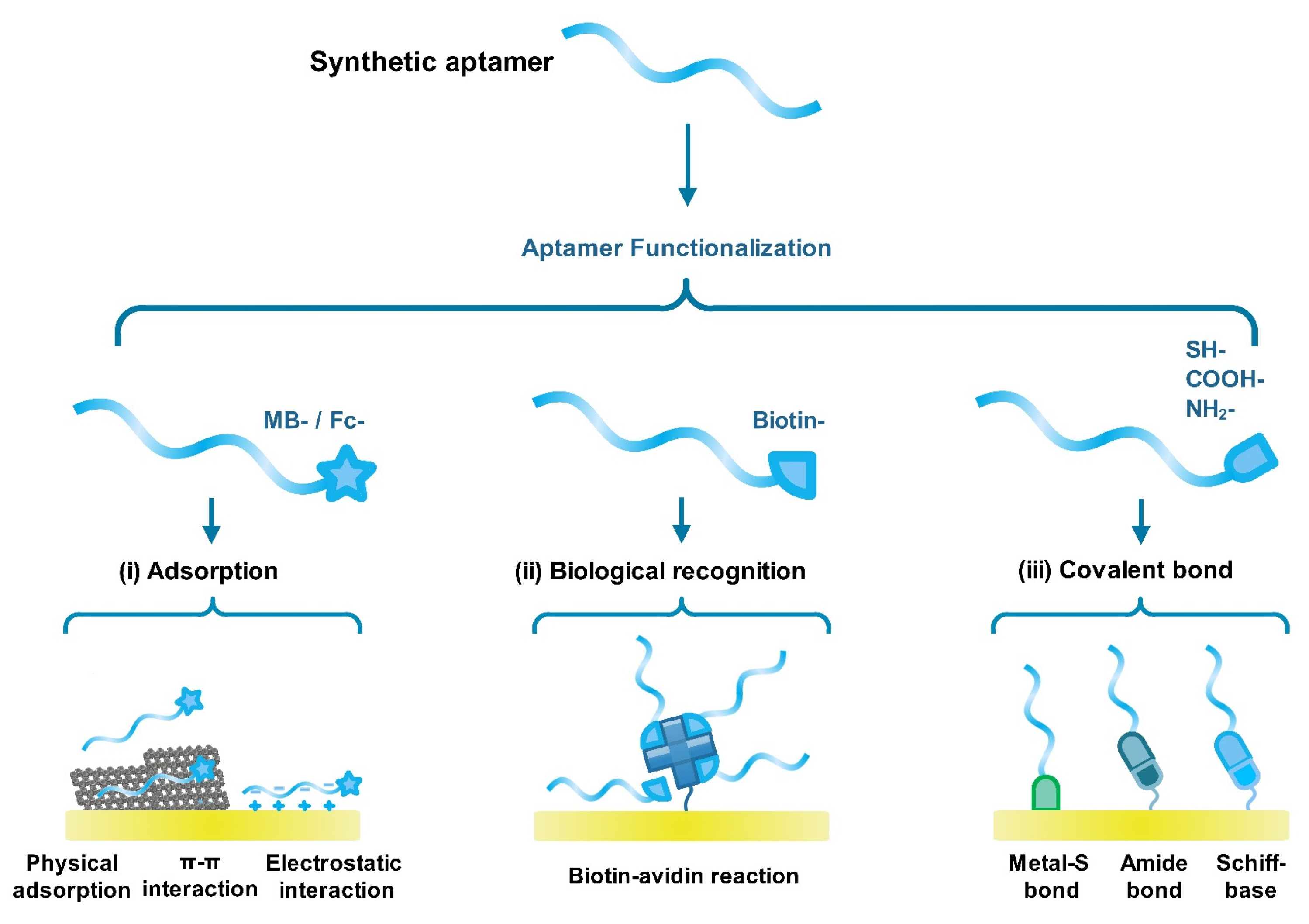
2. Immobilizations of Aptamers on the Surface of Electrode

How to efficiently immobilize aptamers on the electrode surface is a key step in the construction of electrochemical aptasensors, which greatly affects the sensing performance and application fields [20,21]. In general, electrochemical indicator- (such as methylene blue, ferrocene), thiol-, biotin-, and carboxyl-labeled aptamers have been synthesized by companies, which are designed for sensing purposes. Nowadays, three methods including adsorption, biological recognition, and covalent bonds have been widely used to construct electrochemical aptasensors (Scheme 1). The advantages and disadvantages of these immobilization methods are listed in Table 1 [22,23,24].

2.1. Adsorption Method

As a nucleic acid sequence, aptamers usually have negative charges due to their phosphate backbone. Therefore, aptamers easily adsorb on the surface of nanomaterials with positive charges via electrostatic reaction [10,34], such as metal–organic frameworks (MOFs) [35], mesoporous silica nanoparticles (MSNs) [36], and magnetic nanoparticles (MNPs) [37]. Moreover, aptamers also easily adsorb on the modified electrode surface or nanomaterials’ surface through physical adsorption or π–π interaction, such as graphene [38], carbon nanotubes [39], single-layered MoS2 [40], noble metal nanoparticles [41], etc. This immobilization method is simple, fast, and without chemical modification. However, this method has obvious shortcomings including easy detachments (sensitivity to pH, ionic strength, temperature, etc.), low density, and poor orientation on the sensing interfaces [26,27].

2.2. Biological Recognition

The biological recognition system has been recently employed to immobilize aptamers on the electrode surface and nanomaterials’ surface through affinity labeling strategies, such as the biotin–streptavidin recognition reaction [42]. Generally, one streptavidin molecule has four identical subunits, which can specifically bind up to four biotin molecules. The binding constant (Ka) of biotin–streptavidin is about 1015 mol−1, suggesting that this biological recognition is specific and efficient [43]. However, the poor conductivity of biotin and streptavidin molecules often leads to a decrease in the signal of electrochemical aptasensors, resulting in low detection sensitivity [28,29,30].

2.3. Covalent Bond

The covalent bond is a popular method for the construction of electrochemical aptasensors. To achieve this purpose, it usually requires the synthesized aptamer with specific functional groups such as thiol, amino, carboxylic acid, or phosphate groups [44,45]. Correspondingly, noble metal nanoparticles or various active groups including hydroxyl and carboxyl are introduced to the electrode surface. Through a chemical reaction, an aptamer can be efficiently and controllably immobilized on the electrode surface. This method for the construction of electrochemicals exhibits several advantages of good stability, high binding efficiency, easy regeneration, and a controllable process [31,32,33]. Meanwhile, this method required chemical modification, complicated steps, and introduced other interference factors [46,47].
Besides immobilization methods, experimental conditions are important factors to greatly affect the detection performance of electrochemical aptasensors, such as pH value, aptamer concentration, binding time between aptamer and target molecules, elution time, and temperature [48,49,50]. It should be noted that the experimental conditions are generally optimized according to the designed detection strategies and the properties of introduced nanomaterials and target molecules. Taking the pH value effect as an example, Liu et al. found that the electrochemical signal of the designed aptasensor reached a maximum value when the pH value was 7.0. Low and high pH could affect the binding efficiency of the aptamer and dopamine [51]. In another work, the optimized pH value of the developed aptasensor for Aflatoxin B1 detection was about 4.0, which is greatly dependent on the properties of the introduced titanium carbide/carboxylated graphene oxide-poly(4-vinyl pyridine) nanocomposites [52]. For the temperature effect, many electrochemical aptasensors are typically used at room temperature. Especially, electrochemical aptasensors for biological molecules’ detection are usually used at 37 degrees Celsius, the optimized temperature, which is close to normal human body temperature [53,54]. Therefore, suitable experimental conditions could result in better detection performance.

3. Application of Electrochemical Aptasensors for the Detection of Different Target Molecules

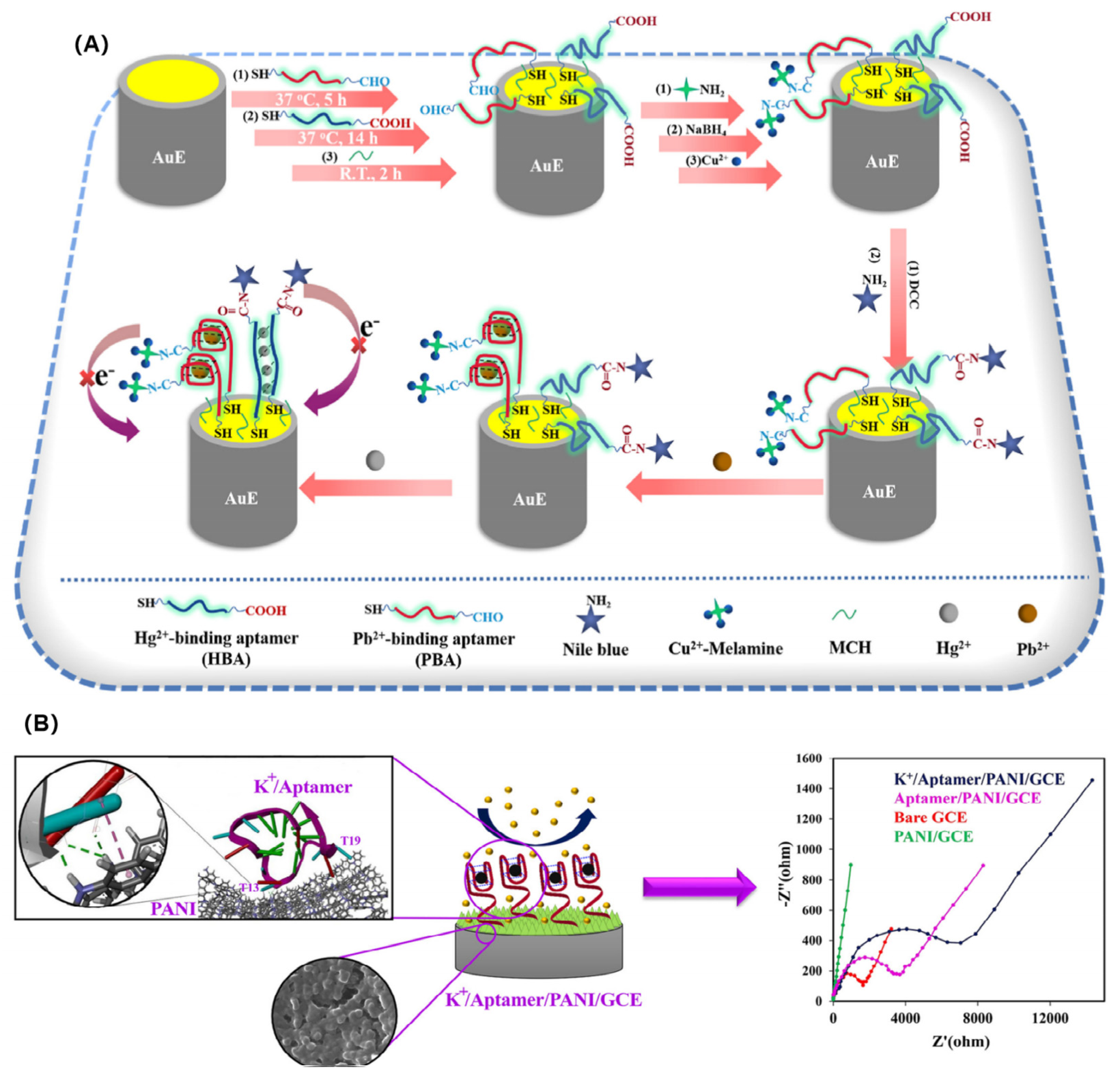
3.1. Detection of Metal Ions

The detection of metal ions is particularly important in both environmental monitoring and biomedical diagnosis [55,56]. In recent years, with the acceleration of industrialization, environmental pollutants have become an increasingly serious problem. Among them, heavy metal ions, as a kind of pollutant, are difficult to degrade under natural conditions and can be accumulated through the food chain to harm human health. Therefore, it is an urgent problem to develop rapid, simple, and efficient sensors for qualitative and quantitative analysis of heavy metal ions in the environment.
Although traditional methods (such as atomic absorption spectroscopy, inductively coupled plasma mass spectrometry, high-performance liquid chromatography, etc.) have achieved high detection performance, the high cost of instruments and the need for specialized operators have limited their widespread application. Compared to traditional analytical methods, electrochemical methods show many advantages for heavy metal ions’ detection due to their real-time detection capability, high sensitivity, fast response time, miniaturization, and integration [57,58]. To further improve detection selectivity in complex environments, electrochemical aptasensors are considered as promising detection methods for the determination of heavy metal ions [1,59]. For example, Zhang et al. designed an electrochemical aptasensor for trace heavy metal ions’ detection based on the Fe(III)-based metal–organic framework (Fe-MOF) and mesoporous Fe3O4@C nanocapsules core-shell nanocomposite (denoted as Fe-MOF@mFe3O4@mC) [60]. The designed nanocomposites showed excellent electrochemical performance and high loading of aptamers, resulting in the high performance of this aptasensor for Pb2+ and As3+ detection. Under optimal conditions, the constructed aptasensor can detect as low as 2.27 pM Pb2+ and 6.73 pM As3+, respectively. Besides the selective determination of metal ions, Gao et al. developed a dual signal-based electrochemical aptasensor for the simultaneous detection of Pb2+ and Hg2+ in environmental water samples by in situ assembling electrochemical signal tags (Cu2+-Melamine and Nile blue) on the terminal of Pb2+-binding aptamer (PBA) and Hg2+-binding aptamer (HBA), respectively (Figure 1A) [61]. Once specifically capturing target Pb2+ and Hg2+, the two electrochemical signal tags are far away from the electrode surface due to the rigidity of PBA-Pb2+ and HBA- Hg2+ secondary structures, leading to electrochemical “signal-off” with the increase in Cu2+-Melamine and Nile blue. Based on this detection mechanism, the detection limits of the developed aptasensor for Pb2+ and Hg2+ analysis are 0.98 pM and 19 pM, respectively. Moreover, the designed aptasensor could achieve the successful determination of Pb2+ and Hg2+ in real water samples, presenting its latent capacity in the monitoring of heavy metal ions.
Metal ion concentration in the body also plays an important physiological role in maintaining the balance of intracellular and extracellular fluids, as well as the excitability of nerves and muscles [62,63]. It has been found that abnormal metal ion concentration is closely related to a variety of nervous system diseases [64]. For example, copper ions (Cu2+), zinc ions (Zn2+), iron ions (Fe3+), and aluminum ions (Al3+) are involved in the pathogenesis of Alzheimer’s disease (AD) [65]. Therefore, it is significant to monitor these metal ions’ concentrations for the early diagnosis of AD patients. For the sensitive and selective determination of Zn2+, Zhao and co-workers designed an electrochemical aptasensor by fixing aptamers on the surface of bi-functional SiO2-Pt@meso-SiO2 core-shell nanoparticles [66]. The developed bi-functional nanoparticles can not only greatly enhance the conductivity of the electrode but also can pre-enrich Zn2+ onto the electrode surface. As a result, this aptasensor can determine the Zn2+ concentration as low as 65 pM. Moreover, this aptasensor can successfully detect Zn2+ in human blood and disrupted human cells. Similarly, Salehan and co-workers proposed an electrochemical aptasensor for potassium ion (K+) analysis through electrochemical impedance spectroscopy [67]. They assembled the selected aptamer [G3(T2AG3)3] onto the surface of a polyaniline (PANI) modified electrode via electrostatic adsorption. In the presence of K+, a single-strand aptamer folds into a G-quadruplex configuration, which blocks the electron transfer between the electrochemical indicator and electrode surface (Figure 1B). As a result, the charge transfer resistance (Rct) increased with the increasing concentration of K+. The linear relationship between ΔRct and the logarithm of K+ concentration is plotted ranging from 10 pM to 60 μM with a detection limit of 3.7 pM. Moreover, this developed aptasensor can efficiently determine K+ in serum samples, suggesting that it is a promising method for metal ion detection.
Figure 1. (A) Fabrication illustration of the dual signal−based electrochemical aptasensor for simultaneous analysis of Pb2+ and Hg2+. (Reproduced with permission from Ref. [61] Copyright 2022, Elsevier). (B) Scheme of the impedimetric biosensor for the determination of potassium ions based on polyaniline/GCE and DNA G-quadruplex conformation. (Reproduced with permission from Ref. [67] Copyright 2022, Elsevier).
Chemosensors 11 00488 g001

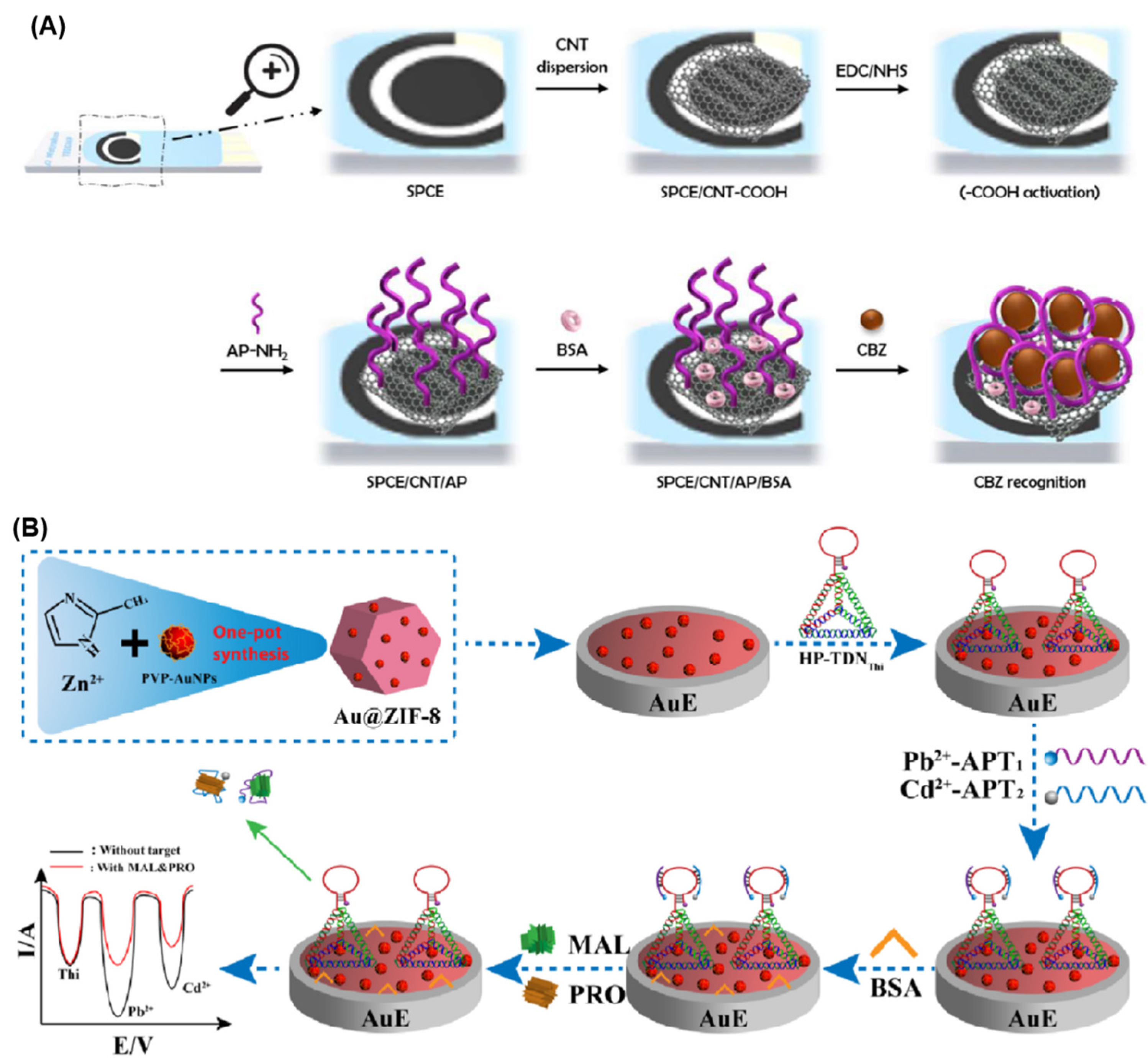
3.2. Detection of Pesticides

Over decades, the over-range and excessive dosage use of pesticides not only causes environmental pollution but also directly or indirectly threatens human health due to the long-term accumulation of residues. Nowadays, gas chromatography (GC), high-performance liquid chromatography (HPLC), GC-mass spectrometry (MS), and fluorescence and electrochemistry methods have been widely used to determine pesticides with accepted sensitivity, selectivity, and efficiency [68]. It is noted that some methods have disadvantages in the analysis of pesticides, such as low sensitivity, poor selectivity, and high cost [69]. Therefore, it is necessary to develop new methods for the sensitive and efficient detection of pesticides.
Electrochemical aptasensors have efficiently addressed these issues in pesticide detection. For example, Venegas and co-workers developed a label-free electrochemical aptasensor for the selective detection of carbendazim (CBZ) [70]. As shown in Figure 2A, the oxidized carbon nanotubes (CNT-COOH) were deposited on the surface of screen-printed carbon electrodes (SPCE) via physical adsorption. Then, amine-labeled aptamer (AP-NH2) was assembled on the surface of SPCE via covalent immobilization. In the presence of CBZ, the corresponding aptamers specifically capture analytes to form secondary structures, causing a decrease in the electrochemical signal due to the blocking of electron transfer. According to this phenomenon, this proposed electrochemical aptasensor can detect values as low as 4.35 nM (0.83 ng/mL) CBZ, which is comparable to the HPLC method. Moreover, this aptasensor can efficiently determine CBZ in tomatoes, proving that the aptasensor can detect pesticides in food samples.
Electrochemical aptasensors also can simultaneously determine pesticides. Liu et al. designed a dual-ratiometric electrochemical aptasensor for the simultaneous detection of malathion (MAL) and profenofos (PRO) [71]. To obtain signal amplification, they firstly fabricated gold nanoparticle (AuNP)-encapsulated zeolitic imidazolate framework (ZIF-8) nanocomposites (Au@ZIF-8) to improve the immobilization amount of hairpin-tetrahedral DNA nanostructures (HP-TDN) and facilitate the electron transfer between the DNA probe and the electrode surface (Figure 2B). Then, metal ions (Pb2+ and Cd2+) served as signal tracers to further amplify detection signal. Pb2+-functionalized MAL aptamer (Pb2+-APT1) and Cd2+-functionalized PRO aptamer (Cd2+-APT2) were co-assembled on the surface of thionine-labeled DNA nanostructures (HP-TDNThi) to specifically recognize MAL and PRO, respectively. With the addition of MAL and PRO, Pb2+-APT1 and Cd2+-APT2 dissociated from the surface of HP-TDNThi, leading to a decrease in both IPb2+/IThi and ICd2+/IThi oxidation current ratios. The detection limits of this aptasensor for MAL and PRO detection can reach 4.3 pg/mL and 13.3 pg/mL under optimized conditions, respectively. The obtained high performance suggested that this work may pave a way to develop aptasensors for multiple pesticides’ detection in food safety and environmental monitoring.

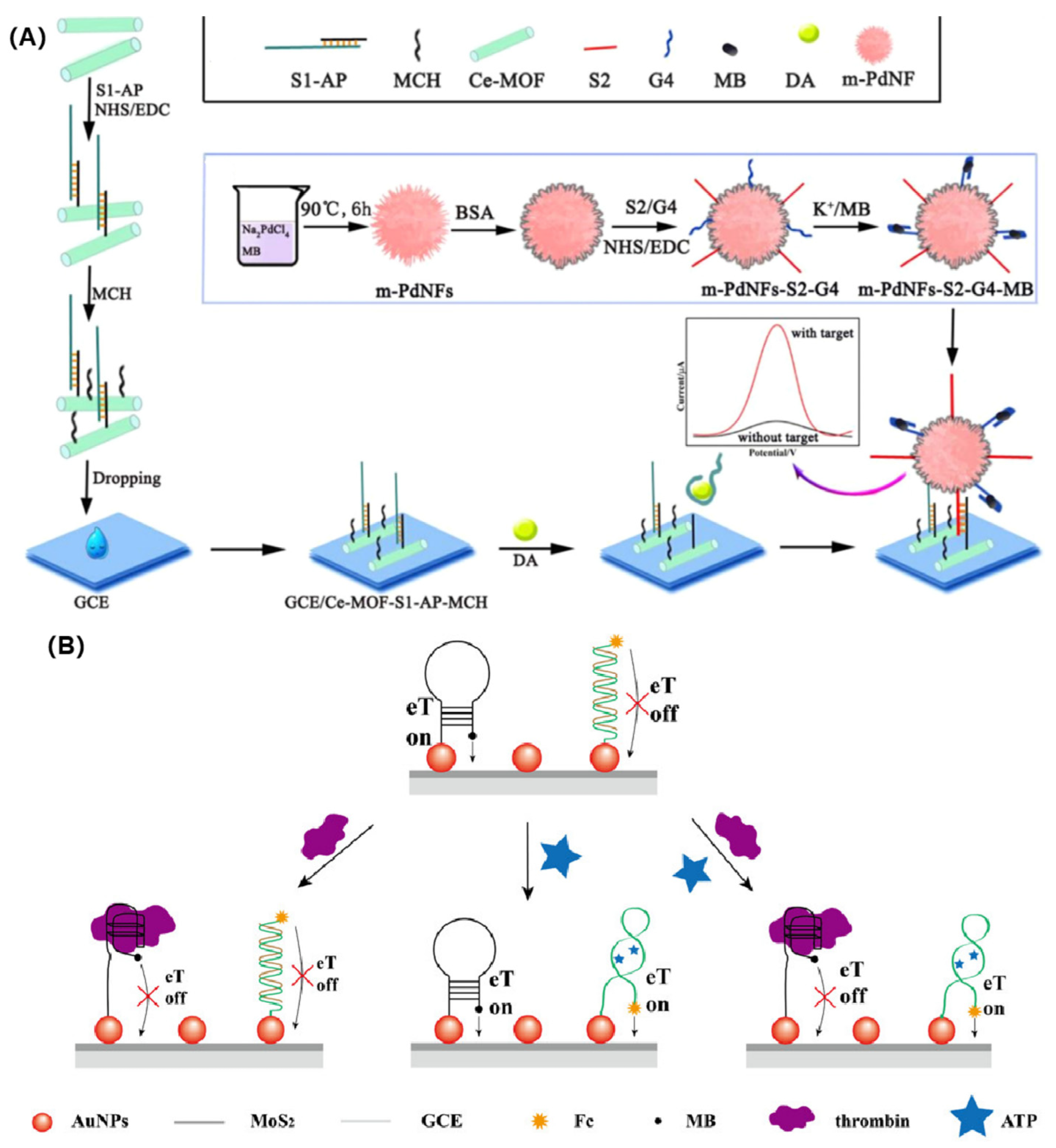
3.3. Detection of Small Biological Molecules

Physiological metabolic activities of living organisms are closely related to various biological small molecules, such as neurotransmitters, adenosine triphosphate (ATP), nicotinamide adenine dinucleotide (NADH), hydrogen peroxide (H2O2), etc. [72,73,74]. Some small molecules including neurotransmitters and H2O2 can be directly detected by electrochemical sensors. However, direct detection is susceptible to interference from other electrochemical active molecules, resulting in low selectivity and sensitivity. Therefore, the biological recognition reaction is a promising tool to construct an electrochemical sensing platform for some biological small molecules’ detection. According to this concept, electrochemical immunosensors and aptasensors have been widely developed to analyze ATP, dopamine (DA), uric acid (AA), 8-hydroxy-2′-deoxyguanosine (8-OHDG), etc. [75,76]. Compared with electrochemical immunosensors, electrochemical aptasensors attracted more and more attention due to their easy design, low cost, and controllable assembly.
Taking DA as an example, many electrochemical biosensors have been developed to directly determine it [77,78]. However, the co-existing AA, norepinephrine, adrenaline, and isoproterenol often affect the analytical performance of those designed biosensors. To improve the sensitivity and selectivity of electrochemical biosensors for dopamine detection, Zhang et al. used aptamer as a recognition unit, methylene blue-integrated m-PdNFs as a signal amplification unit, and Ce-MOF as an electrode-modified unit to construct a novel electrochemical aptasensor [79]. As shown in Figure 3A, a single-stranded DNA (S2) was assembled on the surface of the designed m-PdNFs-G4-MBs nanocomposites to obtain signal probes (m-PdNFs-S2-G4-MBs), which can offer electrochemical signals for detection. In the absence of DA, the anti-DA aptamer was hybridized with a single-stranded DNA (S1), which was further co-immobilized on the surface of the Ce-MOF nanocomposite-modified electrode. At this moment, the m-PdNFs-S2-G4-MBs signal probe cannot be assembled on the electrode surface via a DNA hybridization reaction. As a result, no electrochemical signal was obtained. In the presence of DA, DA preferentially binds with the aptamer to form the DA–aptamer complex, leading to the destruction of the aptamer-S1 double-strand structure. Therefore, the exposed S1 can be hybridized with the m-PdNFs-S2-G4-MBs signal probe to form double-stranded structures, generating significant electrochemical signals. Due to the extraordinary conductivity of Ce-MOF and the signal amplification of m-PdNFs-S2-G4-MBs, the designed electrochemical aptasensor achieved a low detection limit of 6 pM for DA detection. The practical DA detection result is highly consistent with the UPLC-MS method, suggesting that this aptasensor has high sensitivity and specificity.
Another example was given by Su et al. [80]. They constructed an electrochemical aptasensor for the simultaneous detection of ATP and thrombin (Figure 3B). They utilized the advantages of molybdenum disulfide (MoS2) and noble metallic nanoparticles to synthesize MoS2-based nanocomposites. After co-immobilizing anti-ATP and anti-thrombin aptamers on the surface of the MoS2-based nanocomposite-modified electrode, a dual-target electrochemical aptasensor was constructed. Once the binding with ATP and thrombin is completed, the structures of two aptamers are changed, resulting in the labeled ferrocene (Fc) and methylene blue (MB) close to and far away from the electrode surface. Correspondingly, the electrochemical signal increased and decreased with the addition of ATP and thrombin, respectively. In other words, the designed aptasensor featured both “signal-on” and “signal-off” sensing mechanisms for the detection of ATP and thrombin, respectively. Under optimal conditions, this proposed aptasensor can simultaneously determine values as low as 0.74 nM ATP and 0.0012 nM thrombin. Furthermore, they employed the aptasensor to construct an “AND” logic gate platform for ATP and thrombin detection, which demonstrated a general methodology for the advancement of the highly sensitive and selective detection of various aptamer-specific binding targets.

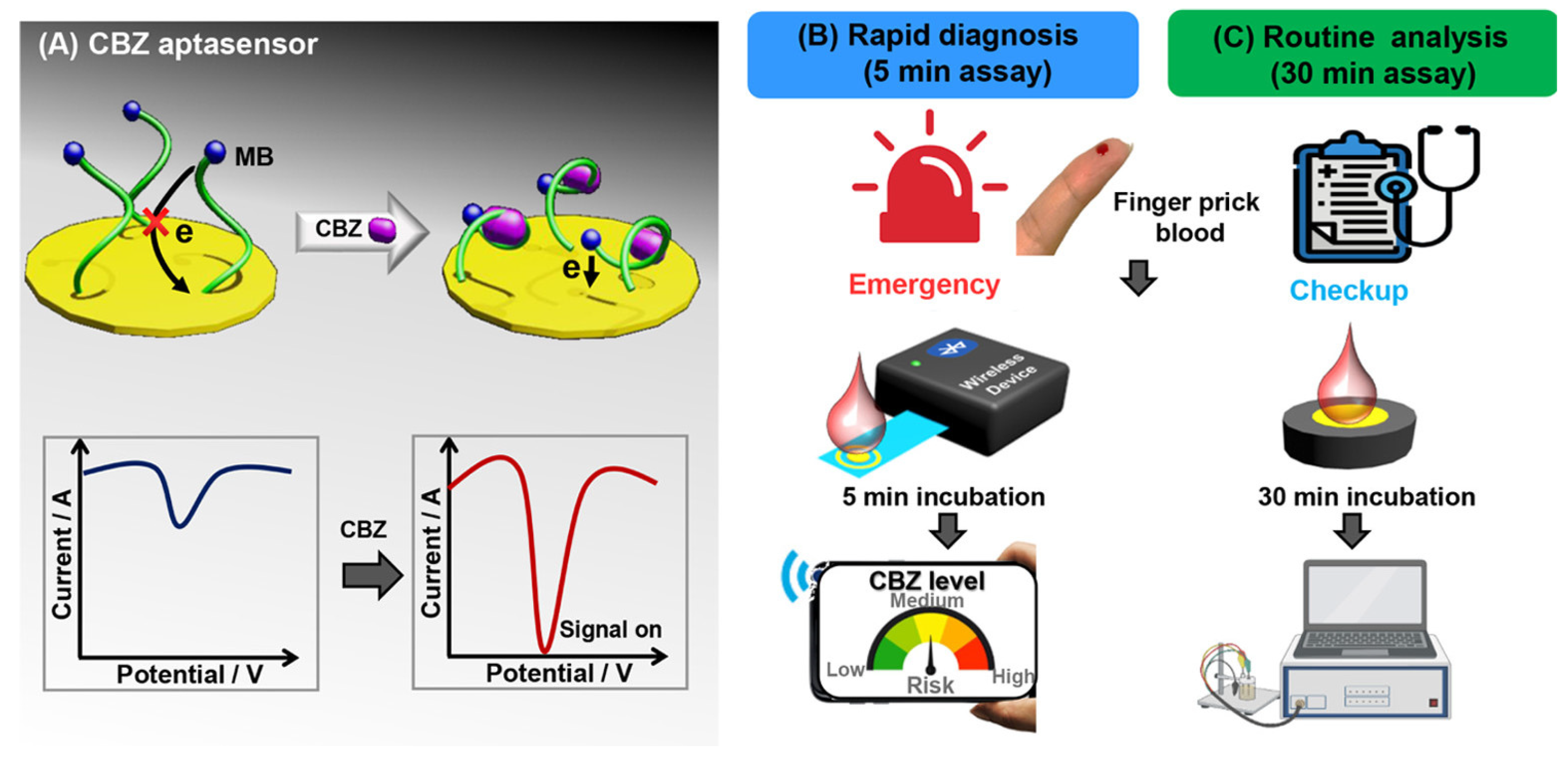
3.4. Detection of Drug and Antibiotic Molecules

As we know, drugs and antibiotics greatly ensure the health of living organisms. However, the abuse of drugs and antibiotics, as well as their concentration-related toxicity, can endanger human health and even lives. Therefore, accurate, fast, and low-cost detection of drugs and antibiotics plays a vital role in disease diagnosis, treatment evaluation, and monitoring after operation.
An electrochemical aptasensor is also a powerful tool for the detection of drugs. For example, Chung et al. chose a de novo aptamer to develop an electrochemical aptasensor for the analysis of the anti-epileptic drug carbamazepine (CBZ) (Figure 4) [81]. The selected aptamer specifically binds with CBZ (Kd < 12 nM) to form a G-quadruplex structure, leading to the labeled MB close to the electrode surface. Correspondingly, the electron transfer between MB and the electrode is facilitated, increasing the electrochemical signal. It should be noted that the designed aptasensor has two functional modules: a 30 min assay for routine monitoring and a 5 min assay for rapid emergency testing. Under optimal conditions, this aptasensor exhibits a wide dynamic range (10 nM to 100 μM) and low detection limits of 1.25 nM and 1.82 nM for 5 min and 30 min analysis, respectively. The practical testing in the pricking blood sample (<50 μL) for CBZ determination proved the clinical applicability of this electrochemical aptasensor, suggesting that it has promising potential in point-of-care testing of personalized CBZ dosing. Using a similar sensing mechanism, cocaine, diclofenac, and amphetamine can be sensitively and selectively detected by electrochemical aptasensors without introducing other signal indicators [82,83,84].
Besides drugs, electrochemical aptasensors are also widely employed to detect antibiotics [85]. A typical example is offered by Sun’s group. They designed an electrochemical aptasensor for the ultrasensitive detection of neomycin by using a dual-signal amplification strategy [86]. In this sensing strategy, Fc-decorated multi-walled carbon nanotubes (MWCNTs) and the silica hybridized mesoporous ferroferric oxide nanoparticles (Fe3O4@SiO2) were co-immobilized on the gold electrode surface to improve the electronic signal, which was used to load more RNA aptamers (Apt1) and facilitate electron transfer. Another RNA aptamer (Apt2) was assembled on the surface of gold nanoparticles to form a nanoprobe for signal amplification. In the presence of neomycin, a classical sandwiched structure was formed on the electrode surface, generating an obvious electrochemical signal. Due to the synergistic effect of different nanomaterials, this electrochemical aptasensor can detect 0.759 nM neomycin with high specificity and selectivity. Moreover, this aptasensor can efficiently determine neomycin in milk. Similarly, Emaminejad’s, Wang’s, Du’s, and Roushani’s groups developed a series of electrochemical aptasensors for the detection of tobramycin [87], kanamycin [88], enrofloxacin [89], and streptomycin [90], respectively.

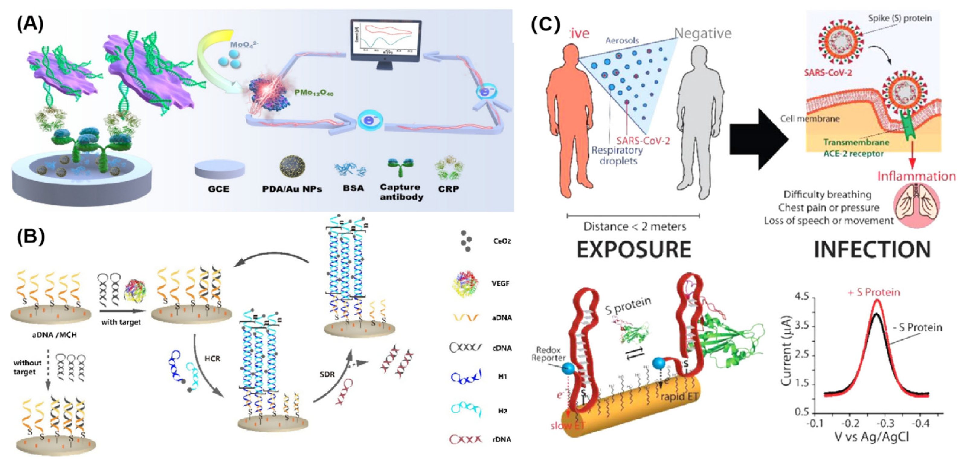
3.5. Detection of Proteins

Immunosensors coupled with different detection techniques have become universal tools for the detection of proteins, which have been widely applied in early diagnosis and clinical treatment of diseases [91]. However, antibody-based immunosensors have some disadvantages including the limited source of antibodies, high cost, disordered assembly on sensing interfaces, and the loss of biological activity [92], which greatly affect their detection performance and limit their further application. Therefore, electrochemical aptasensors have attracted more and more attention in protein detection due to their outstanding advantages, such as low cost, easy preparation, ordered assembly on the sensing interfaces, and anti-environment disturbance ability.
Highly efficient analytical approaches are of great significance for patients to realize early and rapid diagnosis, receive appropriate and timely treatment, and reduce mortality. Currently, C-reactive protein (CRP) has been identified as an angiocardiopathy special indicator for early diagnosis of cardiovascular disease with a high incidence rate [93]. Li and his colleagues have constructed an enzyme-free sandwich-type aptasensor for sensitive CRP detection based on Mn3(PO4)2/CRP aptamer biomineralized nanosheets and polydopamine/Au nanospheres (PDA/Au NPs) (Figure 5A) [94]. The designed Mn3(PO4)2/CRP aptamer biomineralized nanosheets are not only used as recognition units for capturing target CRP, but also serve as signal enhancers to improve electrochemical responses. Coupled with the signal amplification nanoprobe of PDA/Au NPs, the aptasensor exhibits excellent detection performance with a wide linear range (1 pg/mL–1 ng/mL) and a low detection limit (0.37 pg/mL). The repeatability, specificity, and reliability of this aptasensor are superior to that of the traditional immunoturbidimetry assay, suggesting that it has great prospects in clinical diagnostic applications of cardiovascular disease. Vascular endothelial growth factor (VEGF) is a key biomarker of diabetic retinopathy (DR), which can be also determined by electrochemical aptasensors [95]. For instance, Mei et al. designed a noninvasive electrochemical aptasensor to detect VEGF in tears [96]. They developed a cascade signal amplification strategy by combining a hybridization chain reaction (HCR) with CeO2 nanoparticles to improve the analytical performance (Figure 5B). The introduced CeO2 nanoparticles can efficiently catalyze H2O2 to generate electrochemical signals. Under optimal conditions, the aptasensor can detect 1 fg/mL~0.1 ng/mL VEGF with a detection limit of 0.27 fg/mL. Based on the acceptable stability and reproducibility, the proposed aptasensor can accurately analyze VEGF in tears comparable to the ELISA method, indicating that it is a potential tool for early diagnosis and monitoring of DR. Similarly, Li’s, Hosseini’s, and Gao’s teams have constructed various electrochemical sensing platforms coupled with aptamers and a signal amplification strategy for different protein biomarkers’ detection, including thrombin [97], tumor necrosis factor α [98], and tau protein [99], respectively.
Electrochemical aptasensors have also been widely used to monitor infectious diseases, such as SARS-CoV-2 [100], malaria [101], tuberculosis [102], etc. As shown in Figure 5C, Idili et al. have developed a simple electrochemical aptasensor for the detection of SARS CoV-2 S protein in undiluted samples (serum and artificial saliva) [103]. Typically, the added SARS CoV-2 S protein induced the change in the methylene blue derivative-labeled aptamer conformation, resulting in the electrochemical indicator close to the electrode surface. Correspondingly, a concentration-related electrochemical signal is generated for the qualitative and quantitative detection of SARS CoV-2 S protein. This electrochemical aptasensor is simple (single step) and fast (detection time < 5 min), suggesting that it is an ideal device for COVID-19 monitoring. Another typical example was offered by Rahmati et al. They constructed a label-free electrochemical aptasensor for the analysis of SARS-CoV-2 spike glycoprotein based on a copper hydroxide nanorod-modified screen-printed carbon electrode (SPCE) [104]. The obtained SARS-CoV-2 spike glycoprotein–aptamer complex blocked the electron transfer, leading to a decrease in the electrochemical signal. Based on this phenomenon, the designed aptasensor can efficiently detect SARS-CoV-2 spike glycoprotein in buffer, labeled saliva samples, and actual clinical samples. It is noted that the analytical performance of this aptasensor is comparable to the PCR results, proving that it has great potential in rapid and on-site COVID-19 monitoring.
Figure 5. (A) The preparation of the Mn3(PO4)2/aptamer hybrid nanosheet−based electrochemical aptasensor for CRP detection. (Reproduced with permission from Ref. [94] Copyright 2021, Elsevier). (B) Schematic diagram of electrochemical aptasensor coupled with signal amplification strategy for sensitive detection of VEGF. (Reproduced with permission from Ref. [96] Copyright 2021, Elsevier). (C) Schematic of electrochemical aptasensor for the rapid and efficient detection of the SARS-COV-2 spike protein. (Reproduced with permission from Ref. [103] Copyright 2021, American Chemical Society).
Chemosensors 11 00488 g005

3.6. Detection of Exosomes

As nanoscale extracellular vesicles, exosomes play an important role in promoting tumor growth, migration, invasion, and information transfer between cells [105]. Therefore, exosomes are considered as excellent biomarkers for early diagnosis of cancer [106]. However, it is still a challenge to sensitively and selectively detect exosomes due to their low content in biological fluids during the early stages of cancer [107,108]. In recent years, electrochemical aptasensors have been gradually employed to detect exosomes due to their advantages. Coupled with signal amplification strategies, the analytical performance of electrochemical aptasensors can meet the need for exosome detection [109].
Zhang’s group developed an electrochemical aptasensor for exosome detection based on a microelectrode and HCR amplification strategy [110]. They introduced EpCAM aptamers to selectively recognize and capture EpCAM-positive cancerous exosomes. Due to the HCR reaction, a large number of avidin–horseradish peroxidase (HRP) molecules were assembled on the microelectrode surface, which can catalyze the 3,3′,5,5′-tetramethylbenzidine (TMB)+H2O2 reaction strategy to generate a large electrochemical signal. Due to the synergetic effect of the enzyme catalytic reaction, HCR reaction, and microelectrode, this aptasensor has a detection limit as low as 5 × 10² exosomes per milliliter. Moreover, this aptasensor can successfully detect cancerous extracellular vesicles in serum samples of early- and late-stage lung cancer patients. Chang et al. used programmed cell death ligand 1 protein-positive (PD-L1+) exosome as a biomarker to evaluate the diagnosis of non-small cell lung cancer (NSCLC) [111]. They integrated the specific recognition ability of the aptamer, the excellent peroxidase-like catalytic activity of palladium–copper–boron alloy microporous nanospheres (PdCuB MNs), and the high conductivity of Au@CuCl2 nanowires (NWs) to construct an electrochemical aptasensor for detecting PD-L1+ exosomes (Figure 6A). In the presence of PD-L1+ exosomes, a classical sandwiched structure was formed on the electrode surface. The introduced PdCuB MNs efficiently catalyze H2O2 to generate a significant electrochemical signal. According to this result, the aptasensor can detect 1 × 10²–1 × 10⁸ particles/mL exosomes with a low detection limit of 36 particles/mL. Results obtained from clinical samples suggested that this aptasensor has a potential application in the early diagnosis of NSCLC.
Besides captured probes’ immobilized mode, Li et al. developed a dual-aptamer recognition system for precise capture and direct analysis of tumor-specific exosomes without immobilization [112]. They assembled both CD63 and mucin 1 (MUC1) aptamers onto the surface of amino-functionalized Fe3O4@SiO2 nanoparticles to construct dual-aptamer-modified nanoprobes (Fe3O4@SiO2-dual Apt). As shown in Figure 6B, the dual-aptamer-modified nanoprobes can specifically capture target exosomes due to the recognition ability of two aptamers. After magnetic separation, cholesterol-labeled DNA probes (P3) can anchor on the surface of exosomes and activate the hyperbranched hybridization chain reaction, generating a magnetic bead–exosome–hyperbranched DNA superstructure (Fe3O4@SiO2-exosome-HDS). This superstructure can adsorb a large number of Ru(NH3)63+(Ru (III)) molecules, resulting in a few unbound Ru (III) remaining in the supernatant after magnetic separation. Therefore, the redox reaction between Ru (II) and [Fe(CN)6]3− (Fe (III)) is inhibited, leading to a significant increase in IFe(III)/IRu(III). The ratio of the electrochemical signal was related to exosomes, which can be used to quantitatively and qualitatively analyze target exosomes. Consequently, this ratiometric dual-signal electrochemical aptasensor exhibits high reliability, accuracy, and easy manipulation, which can sensitively detect tumor exosomes in complex environments and human serum samples.

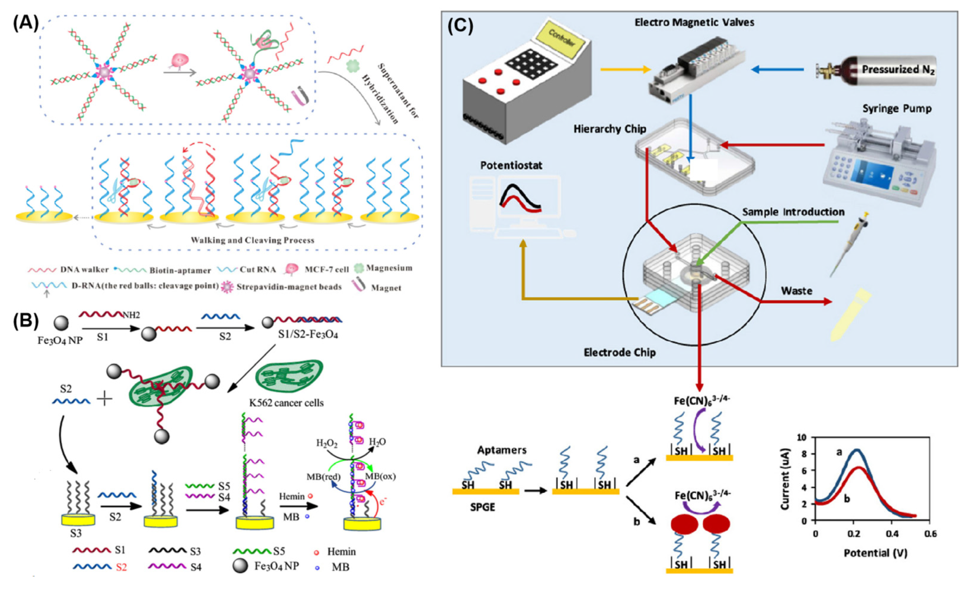
3.7. Detection of Tumor Cells

It is well known that the incidence rate and mortality of cancer have increased year by year, which has brought a huge threat to human health. Thus, the identification and detection of cancer cells in patients’ tumor lesions and circulating blood is crucial for early clinical diagnosis, toxicity monitoring, and public health protection [113,114]. However, conventional clinical, imaging, and other diagnostic methods often have weak sensitivity and specificity due to the complex etiology of tumors, leading to misdiagnosis and missed diagnosis [115,116]. Although a variety of technologies including PCR-based methods [117], cell counting methods [118], and fluorescence determination [119] have been presented, many disadvantages of these methods have limited their wide application, such as expensive fluorescent labeling reagents, cumbersome processes, and the need for advanced instruments and professionals. Therefore, it is necessary to develop reliable, cost-effective, sensitive, and easy to be clinically popularized detection tools for high-precision diagnosis of cancer cells.
Electrochemical aptasensors have received widespread attention in the high-performance detection of tumor cells due to their significant advantages, such as high sensitivity, simplicity, rapid response, reusability, and low cost [120]. An impressive example was given by Cai et al. [121]. As shown in Figure 7A, a double-stranded DNA formed by a biotin-labeled aptamer and DNA walker was assembled on the surface of the streptavidin-modified magnetic microsphere to construct nanoprobe, which was used to recognize and capture target cancer cells. Once capturing target breast cancer MCF-7 cells, an MCF-7–aptamer complex was formed and a DNA walker was released from the surface of the nanoprobe. The released DNA walker (DNAzyme) repeatedly cleaved D-RNA with the help of Mg2+ to produce a large number of short nucleic acids, leading to a great decrease in electrochemical signals. With DNAzyme-assisted signal amplification, the designed aptasensor can analyze as low as 47 cells/mL MCF-7 cells. G-quadruplex DNAzyme is also widely used for signal amplification to construct electrochemical aptasensors for cancer cell detection. Chen and co-workers designed a super sandwich G-quadruplex DNAzyme to construct an electrochemical aptasensor for the highly sensitive detection of cancer cells [122]. In Figure 7B, amino-modified aptamers (S1) and their complementary DNA sequence (S2) were immobilized onto the surface of magnetic beads (Fe3O4 NPs) to form S2/S1/Fe3O4 conjugates, which were used to specifically capture K562 cells. Once encountering K562 cells, the double-stranded S1–S2 structure was dissociated due to the stronger affinity between K562 cells and S1. As a result, S2 was released from the surface of Fe3O4 NPs and hybridized with a capture probe (S3) on the electrode surface. The S2–S3 structure triggered a hybridization cascade between S4 and S5, resulting in the super sandwich structure. In the presence of Hemin, a large number of G-quadruplex DNAzymes were formed on the electrode surface. Finally, the G-quadruplex DNAzyme catalyzes the reduction of H2O2 by MB, leading to a dramatically amplified electrochemical signal. Under optimal conditions, the detection limit of the as-fabricated aptasensor was estimated to be 14 cells/mL K562 cells.
Electrochemical aptasensors are also extensively employed to detect circulating tumor cells (CTCs), which are of great significance for the clinical diagnosis, metastasis, and prognosis of tumors [123,124]. Shaegh et al. reported a novel microfluidic electrochemical aptasensor by coupling a microchip with a screen-printed gold electrode for detecting A549 cells, which is considered as a CTC model [125]. As shown in Figure 7C, the added A549 cells were specifically bound with the aptamer to form a complex on the electrode surface, which blocked the electron transfer between [Fe(CN)6]3−/4− and the electrode surface. As a result, the electrochemical signal is decreased with the increasing concentration of A549 cells. According to this phenomenon, this biosensor can sensitively detect 50~5 × 10⁵ cells/mL A549 cells with a low detection limit of 14 cells/mL. More importantly, this microfluidic aptasensor can effectively determine A549 cells in complex matrices (such as human serum), providing that this sensing platform can be used to detect low-abundance biomarkers obtained from liquid biopsies.
Figure 7. (A) Schematic illustration of the electrochemical aptasensor for MCF-7 cells’ detection based on a DNA walker signal amplification strategy. (Reproduced with permission from Ref. [121] Copyright 2016, Elsevier). (B) Schematic representation of signal-amplified G-quadruplex DNAzyme for ultrasensitive detection of K562 cells. (Reproduced with permission from Ref. [122] Copyright 2015, Elsevier). (C) Schematic illustration of microfluidic electrochemical aptasensor and its electrochemical signal (a) in the absence of and (b) in the presence of A549 cells. (Reproduced with permission from Ref. [125] Copyright 2023, Elsevier).
Chemosensors 11 00488 g007

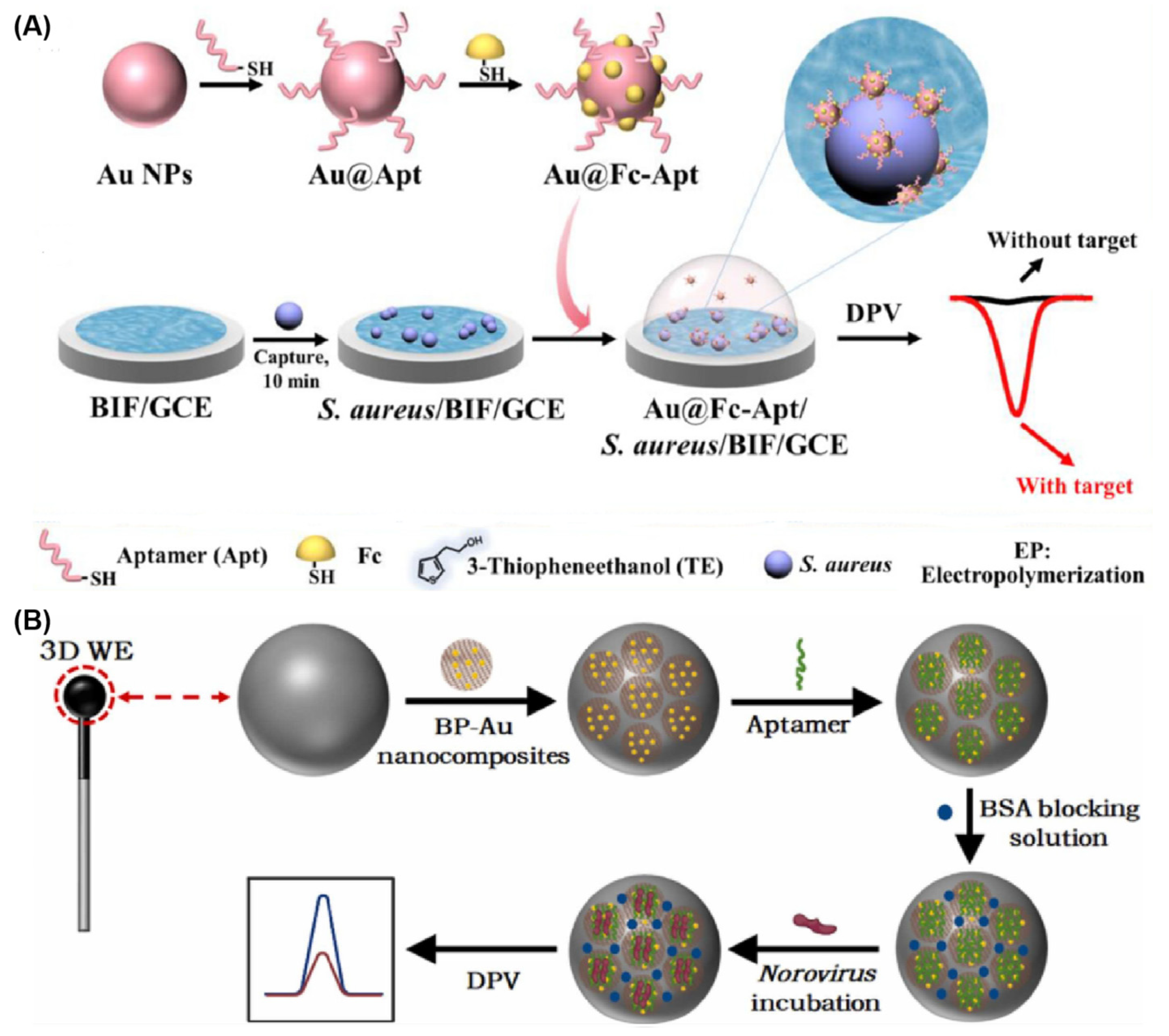
3.8. Detection of Microbial Pathogens

Since the COVID-19 pandemic, infectious diseases have gained people’s attention. Accurate and fast detection of pathogenic microorganisms is of great significance in controlling and preventing microbial infectious diseases. At present, direct smear microscopy, isolation and culture, serological reaction, and PCR are the main methods for the rapid identification and detection of pathogenic microorganisms [126]. However, some limitations of these methods exist, such as being time-consuming, requiring professional personnel and instruments, having low sensitivity, and so on. Therefore, it is urgent to develop simple, rapid, sensitive, and on-site methods for the detection of microbial pathogens.
To meet these requirements, a series of electrochemical aptasensors have been developed for the high-performance analysis of microbial pathogens due to their outstanding advantages. Bian et al. have constructed an electrochemical aptasensor for highly selective and ultrasensitive detection of single-cell levels of bacteria [127]. In Figure 8A, Au NPs modified with aptamers and 6-(Ferrocenyl) hexanethiol molecules (Au@Fc-Apt) and bacteria-imprinted polymer films (BIFs) were employed as the signal nanoprobe and the capture probe, respectively. In the presence of Staphylococcus aureus (S. aureus), a classical sandwiched structure was formed on the electrode surface. As a result, an obvious electrochemical signal is generated from the assembled Au@Fc-Apt signal nanoprobe, which can be used to qualitatively and quantitatively detect bacteria. In view of this, the aptasensor could detect single-cell level and 10 CFU mL−1 S. aureus in an ideal buffer and a complex milk sample, respectively. Moreover, this proposed aptasensor can efficiently distinguish S. aureus from the multiple coexisting interfering bacteria, suggesting that this aptasensor has outstanding selectivity.
Besides bacteria detection, electrochemical aptasensors have also been widely used to detect viruses. For fast identification and detection of norovirus, Jiang et al. developed a 3D electrochemical aptasensor for sensitive detection of norovirus based on a movable spherical working electrode (WE) decorated with phosphorene–gold nanocomposites (BP-AuNCs) [128] (Figure 8B). The design of a movable spherical WE is beneficial for increasing surface area, simplifying sampling, and avoiding cross-contamination. The added norovirus specifically binds with aptamer and induces the change in aptamer structure, leading to the decrease in the electrochemical signal. As expected, the 3D electrochemical aptasensor can detect 1 ng/mL–10 µg/mL norovirus with a detection limit of 0.28 ng/mL. The recovery of this aptasensor for norovirus detection in spiked oyster samples ranges from 97.2% to 103.7%, suggesting that this aptasensor has a potential practical application. The proposed aptasensor for norovirus detection provides a facile, low-cost, highly sensitive, and selective platform, which is potentially applied in the fields of food safety and clinical diagnosis. Besides norovirus, avian influenza virus [129], SARS-CoV-2 virus [130], and dengue virus [131] have also been analyzed by electrochemical aptasensors.

4. Prospects and Challenges of Electrochemical Aptasensors

Nowadays, electrochemical aptasensors have achieved great advances in environmental monitoring, drug safety, biochemical analysis, disease diagnosis, and public safety due to their outstanding advantages of fast response, simple preparation, easy design, high specificity for chemical and biological molecules, excellent chemical stability, and low cost. Inspired by the above exciting progresses, integrating electrochemical aptasensors into portable, wearable, and implantable devices has become a popular trend for their application in POCT and real-time monitoring fields, which has the potential to completely change diagnosis and health management, especially in application scenarios for home users or remote areas where it is difficult to receive hospital nursing care [132]. In addition, with the fast development of nanotechnologies and miniaturization technologies, as well as the urgent need for various detection strategies, electrochemical aptasensors have broad applications and bright prospects in the future.
For achieving these goals, some challenges of electrochemical aptasensors should be solved: (1) For broad applications, more aptamers should be selected, which play a vital role in the detection of various chemical and biological molecules. Nowadays, hundreds of aptamers have been selected to specifically recognize target chemical/biological molecules. However, it is not enough for sensing/biosensing applications in many fields. Therefore, the selection of more aptamers with high binding affinity can promote the development of electrochemical aptasensors. (2) The integration of cutting-edge innovative technologies (e.g., DNAzyme, CRISPR, microfluidic chips, etc.) into electrochemical aptasensors is very prospective to design novel aptasensor devices, which can efficiently enlarge their application [133]. (3) The construction of suitable signal amplification strategies is a vital effect factor to construct high-performance electrochemical aptasensors. Up to now, several signal amplification strategies have been used to construct electrochemical aptasensors, such as loop-mediated isothermal amplification, rolling circle amplification (RCA), HCR, catalyzed hairpin assembly (CHA), enzyme-assisted signal amplification, and nanoprobe-based signal amplification, which can efficiently amplify the detection signal and reach lower detection limits [134]. Therefore, how to choose a signal amplification strategy greatly depends on the analytes and application scenarios. (4) For the POCT and real-time monitoring applications, it is still a challenge to construct a portable and intelligent electrochemical aptasensor by combining the detection instruments and signal communication technologies. To quickly keep up with global health issues, electrochemical aptasensors should gradually being clinically transformed and adapted to market demands. Therefore, the development of home-care, low-cost, real-time, and portable devices has become a popular and attractive research field [135,136]. In a word, electrochemical aptasensors will be an ideal sensing platform for sensitive, selective, real-time, and on-site detection of target molecules in complex environments.

Author Contributions

Conceptualization, R.Y. and S.S.; methodology, R.Y. and J.C.; formal analysis, J.C. and H.M.; investigation, R.Y., H.M., and Y.L.; writing—original draft preparation, R.Y.; writing—review and editing, S.S. and Y.L.; supervision, S.S. and L.W.; project administration, S.S. and L.W.; funding acquisition, L.W. and S.S. All authors have read and agreed to the published version of the manuscript.

Funding

This work is supported by the Leading-edge Technology Program of Jiangsu Natural Science Foundation (Grant No. BK20212012), the “Belt and Road” Innovation Cooperation Project of Jiangsu (Grant No. BZ2022011), the “Six Talents Peak” Foundation of Jiangsu Province (Grant No. SWYY-046), the Natural Science Foundation of the Jiangsu Higher Education Institutions (Grant No. 22KJA150003), and the Natural Science Research Start-up Foundation of Recruiting Talents of Nanjing University of Posts and Telecommunications (Grant No. NY222077).

Institutional Review Board Statement

Not applicable.

Informed Consent Statement

Not applicable.

Data Availability Statement

Not applicable.

Conflicts of Interest

The authors declare no conflict of interest.

References

  1. Umapathi, R.; Ghoreishian, S.M.; Sonwal, S.; Rani, G.M.; Huh, Y.S. Portable electrochemical sensing methodologies for on-site detection of pesticide residues in fruits and vegetables. Coord. Chem. Rev. 2022, 453, 214305. [Google Scholar] [CrossRef]
  2. Baranwal, J.; Barse, B.; Gatto, G.; Broncova, G.; Kumar, A. Electrochemical sensors and their applications: A review. Chemosensors 2022, 10, 363. [Google Scholar] [CrossRef]
  3. Gong, L.; Feng, L.; Zheng, Y.; Luo, Y.; Zhu, D.; Chao, J.; Su, S.; Wang, L. Molybdenum disulfide-based nanoprobes: Preparation and sensing application. Biosensors 2022, 12, 87. [Google Scholar] [CrossRef] [PubMed]
  4. Fabiani, L.; Saroglia, M.; Galata, G.; De Santis, R.; Fillo, S.; Luca, V.; Faggioni, G.; D’Amore, N.; Regalbuto, E.; Salvatori, P.; et al. Magnetic beads combined with carbon black -based screen-printed electrodes for COVID-19: A reliable and miniaturized electrochemical immunosensor for SARS-CoV-2 detection in saliva. Biosens. Bioelectron. 2021, 171, 112686. [Google Scholar] [CrossRef]
  5. Feng, S.; Yan, M.; Xue, Y.; Huang, J.; Yang, X. Electrochemical immunosensor for cardiac troponin I detection based on covalent organic framework and enzyme-catalyzed signal amplification. Anal. Chem. 2021, 93, 13572–13579. [Google Scholar] [CrossRef]
  6. Su, S.; Sun, Q.; Wan, L.; Gu, X.; Zhu, D.; Zhou, Y.; Chao, J.; Wang, L. Ultrasensitive analysis of carcinoembryonic antigen based on MoS2-based electrochemical immunosensor with triple signal amplification. Biosens. Bioelectron. 2019, 140, 77–82. [Google Scholar] [CrossRef]
  7. Zhou, J.; Rossi, J. Aptamers as targeted therapeutics: Current potential and challenges. Nat. Rev. Drug Discov. 2017, 16, 181–202. [Google Scholar] [CrossRef]
  8. Liu, L.; Wang, F.; Ge, Y.; Lo, P.K. Recent developments in aptasensors for diagnostic applications. ACS Appl. Mater. Interfaces 2021, 13, 9329–9358. [Google Scholar] [CrossRef]
  9. He, Y.; He, G.; He, T. Specifically targeted transport of plasma membrane transporters: From potential mechanisms for regulating cell health or disease to applications. Membranes 2021, 11, 736. [Google Scholar] [CrossRef]
  10. Atapour, A.; Khajehzadeh, H.; Shafie, M.; Abbasi, M.; Mosleh-Shirazi, S.; Kasaee, S.R.; Amani, A.M. Gold nanoparticle-based aptasensors: A promising perspective for early-stage detection of cancer biomarkers. Mater. Today Commun. 2022, 30, 103181. [Google Scholar] [CrossRef]
  11. Parihar, A.; Singhal, A.; Kumar, N.; Khan, R.; Khan, M.A.; Srivastava, A.K. Next-generation intelligent Mxene-based electrochemical aptasensors for point-of-care cancer diagnostics. Nano-Micro Lett. 2022, 14, 100. [Google Scholar] [CrossRef]
  12. Zaimbashi, R.; Tajik, S.; Beitollahi, H.; Torkzadeh-Mahani, M. Fabrication of a novel and ultrasensitive label-free electrochemical aptasensor based on gold nanostructure for detection of homocysteine. Biosensors 2023, 13, 244. [Google Scholar] [CrossRef]
  13. Rozenblum, G.T.; Pollitzer, I.G.; Radrizzani, M. Challenges in electrochemical aptasensors and current sensing architectures using flat gold surfaces. Chemosensors 2019, 7, 57. [Google Scholar] [CrossRef]
  14. Rahman, M.M. Progress in Electrochemical biosensing of SARS-CoV-2 Virus for COVID-19 management. Chemosensors 2022, 10, 287. [Google Scholar] [CrossRef]
  15. Hou, Y.; Long, N.; Xu, Q.; Li, Y.; Song, P.; Yang, M.; Wang, J.; Zhou, L.; Sheng, P.; Kong, W. Development of a Nafion-MWCNTs and in-situ generated Au nanopopcorns dual-amplification electrochemical aptasensor for ultrasensitive detection of OTA. Food Chem. 2023, 403, 134375. [Google Scholar] [CrossRef]
  16. Zhang, Z.; Karimi-Maleh, H. Label-free electrochemical aptasensor based on gold nanoparticles/titanium carbide MXene for lead detection with its reduction peak as index signal. Adv. Compos. Hybrid Mater. 2023, 6, 68. [Google Scholar] [CrossRef]
  17. Su, S.; Sun, Q.; Gu, X.; Xu, Y.; Shen, J.; Zhu, D.; Chao, J.; Fan, C.; Wang, L. Two-dimensional nanomaterials for biosensing applications. TrAC, Trends Anal. Chem. 2019, 119, 115610. [Google Scholar] [CrossRef]
  18. Sohouli, E.; Ghalkhani, M.; Zargar, T.; Joseph, Y.; Rahimi-Nasrabadi, M.; Ahmadi, F.; Plonska-Brzezinska, M.E.; Ehrlich, H. A new electrochemical aptasensor based on gold/nitrogen-doped carbon nano-onions for the detection of Staphylococcus aureus. Electrochim. Acta 2022, 403, 139633. [Google Scholar] [CrossRef]
  19. Yang, L.; Liu, X.; Li, L.; Zhang, S.; Zheng, H.; Tang, Y.; Ju, H. A visible light photoelectrochemical sandwich aptasensor for adenosine triphosphate based on MgIn2S4-TiO2 nanoarray heterojunction. Biosens. Bioelectron. 2019, 142, 111487. [Google Scholar] [CrossRef]
  20. Mattarozzi, M.; Toma, L.; Bertucci, A.; Giannetto, M.; Careri, M. Aptamer-based assays: Strategies in the use of aptamers conjugated to magnetic micro- and nanobeads as recognition elements in food control. Anal. Bioanal. Chem. 2022, 414, 63–74. [Google Scholar] [CrossRef] [PubMed]
  21. Shaver, A.; Arroyo-Curras, N. The challenge of long-term stability for nucleic acid-based electrochemical sensors. Curr. Opin. Electrochem. 2022, 32, 100902. [Google Scholar] [CrossRef] [PubMed]
  22. Liu, Z.; Galli, F.; Janssen, K.G.H.; Jiang, L.; van der Linden, H.J.; de Geus, D.C.; Voskamp, P.; Kuil, M.E.; Olsthoorn, R.C.L.; Oosterkamp, T.H.; et al. Stable single-walled carbon nanotube-streptavidin complex for biorecognition. J. Phys. Chem. C 2010, 114, 4345–4352. [Google Scholar] [CrossRef]
  23. Villalonga, A.; Pérez-Calabuig, A.M.; Villalonga, R. Electrochemical biosensors based on nucleic acid aptamers. Anal. Bioanal. Chem. 2020, 412, 55–72. [Google Scholar] [CrossRef] [PubMed]
  24. Fortunati, S.; Rozzi, A.; Curti, F.; Giannetto, M.; Corradini, R.; Careri, M. Novel amperometric genosensor based on peptide nucleic acid (PNA) probes immobilized on carbon nanotubes-screen printed electrodes for the determination of trace levels of non-amplified DNA in genetically modified (GM) soy. Biosens. Bioelectron. 2019, 129, 7–14. [Google Scholar] [CrossRef] [PubMed]
  25. Wang, L.; Wang, H.; Huang, S.; Wu, F.; Niu, X. Electrochemical sensor for detecting streptomycin in milk based on label-free aptamer chain and magnetic adsorption. Food Chem. 2023, 403, 134399. [Google Scholar] [CrossRef] [PubMed]
  26. Acquah, C.; Danquah, M.K.; Yon, J.L.S.; Sidhu, A.; Ongkudon, C.M. A review on immobilised aptamers for high throughput biomolecular detection and screening. Anal. Chim. Acta 2015, 888, 10–18. [Google Scholar] [CrossRef] [PubMed]
  27. Meirinho, S.G.; Dias, L.G.; Peres, A.M.; Rodrigues, L.R. Voltammetric aptasensors for protein disease biomarkers detection: A review. Biotechnol. Adv. 2016, 34, 941–953. [Google Scholar] [CrossRef]
  28. Bharti, A.; Rana, S.; Dahiya, D.; Agnihotri, N.; Prabhakar, N. An electrochemical aptasensor for analysis of MUC1 using gold platinum bimetallic nanoparticles deposited carboxylated graphene oxide. Anal. Chim. Acta 2020, 1097, 186–195. [Google Scholar] [CrossRef] [PubMed]
  29. Lucarelli, F.; Marrazza, G.; Turner, A.P.F.; Mascini, M. Carbon and gold electrodes as electrochemical transducers for DNA hybridisation sensors. Biosens. Bioelectron. 2004, 19, 515–530. [Google Scholar] [CrossRef]
  30. Sassolas, A.; Leca-Bouvier, B.D.; Blum, L.J. DNA biosensors and microarrays. Chem. Rev. 2008, 108, 109–139. [Google Scholar] [CrossRef]
  31. Jo, H.; Her, J.; Lee, H.; Shim, Y.B.; Ban, C. Highly sensitive amperometric detection of cardiac troponin I using sandwich aptamers and screen-printed carbon electrodes. Talanta 2017, 165, 442–448. [Google Scholar] [CrossRef] [PubMed]
  32. Zhou, L.; Wang, M.H.; Wang, J.P.; Ye, Z.Z. Application of biosensor surface immobilization methods for aptamers. Chin. J. Anal. Chem. 2011, 39, 432–438. [Google Scholar] [CrossRef]
  33. Diba, F.S.; Kim, S.; Lee, H.J. Amperometric bioaffinity sensing platform for avian influenza virus proteins with aptamer modified gold nanoparticles on carbon chips. Biosens. Bioelectron. 2015, 72, 355–361. [Google Scholar] [CrossRef]
  34. Palecek, E.; Bartosik, M. Electrochemistry of nucleic acids. Chem. Rev. 2012, 112, 3427–3481. [Google Scholar] [CrossRef] [PubMed]
  35. Song, Y.; Xu, M.; Liu, X.; Li, Z.; Wang, C.; Jia, Q.; Zhang, Z.; Du, M. A label-free enrofloxacin electrochemical aptasensor constructed by a semiconducting CoNi-based metal-organic framework (MOF). Electrochim. Acta 2021, 368, 137609. [Google Scholar] [CrossRef]
  36. Tang, J.; Tang, D.; Niessner, R.; Knopp, D.; Chen, G. Hierarchical dendritic gold microstructure-based aptasensor for ultrasensitive electrochemical detection of thrombin using functionalized mesoporous silica nanospheres as signal tags. Anal. Chim. Acta 2012, 720, 1–8. [Google Scholar] [CrossRef]
  37. Zhang, Y.; Xia, J.; Zhang, F.; Wang, Z.; Liu, Q. A dual-channel homogeneous aptasensor combining colorimetric with electrochemical strategy for thrombin. Biosens. Bioelectron. 2018, 120, 15–21. [Google Scholar] [CrossRef]
  38. Qin, X.; Yin, Y.; Yu, H.; Guo, W.; Pei, M. A novel signal amplification strategy of an electrochemical aptasensor for kanamycin, based on thionine functionalized graphene and hierarchical nanoporous PtCu. Biosens. Bioelectron. 2016, 77, 752–758. [Google Scholar] [CrossRef]
  39. Zhao, P.; Zheng, J.; Liang, Y.; Tian, F.; Peng, L.; Huo, D.; Hou, C. Functionalized carbon nanotube-decorated Mxene nanosheet-enabled microfluidic electrochemical aptasensor for carcinoembryonic antigen determination. ACS Sustain. Chem. Eng. 2021, 9, 15386–15393. [Google Scholar] [CrossRef]
  40. Zhou, Y.; Li, F.; Wu, H.; Chen, Y.; Yin, H.; Ai, S.; Wang, J. Electrochemical aptasensing strategy for kanamycin detection based on target-triggered single-strand DNA adsorption on MoS2 nanosheets and enzymatic signal amplification. Sens. Actuators B 2019, 296, 126664. [Google Scholar] [CrossRef]
  41. Gu, C.; Yang, L.; Wang, M.; Zhou, N.; He, L.; Zhang, Z.; Du, M. A bimetallic (Cu-Co) Prussian Blue analogue loaded with gold nanoparticles for impedimetric aptasensing of ochratoxin A. Microchim. Acta 2019, 186, 343. [Google Scholar] [CrossRef]
  42. Taskinen, B.; Zauner, D.; Lehtonen, S.I.; Koskinen, M.; Thomson, C.; Kahkonen, N.; Kukkurainen, S.; Maatta, J.A.E.; Ihalainen, T.O.; Kulomaa, M.S.; et al. Switchavidin: Reversible biotin-avidin-biotin bridges with high affinity and specificity. Bioconjug. Chem. 2014, 25, 2233–2243. [Google Scholar] [CrossRef]
  43. Kim, D.S.; Park, H.J.; Park, J.E.; Shin, J.K.; Kang, S.W.; Seo, H.I.; Lim, G. MOSFET-type biosensor for detection of streptavidin-biotin protein complexes. Sens. Mater. 2005, 17, 259–268. [Google Scholar]
  44. Balamurugan, S.; Obubuafo, A.; Soper, S.A.; Spivak, D.A. Surface immobilization methods for aptamer diagnostic applications. Anal. Bioanal. Chem. 2008, 390, 1009–1021. [Google Scholar] [CrossRef]
  45. Thapa, K.; Liu, W.; Wang, R. Nucleic acid-based electrochemical biosensor: Recent advances in probe immobilization and signal amplification strategies. Wiley Interdiscip. Rev. Nanomed. Nanobiotechnol. 2022, 14, e1765. [Google Scholar] [CrossRef] [PubMed]
  46. Du, Y.; Li, B.; Wang, E. “Fitting” makes “Sensing” simple: Label-free detection strategies based on nucleic acid aptamers. Acc. Chem. Res. 2013, 46, 203–213. [Google Scholar] [CrossRef] [PubMed]
  47. Pilehvar, S.; Reinemann, C.; Bottari, F.; Vanderleyden, E.; Van Vlierberghe, S.; Blust, R.; Strehlitz, B.; De Wael, K. A joint action of aptamers and gold nanoparticles chemically trapped on a glassy carbon support for the electrochemical sensing of ofloxacin. Sens. Actuators B 2017, 240, 1024–1035. [Google Scholar] [CrossRef]
  48. Ganguly, A.; Lin, K.C.; Muthukumar, S.; Prasad, S. Autonomous, real-time monitoring electrochemical aptasensor for circadian tracking of cortisol hormone in sub-microliter volumes of passively eluted human sweat. ACS Sens. 2021, 6, 63–72. [Google Scholar] [CrossRef] [PubMed]
  49. Li, C.; Li, J.; Yang, X.; Gao, L.; Jing, L.; Ma, X. A label-free electrochemical aptasensor for sensitive myoglobin detection in meat. Sens. Actuators B 2017, 242, 1239–1245. [Google Scholar] [CrossRef]
  50. Tu, C.; Dai, Y.; Xu, K.; Qi, M.; Wang, W.; Wu, L.; Wang, A. Determination of tetracycline in water and honey by iron (II, III)/aptamer-based magnetic solid-phase extraction with high-performance liquid chromatography analysis. Anal. Lett. 2019, 52, 1653–1669. [Google Scholar] [CrossRef]
  51. Liu, S.; Xing, X.; Yu, J.; Lian, W.; Li, J.; Cui, M.; Huang, J. A novel label-free electrochemical aptasensor based on graphene-polyaniline composite film for dopamine determination. Biosens. Bioelectron. 2012, 36, 186–191. [Google Scholar] [CrossRef]
  52. Guo, W.; Umar, A.; Algadi, H.; Albargi, H.; Ibrahim, A.A.; Cui, K.; Wang, L.; Pei, M.; Wang, Y. Design of a unique “ON/OFF” switch electrochemical aptasensor driven by the pH for the detection of Aflatoxin B1 in acid solutions based on titanium carbide/carboxylated graphene oxide- poly(4-vinyl pyridine)/aptamer composite. Microchem. J. 2021, 169, 106548. [Google Scholar] [CrossRef]
  53. Sun, Y.; Jin, H.; Jiang, X.; Gui, R. Black phosphorus nanosheets adhering to thionine-doped 2D MOF as a smart aptasensor enabling accurate capture and ratiometric electrochemical detection of target microRNA. Sens. Actuators B 2020, 309, 127777. [Google Scholar] [CrossRef]
  54. Wang, S.; Chen, S.; Shang, K.; Gao, X.; Wang, X. Sensitive electrochemical detection of cholesterol using a portable paper sensor based on the synergistic effect of cholesterol oxidase and nanoporous gold. Int. J. Biol. Macromol. 2021, 189, 356–362. [Google Scholar] [CrossRef] [PubMed]
  55. Khizar, S.; Zine, N.; Jaffrezic-Renault, N.; Elaissari, A. Prospective analytical role of sensors for environmental screening and monitoring. TrAC Trends Anal. Chem. 2022, 157, 116751. [Google Scholar] [CrossRef]
  56. Mohanty, S.; Ghosh, S.; Bal, B.; Das, A.P. A review of biotechnology processes applied for manganese recovery from wastes. Rev. Environ. Sci. Bio/Technol. 2018, 17, 791–811. [Google Scholar] [CrossRef]
  57. Mohammed, M.Q.; Ismail, H.K.; Alesary, H.F.; Barton, S. Use of a Schiff base-modified conducting polymer electrode for electrochemical assay of Cd(II) and Pb(II) ions by square wave voltammetry. Chem. Pap. 2022, 76, 715–729. [Google Scholar] [CrossRef]
  58. Alshawi, J.M.S.; Mohammed, M.Q.; Alesary, H.F.; Ismail, H.K.; Barton, S. Voltammetric determination of Hg2+, Zn2+, and Pb2+ ions using a PEDOT/NTA-modified electrode. ACS Omega 2022, 7, 20405–20419. [Google Scholar] [CrossRef] [PubMed]
  59. Bansod, B.; Kumar, T.; Thakur, R.; Rana, S.; Singh, I. A review on various electrochemical techniques for heavy metal ions detection with different sensing platforms. Biosens. Bioelectron. 2017, 94, 443–455. [Google Scholar] [CrossRef] [PubMed]
  60. Zhang, Z.; Ji, H.; Song, Y.; Zhang, S.; Wang, M.; Jia, C.; Tian, J.Y.; He, L.; Zhang, X.; Liu, C.S. Fe(III)-based metal-organic framework-derived core-shell nanostructure: Sensitive electrochemical platform for high trace determination of heavy metal ions. Biosens. Bioelectron. 2017, 94, 358–364. [Google Scholar] [CrossRef]
  61. Gao, F.; Zhan, F.; Li, S.; Antwi-Mensah, P.; Niu, L.; Wang, Q. Dual signal-based electrochemical aptasensor for simultaneous detection of Lead(II) and Mercury(II) in environmental water samples. Biosens. Bioelectron. 2022, 209, 114280. [Google Scholar] [CrossRef]
  62. Jomova, K.; Makova, M.; Alomar, S.Y.; Alwasel, S.H.; Nepovimova, E.; Kuca, K.; Rhodes, C.J.; Valko, M. Essential metals in health and disease. Chem.-Biol. Interact. 2022, 367, 110173. [Google Scholar] [CrossRef]
  63. Xu, M.; Xing, J.; Yuan, B.; He, L.; Lu, L.; Chen, N.; Cai, P.; Wu, A.; Li, J. Organic small-molecule fluorescent probe-based detection for alkali and alkaline earth metal ions in biological systems. J. Mater. Chem. B 2023, 11, 3295–3306. [Google Scholar] [CrossRef]
  64. Guo, F.; Zylinska, L.; Boczek, T. Role of metal ions in central nervous system: Physiology and pathophysiology. Front. Cell. Neurosci. 2022, 16, 1093224. [Google Scholar] [CrossRef]
  65. Chen, W.T.; Liao, Y.H.; Yu, H.M.; Cheng, I.H.; Chen, Y.R. Distinct effects of Zn2+, Cu2+, Fe3+, and Al3+ on Amyloid-beta stability, oligomerization, and aggregation. J. Biol. Chem. 2011, 286, 9646–9656. [Google Scholar] [CrossRef]
  66. Li, Z.; Liu, M.; Fan, L.; Ke, H.; Luo, C.; Zhao, G. A highly sensitive and wide-ranged electrochemical zinc(II) aptasensor fabricated on core-shell SiO2-Pt@meso-SiO2. Biosens. Bioelectron. 2014, 52, 293–297. [Google Scholar] [CrossRef]
  67. Salehan, P.; Ensafi, A.A.; Mousaabadi, K.Z.; Ghasemi, J.B.; Aghaee, E.; Rezaei, B. A theoretical and experimental study of polyaniline/GCE and DNA G-quadruplex conformation as an impedimetric biosensor for the determination of potassium ions. Chemosphere 2022, 292, 133460. [Google Scholar] [CrossRef] [PubMed]
  68. Gruber, B.; David, F.; Sandra, P. Capillary gas chromatography-mass spectrometry: Current trends and perspectives. TrAC Trends Anal. Chem. 2020, 124, 115475. [Google Scholar] [CrossRef]
  69. Fuyal, M.; Giri, B. A combined system of paper device and portable spectrometer for the detection of pesticide residues. Food Anal. Methods 2020, 13, 1492–1502. [Google Scholar] [CrossRef]
  70. Venegas, C.J.; Rodríguez, L.; Sierra-Rosales, P. Selective Label-Free Electrochemical Aptasensor Based on Carbon Nanotubes for Carbendazim Detection. Chemosenros 2023, 11, 117. [Google Scholar] [CrossRef]
  71. Li, J.S.; Yang, F.Z.; Chen, X.F.; Fang, H.G.; Zha, C.Y.; Huang, J.C.; Sun, X.; Ahmed, M.B.M.; Guo, Y.M.; Liu, Y. Dual-ratiometric aptasensor for simultaneous detection of malathion and profenofos based on hairpin tetrahedral DNA nanostructures. Biosens. Bioelectron. 2023, 227, 114853. [Google Scholar] [CrossRef]
  72. Krishnan, S.K.; Singh, E.; Singh, P.; Meyyappan, M.; Nalwa, H.S. A review on graphene-based nanocomposites for electrochemical and fluorescent biosensors. RSC Adv. 2019, 9, 8778–8881. [Google Scholar] [CrossRef] [PubMed]
  73. Sassetti, E.; Clausen, M.H.; Laraia, L. Small-molecule inhibitors of reactive oxygen species production. J. Med. Chem. 2021, 64, 5252–5275. [Google Scholar] [CrossRef] [PubMed]
  74. Sinha, A.; Lu, X.; Wu, L.; Tan, D.; Li, Y.; Chen, J.; Jain, R. Voltammetric sensing of biomolecules at carbon based electrode interfaces: A review. TrAC Trends Anal. Chem. 2018, 98, 174–189. [Google Scholar] [CrossRef]
  75. Shen, W.J.; Zhuo, Y.; Chai, Y.Q.; Han, J.; Li, E.K.; Yuan, R. An enzyme-free signal amplified strategy based on hollow platinum nanochains catalyzed oxidation of uric acid for electrochemical aptasensor construction. Electrochim. Acta 2014, 143, 240–246. [Google Scholar] [CrossRef]
  76. Jia, L.P.; Wang, L.J.; Ma, R.N.; Shang, L.; Zhang, W.; Xue, Q.W.; Wang, H.S. An electrochemical aptasensor for the highly sensitive detection of 8-hydroxy-2′-deoxyguanosine based on the hybridization chain reaction. Talanta 2018, 179, 414–419. [Google Scholar] [CrossRef]
  77. Su, S.; Sun, H.; Xu, F.; Yuwen, L.; Wang, L. Highly sensitive and selective determination of dopamine in the presence of ascorbic acid using gold nanoparticles-decorated MoS2 nanosheets modified electrode. Electroanalysis 2013, 25, 2523–2529. [Google Scholar] [CrossRef]
  78. Su, S.; Hao, Q.; Yan, Z.; Dong, R.; Yang, R.; Zhu, D.; Chao, J.; Zhou, Y.; Wang, L. A molybdenum disulfide@methylene blue nanohybrid for electrochemical determination of microRNA-21, dopamine and uric acid. Microchim. Acta 2019, 186, 607. [Google Scholar] [CrossRef]
  79. Zhang, C.; You, X.; Li, Y.; Zuo, Y.; Wang, W.; Li, D.; Huang, S.; Hu, H.; Yuan, F.; Shao, F.; et al. A novel electrochemical aptasensor for serum dopamine detection based on methylene blue-integrated m-PdNFs signal material. Sens. Actuators B 2022, 354, 131233. [Google Scholar] [CrossRef]
  80. Su, S.; Sun, H.; Cao, W.; Chao, J.; Peng, H.; Zuo, X.; Yuwen, L.; Fan, C.; Wang, L. Dual-target electrochemical biosensing based on DNA structural switching on gold nanoparticle-decorated MoS2 nanosheets. ACS Appl. Mater. Interfaces 2016, 8, 6826–6833. [Google Scholar] [CrossRef]
  81. Chung, S.; Singh, N.K.; Gribkoff, V.K.; Hall, D.A. Electrochemical carbamazepine aptasensor for therapeutic drug monitoring at the point of care. ACS Omega 2022, 7, 39097–39106. [Google Scholar] [CrossRef] [PubMed]
  82. Du, Y.; Chen, C.; Yin, J.; Li, B.; Zhou, M.; Dong, S.; Wang, E. Solid-state probe based electrochemical aptasensor for cocaine: A potentially convenient, sensitive, repeatable, and integrated sensing platform for drugs. Anal. Chem. 2010, 82, 1556–1563. [Google Scholar] [CrossRef]
  83. Derikvand, H.; Roushani, M.; Abbasi, A.R.; Derikvand, Z.; Azadbakht, A. Design of folding-based impedimetric aptasensor for determination of the nonsteroidal anti-inflammatory drug. Anal. Biochem. 2016, 513, 77–86. [Google Scholar] [CrossRef]
  84. Soni, S.; Jain, U.; Burke, D.H.; Chauhan, N. A label free, signal off electrochemical aptasensor for amphetamine detection. Surf. Interfaces 2022, 31, 102023. [Google Scholar] [CrossRef]
  85. Evtugyn, G.; Porfireva, A.; Tsekenis, G.; Oravczova, V.; Hianik, T. Electrochemical aptasensors for antibiotics detection: Recent achievements and applications for monitoring food safety. Sensors 2022, 22, 3684. [Google Scholar] [CrossRef]
  86. Li, F.; Gao, X.; Wang, X.; Guo, Y.; Sun, X.; Yang, Q.; Zhang, Y. Ultrasensitive sandwich RNA-aptasensor based on dual-signal amplification strategy for highly sensitive neomycin detection. Food Control 2022, 131, 108445. [Google Scholar] [CrossRef]
  87. Lin, S.; Cheng, X.; Zhu, J.; Wang, B.; Jelinek, D.; Zhao, Y.; Wu, T.Y.; Horrillo, A.; Tan, J.; Yeung, J.; et al. Wearable microneedle-based electrochemical aptamer biosensing for precision dosing of drugs with narrow therapeutic windows. Sci. Adv. 2022, 8, eabq4539. [Google Scholar] [CrossRef] [PubMed]
  88. Huang, S.; Gan, N.; Zhang, X.; Wu, Y.; Shao, Y.; Jiang, Z.; Wang, Q. Portable fluoride-selective electrode as signal transducer for sensitive and selective detection of trace antibiotics in complex samples. Biosens. Bioelectron. 2019, 128, 113–121. [Google Scholar] [CrossRef]
  89. Wang, M.; Hu, M.; Liu, J.; Guo, C.; Peng, D.; Jia, Q.; He, L.; Zhang, Z.; Du, M. Covalent organic framework-based electrochemical aptasensors for the ultrasensitive detection of antibiotics. Biosens. Bioelectron. 2019, 132, 8–16. [Google Scholar] [CrossRef]
  90. Ghanbari, K.; Roushani, M. A novel electrochemical aptasensor for highly sensitive and quantitative detection of the streptomycin antibiotic. Bioelectrochemistry 2018, 120, 43–48. [Google Scholar] [CrossRef] [PubMed]
  91. Filik, H.; Avan, A.A. Nanostructures for nonlabeled and labeled electrochemical immunosensors: Simultaneous electrochemical detection of cancer markers: A review. Talanta 2019, 205, 120153. [Google Scholar] [CrossRef]
  92. Arshavsky-Graham, S.; Heuer, C.; Jiang, X.; Segal, E. Aptasensors versus immunosensors-Which will prevail? Eng. Life Sci. 2022, 22, 319–333. [Google Scholar] [CrossRef] [PubMed]
  93. Xifre-Perez, E.; Ferre-Borrull, J.; Marsal, L.F. Oligonucleotic probes and immunosensors based on nanoporous anodic alumina for screening of diseases. Adv. Mater. Technol. 2022, 7, 2101591. [Google Scholar] [CrossRef]
  94. Tan, X.; Sun, X.; Li, Y.; Zeng, Y.; Gong, J.; Wang, Z.; An, Y.; Li, H. Biomineralized Mn3(PO4)2/aptamer nanosheets for enhanced electrochemical determination of C-reactive protein. Sens. Actuators B 2021, 333, 129510. [Google Scholar] [CrossRef]
  95. Mei, C.; Zhang, Y.; Pan, L.; Dong, B.; Chen, X.; Gao, Q.; Xu, H.; Xu, W.; Fang, H.; Liu, S.; et al. A One-step electrochemical aptasensor based on signal amplification of metallo nanoenzyme particles for vascular endothelial growth factor. Front. Bioeng. Biotechnol. 2022, 10, 850412. [Google Scholar] [CrossRef] [PubMed]
  96. Mei, C.; Pan, L.; Xu, W.; Xu, H.; Zhang, Y.; Li, Z.; Dong, B.; Ke, X.; McAlinden, C.; Yang, M.; et al. An ultrasensitive reusable aptasensor for noninvasive diabetic retinopathy diagnosis target on tear biomarker. Sens. Actuators B 2021, 345, 130398. [Google Scholar] [CrossRef]
  97. Qing, M.; Sun, Z.; Wang, L.; Du, S.Z.; Zhou, J.; Tang, Q.; Luo, H.Q.; Li, N.B. CRISPR/Cas12a-regulated homogeneous electrochemical aptasensor for amplified detection of protein. Sens. Actuators B 2021, 348, 130713. [Google Scholar] [CrossRef]
  98. Ghalehno, M.H.; Mirzaei, M.; Torkzadeh-Mahani, M. Electrochemical aptasensor for tumor necrosis factor alpha using aptamer-antibody sandwich structure and cobalt hexacyanoferrate for signal amplification. J. Iran. Chem. Soc. 2019, 16, 1783–1791. [Google Scholar] [CrossRef]
  99. Wei, J.; Qiu, Z.; Yu, D.; Yin, Y.; Tang, Q.; Liao, X.; Zhang, G.; Liu, Z.; Gao, F. DNAzyme-driven tripedal DNA walker triggered hybridization chain reaction for label-free electrochemical detection of Alzheimer’s tau protein. Sens. Actuators B 2023, 384, 133656. [Google Scholar] [CrossRef]
  100. Biswas, G.C.; Choudhury, S.; Rabbani, M.M.; Das, J. A review on potential electrochemical point-of-care tests targeting pandemic infectious disease detection: COVID-19 as a reference. Chemosensors 2022, 10, 269. [Google Scholar] [CrossRef]
  101. Liu, Y.; Tuleouva, N.; Ramanculov, E.; Revzin, A. Aptamer-based electrochemical biosensor for interferon gamma detection. Anal. Chem. 2010, 82, 8131–8136. [Google Scholar] [CrossRef] [PubMed]
  102. Thakur, H.; Kaur, N.; Sabherwal, P.; Sareen, D.; Prabhakar, N. Aptamer based voltammetric biosensor for the detection of Mycobacterium tuberculosis antigen MPT64. Microchim. Acta 2017, 184, 1915–1922. [Google Scholar] [CrossRef]
  103. Idili, A.; Parolo, C.; Alvarez-Diduk, R.; Merkoci, A. Rapid and efficient detection of the SARS-CoV-2 spike protein using an electrochemical aptamer-based sensor. ACS Sens. 2021, 6, 3093–3101. [Google Scholar] [CrossRef]
  104. Rahmati, Z.; Roushani, M.; Hosseini, H.; Choobin, H. Label-free electrochemical aptasensor for rapid detection of SARS-CoV-2 spike glycoprotein based on the composite of Cu(OH)2 nanorods arrays as a high-performance surface substrate. Bioelectrochemistry 2022, 146, 108106. [Google Scholar] [CrossRef]
  105. Chen, Z.; Wang, X. The role and application of exosomes and their cargos in reproductive diseases: A systematic review. Vet. Sci. 2022, 9, 706. [Google Scholar] [CrossRef]
  106. Pan, H.; Dong, Y.; Gong, L.; Zhai, J.; Song, C.; Ge, Z.; Su, Y.; Zhu, D.; Chao, J.; Su, S.; et al. Sensing gastric cancer exosomes with MoS2-based SERS aptasensor. Biosens. Bioelectron. 2022, 215, 114553. [Google Scholar] [CrossRef]
  107. Mei, K.; Yan, T.; Wang, Y.; Rao, D.; Peng, Y.; Wu, W.; Chen, Y.; Ren, M.; Yang, J.; Wu, S.; et al. Magneto-nanomechanical array biosensor for ultrasensitive detection of oncogenic exosomes for early diagnosis of cancers. Small 2023, 19, 2205445. [Google Scholar] [CrossRef] [PubMed]
  108. Wu, Q.; Ding, Q.; Lin, W.; Weng, Y.; Feng, S.; Chen, R.; Chen, C.; Qiu, S.; Lin, D. Profiling of tumor cell-delivered exosome by surface enhanced raman spectroscopy-based biosensor for evaluation of nasopharyngeal cancer radioresistance. Adv. Healthc. Mater. 2022, 12, 2202482. [Google Scholar] [CrossRef] [PubMed]
  109. Huang, R.; He, L.; Xia, Y.; Xu, H.; Liu, C.; Xie, H.; Wang, S.; Peng, L.; Liu, Y.; Liu, Y.; et al. A sensitive aptasensor based on a hemin/G-quadruplex-assisted signal amplification strategy for electrochemical detection of gastric cancer exosomes. Small 2019, 15, 1900735. [Google Scholar] [CrossRef]
  110. Zhang, W.; Tian, Z.; Yang, S.; Rich, J.; Zhao, S.; Klingeborn, M.; Huang, P.H.; Li, Z.; Stout, A.; Murphy, Q.; et al. Electrochemical micro-aptasensors for exosome detection based on hybridization chain reaction amplification. Microsyst. Nanoeng. 2021, 7, 63. [Google Scholar] [CrossRef]
  111. Chang, L.; Wu, H.; Chen, R.; Sun, X.; Yang, Y.; Huang, C.; Ding, S.; Liu, C.; Cheng, W. Microporous PdCuB nanotag-based electrochemical aptasensor with Au@CuCl2 nanowires interface for ultrasensitive detection of PD-L1-positive exosomes in the serum of lung cancer patients. J. Nanobiotechnol. 2023, 21, 86. [Google Scholar] [CrossRef]
  112. Yang, L.; Yin, X.; An, B.; Li, F. Precise capture and direct quantification of tumor exosomes via a highly efficient dual-aptamer recognition-assisted ratiometric immobilization-free electrochemical strategy. Anal. Chem. 2021, 93, 1709–1716. [Google Scholar] [CrossRef]
  113. Tan, P.; Chen, X.; Zhang, H.; Wei, Q.; Luo, K. Artificial intelligence aids in development of nanomedicines for cancer management. Semin. Cancer Biol. 2023, 89, 61–75. [Google Scholar] [CrossRef] [PubMed]
  114. Fitzgerald, R.C.; Antoniou, A.C.; Fruk, L.; Rosenfeld, N. The future of early cancer detection. Nat. Med. 2022, 28, 666–677. [Google Scholar] [CrossRef] [PubMed]
  115. Xie, X.; Fu, C.C.; Lv, L.; Ye, Q.; Yu, Y.; Fang, Q.; Zhang, L.; Hou, L.; Wu, C. Deep convolutional neural network-based classification of cancer cells on cytological pleural effusion images. Mod. Pathol. 2022, 35, 609–614. [Google Scholar] [CrossRef] [PubMed]
  116. de Beur, S.M.J.; Minisola, S.; Xia, W.B.; Abrahamsen, B.; Body, J.J.; Brandi, M.L.; Clifton-Bligh, R.; Collins, M.; Florenzano, P.; Houillier, P.; et al. Global guidance for the recognition, diagnosis, and management of tumor-induced osteomalacia. J. Intern. Med. 2023, 293, 309–328. [Google Scholar] [CrossRef]
  117. Chen, X.; Zhou, F.; Li, X.; Yang, G.; Zhang, L.; Ren, S.; Zhao, C.; Deng, Q.; Li, W.; Gao, G.; et al. Folate receptor-positive circulating tumor cell detected by LT-PCR based method as a diagnostic biomarker for non-small cell lung cancer. J. Clin. Oncol. 2015, 33, 11032. [Google Scholar] [CrossRef]
  118. Choi, H.; Kim, K.B.; Jeon, C.S.; Hwang, I.; Lee, S.; Kim, H.K.; Kim, H.C.; Chung, T.D. A label-free DC impedance-based microcytometer for circulating rare cancer cell counting. Lab Chip 2013, 13, 970–977. [Google Scholar] [CrossRef]
  119. Yin, J.; He, X.; Wang, K.; Xu, F.; Shangguan, J.; He, D.; Shi, H. Label-free and turn-on aptamer strategy for cancer cells detection based on a DNA-silver nanocluster fluorescence upon recognition-induced hybridization. Anal. Chem. 2013, 85, 12011–12019. [Google Scholar] [CrossRef] [PubMed]
  120. Kivrak, E.; Ince-Yardimci, A.; Ilhan, R.; Kirmizibayrak, P.B.; Yilmaz, S.; Kara, P. Aptamer-based electrochemical biosensing strategy toward human non-small cell lung cancer using polyacrylonitrile/polypyrrole nanofibers. Anal. Bioanal. Chem. 2020, 412, 7851–7860. [Google Scholar] [CrossRef]
  121. Cai, S.; Chen, M.; Liu, M.; He, W.; Liu, Z.; Wu, D.; Xia, Y.; Yang, H.; Chen, J. A signal amplification electrochemical aptasensor for the detection of breast cancer cell via free-running DNA walker. Biosens. Bioelectron. 2016, 85, 184–189. [Google Scholar] [CrossRef] [PubMed]
  122. Lu, C.Y.; Xu, J.J.; Wang, Z.H.; Chen, H.Y. A novel signal-amplified electrochemical aptasensor based on supersandwich G-quadruplex DNAzyme for highly sensitive cancer cell detection. Electrochem. Commun. 2015, 52, 49–52. [Google Scholar] [CrossRef]
  123. Ring, A.; Nguyen-Sträuli, B.D.; Wicki, A.; Aceto, N. Biology, vulnerabilities and clinical applications of circulating tumour cells. Nat. Rev. Cancer 2023, 23, 95–111. [Google Scholar] [CrossRef] [PubMed]
  124. Eslami-S, Z.; Cortés-Hernández, L.E.; Thomas, F.; Pantel, K.; Alix-Panabières, C. Functional analysis of circulating tumour cells: The KEY to understand the biology of the metastatic cascade. Br. J. Cancer 2022, 127, 800–810. [Google Scholar] [CrossRef] [PubMed]
  125. Khaksari, S.; Ameri, A.R.; Taghdisi, S.M.; Sabet, M.; Bami, S.M.J.G.; Abnous, K.; Shaegh, S.A.M. A microfluidic electrochemical aptasensor for highly sensitive and selective detection of A549 cells as integrin α6β4 -containing cell model via IDA aptamers. Talanta 2023, 252, 123781. [Google Scholar] [CrossRef]
  126. Reis, H.J.; Wang, L.; Verano-Braga, T.; Pimenta, A.M.C.; Kalman, J.; Bogats, G.; Babik, B.; Vieira, L.B.; Teixeira, A.L.; Mukhamedyarov, M.A.; et al. Evaluation of post-surgical cognitive function and protein fingerprints in the cerebro-spinal fluid utilizing surface-enhanced laser desorption/ionization time-of-flight mass-spectrometry (SELDI-TOF MS) after coronary artery bypass grafting: Review of proteomic analytic tools and introducing a new syndrome. Curr. Med. Chem. 2011, 18, 1019–1037. [Google Scholar]
  127. Lin, X.H.; Liu, P.P.; Yan, J.; Luan, D.L.; Sun, T.; Bian, X.J. Dual synthetic receptor-based sandwich electrochemical sensor for highly selective and ultrasensitive detection of pathogenic bacteria at the single-cell level. Anal. Chem. 2023, 95, 5561–5567. [Google Scholar] [CrossRef]
  128. Jiang, H.; Sun, Z.; Zhang, C.; Weng, X. 3D-architectured aptasensor for ultrasensitive electrochemical detection of norovirus based on phosphorene-gold nanocomposites. Sens. Actuators B 2022, 354, 131232. [Google Scholar] [CrossRef]
  129. Lee, I.; Kim, S.E.; Lee, J.; Woo, D.H.; Lee, S.; Pyo, H.; Song, C.S.; Lee, J. A self-calibrating electrochemical aptasensing platform: Correcting external interference errors for the reliable and stable detection of avian influenza viruses. Biosens. Bioelectron. 2020, 152, 112010. [Google Scholar] [CrossRef]
  130. Rahmati, Z.; Roushani, M. SARS-CoV-2 virus label-free electrochemical nanohybrid MIP-aptasensor based on Ni3(BTC)2 MOF as a high-performance surface substrate. Microchim. Acta 2022, 189, 287. [Google Scholar] [CrossRef]
  131. Rashid, S.; Nawaz, M.H.; Marty, J.L.; Hayat, A. Label free ultrasensitive detection of NS1 based on electrochemical aptasensor using polyethyleneimine aggregated AuNPs. Microchem. J. 2020, 158, 105285. [Google Scholar] [CrossRef]
  132. Tu, J.; Torrente-Rodriguez, R.M.; Wang, M.; Gao, W. The era of digital health: A review of portable and wearable affinity biosensors. Adv. Funct. Mater. 2020, 30, 1906713. [Google Scholar] [CrossRef]
  133. Wu, J.; Liu, H.; Chen, W.; Ma, B.; Ju, H. Device integration of electrochemical biosensors. Nat. Rev. Bioeng. 2023, 1, 346–360. [Google Scholar] [CrossRef] [PubMed]
  134. Li, J.; Macdonald, J. Advances in isothermal amplification: Novel strategies inspired by biological processes. Biosens. Bioelectron. 2015, 64, 196–211. [Google Scholar] [CrossRef]
  135. Kulkarni, M.B.; Ayachit, N.H.; Aminabhavi, T.M. Recent advancements in nanobiosensors: Current trends, challenges, applications, and future scope. Biosensors 2022, 12, 892. [Google Scholar] [CrossRef] [PubMed]
  136. Li, S.; Zhang, H.; Zhu, M.; Kuang, Z.; Li, X.; Xu, F.; Miao, S.; Zhang, Z.; Lou, X.; Li, H.; et al. Electrochemical biosensors for whole blood analysis: Recent progress, challenges, and future perspectives. Chem. Rev. 2023, 123, 7953–8039. [Google Scholar] [CrossRef] [PubMed]
Scheme 1. A general scheme of the modification and immobilization of aptamers on sensing interfaces.
Chemosensors 11 00488 sch001
Figure 2. (A) The fabrication of a label-free electrochemical aptasensor for CBZ detection. (Reproduced with permission from Ref. [70] Copyright 2023, MDPI AG). (B) Scheme of the dual-ratiometric electrochemical aptasensing platform for the simultaneous detection of MAL and PRO. (Reproduced with permission from Ref. [71] Copyright 2023, Elsevier).
Chemosensors 11 00488 g002
Figure 3. (A) Schematic diagram of a novel electrochemical aptasensor for dopamine detection based on MB-integrated m-PdNFs signal material. (Reproduced with permission from Ref. [79] Copyright 2022, Elsevier). (B) Schematic description of a dual-target electrochemical biosensor for ATP and thrombin detection based on DNA structural switching. (Reproduced with permission from Ref. [80] Copyright 2016, American Chemical Society).
Chemosensors 11 00488 g003
Figure 4. (A) Sensing schematic of the electrochemical aptasensor and its workflow for (B) 5 min rapid assay and (C) 30 min routine analysis for CBZ analysis. (Reproduced with permission from Ref. [81] Copyright 2022, American Chemical Society).
Chemosensors 11 00488 g004
Figure 6. (A) Schematic illustration of a nanomaterial-based electrochemical aptasensor for PD-L1+ exosome analysis. (Reproduced with permission from Ref. [111] Copyright 2023, Springer Nature). (B) Schematic illustration of a ratiometric immobilization-free electrochemical aptasensor for precise capture and direct quantification of (a) in the absence and (b) in the presence of the tumor exosomes. (Reproduced with permission from Ref. [112] Copyright 2021, American Chemical Society).
Chemosensors 11 00488 g006
Figure 8. (A) Schematic preparation of the electrochemical aptasensor for highly selective and ultrasensitive detection of S. aureus. (Reproduced with permission from Ref. [127] Copyright 2023, American Chemical Society). (B) Workflow scheme for the electrode functionalization and aptasensing of norovirus based on BP-AuNCs. (Reproduced with permission from Ref. [128] Copyright 2022, Elsevier).
Chemosensors 11 00488 g008
Table 1. Immobilization methods for the construction of electrochemical aptasensors.
Immobilization MethodsTechnique PrinciplesAdvantagesDisadvantagesReferences
adsorption methodelectrostatic interaction, physical adsorption, π–π interactionno chemical modification, simple, fasteasy detachment, low density, poor orientation[25,26,27]
biological recognitionbiotin–avidin reactionmild conditions, high immobilization efficiency, orderly assemblydecrease in electrochemical signal[28,29,30]
covalent bondmetal-S bond, amide bond, Schiff-basegood stability, high recognition activity, easy regeneration, adjustable processcomplex chemical modification, introduced interferences[31,32,33]
Disclaimer/Publisher’s Note: The statements, opinions and data contained in all publications are solely those of the individual author(s) and contributor(s) and not of MDPI and/or the editor(s). MDPI and/or the editor(s) disclaim responsibility for any injury to people or property resulting from any ideas, methods, instructions or products referred to in the content.

