
Current Trends in the Use of Semiconducting Materials for Electrochemical Aptasensing
 , ,
, ,
Abstract
:1. Introduction
2. Carbon Allotropes within Electrochemical Aptasensing
3. Inorganic Metal-Oxide-Based Semiconductors
4. Other Inorganic Semiconductors (PbS, CdS, ZnS, CdT)
5. Organic Semiconductors


| Analyte | Sensing Surface | Detection Method | LOD | Linear Range | References |
|---|---|---|---|---|---|
| Carbon-Based Nanomaterials | |||||
| MPT64 antigen | GNPs/C 60/PAN | DPV | 20 fg/mL | 0.02–1000 pg/mL | [38] |
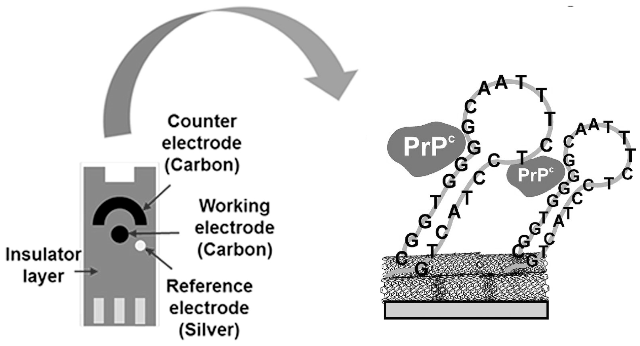
| PrPC | MWCNTs | EQCM | 50 pM | 0.1–5 nM | [41] |
| PrPC (103–230) | GE/MWCNTs/PAMAM G4 | CV | 0.5 pM | 1–10 μM | [42] |
| PrPC (103–231) | GE/PPY/PAMAM G4 | CV | 0.8 pM | 1–10 μM | [43] |
| Interleukin 6 (IL-6) | GCE/MWCNTs/CoHCF/AuNPs | DPV | 0.17 pg/mL | 0.5–1000 pg/mL | [44] |
| Adenosine | SPGE/PtCu/MWCNTs | CV | 1 nM | 10 nM–1 μM | [45] |
| Escheria coli | GCE/SWCNTs | Potentiometry | 6 CFU/mL (Milk); 26 CFU/mL (Juice) | 4–104 CFU/mL | [47] |
| Salmonella typhimurium | GCE/SWCNTs | Potentiometry | 1 CFU/mL | 0.2–103 CFU/mL | [46] |
| Salmonella typhimurium | MWCNTs/ITO | EIS | 55 CFU/mL | 55–5.5 × 10⁶ CFU/mL | [48] |
| Salmonella enteritidis | MWCNTs/ITO | EIS | 67 CFU/mL | 67–6.7 × 10⁵ CFU/mL | [48] |
| Salmonella typhimurium | GCE/rGO/CNTs | DPV | 10 CFU/mL | 10–108 CFU/mL | [81] |
| Salmonella ATCC 50761 | GCE/rGO/MWCNTs | EIS | 25 CFU/mL | 75–7.5 × 10⁵ CFU/mL | [82] |
| Thrombin | ITO/TiO2/CQDs | Photocurrent | 0.83 pM | 1–250 pM | [51] |
| Myoglobin | SPE/rGO/CNTs | CV | 0.34 ng/mL | 1 ng/mL–4 µg/mL | [83] |
| Lysozyme | MWCNTs-N-CQDs-chitosan/GCE | DPV | 4.26 fM | 1 fM–100 nM | [84] |
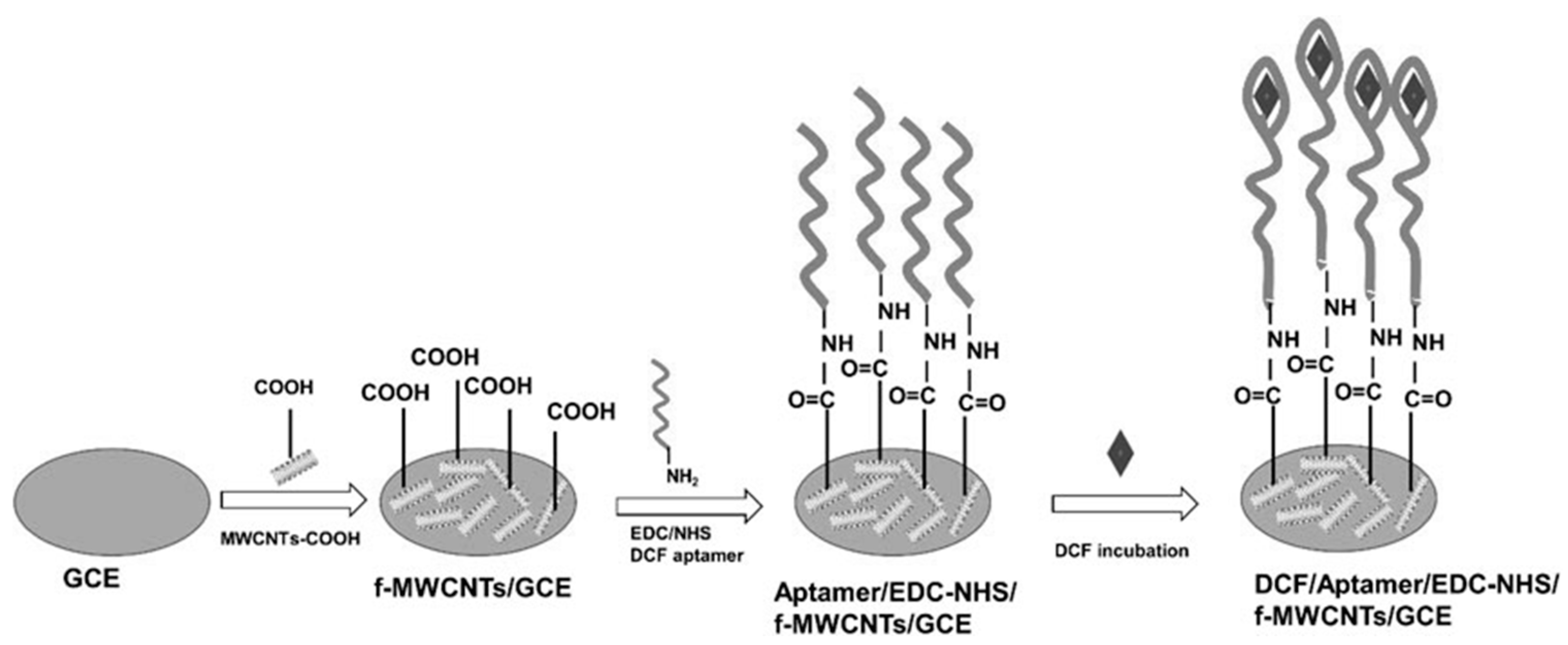
| Diclofenac | GCE/MWCNTs | EIS | 162 fM | 250 fM–1 pM | [49] |
| Sulfamethazine | GCE/CQDs/WS2 | DPV | 4 pM | 10 pM–1μM | [50] |
| Kanamycin | GCE/CB/Chitosan/Oligolactide | EIS | 0.3 nM | 0.7–50 nM | [52] |
| Oxytetracycline | MWCNTs/AuNPs/CS/AuNPs/rGO/AuNPs | DPV | 30 pM | 1–540 nM | [85] |
| Inorganic Metal-Oxide-Based Semiconductors | |||||
| Escherichia coli O157:H7 | IDEs/TiO2 | Amperometry | 0.1 pM | 1 pM–10 μM | [58] |
| Salmonella typhimurium | GCE/TiO2/rGO | DPV | 10 CFU/mL | 10–108 CFU/mL | [60] |
| Staphylococcus aureus | FTO/g-C3N4/NiO | PEC | 24 CFU/mL | 102–106 CFU/mL | [24] |
| Lysozyme | GE/TiO2@PPAA | EIS | 0.015 ng/mL | 0.05–100 ng/mL | [61] |
| PSA | GCE/TiO2(200)/rGO | EIS | 1 pg/mL | 0.003–1000 ng/mL | [59] |
| MUC1 | TiO2NT/aptamer/c-DNA@QD | Photocurrent | 0.52 nM | 0.002–0.2 µM | [86] |
| BRCA1 gene | Insulin stabilized Ag–Au@nanoclusters | FET | 3 aM | 10 aM–1 nM | [87] |
| Progesterone | SPCE/GQDs/NiO/AuNFs/f-MWCNTs | DPV | 1.86 pM | 0.01–1000 nM | [25] |
| Adenosine | FTO/TiO2/AuNPs | SWV | 0.42 fM | 1 fM–100 nM | [63] |
| Thrombin | GE/ZnO | EIS/CV | 3 pM/10 pM | 3 pM–100 nM | [64] |
| Thrombin | Sb-doped BaSrFeO3-δ | CV | 0.02 pM | 0.05 pM–0.3 nM | [88] |
| Thrombin, Lysozyme | GE/CdS/PbS QDs | SWV | 20 ng/mL | 20–500 ng/mL | [69] |
| Troponin I | SPCE/3DLSG_MoS2_Ag NPs-2.0 hybrid | EIS | 0.1 fM | 10−16–10−10 M | [89] |
| Ochratoxin A | GCE/ZnO/AuNPs/Nafion | DPV | 0.05 pg/mL | 0.1–30 × 103 pg/mL | [65] |
| Aflatoxin B1 | GCE/CQDs/Cu2O | DPV | 0.9 Ag/mL | 3 Ag/mL–1.9 μg/mL | [66] |
| Tetracycline | GCE/MWCNTs/WO3@AuNPs | DPV | 48 pM | 0.1 nM–100 nM | [23] |
| Kanamycin | PE/MWCNTs@TiO2/Thi | ECL | 0.049 ng/mL | 0.1–105 ng/mL | [90] |
| Organic Semiconductors | |||||
| Carcinoembryonic antigens | PE-graphene/PEDOT:PSS | EIS | 0.45 ng/mL | 0.77–14 ng/mL | [73] |
| Dopamine | GCE/GR/PANI | SWV | 1.98 pM | 0.007–90 nM | [74] |
| Prostate cancer marker AMACR/P504S | PPy/PEG | SWV | 0.15 fM | 1 fM–100 nM | [75] |
| MUC1 | EDOT/ProDOT(COOH)2 | CV | 418 fM | 709 fM–7.09 nM | [77] |
| Aflatoxin M1 | GCE/NR@pillar[5]arene (P[5]A-COOH) | EIS | 0.5 ng/L | 5–120 ng/L | [79] |
| Idarubicin | GCE/Azure B | EIS | 0.3 fM | 1 fM–100 nM | [80] |
| Idarubicin | GCE/Azure B | CV | 0.3 fM | 1 fM–0.1 nM | [80] |
6. Conclusions
Author Contributions
Funding
Institutional Review Board Statement
Informed Consent Statement
Data Availability Statement
Conflicts of Interest
References
- Zhuo, Z.; Yu, Y.; Wang, M.; Li, J.; Zhang, Z.; Liu, J.; Wu, X.; Lu, A.; Zhang, G.; Zhang, B. Recent advances in SELEX technology and aptamer applications in biomedicine. Int. J. Mol. Sci. 2017, 18, 2142. [Google Scholar] [CrossRef] [PubMed] [Green Version]
- Tuerk, C.; Gold, L. Systematic evolution of ligands by exponential enrichment: RNA ligands to bacteriophage T4 DNA polymerase. Science 1990, 249, 505–510. [Google Scholar] [CrossRef]
- Ellington, A.D.; Szostak, J.W. In vitro selection of RNA molecules that bind specific ligands. Nature 1990, 346, 818–822. [Google Scholar] [CrossRef]
- Arshavsky-Graham, S.; Heuer, C.; Jiang, X.; Segal, E. Aptasensors versus immunosensors-Which will prevail? Eng. Life Sci. 2022, 22, 319–333. [Google Scholar] [CrossRef] [PubMed]
- Kang, D.; Zuo, X.; Yang, R.; Xia, F.; Plaxco, K.W.; White, R.J. Comparing the properties of electrochemical-based DNA sensors employing different redox tags. Anal. Chem. 2009, 81, 9109–9113. [Google Scholar] [CrossRef] [PubMed] [Green Version]
- Catanante, G.; Mishra, R.K.; Hayat, A.; Marty, J.-L. Sensitive analytical performance of folding based biosensor using methylene blue tagged aptamers. Talanta 2016, 153, 138–144. [Google Scholar] [CrossRef] [PubMed]
- Yang, S.; Li, H.; Xu, L.; Deng, Z.; Han, W.; Liu, Y.; Jiang, W.; Zu, Y. Oligonucleotide aptamer-mediated precision therapy of hematological malignancies. Mol. Ther. Nucleic Acids 2018, 13, 164–175. [Google Scholar] [CrossRef] [Green Version]
- Bernat, V.; Disney, M.D. RNA structures as mediators of neurological diseases and as drug targets. Neuron 2015, 87, 28–46. [Google Scholar] [CrossRef] [Green Version]
- Villalonga, A.; Mayol, B.; Villalonga, R.; Vilela, D. Electrochemical aptasensors for clinical diagnosis. A review of the last five years. Sens. Actuat. B Chem. 2022, 369, 132318. [Google Scholar] [CrossRef]
- Ikebukuro, K.; Kiyohara, C.; Sode, K. Electrochemical detection of protein using a double aptamer sandwich. Anal. Lett. 2004, 37, 2901–2909. [Google Scholar] [CrossRef]
- Hianik, T.; Wang, J. Electrochemical aptasensors—Recent achievements and perspectives. Electroanalysis 2009, 21, 1223–1235. [Google Scholar] [CrossRef]
- Abd-Ellatief, R.; Abd-Ellatief, M.R. Electrochemical aptasensors: Current status and future perspectives. Diagnostics 2021, 11, 104. [Google Scholar] [CrossRef]
- Bard, A.J.; Faulkner, L.R.; White, H.S. Electrochemical Methods: Fundamentals and Applications, 3rd ed.; Wiley: Hoboken, NJ, USA, 2022; ISBN 9781119334071. [Google Scholar]
- Girault, H.H. Analytical and Physical Electrochemistry, 1st ed.; EPFL Press: Lausanne, Switzerland, 2004; p. 315. [Google Scholar]
- Renedo, O.D.; Alonso-Lomillo, M.A.; Martínez, M.J.A. Recent developments in the field of screen-printed electrodes and their related applications. Talanta 2007, 73, 202–219. [Google Scholar] [CrossRef] [PubMed]
- García-Miranda Ferrari, A.; Rowley-Neale, S.J.; Banks, C.E. Screen-printed electrodes: Transitioning the laboratory in-to-the field. Talanta Open 2021, 3, 100032. [Google Scholar] [CrossRef]
- Evtugyn, G.; Porfireva, A.; Shamagsumova, R.; Hianik, T. Advances in electrochemical aptasensors based on carbon nanomaterials. Chemosensors 2020, 8, 96. [Google Scholar] [CrossRef]
- Lai, H.; Ming, P.; Wu, M.; Wang, S.; Sun, D.; Zhai, H. An electrochemical aptasensor based on P-Ce-MOF@MWCNTs as signal amplification strategy for highly sensitive detection of zearalenone. Food Chem. 2023, 423, 136331. [Google Scholar] [CrossRef]
- Fujishima, A.; Honda, K. Electrochemical photolysis of water at a semiconductor electrode. Nature 1972, 238, 37–38. [Google Scholar] [CrossRef] [PubMed]
- Bertel, L.; Miranda, D.A.; García-Martín, J.M. Nanostructured titanium dioxide surfaces for electrochemical biosensing. Sensors 2021, 21, 6167. [Google Scholar] [CrossRef]
- Shetti, N.P.; Bukkitgar, S.D.; Reddy, K.R.; Reddy, C.V.; Aminabhavi, T.M. ZnO-based nanostructured electrodes for electrochemical sensors and biosensors in biomedical applications. Biosens. Bioelectron. 2019, 141, 111417. [Google Scholar] [CrossRef] [PubMed]
- Bukkitgar, S.D.; Kumar, S.; Pratibha; Singh, S.; Singh, V.; Raghava Reddy, K.; Sadhu, V.; Bagihalli, G.B.; Shetti, N.P.; Venkata Reddy, C.; et al. Functional nanostructured metal oxides and its hybrid electrodes—Recent advancements in electrochemical biosensing applications. Microchem. J. 2020, 159, 105522. [Google Scholar] [CrossRef]
- Song, J.; Lin, X.; Jiang, N.; Huang, M. Carbon-doped WO3 electrochemical aptasensor based on Box-Behnken strategy for highly-sensitive detection of tetracycline. Food Chem. 2022, 367, 130564. [Google Scholar] [CrossRef]
- Chen, X.; Yang, Z.; Ai, L.; Zhou, S.; Fan, H.; Ai, S. Signal-off photoelectrochemical aptasensor for S. aureus detection based on graphite-like carbon nitride decorated with nickel oxide. Electroanalysis 2022, 34, 310–315. [Google Scholar] [CrossRef]
- Samie, H.A.; Arvand, M. Label-free electrochemical aptasensor for progesterone detection in biological fluids. Bioelectrochemistry 2020, 133, 107489. [Google Scholar] [CrossRef] [PubMed]
- Mohan, S.; Srivastava, P.; Maheshwari, S.N.; Sundar, S.; Prakash, R. Nano-structured nickel oxide based DNA biosensor for detection of visceral leishmaniasis (Kala-azar). Analyst 2011, 136, 2845–2851. [Google Scholar] [CrossRef] [Green Version]
- Georgakilas, V.; Perman, J.A.; Tucek, J.; Zboril, R. Broad family of carbon nanoallotropes: Classification, chemistry, and applications of fullerenes, carbon dots, nanotubes, graphene, nanodiamonds, and combined superstructures. Chem. Rev. 2015, 115, 4744–4822. [Google Scholar] [CrossRef]
- Rahman, M.A.; Kumar, P.; Park, D.-S.; Shim, Y.-B. Electrochemical sensors based on organic conjugated polymers. Sensors 2008, 8, 118–141. [Google Scholar] [CrossRef]
- Hopkins, J.; Fidanovski, K.; Lauto, A.; Mawad, D. All-organic semiconductors for electrochemical biosensors: An overview of recent progress in material design. Front. Bioeng. Biotechnol. 2019, 7, 237. [Google Scholar] [CrossRef]
- Kim, K.; Yoo, H.; Lee, E.K. New opportunities for organic semiconducting polymers in biomedical applications. Polymers 2022, 14, 2960. [Google Scholar] [CrossRef] [PubMed]
- Hirsch, A. The era of carbon allotropes. Nat. Mater. 2010, 9, 868–871. [Google Scholar] [CrossRef]
- Yang, X.; Ebrahimi, A.; Li, J.; Cui, Q. Fullerene-biomolecule conjugates and their biomedicinal applications. Int. J. Nanomed. 2014, 9, 77–92. [Google Scholar] [CrossRef] [Green Version]
- Pilehvar, S.; De Wael, K. Recent advances in electrochemical biosensors based on fullerene-C60 nano-structured platforms. Biosensors 2015, 5, 712–735. [Google Scholar] [CrossRef] [Green Version]
- Bardhan, N.M. 30 years of advances in functionalization of carbon nanomaterials for biomedical applications: A practical review. J. Mater. Res. 2017, 32, 107–127. [Google Scholar] [CrossRef] [Green Version]
- Thirumalraj, B.; Palanisamy, S.; Chen, S.-M.; Lou, B.-S. Preparation of highly stable fullerene C60 decorated graphene oxide nanocomposite and its sensitive electrochemical detection of dopamine in rat brain and pharmaceutical samples. J. Colloid Interface Sci. 2016, 462, 375–381. [Google Scholar] [CrossRef] [PubMed]
- Compton, R.G.; Spackman, R.A.; Wellington, R.G.; Green, M.L.H.; Turner, J. A C60 modified electrode: Electrochemical formation of tetra-butylammonium salts of C60 anions. J. Electroanal. Chem. 1992, 327, 337–341. [Google Scholar] [CrossRef]
- Cassell, A.M.; Scrivens, W.A.; Tour, J.M. Assembly of DNA/fullerene hybrid materials. Angew. Chem. Int. Ed. Engl. 1998, 37, 1528–1531. [Google Scholar] [CrossRef]
- Bai, L.; Chen, Y.; Bai, Y.; Chen, Y.; Zhou, J.; Huang, A. Fullerene-doped polyaniline as new redox nanoprobe and catalyst in electrochemical aptasensor for ultrasensitive detection of Mycobacterium tuberculosis MPT64 antigen in human serum. Biomaterials 2017, 133, 11–19. [Google Scholar] [CrossRef]
- Ferrier, D.C.; Honeychurch, K.C. Carbon nanotube (CNT)-based biosensors. Biosensors 2021, 11, 486. [Google Scholar] [CrossRef]
- Hianik, T. Aptamer-based biosensors. In Encyclopedia of Interfacial Chemistry: Surface Science and Electrochemistry; Wandelt, K., Ed.; Elsevier: Amsterdam, The Netherlands, 2018; Volume 7, pp. 11–19. [Google Scholar]
- Hianik, T.; Porfireva, A.; Grman, I.; Evtugyn, G. EQCM biosensors based on DNA aptamers and antibodies for rapid detection of prions. Protein Pept. Lett. 2009, 16, 363–367. [Google Scholar] [CrossRef] [PubMed]
- Miodek, A.; Castillo, G.; Hianik, T.; Korri-Youssoufi, H. Electrochemical aptasensor of human cellular prion based on multiwalled carbon nanotubes modified with dendrimers: A platform for connecting redox markers and aptamers. Anal. Chem. 2013, 85, 7704–7712. [Google Scholar] [CrossRef]
- Miodek, A.; Castillo, G.; Hianik, T.; Korri-Youssoufi, H. Electrochemical aptasensor of cellular prion protein based on modified polypyrrole with redox dendrimers. Biosens. Bioelectron. 2014, 56, 104–111. [Google Scholar] [CrossRef]
- Li, Y.; Hua, X.; Wang, J.; Jin, B. cMWCNT/CoHCF/AuNPs nanocomposites aptasensor for electrochemical detection of interleukin-6. Talanta Open 2023, 7, 100188. [Google Scholar] [CrossRef]
- Cao, S.; Zhao, H.; Chen, K.; Zhou, F.; Lan, M. An electrochemical aptasensor based on multi-walled carbon nanotubes loaded with PtCu nanoparticles as signal label for ultrasensitive detection of adenosine. Anal. Chim. Acta 2023, 1260, 341212. [Google Scholar] [CrossRef] [PubMed]
- Zelada-Guillén, G.A.; Riu, J.; Düzgün, A.; Rius, F.X. Immediate detection of living bacteria at ultralow concentrations using a carbon nanotube based potentiometric aptasensor. Angew. Chem. Int. Ed. Engl. 2009, 48, 7334–7337. [Google Scholar] [CrossRef] [PubMed]
- Zelada-Guillén, G.A.; Blondeau, P.; Rius, F.X.; Riu, J. Carbon nanotube-based aptasensors for the rapid and ultrasensitive detection of bacteria. Methods 2013, 63, 233–238. [Google Scholar] [CrossRef] [PubMed]
- Hasan, M.R.; Pulingam, T.; Appaturi, J.N.; Zifruddin, A.N.; Teh, S.J.; Lim, T.W.; Ibrahim, F.; Leo, B.F.; Thong, K.L. Carbon nanotube-based aptasensor for sensitive electrochemical detection of whole-cell Salmonella. Anal. Biochem. 2018, 554, 34–43. [Google Scholar] [CrossRef] [PubMed]
- Zou, Y.; Griveau, S.; Ringuedé, A.; Bedioui, F.; Richard, C.; Slim, C. Functionalized multi-walled carbon nanotube-based aptasensors for diclofenac detection. Front. Chem. 2021, 9, 812909. [Google Scholar] [CrossRef]
- Wang, Y.; Gong, C.; Zhu, Y.; Wang, Q.; Geng, L. Signal-on electrochemical aptasensor for sensitive detection of sulfamethazine based on carbon quantum dots/tungsten disulfide nanocomposites. Electrochim. Acta 2021, 393, 139054. [Google Scholar] [CrossRef]
- Cheng, W.; Pan, J.; Yang, J.; Zheng, Z.; Lu, F.; Chen, Y.; Gao, W. A photoelectrochemical aptasensor for thrombin based on the use of carbon quantum dot-sensitized TiO2 and visible-light photoelectrochemical activity. Microchim. Acta 2018, 185, 263. [Google Scholar] [CrossRef]
- Kulikova, T.; Gorbatchuk, V.; Stoikov, I.; Rogov, A.; Evtugyn, G.; Hianik, T. Impedimetric determination of kanamycin in milk with aptasensor based on carbon black-oligolactide composite. Sensors. 2020, 20, 4738. [Google Scholar] [CrossRef]
- Song, H.; Zhang, Y.; Wang, S.; Huang, K.; Luo, Y.; Zhang, W.; Xu, W. Label-free polygonal-plate fluorescent-hydrogel biosensor for ultrasensitive microRNA detection. Sens. Actuators B Chem. 2020, 306, 127554. [Google Scholar] [CrossRef]
- Hernández-Cancel, G.; Suazo-Dávila, D.; Medina-Guzmán, J.; Rosado-González, M.; Díaz-Vázquez, L.M.; Griebenow, K. Chemically glycosylation improves the stability of an amperometric horseradish peroxidase biosensor. Anal. Chim. Acta 2015, 854, 129–139. [Google Scholar] [CrossRef] [PubMed] [Green Version]
- Visa, M.; Andronic, L.; Enesca, A. Behavior of the new composites obtained from fly ash and titanium dioxide in removing of the pollutants from wastewater. Appl. Surf. Sci. 2016, 388, 359–369. [Google Scholar] [CrossRef]
- Şerban, I.; Enesca, A. Metal oxides-based semiconductors for biosensors applications. Front. Chem. 2020, 8, 354. [Google Scholar] [CrossRef]
- Ma, X.; Li, X.; Zhang, W.; Meng, F.; Wang, X.; Qin, Y.; Zhang, M. Carbon-based nanocomposite smart sensors for the rapid detection of mycotoxins. Nanomaterials 2021, 11, 2851. [Google Scholar] [CrossRef]
- Nadzirah, S.; Azizah, N.; Hashim, U.; Gopinath, S.C.B.; Kashif, M. Titanium dioxide nanoparticle-based interdigitated electrodes: A novel current to voltage DNA biosensor recognizes E. coli O157:H7. PLoS ONE 2015, 10, e0139766. [Google Scholar] [CrossRef]
- Karimipour, M.; Heydari-Bafrooei, E.; Sanjari, M.; Johansson, M.B.; Molaei, M. A glassy carbon electrode modified with TiO2(200)-rGO hybrid nanosheets for aptamer based impedimetric determination of the prostate specific antigen. Mikrochim. Acta 2018, 186, 33. [Google Scholar] [CrossRef] [PubMed]
- Muniandy, S.; Teh, S.J.; Appaturi, J.N.; Thong, K.L.; Lai, C.W.; Ibrahim, F.; Leo, B.F. A reduced graphene oxide-titanium dioxide nanocomposite based electrochemical aptasensor for rapid and sensitive detection of Salmonella enterica. Bioelectrochemistry 2019, 127, 136–144. [Google Scholar] [CrossRef] [PubMed]
- Zhang, Z.; Zhang, S.; He, L.; Peng, D.; Yan, F.; Wang, M.; Zhao, J.; Zhang, H.; Fang, S. Feasible electrochemical biosensor based on plasma polymerization-assisted composite of polyacrylic acid and hollow TiO2 spheres for sensitively detecting lysozyme. Biosens. Bioelectron. 2015, 74, 384–390. [Google Scholar] [CrossRef]
- Erdem, A.; Eksin, E.; Muti, M. Chitosan–graphene oxide based aptasensor for the impedimetric detection of lysozyme. Coll. Surf. B: Biointerfaces 2014, 115, 205–211. [Google Scholar] [CrossRef]
- Wang, X.; Qin, Y.; Zhang, X.; Leng, Y.; Chen, Z. Au/TiO2 nanorod arrays-based electrochemical aptasensor for ultrasensitive detection of adenosine. Microchem. J. 2023, 190, 108695. [Google Scholar] [CrossRef]
- Evtugyn, G.; Cherkina, U.; Porfireva, A.; Danzberger, J.; Ebner, A.; Hianik, T. Electrochemical aptasensor based on ZnO modified gold electrode. Electroanalysis 2013, 25, 1855–1863. [Google Scholar] [CrossRef]
- Zhang, S.; Wang, Y.; Sheng, Q.; Yue, T. Electrochemical aptasensor based on ZnO-Au nanocomposites for the determination of ochratoxin A in wine and beer. Processes 2023, 11, 864. [Google Scholar] [CrossRef]
- Rahimi, F.; Roshanfekr, H.; Peyman, H. Ultra-sensitive electrochemical aptasensor for label-free detection of Aflatoxin B1 in wheat flour sample using factorial design experiments. Food Chem. 2021, 343, 128436. [Google Scholar] [CrossRef] [PubMed]
- Grabowska, I.; Hepel, M.; Kurzątkowska-Adaszyńska, K. Advances in design strategies of multiplex electrochemical aptasensors. Sensors 2021, 22, 161. [Google Scholar] [CrossRef] [PubMed]
- Evtugyn, G.; Belyakova, S.; Porfireva, A.; Hianik, T. Electrochemical aptasensors based on hybrid metal-organic frameworks. Sensors 2020, 20, 6963. [Google Scholar] [CrossRef]
- Hansen, J.A.; Wang, J.; Kawde, A.-N.; Xiang, Y.; Gothelf, K.V.; Collins, G. Quantum-dot/aptamer-based ultrasensitive multi-analyte electrochemical biosensor. J. Am. Chem. Soc. 2006, 128, 2228–2229. [Google Scholar] [CrossRef]
- Pavel, I.-A.; Lakard, S.; Lakard, B. Flexible sensors based on conductive polymers. Chemosensors 2022, 10, 97. [Google Scholar] [CrossRef]
- Fan, X.; Nie, W.; Tsai, H.; Wang, N.; Huang, H.; Cheng, Y.; Wen, R.; Ma, L.; Yan, F.; Xia, Y. PEDOT:PSS for flexible and stretchable electronics: Modifications, strategies, and applications. Adv. Sci. 2019, 6, 1900813. [Google Scholar] [CrossRef] [Green Version]
- Malinauskas, A. Self-doped polyanilines. J. Power Sources 2004, 126, 214–220. [Google Scholar] [CrossRef]
- Yen, Y.-K.; Chao, C.-H.; Yeh, Y.-S. A Graphene-PEDOT:PSS modified paper-based aptasensor for electrochemical impedance spectroscopy detection of tumor marker. Sensors 2020, 20, 1372. [Google Scholar] [CrossRef]
- Liu, S.; Xing, X.; Yu, J.; Lian, W.; Li, J.; Cui, M.; Huang, J. A novel label-free electrochemical aptasensor based on graphene–polyaniline composite film for dopamine determination. Biosens. Bioelectron. 2012, 36, 186–191. [Google Scholar] [CrossRef] [PubMed]
- Jolly, P.; Miodek, A.; Yang, D.-K.; Chen, L.-C.; Lloyd, M.D.; Estrela, P. Electro-engineered polymeric films for the development of sensitive aptasensors for prostate cancer marker detection. ACS Sens. 2016, 1, 1308–1314. [Google Scholar] [CrossRef] [Green Version]
- Šafaříková, E.; Švihálková, Š.L.; Stříteský, S.; Kubala, L.; Vala, M.; Weiter, M.; Víteček, J. Evaluation and improvement of organic semiconductors’ biocompatibility towards fibroblasts and cardiomyocytes. Sens. Actuators B Chem. 2018, 260, 418–425. [Google Scholar] [CrossRef]
- Haya, G.; Runsewe, D.O.; Otakpor, M.U.; Pohlman, G.E.; Towne, A.; Betancourt, T.; Irvin, J.A. Functionalized thiophene-based aptasensors for the electrochemical detection of Mucin-1. ACS Appl. Polym. Mater. 2023, 5, 1208–1218. [Google Scholar] [CrossRef]
- Barsan, M.M.; Ghica, M.E.; Brett, C.M.A. Electrochemical sensors and biosensors based on redox polymer/carbon nanotube modified electrodes: A review. Anal. Chim. Acta 2015, 881, 1–23. [Google Scholar] [CrossRef] [PubMed]
- Smolko, V.; Shurpik, D.; Porfireva, A.; Evtugyn, G.; Stoikov, I.; Hianik, T. Electrochemical aptasensor based on Poly(Neutral Red) and carboxylated Pillar [5]arene for sensitive determination of Aflatoxin M1. Electroanalysis 2018, 30, 486–496. [Google Scholar] [CrossRef]
- Goida, A.; Kuzin, Y.; Evtugyn, V.; Porfireva, A.; Evtugyn, G.; Hianik, T. Electrochemical sensing of idarubicin—DNA interaction using electropolymerized azure B and methylene blue mediation. Chemosensors 2022, 10, 33. [Google Scholar] [CrossRef]
- Appaturi, J.N.; Pulingam, T.; Thong, K.L.; Muniandy, S.; Ahmad, N.; Leo, B.F. Rapid and sensitive detection of Salmonella with reduced graphene oxide-carbon nanotube based electrochemical aptasensor. Anal. Biochem. 2020, 589, 113489. [Google Scholar] [CrossRef]
- Jia, F.; Duan, N.; Wu, S.; Dai, R.; Wang, Z.; Li, X. Impedimetric Salmonella aptasensor using a glassy carbon electrode modified with an electrodeposited composite consisting of reduced graphene oxide and carbon nanotubes. Microchim. Acta 2016, 183, 337–344. [Google Scholar] [CrossRef]
- Kumar, V.; Shorie, M.; Ganguli, A.K.; Sabherwal, P. Graphene-CNT nanohybrid aptasensor for label free detection of cardiac biomarker myoglobin. Biosens. Bioelectr. 2015, 72, 56–60. [Google Scholar] [CrossRef]
- Beiki, T.; Najafpour-Darzi, G.; Mohammadi, M.; Shakeri, M.; Boukherroub, R. Fabrication of a novel electrochemical biosensor based on a molecular imprinted polymer-aptamer hybrid receptor for lysozyme determination. Anal. Bioanal. Chem. 2022, 415, 899–911. [Google Scholar] [CrossRef] [PubMed]
- Akbarzadeh, S.; Khajehsharifi, H.; Hajihosseini, S. Detection of oxytetracycline using an electrochemical label-free aptamer-based biosensor. Biosensors 2022, 12, 468. [Google Scholar] [CrossRef] [PubMed]
- Tian, J.; Huang, T.; Jusheng Lu, J. Photoelectrochemical aptasensor for mucin 1 based on DNA/aptamer linking of quantum dots and TiO2 nanotube arrays. Anal. Meth. 2016, 8, 2375–2382. [Google Scholar] [CrossRef]
- Majd, S.M.; Mirzapour, F.; Shamsipur, M.; Manouchehri, I.; Babaee, E.; Pashabadi, A.; Moradian, R. Design of a novel aptamer/molecularly imprinted polymer hybrid modified Ag–Au@Insulin nanoclusters/Au-gate-based MoS2 nanosheet field-effect transistor for attomolar detection of BRCA1 gene. Talanta 2023, 257, 124394. [Google Scholar] [CrossRef]
- Rauf, S.; Tayyab, Z.; Pan, X.; Yang, Y.; Hussain, A.; Yunzhi, H.; Amin, N.; Rauf, S.; Salem, A.; Xu, W. Highly electroactive Sb-doped BaSrFeO3-d semiconductor as a label-free electrochemical aptasensing platform for thrombin detection. IEEE Sens. J. 2023, 23, 9077–9085. [Google Scholar] [CrossRef]
- Vasudevan, M.; Remesh, S.; Perumal, V.; Raja, P.B.; Ibrahim, M.N.M.; Gopinath, S.C.B.; Karuppanan, S.; Ovinis, M. Facile synthesis of MoS2 nanoflower-Ag NPs grown on lignin-derived graphene for Troponin I aptasensing. Chem. Eng. J. 2023, 468, 143613. [Google Scholar] [CrossRef]
- Cheng, S.; Zhang, H.; Huang, J.; Xu, R.; Suna, X.; Guo, Y. Highly sensitive electrochemiluminescence aptasensor based on dual-signal amplification strategy for kanamycin detection. Sci. Total Environ. 2020, 737, 139785. [Google Scholar] [CrossRef] [PubMed]
- Mehlhorn, A.; Rahimi, P.; Joseph, Y. Aptamer-based biosensors for antibiotic detection: A review. Biosensors 2018, 8, 54. [Google Scholar] [CrossRef] [PubMed] [Green Version]


| Material | Advantages | Disadvantages |
|---|---|---|
| Metal system | Excellent electrical conductivity, great chemical stability | Hydrophobic, surface electrons can lead to denaturing of biomolecules |
| Metal oxides | Low cost, easy to prepare, wide potential window (transition metal oxides), tunable composition, hydrophilic | Lower conductivity, high recombination rate (especially for photoelectrochemical applications) |
| WO3 | Non-stoichiometric properties, good electron transport, stable in acid conditions, moderate hole-diffusion length (approx. 150 nm) especially for photoelectrochemical applications | Unstable at pH > 4 |
| ZnO | Good stability, environmentally friendly, inexpensive, high carrier mobility | ZnO NPs are ion shedding particles and zinc ions can lead to oxidative stress |
| TiO2 | Highly stable over a range of pH values in aqueous environments, even under illumination, good electrochemical activity, non-toxic | Low absorption in the visible light (especially for photoelectrochemical applications) |
| CuO | Good electrochemical activity and the ability to stimulate electron transfer reactions at lower potentials | Potentially toxic effects in the case of CuO nanoparticles |
| NiO | High isoelectric point, porous morphology, small crystallite size, chemical stability | The charge acceptance at temperatures above 35 °C is not good enough |
| Conductive polymers | Filming has good flexibility, unique solution processibility, biocompatible | Low conductivity, lower electrochemical stability |
| Carbon systems | Large specific surface area, high conductivity, electrochemical stability, low cost | Poor dispersity |
| Graphene | Large specific area, excellent electrical properties, high control on functionalization | Susceptibility to oxidative environments, hydrophobic, high cost |
| Graphene oxide | High dispersibility in water (hydrophilic), abundant presence of oxygenated groups—binding sites | Lower electrical conductivity, surface random functionalization |
| Reduced graphene oxide | High electrical conductivity, good control on functionalization, less expensive than graphene | Low density of oxygen containing groups, lower colloidal stability, hydrophobic |
| Carbon nanotubes | Large surface area, excellent electrical properties | High cost, complex preparation, potential toxic |
Disclaimer/Publisher’s Note: The statements, opinions and data contained in all publications are solely those of the individual author(s) and contributor(s) and not of MDPI and/or the editor(s). MDPI and/or the editor(s) disclaim responsibility for any injury to people or property resulting from any ideas, methods, instructions or products referred to in the content. |
© 2023 by the authors. Licensee MDPI, Basel, Switzerland. This article is an open access article distributed under the terms and conditions of the Creative Commons Attribution (CC BY) license (https://creativecommons.org/licenses/by/4.0/).
Share and Cite
Bousiakou, L.; Al-Dosary, O.; Economou, A.; Subjakova, V.; Hianik, T. Current Trends in the Use of Semiconducting Materials for Electrochemical Aptasensing. Chemosensors 2023, 11, 438. https://doi.org/10.3390/chemosensors11080438
Bousiakou L, Al-Dosary O, Economou A, Subjakova V, Hianik T. Current Trends in the Use of Semiconducting Materials for Electrochemical Aptasensing. Chemosensors. 2023; 11(8):438. https://doi.org/10.3390/chemosensors11080438
Chicago/Turabian StyleBousiakou, Leda, Omar Al-Dosary, Anastasios Economou, Veronika Subjakova, and Tibor Hianik. 2023. "Current Trends in the Use of Semiconducting Materials for Electrochemical Aptasensing" Chemosensors 11, no. 8: 438. https://doi.org/10.3390/chemosensors11080438