
Fluorescent Quantum Dots and Its Composites for Highly Sensitive Detection of Heavy Metal Ions and Pesticide Residues: A Review


Abstract
:1. Introduction
2. QD-Based Fluorescence Sensors
3. Mechanisms of Fluorescence Sensors
3.1. Fluorescence Resonance Energy Transfer (FRET)
- (i)
- Original FRET process destruction
- (ii)
- New FRET system formation
- (iii)
- Analyte-based FRET system activation
3.2. Photo-Induced Electron Transfer (PET)
3.3. Internal Filtration Effects (IFEs)
3.4. Aggregation Effects (AEs)
4. QDs as Fluorescence Sensors
4.1. QDs for Detection of Heavy Metal Ions
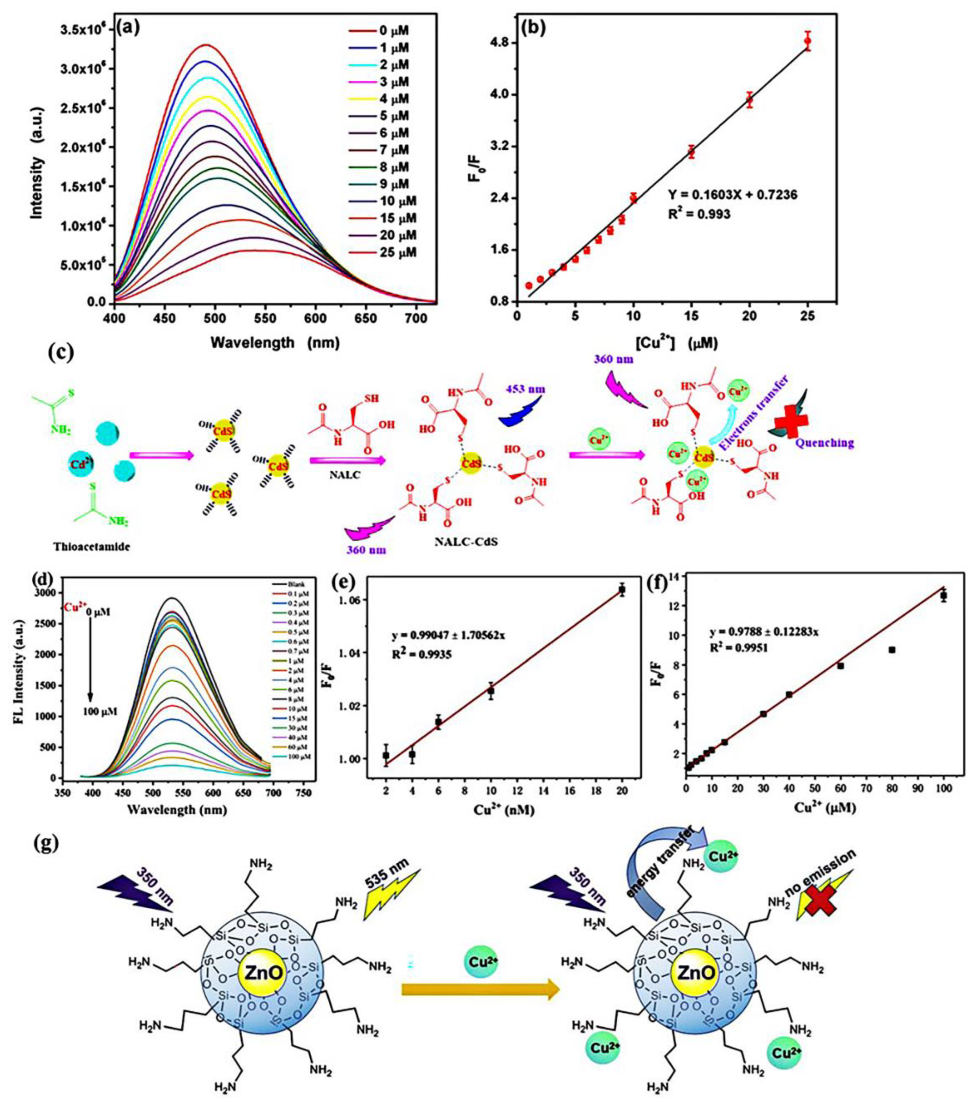
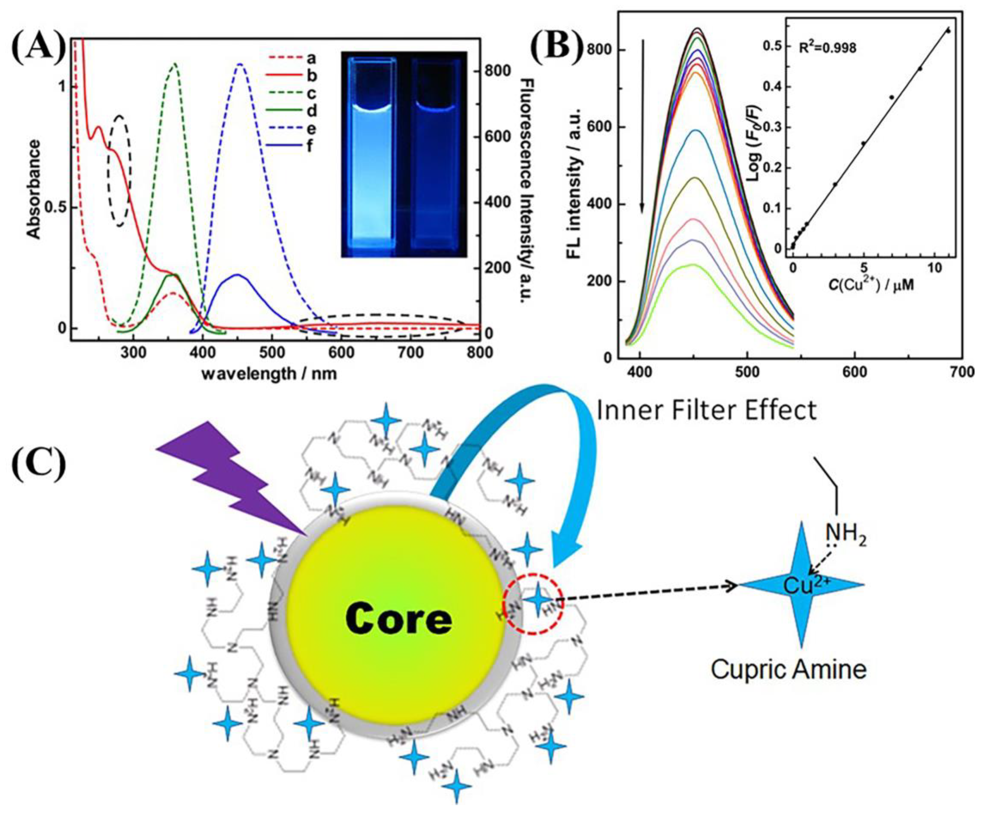
4.1.1. Copper(II) Ions
4.1.2. Mercury Ions
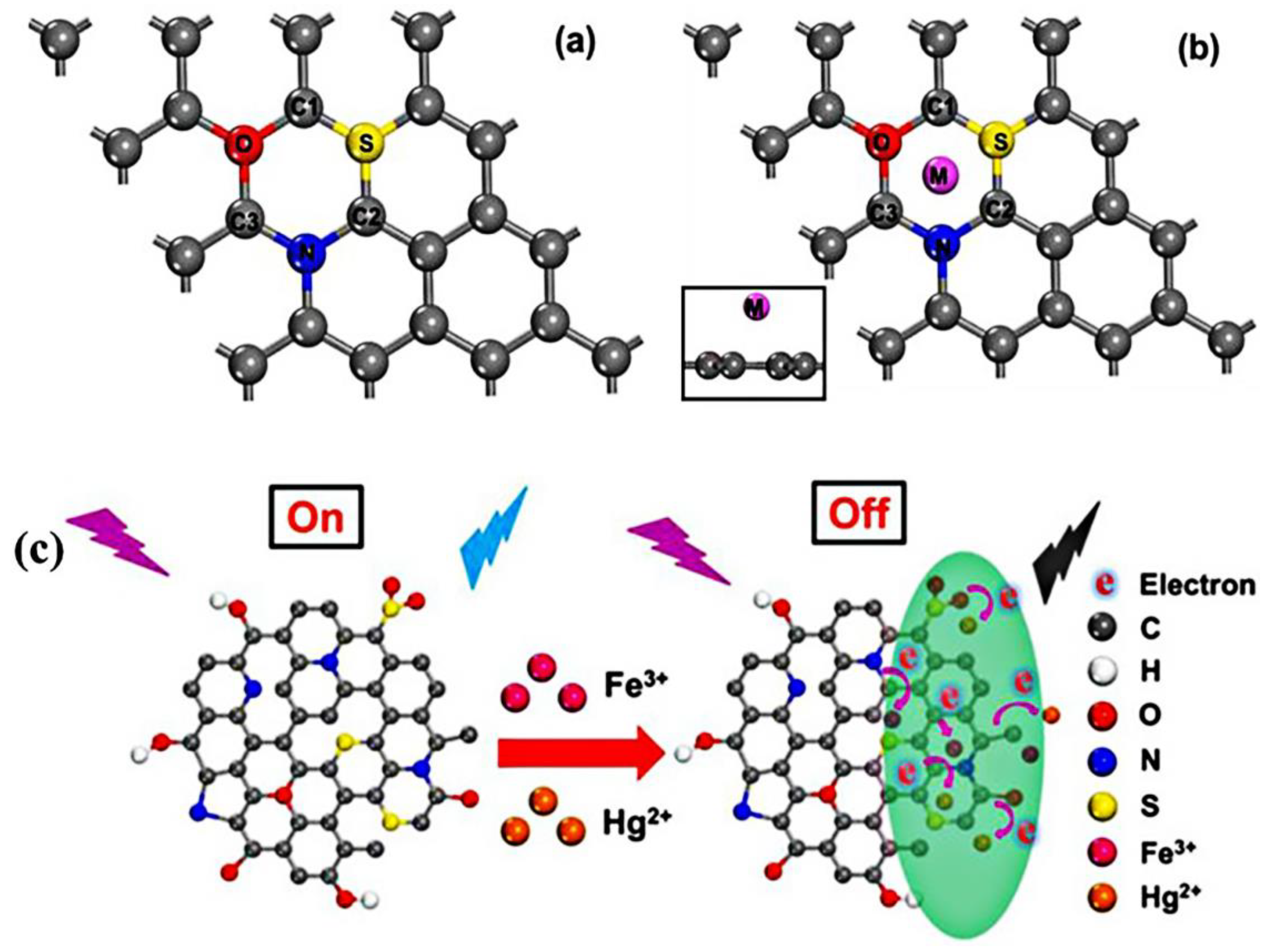
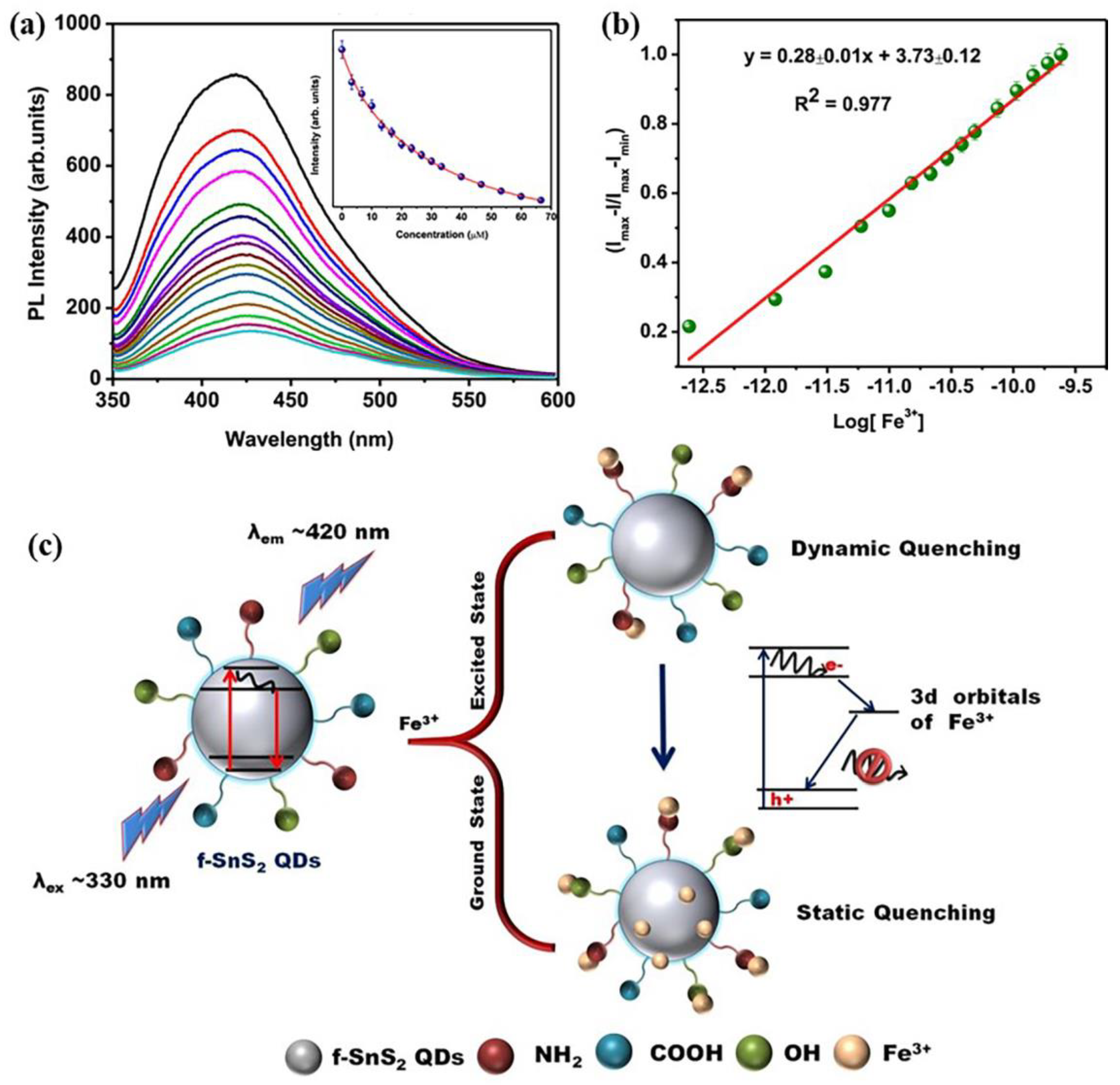
4.1.3. Iron(III) Ions
4.1.4. Lead(II) Ions
4.1.5. Other Metal Ions
4.2. QDs as Fluorescence Sensor for Pesticide Residues
4.2.1. Single-Irradiation Fluorescence Sensor
4.2.2. Ratiometric Fluorescence Sensors
4.3. Colorimetric Sensor
5. Conclusions and Outlook
Author Contributions
Funding
Institutional Review Board Statement
Informed Consent Statement
Data Availability Statement
Acknowledgments
Conflicts of Interest
References
- Sonali, J.M.I.; Subhashree, S.; Kumar, P.S.; Gayathri, K.V. New analytical strategies amplified with carbon-based nanomaterial for sensing food pollutants. Chemosphere 2022, 295, 133847. [Google Scholar]
- Lan, L.Y.; Yao, Y.; Ping, J.F.; Ying, Y.B. Recent progress in nanomaterial-based optical aptamer assay for the detection of food chemical contaminants. ACS Appl. Mater. Interfaces 2017, 9, 23287–23301. [Google Scholar] [CrossRef] [PubMed]
- Zhang, K.; Li, H.Y.; Wang, W.J.; Cao, J.X.; Gan, N.; Han, H.Y. Application of multiplexed aptasensors in food contaminants detection. ACS Sens. 2021, 5, 3721–3738. [Google Scholar] [CrossRef] [PubMed]
- Xu, M.M.; Wang, X.Y.; Liu, X.P. Detection of heavy metal ions by ratiometric photoelectric sensor. J. Agric. Food Chem. 2022, 70, 11468–11480. [Google Scholar] [CrossRef] [PubMed]
- Huang, Y.F.; Peng, J.H.; Huang, X.J. Allylthiourea functionalized magnetic adsorbent for the extraction of cadmium, copper and lead ions prior to their determination by atomic absorption spectrometry. Microchim. Acta 2019, 186, 5. [Google Scholar] [CrossRef]
- Mir, S.A.; Dar, B.N.; Mir, M.; Sofi, S.A.; Shah, M.A.; Sidiq, T.; Sunooj, K.V.; Hamdani, A.M.; Khaneghah, A.M. Current strategies for the reduction of pesticide residues in food products. J. Food Compos. Anal. 2022, 106, 104274. [Google Scholar] [CrossRef]
- Panda, S.; Jadav, A.; Panda, N.; Mohapatra, S. A novel carbon quantum dot-based fluorescent nanosensor for selective detection of flumioxazin in real samples. New J. Chem. 2018, 42, 2074–2080. [Google Scholar] [CrossRef]
- Xu, L.Y.; Abd El-Aty, A.M.; Eun, J.B.; Shim, J.H.; Zhao, J.; Lei, X.M.; Gao, S.; She, Y.X.; Jin, F.; Wang, J.; et al. Recent advances in rapid detection techniques for pesticide residue:A review. J. Agric. Food Chem. 2022, 70, 13093–13117. [Google Scholar] [CrossRef]
- May, B.M.; Bambo, M.F.; Hosseini, S.S.; Sidwaba, U.; Nxumalo, E.N.; Mishra, A.K. A review on I-III-VI ternary quantum dots for fluorescence detection of heavy metals ions in water: Optical properties, synthesis and application. RSC Adv. 2022, 12, 11216–11232. [Google Scholar] [CrossRef]
- Wu, Q.H.; Li, Y.P.; Wang, C.; Liu, Z.M.; Zang, X.H.; Zhou, X.; Wang, Z. Dispersive liquid–liquid microextraction combined with high performance liquid chromatography–fluorescence detection for the determination of carbendazim and thiabendazole in environmental samples. Anal. Chim. Acta 2009, 638, 139–145. [Google Scholar] [CrossRef]
- Chen, X.; Lin, M.S.; Sun, L.; Xu, T.T.; Lai, K.Q.; Huang, M.Z.; Lin, H.T. Detection and quantification of carbendazim in Oolong tea by surface-enhanced Raman spectroscopy and gold nanoparticle substrates. Food Chem. 2019, 293, 271–277. [Google Scholar] [CrossRef] [PubMed]
- Gao, X.Y.; Gao, Y.; Bian, C.C.; Ma, H.Y.; Liu, H.L. Electroactive nanoporous gold driven electrochemical sensor for the simultaneous detection of carbendazim and methyl parathion. Electrochim. Acta 2019, 310, 78–85. [Google Scholar] [CrossRef]
- Mahdavi, V.; Ghorbani-Paji, F.; Ramezani, M.K.; Ghassempour, A.; Aboul-Enein, H.Y. Dissipation of carbendazim and its metabolites in cucumber using liquid chromatography tandem mass spectrometry. Int. J. Environ. Anal. Chem. 2019, 99, 968–976. [Google Scholar] [CrossRef]
- Zhao, G.; Wang, X.; Liu, G.; Thuy, N.T.D. A disposable and flexible electrochemical sensor for the sensitive detection of heavy metals based on a one-step laser-induced surface modification: A new strategy for the batch fabrication of sensors. Sens. Actuator B Chem. 2022, 350, 130834. [Google Scholar] [CrossRef]
- Wu, P.; Zhao, T.; Wang, S.L.; Hou, X.D. Semicondutor quantum dots-based metal ion probes. Nanoscale 2014, 6, 43–64. [Google Scholar] [CrossRef]
- Kazemifard, N.; Ensafi, A.A.; Dehkordi, Z.S. A review of the incorporation of QDs and imprinting technology in optical sensors-imprinting methods and sensing responses. New J. Chem. 2021, 45, 10170–10198. [Google Scholar] [CrossRef]
- Sharma, G.; Kumar, A.; Naushad, M.; Kumar, A.; Al-Muhtaseb, A.H.; Dhiman, P.; Ghfar, A.A.; Stadler, F.J.; Khan, M.R. Photoremediation of toxic dye from aqueous environment using monometallic and bimetallic quantum dots-based nanocomposites. J. Clean. Prod. 2018, 172, 2919–2930. [Google Scholar] [CrossRef]
- Iga, A.M.; Robertson, J.H.P.; Winslet, M.C.; Seifalian, A.M. Clinical potential of quantum dots. J. Biomed. Biotechnol. 2007, 1, 76087. [Google Scholar] [CrossRef] [Green Version]
- Farzin, M.A.; Abdoos, H. A critical review on quantum dots: From synthesis toward applications in electrochemical biosensors for determination of disease-related biomolecules. Talanta 2021, 224, 121828. [Google Scholar] [CrossRef]
- Shellaiah, M.; Sun, K.W. Review on sensing applications of perovskite nanomaterials. Chemosensors 2020, 8, 55. [Google Scholar] [CrossRef]
- Sanjayan, C.G.; Jyothi, M.S.; Balakrishna, R.G. Stabilization of CsPbBr3 quantum dots for photocatalysis, imaging and optical sensing in water and biological medium: A review. J. Mater. Chem. C 2022, 10, 6935–6956. [Google Scholar]
- Ren, X.X.; Zhang, X.; Xie, H.X.; Cai, J.H.; Wang, C.H.; Chen, E.G.; Xu, S.; Ye, Y.; Sun, J.; Yan, Q.; et al. Perovskite quantumdots for emerging displays: Recent progress and perspectives. Nanomaterials 2022, 12, 2243. [Google Scholar] [CrossRef] [PubMed]
- Rakshit, S.; Piatkowski, P.; Mora-Sero, I.; Douhal, A. Combining perovskites and quantum dots: Synthesis, characterization, and applications in solar cells, LEDs, and photodetectors. Adv. Opt. Mater. 2022, 10, 2102566. [Google Scholar] [CrossRef]
- Shellaiah, M.; Awasthi, K.; Chandran, S.; Aazaad, B.; Sun, K.W.; Ohta, N.; Wu, S.P.; Lin, M.C. Methylammonium tin tribromide quantum dots for heavy metal ion detection and cellular imaging. ACS Appl. Nano Mater. 2022, 5, 2859–2874. [Google Scholar] [CrossRef]
- Ai, B.; Fan, Z.W.; Wong, Z.J. Plasmonic-perovskite solar cells, light emitters, and sensors. Microsyst. Nanoeng. 2022, 8, 5. [Google Scholar] [CrossRef]
- Yang, Y.Y.; Han, A.L.; Hao, S.J.; Li, X.; Luo, X.Y.; Fang, G.Z.; Liu, J.F.; Wang, S. Fluorescent methylammonium lead halide perovskite quantum dots as a sensing material for the detection of polar organochlorine pesticide residues. Analyst 2022, 147, 3258. [Google Scholar] [CrossRef]
- Kaur, A.; Pandey, K.; Kaur, R.; Vashishat, N.; Kaur, M. Nanocomposites of carbon quantum dots and graphene quantum dots: Environmental applications as sensors. Chemosensors 2022, 10, 367. [Google Scholar] [CrossRef]
- Reshma, V.G.; Mohanan, P.V. Quantum dots: Applications and safety consequences. J. Lumin. 2018, 205, 287–298. [Google Scholar] [CrossRef]
- Resch-Genger, U.; Grabolle, M.; Cavaliere-Jaricot, S.; Nitschke, R.; Nann, T. Quantum dots versus organic dyes as fluorescent labels. Nat. Methods 2008, 5, 763–775. [Google Scholar] [CrossRef]
- Wang, J.R.; Yu, J.L.; Wang, X.Y.; Wang, L.Y.; Li, B.W.; Shen, D.Z.; Kang, Q.; Chen, L.X. Functional ZnS:Mn(II) quantum dot modified with L-cysteine and 6-mercaptonicotinic acid as a fluorometric probe for copper (II). Microchim. Acta 2018, 185, 420. [Google Scholar] [CrossRef]
- Zhang, Y.J.; Liu, B.; Liu, Z.M.; Li, J.K. Research progress in the synthesis and biological application of quantum dots. New J. Chem. 2022, 46, 20515–20539. [Google Scholar] [CrossRef]
- Zhu, C.Y.; Chen, Z.; Gao, S.; Goh, B.L.; Bin Samsudin, I.; Lwe, K.W.; Wu, Y.L.; Wu, C.S.; Su, X.D. Recent advances in non-toxic quantum dots and their biomedical applications. Prog. Nat. Sci. Mater. Int. 2020, 29, 628–640. [Google Scholar] [CrossRef]
- Zou, L.; Gu, Z.H.; Sun, M. Review of the application of quantum dots in the heavy-metal detection. Toxicol. Environ. Chem. 2015, 97, 477–490. [Google Scholar] [CrossRef]
- Wang, X.Y.; Kong, L.B.; Zhou, S.Q.; Ma, C.Y.; Lin, W.C.; Sun, X.Y.; Kirsanov, D.; Legin, A.; Wan, H.; Wang, P. Development of QDs-based nanosensors for heavy metal detection: A review on transducer principles and in-situ detection. Talanta 2022, 239, 122903. [Google Scholar] [CrossRef] [PubMed]
- Mukherjee, S.; Bhattacharyya, S.; Ghosh, K.; Pal, S.; Halder, A.; Naseri, M.; Mohammadniaei, M.; Sarkar, S.; Ghosh, A.; Sun, Y.; et al. Sensory development for heavy metal detection: A review on translation from conventional analysis to field-portable sensor. Trends Food Sci. Technol. 2021, 109, 674–689. [Google Scholar] [CrossRef]
- Nsibande, S.A.; Forbes, P.B.C. Fluorescence detection of pesticides using quantum dot materials—A review. Anal. Chim. Acta 2016, 945, 9–22. [Google Scholar] [CrossRef] [PubMed]
- Nazir, F.; Asad, M.; Fatima, L.; Bokhari, A.; Majeed, S.; Fatima, B.; Mohammed, A.A.A.; Karri, R.R. Silica quantum dots; an optical nanosensing approach for trace detection of pesticides in environmental and biological samples. Environ. Res. 2023, 231, 116147. [Google Scholar] [CrossRef] [PubMed]
- Anand, K.V.; Subala, A.S.; Sumathi, K.S.; Merin, S.A.L. A review on the removal of dye, pesticide and pathogens from waste water using quantum dots. Eur. J. Adv. Chem. Res. 2020, 1, 14. [Google Scholar] [CrossRef]
- He, K.Y.; Wang, L.; Xu, X.H. Chapter 17—Chemical sensing of pesticides in water. In Advanced Sensor Technology; Elsevier: Amsterdam, The Netherlands, 2023; pp. 647–668. [Google Scholar]
- Caroleo, F.; Magna, G.; Naitana, M.L.; Di Zazzo, L.; Martini, R.; Pizzoli, F.; Muduganti, M.; Lvova, L.; Mandoj, F.; Nardis, S.; et al. Advances in optical sensors for persistent organic pollutant environmental monitoring. Sensors 2022, 22, 2649. [Google Scholar] [CrossRef] [PubMed]
- Chen, S.Y.; Yun, S.N.; Liu, Y.J.; Yu, R.J.; Tu, Q.; Wang, J.Y.; Yuan, M.S. A highly selective and sensitive CdS fluorescent quantum dot for the simultaneous detection of multiple pesticides. Analyst 2022, 147, 3258–3265. [Google Scholar] [CrossRef]
- Spanhel, L.; Haase, M.; Weller, H.; Henglein, A. Photochemistry of colloidal semiconductors surface modification and stability of strong luminescing CdS particles. J. Am. Chem. Soc. 1987, 109, 5649–5655. [Google Scholar] [CrossRef]
- Chen, Y.F.; Rosenzweig, Z. Luminescent CdS quantum dots as selective ion probes. Anal. Chem. 2002, 74, 5132–5138. [Google Scholar] [CrossRef] [PubMed]
- Zhu, J.; Zhao, Z.J.; Li, J.J.; Zhao, J.W. CdTe quantum dot-based fluorescent probes for selective detection of Hg (II): The effect of particle size. Spectrochim. Acta A Mol. Biomol. Spectrosc. 2017, 177, 140–146. [Google Scholar] [CrossRef] [PubMed]
- Uppa, Y.; Kulchat, S.; Ngamdee, K.; Pradublai, K.; Tuntulani, T.; Ngeontae, W. Silver ion modulated CdS quantum dots for highly selective detection of trace Hg2+. J. Lumin. 2016, 178, 437–445. [Google Scholar] [CrossRef]
- Han, S.Q.; Liu, J.L.; Gan, Z.G.; Liang, J.G.; Zhao, S.M. Application of luminescent BSA-capped CdS quantum dots as a fluorescence probe for the detection of Cu2+. J. Chin. Chem. Soc.-Taip. 2008, 55, 1069–1073. [Google Scholar] [CrossRef]
- Shellaiah, M.; Sun, K.W. Review on carbon dot-based fluorescent detection of biothiols. Biosensors 2023, 13, 335. [Google Scholar] [CrossRef]
- Khan, Z.G.; Patil, P.O. A comprehensive review on carbon dots and graphene quantum dots based fluorescent sensor for biothiols. Microchem. J. 2020, 157, 105011. [Google Scholar] [CrossRef]
- Gonzalez-Gonzalez, R.B.; Morales-Murillo, M.B.; Martinez-Prado, M.A.; Melchor-Martinez, E.M.; Ahmed, I.; Bilal, M.; Parra-Saldivar, R.; Iqbal, H.M.N. Carbon dots-based nanomaterials for fluorescent sensing of toxic elements in environmental samples: Strategies for enhanced performance. Chemosphere 2022, 300, 134515. [Google Scholar] [CrossRef]
- Kamat, P.V. Photochemistry on nonreactive and reactive (semiconductor) surfaces. Chem. Rev. 1993, 93, 267–300. [Google Scholar] [CrossRef]
- Mehta, V.N.; Desai, M.L.; Basu, H.; Singhal, R.K.; Kailasa, S.K. Recent developments on fluorescent hybrid nanomaterials for metal ions sensing and bioimaging applications: A review. J. Mol. Liq. 2021, 333, 115950. [Google Scholar] [CrossRef]
- Ghali, M. Static quenching of bovine serum albumin conjugated with small size CdS nanocrystalline quantum dots. J. Lumin. 2010, 130, 1254–1257. [Google Scholar] [CrossRef]
- Xu, Z.Y.; Wang, Y.Z.; Zhang, J.R.; Shi, C.; Yang, X.T. A highly sensitive and selective fluorescent probe using MPA-InP/ZnS QDs for detection of trace amounts of Cu2+ in water. Foods 2021, 10, 2777. [Google Scholar] [CrossRef] [PubMed]
- Lee, S.; Kang, S.H. Wavelength-dependent metal-enhanced fluorescence biosensors via resonance energy transfer modulation. Biosensors 2023, 13, 376. [Google Scholar] [CrossRef]
- Wang, K.; Dong, E.F.; Fang, M.; Zhu, W.J.; Li, C. Construction of hybrid fluorescent sensor for Cu2+ detection using fluorescein- functionalized CdS quantum dots via FRET. J. Fluoresc. 2022, 32, 1099–1107. [Google Scholar] [CrossRef] [PubMed]
- Huang, D.W.; Niu, C.G.; Ruan, M.; Wang, X.Y.; Zeng, G.M.; Deng, C.H. Highly sensitive strategy for Hg2+ detection in environmental water samples using long lifetime fluorescence quantum dots and gold nanoparticles. Environ. Sci. Technol. 2013, 47, 4392–4398. [Google Scholar] [CrossRef]
- Wang, J.; Song, F.J.; Ai, Y.L.; Hu, S.W.; Huang, Z.Z.; Zhong, W.Y. A simple FRET system using two-color CdTe quantum dots assisted by cetyltrimethylammonium bromide and its application to Hg(II) detection. Luminescence 2019, 34, 205–211. [Google Scholar] [CrossRef]
- Li, P.; Luo, C.; Chen, X.X.; Huang, C.B. An off–on fluorescence aptasensor for trace thrombin detection based on FRET between CdS QDs and AuNPs. RSC Adv. 2022, 12, 35763–35769. [Google Scholar] [CrossRef]
- Zhang, K.; Zhang, J.M. A fluorescent probe for the detection of Hg2+ based on rhodamine derivative and modified CdTe quantum dots. Res. Chem. Intermed. 2020, 46, 987–997. [Google Scholar] [CrossRef]
- Liu, B.Y.; Fang, Z.; Wu, G.F.; Wu, S.Z. Nanoparticles as scaffolds for FRET-based ratiometric detection of mercury ions in water with QDs as donors. Analyst 2012, 137, 3717. [Google Scholar] [CrossRef]
- Zhao, R.J.; Wang, Z.Z.; Tian, X.; Shu, H.; Yang, Y.; Xiao, X.C.; Wang, Y.D. Excellent fluorescence detection of Cu2+ in water system using N-acetyl-L-cysteines modified CdS quantum dots as fluorescence probe. Nanotechnology 2021, 32, 405707. [Google Scholar] [CrossRef]
- Wang, Z.Z.; Xiao, X.C.; Zou, T.; Yang, Y.; Xing, X.X.; Zhao, R.Z.; Wang, Z.D.; Wang, Y.D. Citric acid capped CdS quantum dots for fluorescence detection of copper ions (II) in aqueous solution. Nanomaterials 2019, 9, 32. [Google Scholar] [CrossRef] [PubMed] [Green Version]
- Zou, T.; Xing, X.X.; Yang, Y.; Wang, Z.Z.; Wang, Z.D.; Zhao, R.Z.; Zhang, X.; Wang, Y.D. Water-soluble ZnO quantum dots modified by (3-aminopropyl) triethoxysilane: The promising fluorescent probe for the selective detection of Cu2+ ion in drinking water. J. Alloys Compd. 2020, 825, 153904. [Google Scholar] [CrossRef]
- Liu, X.; Yang, Y.; Xing, X.X.; Wang, Y.D. Grey level replaces fluorescent intensity: Fluorescent paper sensor based on ZnO nanoparticles for quantitative detection of Cu2+ without photoluminescence spectrometer. Sens. Actuators B Chem. 2018, 255, 2356–2366. [Google Scholar] [CrossRef]
- Liu, J.M.; Lin, L.P.; Wang, X.X.; Lin, S.Q.; Cai, W.L.; Zhang, L.H.; Zheng, Z.Y. Highly selective and sensitive detection of Cu2+ with lysine enhancing bovine serum albumin modified-carbon dots fluorescent probe. Analyst 2012, 137, 2637. [Google Scholar] [CrossRef]
- Dong, Y.Q.; Wang, R.X.; Li, G.; Chen, C.Q.; Chi, Y.W.; Chen, G.N. Polyamine-functionalized carbon quantum dots as fluorescent probes for selective and sensitive detection of copper ions. Anal. Chem. 2012, 84, 6220–6224. [Google Scholar] [CrossRef]
- Wang, Z.M.; Zhou, C.; Wu, S.W.; Sun, C.Y. Ion-imprinted polymer modified with carbon quantum dots as a highly sensitive copper (II) ion probe. Polymers 2021, 13, 1376. [Google Scholar] [CrossRef]
- Jiang, X.M.; Kou, Y.X.; Lu, J.J.; Xue, Y.Y.; Wang, M.J.; Tian, B.W.; Tan, L. Fluorescence “on-off-on” assay of copper ions and EDTA using amino-functionalized graphene quantum dots. J. Fluoresc. 2020, 30, 301–308. [Google Scholar] [CrossRef]
- Liu, Y.F.; Zhu, T.; Deng, M.; Tang, X.S.; Han, S.; Liu, A.P.; Bai, Y.Z.; Qu, D.R.; Huang, X.B.; Qiu, F. Selective and sensitive detection of copper (II) based on fluorescent zinc-doped AgInS2 quantum dots. J. Lumin. 2018, 201, 182–188. [Google Scholar] [CrossRef]
- Jiang, P.; Li, S.L.; Ha, M.L.; Liu, Y.; Chen, Z.L. Biocompatible Ag2S quantum dots for highly sensitive detection of copper ions. Analyst 2019, 144, 2604–2610. [Google Scholar] [CrossRef]
- Zhou, Q.X.; Li, Z.F.; Wang, Q.F.; Peng, L.; Luo, S.H.; Gu, F.L. Polymer-capped CdSe/ZnS quantum dots for the sensitive detection of Cu2+ and Hg2+ and the quenching mechanism. Anal. Methods 2021, 13, 2305–2312. [Google Scholar] [CrossRef]
- Xie, Y.F.; Jiang, Y.J.; Zou, H.Y.; Wang, J.; Huang, C.Z. Discrimination of copper and silver ions based on the label-free quantum dots. Talanta 2020, 220, 121430. [Google Scholar] [CrossRef] [PubMed]
- Liu, Y.J.; Zhu, Y.; Liu, X.Y.; Dong, L.M.; Zheng, Q.L.; Kang, S.; He, Y.H.; Wang, J.; Abd El-Aty, A.M. CdSe/ZnS QDs embedded polyethersulfone fluorescence composite membrane for sensitive detection of copper ions in various drinks. J. Environ. Sci. Health Part B 2023, 58, 120–123. [Google Scholar] [CrossRef] [PubMed]
- Passos, S.G.B.; Kunst, T.H.; Freitas, D.V.; Navarro, M. Paired electrosynthesis of ZnSe/ZnS quantum dots and Cu2+ detection by fluorescence quenching. J. Lumin. 2020, 228, 117611. [Google Scholar] [CrossRef]
- Omer, K.M. Highly passivated phosphorous and nitrogen co-doped carbon quantum dots and fluorometric assay for detection of copper ions. Anal. Bioanal. Chem. 2018, 410, 6331–6336. [Google Scholar] [CrossRef] [PubMed]
- Liao, S.; Huang, X.Q.; Yang, H.; Chen, X.Q. Nitrogen-doped carbon quantum dots as a fluorescent probe to detect copper ions, glutathione, and intracellular pH. Anal. Bioanal. Chem. 2018, 410, 7701–7710. [Google Scholar] [CrossRef]
- El-Malla, S.F.; Elshenawy, E.A.; Hammad, S.F.; Mansour, F.R. Rapid microwave synthesis of N, S-doped carbon quantum dots as a novel turn off-on sensor for label-free determination of copper and etidronate disodium. Anal. Chim. Acta 2022, 1197, 339491. [Google Scholar] [CrossRef]
- Preethi, M.; Viswanathan, C.; Ponpandian, N. Fluorescence quenching mechanism of P-doped carbon quantum dots as fluorescent sensor for Cu2+ ions. Colloids Surf. A Physicochem. Eng. Asp. 2022, 653, 129942. [Google Scholar] [CrossRef]
- Gao, B.; Chen, D.; Gu, B.L.; Wang, T.; Wang, Z.H.; Xie, F.; Yang, Y.S.; Guo, Q.L.; Wang, G. Facile and highly effective synthesis of nitrogen-doped graphene quantum dots as a fluorescent sensing probe for Cu2+ detection. Curr. Appl. Phys. 2020, 20, 538–544. [Google Scholar] [CrossRef]
- Wang, F.X.; Gu, Z.Y.; Lei, W.; Wang, W.J.; Xia, X.F.; Hao, Q.L. Graphene quantum dots as a fluorescent sensing platform for highly efficient detection of copper(II) ions. Sens. Actuators B Chem. 2014, 190, 516–522. [Google Scholar] [CrossRef]
- Liu, X.; Han, J.; Hou, X.D.; Altincicek, F.; Oncel, N.; Pierce, D.; Wu, X.; Zhao, J.X. One-pot synthesis of graphene quantum dots using humic acid and its application for copper (II) ion detection. J. Mater. Sci. 2021, 56, 4991–5005. [Google Scholar] [CrossRef]
- Yao, J.; Wang, L. Graphene quantum dots as nanosensor for rapid and label-free dual detection of Cu2+ and tiopronin by means of fluorescence “on-off-on” switching: Mechanism and molecular logic gate. New J. Chem. 2021, 45, 20649–20659. [Google Scholar] [CrossRef]
- Li, W.T.; Jiang, N.J.; Zhang, L.M.; Chen, Y.Q.; Gao, J.; Zhang, J.H.; Yang, B.S.; He, J.X. Facile synthesis of aminated graphene quantum dots for promising and selective detection of cobalt and copper ions in aqueous media. Molecules 2022, 27, 7844. [Google Scholar] [CrossRef] [PubMed]
- Tam, T.V.; Choi, W.M. One-pot synthesis of highly fluorescent amino-functionalized graphene quantum dots for effective detection of copper ions. Curr. Appl. Phys. 2018, 18, 1255–1260. [Google Scholar] [CrossRef]
- Choudhary, Y.S.; Nageswaran, G. Branched mercapto acid capped CdTe quantum dots as fluorescence probes for Hg2+ detection. Sens. Bio-Sens. Res. 2019, 23, 100278. [Google Scholar] [CrossRef]
- Kaur, J.; Komal; Renu; Kumar, V.; Tikoo, K.B.; Bansal, S.; Kaushik, A.; Singhal, S. Glutathione modified fluorescent CdS QDs synthesized using environmentally benign pathway for detection of mercury ions in aqueous phase. J. Fluoresc. 2020, 30, 773–785. [Google Scholar] [CrossRef] [PubMed]
- You, J.Q.; Yan, D.; He, Y.; Zhou, J.G.; Ge, Y.L.; Song, G.W. Polyethyleneimine-protected Ag2S quantum dots for near-infrared fluorescence-enhanced detection of trace-level Hg2+ in water. J. Water Chem. Technol. 2020, 42, 36–44. [Google Scholar] [CrossRef]
- Granados-Oliveros, G.; Pineros, B.S.G.; Calderon, F.G.O. CdSe/ZnS quantum dots capped with oleic acid and L-glutathione: Structural properties and application in detection of Hg2+. J. Mol. Struct. 2022, 1254, 132293. [Google Scholar] [CrossRef]
- Yang, Y.; Xiao, X.C.; Xing, X.X.; Wang, Z.Z.; Zou, T.; Wang, Z.D.; Wang, Y.D. One-pot synthesis of N-doped graphene quantum dots as highly sensitive fluorescent sensor for detection of mercury ions water solutions. Mater. Res. Express 2019, 6, 095615. [Google Scholar] [CrossRef]
- Yang, Y.; Zou, T.; Wang, Z.Z.; Xing, X.X.; Peng, S.J.; Zhao, R.J.; Zhang, X.; Wang, Y.D. The fluorescent quenching mechanism of N and S co-doped graphene quantum dots with Fe3+ and Hg2+ ions and their application as a novel fluorescent sensor. Nanomaterials 2019, 9, 738. [Google Scholar] [CrossRef] [Green Version]
- Yang, Y.; Xiao, X.C.; Xing, X.X.; Wang, Z.Z.; Zou, T.; Wang, Z.D.; Zhao, R.J.; Wang, Y.D. Rhodamine B assisted graphene quantum dots flourescent sensor system for sensitive recognition of mercury ions. J. Lumin. 2019, 207, 273–281. [Google Scholar] [CrossRef]
- Khose, R.V.; Chakraborty, G.; Bondarde, M.P.; Wadekar, P.H.; Ray, A.K.; Some, S. Red-fluorescent graphene quantum dots from guava leaf as a turn-off probe for sensing aqueous Hg(II). New J. Chem. 2021, 45, 4617–4625. [Google Scholar] [CrossRef]
- Llaver, M.; Barrionuevo, S.D.; Prieto, E.; Wuilloud, R.G.; Ibanez, F.J. Functionalized graphene quantum dots obtained from graphene foams used for highly selective detection of Hg2+ in real samples. Anal. Chim. Acta 2022, 1232, 340422. [Google Scholar] [CrossRef] [PubMed]
- Abdolmohammad-Zadeh, H.; Azari, Z.; Pourbasheer, E. Fluorescence resonance energy transfer between carbon quantum dots and silver nanoparticles: Application to mercuric ion sensing. Spectrochim. Acta Part A Mol. Biomol. Spectrosc. 2021, 245, 118924. [Google Scholar] [CrossRef] [PubMed]
- Dhandapani, E.; Maadeswaran, P.; Raj, R.M.; Raj, V.; Kandiah, K.; Duraisamy, N. A potential forecast of carbon quantum dots (CQDs) as an ultrasensitive and selective fluorescence probe for Hg (II) ions sensing. Mater. Sci. Eng. B 2023, 287, 116098. [Google Scholar] [CrossRef]
- Hao, Y.Q.; Yu, L.Y.; Li, T.T.; Chen, L.H.; Han, X.; Chai, F. The synthesis of carbon dots by folic acid and utilized as sustainable probe and paper sensor for Hg2+ sensing and cellular imaging. Spectrochim. Acta A Mol. Biomol. Spectrosc. 2023, 285, 121865. [Google Scholar] [CrossRef]
- Dewangan, L.; Chawre, Y.; Korram, J.; Karbhal, I.; Nagwanshi, R.; Jain, V.; Satnami, M.L. N-doped, silver, and cerium co-doped carbon quantum dots based sensor for detection of Hg2+ and captopril. Microchem. J. 2022, 182, 107867. [Google Scholar] [CrossRef]
- Qi, H.Y.; Sun, X.N.; Jing, T.; Li, J.L.; Li, J. Integration detection of mercury(II) and GSH with a fluorescent “on-off-on” switch sensor based on nitrogen, sulfur co-doped carbon dots. RSC Adv. 2022, 12, 1989–1997. [Google Scholar] [CrossRef]
- Xing, X.X.; Yang, Y.; Zou, T.; Wang, Z.Z.; Wang, Z.D.; Zhao, R.J.; Zhang, X.; Wang, Y.D. Thioglycolic acid-capped ZnSe quantum dots as nanoprobe for cobalt(II) and iron(III) via measurement of grey level, UV-vis spectra and dynamic light scattering. Microchim. Acta 2019, 186, 444. [Google Scholar] [CrossRef]
- Ma, J.; Yu, H.H.; Jiang, X.; Luo, Z.Z.; Zheng, Y. High sensitivity label-free detection of Fe3+ ion in aqueous solution using fluorescent MoS2 quantum dots. Sens. Actuators B Chem. 2019, 281, 989–997. [Google Scholar] [CrossRef]
- Huang, C.; Wang, H.W.; Xu, Y.J.; Ma, S.J.; Gong, B.L.; Ou, J.J. Carbon dot-functionalized macroporous adsorption resin for bifunctional ultra-sensitive detection and fast removal of iron(III) ions. Anal. Methods 2022, 14, 3727. [Google Scholar] [CrossRef]
- Zhou, M.J.; Guo, J.J.; Yang, C.X. Ratiometric fluorescence sensor for Fe3+ ions detection based on quantum dot-doped hydrogel optical fiber. Sens. Actuators B Chem. 2018, 264, 52–58. [Google Scholar] [CrossRef]
- Kuang, Y.S.; Wang, X.; Tian, X.K.; Yang, C.; Li, Y.; Nie, Y.L. Silica-embedded CdTe quantum dots functionalized with rhodamine derivative for instant visual detection of ferric ions in aqueous media. J. Photochem. Photobiol. A Chem. 2019, 372, 140–146. [Google Scholar] [CrossRef]
- Srivastava, R.R.; Singh, V.K.; Srivastava, A. Facile synthesis of highly fluorescent water-soluble SnS2 QDs for effective detection of Fe3+ and unveiling its fluorescence quenching mechanism. Opt. Mater. 2020, 109, 110337. [Google Scholar] [CrossRef]
- Wu, D.M.; Qu, C.J.; Wang, J.Z.; Yang, R.; Qu, L.B. Highly sensitive and selective fluorescence sensing and imaging of Fe3+ based on a novel nitrogen-doped graphene quantum dots. Luminescence 2021, 36, 1592–1599. [Google Scholar] [CrossRef]
- Wang, Z.H.; Chen, D.; Gu, B.L.; Gao, B.; Liu, Z.D.; Yang, Y.S.; Guo, Q.L.; Zheng, X.H.; Wang, G. Yellow emissive nitrogen-doped graphene quantum dots as a label-free fluorescent probe for Fe3+ sensing and bioimaging. Diam. Relat. Mater. 2020, 104, 107749. [Google Scholar] [CrossRef]
- Lou, Y.; Ji, J.Y.; Qin, A.M.; Liao, L.; Li, Z.Y.; Chen, S.P.; Zhang, K.Y.; Ou, J. Cane molasses graphene quantum dots passivated by PEG functionalization for detection of metal ions. ACS Omega 2020, 5, 6763–6772. [Google Scholar] [CrossRef] [Green Version]
- Zhou, H.; Ou, M.G.; Sun, D.H.; Yang, C.L. Facile preparation of highly fluorescent nitrogen-doped graphene quantum dots for sensitive Fe3+ detection. Opt. Laser Technol. 2022, 156, 108542. [Google Scholar] [CrossRef]
- Wu, S.W.; Zhou, C.; Ma, C.M.; Yin, Y.Z.; Sun, C.Y. Carbon quantum dots-based fluorescent hydrogel hybrid platform for sensitive detection of iron ions. J. Chem. 2022, 2022, 3737646. [Google Scholar] [CrossRef]
- Huang, M.Q.; Tong, C.L. A dual-emission ratiometric fluorescence probe for highly selective and simultaneous detection of tetracycline and ferric ions in environmental water samples based on a boron-doped carbon quantum dot/CdTe-Eu3+ composite. Environ. Sci. Nano 2022, 9, 1712–1723. [Google Scholar] [CrossRef]
- Sohal, N.; Maity, B.; Basu, S. Transformation of bulk MnO2 to fluorescent quantum dots for selective and sensitive detection of ferric ions and ascorbic acid by turn-off-on strategy. J. Photochem. Photobiol. A Chem. 2023, 434, 114280. [Google Scholar] [CrossRef]
- Lv, S.H.; Zhang, S.S.; Zuo, J.J.; Liang, S.; Liu, L.P.; Wei, D.Q. The efficient detection of Fe3+ by sulfonamidated lignin composite carbon quantum dots. Polym. Eng. Sci. 2023, 2023, 26295. [Google Scholar] [CrossRef]
- Lai, C.M.; Lin, S.M.; Xiong, L.; Wu, Y.X.; Liu, C.; Jin, Y.Q. High quantum yield nitrogen-doped carbon quantum dots: Green synthesis and application as “on-off” fluorescent sensors for specific Fe3+ ions detection and cell imaging. Diam. Relat. Mater. 2023, 133, 109702. [Google Scholar] [CrossRef]
- Singh, S.; Kansal, S.K. Dual Fluorometric detection of Fe3+ and Hg2+ ions in an aqueous medium using carbon quantum dots as a “turn-off” fluorescence sensor. J. Fluoresc. 2022, 32, 1143–1154. [Google Scholar] [CrossRef] [PubMed]
- Kang, K.M.; Liu, B.Y.; Yue, G.; Ren, H.W.; Zheng, K.Y.; Wang, L.M.; Wang, Z.Q. Preparation of carbon quantum dots from ionic liquid modified biomass for the detection of Fe3+ and Pd2+ in environmental water. Ecotoxicol. Environ. Saf. 2023, 255, 114795. [Google Scholar] [CrossRef] [PubMed]
- Kang, S.; Han, H.; Lee, K.; Kim, K.M. Ultrasensitive detection of Fe3+ ions using functionalized graphene quantum dots fabricated by a one-step pulsed laser ablation process. ACS Omega 2022, 7, 2074–2081. [Google Scholar] [CrossRef] [PubMed]
- Singh, H.; Bamrah, A.; Bhardwaj, S.K.; Deep, A.; Khatri, M.; Kim, K.H.; Bhardwaj, N. Nanomaterial-based fluorescent sensors for the detection of lead ions. J. Hazard. Mater. 2021, 407, 124379. [Google Scholar] [CrossRef]
- Shi, H.L.; Zhao, Q.; Zhou, C.H.; Jia, N.Q. Nitrogen and sulfur co-doped carbon quantum dots as fluorescence sensor for detection of lead ion. Chin. J. Anal. Chem. 2022, 50, 63–68. [Google Scholar] [CrossRef]
- Hadidi, M.; Ahour, F.; Keshipour, S. Electrochemical determination of trace amounts of lead ions using D-penicillamine-functionalized graphene quantum dot-modified glassy carbon electrode. J. Iran. Chem. Soc. 2021, 19, 1179–1189. [Google Scholar] [CrossRef]
- Wu, H.M.; Liang, J.G.; Han, H.Y. A novel method for the determination of Pb2+ based on the quenching of the fluorescence of CdTe quantum dots. Microchim. Acta 2008, 161, 81–86. [Google Scholar] [CrossRef]
- Zhong, W.; Zhang, C.; Gao, Q.; Li, H. Highly sensitive detection of lead(II) ion using multicolor CdTe quantum dots. Microchim. Acta 2012, 176, 101–107. [Google Scholar] [CrossRef]
- Mabrouk, S.; Rinnert, H.; Balan, L.; Jasniewski, J.; Medjahdi, G.; Ben Chaabane, R.; Schneider, R. Aqueous synthesis of core/shell/shell ZnSeS/Cu:ZnS/ZnS quantum dots and their use as a probe for the selective photoluminescent detection of Pb2+ in water. J. Photochem. Photobiol. A 2022, 431, 114050. [Google Scholar] [CrossRef]
- Cheng, S.S.; Hou, D.Y.; Li, C.; Liu, S.; Zhang, C.L.; Kong, Q.Q.; Ye, M.Q.; Wu, S.; Xian, Y.Z. Dual-ligand functionalized Ag2S quantum dots for turn-on detection of lead(II) ions in mineral samples based on aggregation-induced enhanced emission. ChemistrySelect 2021, 6, 4063–4066. [Google Scholar] [CrossRef]
- Bamrah, A.; Singh, H.; Singh, S.; Bhardwaj, S.K.; Khatri, M.; Deep, A.; Bhardwaj, N. Surface-functionalized fluorescent carbon dots (CDs) for dual-mode detection of lead ions. Chem. Pap. 2022, 76, 6193–6203. [Google Scholar] [CrossRef]
- Babu, A.M.; Bijoy, G.; Keerthana, P.; Varghese, A. Fluorescent detection of Pb2+ pollutant in water samples with the help of Delonix regia leaf-derived CQDs. Synth. Met. 2023, 291, 117211. [Google Scholar] [CrossRef]
- Qi, Y.X.; Zhang, M.; Fu, Q.Q.; Liu, R.; Shi, G.Y. Highly sensitive and selective fluorescent detection of cerebral lead(II) based on graphene quantum dot conjugates. Chem. Commun. 2013, 49, 10599–10601. [Google Scholar] [CrossRef] [PubMed]
- Qian, Z.S.; Shan, X.Y.; Chai, L.J.; Chen, J.R.; Peng, H. A fluorescent nanosensor based on graphene quantum dots-aptamer probe and graphene oxide platform for detection of lead (II) ion. Biosens. Bioelectron. 2015, 68, 225–231. [Google Scholar] [CrossRef]
- Sebastian, D.; Kala, R.; Parvathy, K.P.N.; Savitha, D.P. A fluorescent probe based on visible light-emitting functionalized graphene quantum dots for the sensitive and selective detection of Pb (II) ions. J. Mater. Sci. 2021, 56, 18126–18146. [Google Scholar] [CrossRef]
- Sharma, P.; Mehata, M.S. Rapid sensing of lead metal ions in an aqueous medium by MoS2 quantum dots fluorescence turn-off. Mater. Res. Bull. 2020, 131, 110978. [Google Scholar] [CrossRef]
- Cai, Z.X.; Shi, B.Q.; Zhao, L.; Ma, M.H. Ultrasensitive and rapid lead sensing in water based on environmental friendly and high luminescent l-glutathione-capped-ZnSe quantum dots. Spectrochim. Acta A Mol. Biomol. Spectrosc. 2012, 97, 909–914. [Google Scholar] [CrossRef]
- Zhou, J.R.; Li, B.W.; Qi, A.J.; Shi, Y.J.; Qi, J.; Xu, H.Z.; Chen, L.X. ZnSe quantum dot based ion imprinting technology for fluorescence detecting cadmium and lead ions on a three-dimensional rotary paper-based microfluidic chip. Sens. Actuators B Chem. 2020, 305, 127462. [Google Scholar] [CrossRef]
- Sefid-Sefidehkhan, Y.; Jouyban, A.; Rahimpour, E. Fluorescence turn-off sensing of lead and gentamicin based on phosphorus and chlorine co-doped carbon dots. Microchem. J. 2022, 181, 107753. [Google Scholar] [CrossRef]
- Kumar, A.; Chowdhuri, A.R.; Laha, D.; Mahto, T.K.; Karmakar, P.; Sahu, S.K. Green synthesis of carbon dots from Ocimum sanctum for effective fluorescent sensing of Pb2+ ions and live cell imaging. Sens. Actuators B Chem. 2017, 242, 679–686. [Google Scholar] [CrossRef]
- Tabaraki, R.; Abdi, O. Fluorescent sensing of Pb2+ by microwave-assisted synthesized N-doped carbon dots: Application of response surface methodology and Doehlert design. J. Iran. Chem. Soc. 2020, 17, 839–846. [Google Scholar] [CrossRef]
- Bandi, R.; Dadigala, R.; Gangapuram, B.R.; Guttena, V. Green synthesis of highly fluorescent nitrogen-Doped carbon dots from Lantana camara berries for effective detection of lead(II) and bioimaging. J. Photochem. Photobiol. B Biol. 2018, 178, 330–338. [Google Scholar] [CrossRef]
- Sebastian, D.; Soman, S.; Kala, R. A nanohybrid system based on covalently functionalized graphene quantum dots with dithienopyrrole derivative for the sensitive and selective fluorometric detection of Pb2+ ions. Luminescence 2021, 36, 1743–1750. [Google Scholar] [CrossRef]
- Niu, X.F.; Zhong, Y.B.; Chen, R.; Wang, F.; Liu, Y.J.; Luo, D. A “turn-on” fluorescence sensor for Pb2+ detection based on graphene quantum dots and gold nanoparticles. Sens. Actuators B Chem. 2018, 255, 1577–1581. [Google Scholar] [CrossRef]
- Bian, S.Y.; Shen, C.; Hua, H.; Zhou, L.; Zhu, H.L.; Xi, F.N.; Liu, J.Y.; Dong, X.P. One-pot synthesis of sulfur-doped graphene quantum dots as a novel fluorescent probe for highly selective and sensitive detection of lead(II). RSC Adv. 2016, 6, 69977–69983. [Google Scholar] [CrossRef]
- Wang, Z.Z.; Yang, Y.; Zou, T.; Xing, X.X.; Zhao, R.J.; Wang, Y.D. Novel method for the qualitative identification of chromium ions (III) using L-aspartic acid stabilized CdS quantum dots. J. Phys. Chem. Solids 2020, 136, 109160. [Google Scholar] [CrossRef]
- Liu, X.; Yang, Y.; Li, Q.; Wang, Z.Z.; Xing, X.X.; Wang, Y.D. Portably colorimetric paper sensor based on ZnS quantum dots for semi-quantitative detection of Co2+ through the measurement of grey level. Sens. Actuators B Chem. 2018, 260, 1068–1075. [Google Scholar] [CrossRef]
- Wang, Z.Z.; Xing, X.X.; Yang, Y.; Zhao, R.J.; Zou, T.; Wang, Z.D.; Wang, Y.D. One-step hydrothermal synthesis of thioglycolic acid capped CdS quantum dots as fuorescence determination of cobalt ion. Sci. Rep. 2018, 8, 8953. [Google Scholar] [CrossRef] [Green Version]
- Tong, Z.; Xing, X.X.; Yang, Y.; Hong, P.; Wang, Z.D.; Zhao, R.J.; Zhang, X.; Peng, S.J.; Wang, Y.D. Fluorescent ZnO quantum dots synthesized with urea for the selective detection of Cr6+ ion in water with a wide range of concentrations. Methods Appl. Fluoresc. 2019, 7, 035007. [Google Scholar] [CrossRef] [PubMed]
- Yang, Y.; Zou, T.; Zhao, R.J.; Kong, Y.L.; Su, L.F.; Ma, D.; Xiao, X.C.; Wang, Y.D. Fluorescence ‘turn-on’ probe for Al3+ detection in water based on ZnS/ZnO quantum dots with excellent selectivity and stability. Nanotechnology 2021, 32, 375001. [Google Scholar] [CrossRef] [PubMed]
- Wang, Z.Z.; Xiao, X.C.; Yang, Y.; Zou, T.; Xing, X.X.; Zhao, R.J.; Wang, Z.D.; Wang, Y.D. L-aspartic acid capped CdS quantum dots as a high performance fluorescence assay for sliver ions (I) detection. Nanomaterials 2019, 9, 1165. [Google Scholar] [CrossRef] [Green Version]
- Xia, Y.S.; Cao, C.; Zhu, C.Q. Two distinct photoluminescence responses of CdTe quantum dots to Ag (I). J. Lumin. 2008, 128, 166–172. [Google Scholar] [CrossRef]
- Chen, B.; Liu, J.J.; Yang, T.; Chen, L.; Hou, J.; Feng, C.H.; Huang, C.Z. Development of a portable device for Ag+ sensing using CdTe QDs as fluorescence probe via an electron transfer process. Talanta 2018, 191, 357–363. [Google Scholar] [CrossRef]
- Liu, Y.F.; Gao, Z.J.; Shao, X.W.; Yang, J.P.; Tang, X.S.; Wang, J.; Chen, W.W.; Lin, H.; Deng, M.; Zhu, T. A turn-on ratiometric fluorescent nanoprobe based on AgInZnS and nitrogen-doped graphene quantum dots for Cd2+ detection in lake water. J. Mater. Sci. 2022, 57, 17336–17346. [Google Scholar] [CrossRef]
- Xu, H.; Miao, R.; Fang, Z.; Zhong, X.H. Quantum dot-based “turn-on” fluorescent probe for detection of zinc and cadmium ions in aqueous media. Anal. Chim. Acta 2011, 687, 82–88. [Google Scholar] [CrossRef]
- Yan, Z.H.; Yao, W.; Mai, K.; Huang, J.Q.; Wan, Y.T.; Huang, L.; Cai, B.; Liu, Y. A highly selective and sensitive “on–off” fluorescent probe for detecting cadmium ions and L-cysteine based on nitrogen and boron co-doped carbon quantum dots. RSC Adv. 2022, 12, 8202–8210. [Google Scholar] [CrossRef]
- Zhu, L.L.; Shen, D.K.; Luo, K.H. Triple-emission nitrogen and boron co-doped carbon quantum dots from lignin: Highly fluorescent sensing platform for detection of hexavalent chromium ions. J. Colloid Interface Sci. 2022, 617, 557–567. [Google Scholar] [CrossRef]
- Hu, Q.; Li, T.; Gao, L.; Gong, X.J.; Rao, S.Q.; Fang, W.M.; Gu, R.X.; Yang, Z.Q. Ultrafast and energy-saving synthesis of nitrogen and chlorine co-doped carbon nanodots via neutralization heat for selective detection of Cr(VI) in aqueous phase. Sensors 2018, 18, 3416. [Google Scholar] [CrossRef] [Green Version]
- Wang, C.L.; Xu, J.; Li, H.Z.; Zhao, W.L. Tunable multicolour S/N co-doped carbon quantum dots synthesized from waste foam and application to detection of Cr3+ ions. Luminscence 2020, 35, 1373–1383. [Google Scholar] [CrossRef]
- Mohamed, N.B.H.; Ben Brahim, N.; Mrad, R.; Haouari, M.; Ben Chaabane, R.; Negrerie, M. Use of MPA-capped CdS quantum dots for sensitive detection and quantification of Co2+ ions in aqueous solution. Anal. Chim. Acta 2018, 1028, 50–58. [Google Scholar] [CrossRef] [PubMed]
- Zikalala, N.; Parani, S.; Oluwafemi, O.S. Facile aqueous synthesis of ZnInS quantum dots and its application for selective detection of Co2+ ions. Nanotechnology 2021, 32, 295503. [Google Scholar] [CrossRef] [PubMed]
- Song, Y.X.; Xia, X.F.; Xiao, Z.W.; Zhao, Y.; Yan, M.J.; Li, J.Y.; Li, H.Q.; Liu, X.T. Synthesis of N, S co-doped carbon dots for fluorescence turn-on detection of Fe2+ and Al3+ in a wide pH range. J. Mol. Liq. 2022, 368, 120663. [Google Scholar] [CrossRef]
- Peng, C.F.; Zhang, Y.Y.; Qian, Z.J.; Xie, Z.J. Fluorescence sensor based on glutathione capped CdTe QDs for detection of Cr3+ ions in vitamins. Food Sci. Hum. Wellness 2018, 7, 71–76. [Google Scholar] [CrossRef]
- Oliveira, J.J.P.; Carneiro, S.V.; Cruz, A.A.C.; Fechine, L.M.U.D.; Michea, S.; Antunes, R.A.; Neto, M.L.A.; Moura, T.A.; Cesar, C.L.; Carvalho, H.F. Determination of Co2+ ions in blood samples: A multi-way sensing based on NH2-rich carbon quantum dots. Dye Pigment. 2023, 215, 11253. [Google Scholar] [CrossRef]
- Jing, N.; Tian, M.; Wang, Y.T.; Zhang, Y. Nitrogen-doped carbon dots synthesized from acrylic acid and ethylenediamine for simple and selective determination of cobalt ions in aqueous media. J. Lumin. 2019, 206, 169–175. [Google Scholar] [CrossRef]
- Wang, C.J.; Sun, Q.; Li, C.X.; Tang, D.B.; Shi, H.X.; Liu, E.Z.; Guo, P.Q.; Xue, W.M.; Fan, J. Biocompatible double emission boron nitrogen co-doped carbon quantum dots for selective and sensitive detection of Al3+ and Fe2+. Mater. Res. Bull. 2022, 155, 111970. [Google Scholar] [CrossRef]
- Wu, Q.A.; Zhou, M.; Shi, J.; Li, Q.J.; Yang, M.Y.; Zhang, Z.X. Synthesis of water-soluble Ag2S quantum dots with fluorescence in the second near-infrared window for turn-on detection of Zn(II) and Cd(II). Anal. Chem. 2017, 89, 6616–6623. [Google Scholar] [CrossRef]
- Dastidar, D.G.; Mukherjee, P.; Ghosh, D.; Banerjee, D. Carbon quantum dots prepared from onion extract as fluorescence turn-on probes for selective estimation of Zn2+ in blood plasma. Colloids Surf. A Physicochem. Eng. Asp. 2021, 611, 125781. [Google Scholar] [CrossRef]
- Zhou, Z.Q.; Yang, L.Y.; Liao, Y.P.; Wu, H.Y.; Zhou, X.H.; Huang, S.; Liu, Y.; Xiao, Q. A Mn:ZnSe quantum dot-based turn-on fluorescent sensor for the highly selective and sensitive detection of Cd2+. Anal. Methods 2020, 12, 552–556. [Google Scholar] [CrossRef]
- Zhang, X.; Liao, X.F.; Hou, Y.J.; Jia, B.Y.; Fu, L.Z.; Jia, M.X.; Zhou, L.D.; Lu, J.H.; Kong, W.J. Recent advances in synthesis and modification of carbon dots for optical sensing of pesticides. J. Hazard. Mater. 2022, 422, 126881. [Google Scholar] [CrossRef] [PubMed]
- Mahajan, M.R.; Patil, P.O. Design of zero-dimensional graphene quantum dots based nanostructures for the detection of organophosphorus pesticides in food and water: A review. Inorg. Chem. Commun. 2022, 144, 109883. [Google Scholar] [CrossRef]
- Maluleke, R.; Oluwafemi, O.S. Synthetic approaches, modification strategies and the application of quantum dots in the sensing of priority pollutants. Appl. Sci. 2021, 11, 11580. [Google Scholar] [CrossRef]
- Yang, Y.X.; Huo, D.Q.; Wu, H.X.; Wang, X.F.; Yang, J.S.; Bian, M.H.; Ma, Y.; Hou, C.J. N, P-doped carbon quantum dots as a fluorescent sensing platform for carbendazim detection based on fluorescence resonance energy transfer. Sens. Actuators B Chem. 2018, 274, 296–303. [Google Scholar] [CrossRef]
- Yang, Y.; Xing, X.X.; Zou, T.; Wang, Z.D.; Zhao, R.J.; Hong, P.; Peng, S.J.; Zhang, X.; Wang, Y.D. A novel and sensitive ratiometric fluorescence assay for carbendazim based on N-doped carbon quantum dots and gold nanocluster nanohybrid. J. Hazard. Mater. 2020, 386, 121958. [Google Scholar] [CrossRef]
- Yang, Y.; Wei, Q.Y.; Zou, T.; Kong, Y.L.; Su, L.F.; Ma, D.; Wang, Y.D. Dual-emission ratiometric fluorescent detection of dinotefuran based on sulfur-doped carbon quantum dots and copper nanocluster hybrid. Sens. Actuators B Chem. 2020, 321, 128534. [Google Scholar] [CrossRef]
- Bera, M.K.; Mohapatra, S. Ultrasensitive detection of glyphosate through effective photoelectron transfer between CdTe and chitosan derived carbon dot. Colloids Surf. A Physicochem. Eng. Asp. 2020, 596, 124710. [Google Scholar] [CrossRef]
- Cheng, C.H.; Peng, F.; Hou, W.L.; Wang, X.P. Selective and sensitive detection of malathion pesticide with CdSe quantum dots as ligand-exchange probes. Environ. Eng. Sci. 2021, 39, 453–459. [Google Scholar] [CrossRef]
- Yang, L.M.; Zhang, X.H.; Jiang, L. Determination of organophosphorus pesticides in fortified tomatoes by fluorescence quenching of cadmium selenium-zinc sulfide quantum dots. Anal. Lett. 2019, 52, 729–744. [Google Scholar] [CrossRef]
- Nair, R.V.; Chandran, P.R.; Mohamed, A.P.; Pillai, S. Sulphur-doped graphene quantum dot based fluorescent turn-on aptasensor for selective and ultrasensitive detection of omethoate. Anal. Chim. Acta 2021, 1181, 338893. [Google Scholar] [CrossRef] [PubMed]
- Nair, R.V.; Thomas, R.T.; Mohamed, A.P.; Pillai, S. Fluorescent turn-off sensor based on sulphur-doped graphene quantum dots in colloidal and film forms for the ultrasensitive detection of carbamate pesticides. Microchem. J. 2020, 157, 104971. [Google Scholar] [CrossRef]
- Chen, L.; Lu, J.; Luo, M.; Yu, H.; Chen, X.J.; Deng, J.G.; Hou, X.T.; Hao, E.W.; Wei, J.C.; Li, P. A ratiometric fluorescent sensing system for the selective and ultrasensitive detection of pesticide residues via the synergetic effects of copper nanoclusters and carbon quantum dots. Food Chem. 2022, 379, 132139. [Google Scholar] [CrossRef]
- Shahdost-fard, F.; Fahimi-Kashani, N.; Hormozi-nezhad, M.R. A ratiometric fluorescence nanoprobe using CdTe QDs for fast detection of carbaryl insecticide in apple. Talanta 2020, 221, 121467. [Google Scholar] [CrossRef] [PubMed]
- Li, H.Q.; Deng, R.; Tavakoli, H.M.; Li, X.C.; Li, X.J. Ultrasensitive detection of acephate based on carbon quantum dot-mediated fluorescence inner filter effects. Analyst 2022, 147, 5462–5469. [Google Scholar] [CrossRef]
- Chandra, S.; Bano, D.; Sahoo, K.; Kumar, D.; Kumar, V.; Yadav, P.K.; Hasan, S.H. Synthesis of fluorescent carbon quantum dots from Jatropha fruits and their application in fluorometric sensor for the detection of chlorpyrifos. Microchem. J. 2022, 172, 106953. [Google Scholar] [CrossRef]
- Lv, S.C.; Liu, D.H.; Liu, X.K.; Zhou, Z.Q.; Wang, P. Aminophenol functionalized carbon quantum dots as fluorescent sensor for nitroalkenes. Microchem. J. 2023, 189, 108569. [Google Scholar] [CrossRef]
- Luo, J.M.; Li, S.H.; Pang, C.H.; Wang, M.Y.; Ma, X.H.; Zhang, C.H. Highly selective fluorescence probe for imidacloprid measurement based on fluorescence resonance energy transfer. Microchem. J. 2022, 175, 107172. [Google Scholar] [CrossRef]


| QD-Based Sensors | Analyte | Linearity | LOD (nM) | Applications | Mechanism | Ref. |
|---|---|---|---|---|---|---|
| AgInS2 QDs | Cu2+ | 0–340 μM | 27.3 | Drinking/natural water | FRET | [69] |
| Ag2S QDs | Cu2+ | 0.025–10 μM | 27.6 | Drinking water | PET | [70] |
| CdSe/ZnS QDs | Cu2+, Hg2+ | 0.02–0.7 μM | 6.94 | ~ | PET | [71] |
| MPA-InP/ZnS QDs | Cu2+ | 0–1000 nM | 0.22 | River/tap/purified/mineral/drinking water, beverages | SQE | [53] |
| TGA-CdTe QDs | Cu2+, Ag+ | 40–60 nM | 35 | Urine | SQE | [72] |
| CdSe/ZnS QDs@PESM | Cu2+ | 0.15–15 μM | 67 | Tap water, baijiu, orange juice, beer, milk | SQE | [73] |
| ZnSe/ZnS-MPA QDs | Cu2+ | 0.049–59 μM | 170 | ~ | SQE | [74] |
| P, N-CQDs | Cu2+ | 4–400 nM | 1.5 | Water | IFE | [75] |
| N-CQDs | Cu2+, GSH, pH | 0.1–40 μM | 90 | Cellular imaging, natural water | SQE | [76] |
| N, S-CQDs | Cu2+, EDT | 5–125 μM | ~ | Natural water | IFE | [77] |
| P-CQDs | Cu2+ | 0–260 μM | 32 | Tap water | IFE | [78] |
| N–GQDs | Cu2+ | 0–10 μM | 57 | ~ | SQE | [79] |
| GQDs | Cu2+ | 1–40 μM | 440 | ~ | ACQ | [80] |
| GQDs | Cu2+ | 0–15 μM | 226 | River water | SQE | [81] |
| GQDs | Cu2+, MPG | 0.01–0.5 μM | 2.5 | ~ | PET | [82] |
| N–GQDs | Cu2+, Co2+ | 0.2–1 μM | ~ | ~ | ~ | [83] |
| a-GQDs | Cu2+ | 0–100 μM | 5.6 | ~ | PET | [84] |
| QDs-Based Sensors | Analyte(s) | Linearity | LOD (nM) | Applications | Mechanisms | Ref. |
|---|---|---|---|---|---|---|
| CdTe@3-MIBA | Hg2+ | 1.5–100 nM | 1.5 ± 0.5 | ~ | ACQ, DQE | [85] |
| GSH@CdS QDs | Hg2+ | 10–1000 nM/ 1–20 µM | 0.54 | Tap/water | ACQ, DQE | [86] |
| CdSe/ZnS QDs | Hg2+, Cu2+ | 0.1–1.4 μM | 20.58 | ~ | ACQ | [71] |
| PEI-Ag2S | Hg2+ | 5–625 nM | 5 | Lake water | ACQ | [87] |
| CdSe/ZnS/OA | Hg2+ | 0.3–6.1 µM | 74.8 | ~ | ACQ | [88] |
| CdSe/ZnS/GSH | Hg2+ | 0.3–6.1 µM | 54.8 | ~ | ACQ | [88] |
| N-GQDs | Hg2+ | 1–1000 nM | 0.45 | ~ | DQE | [89] |
| N, S-GQDs | Hg2+, Fe3+ | 1–30 nM | 0.27 | Drinking water | DQE | [90] |
| RhB-GQDs | Hg2+ | 0–1000 nM | 0.16 | Drinking water | DQE | [91] |
| GQDs | Hg2+ | 0–380 µM | 82 | ~ | DQE | [92] |
| 8-HQ-GQDs | Hg2+ | 0–200 nM | 2.4 | Tap/lake/underground/dam water | DQE | [93] |
| CQDs–Ag NPs | Hg2+ | 0.5–500 nM | 0.1 | Lake water, wastewater, green/Galin/Lahijan tea | FRET | [94] |
| CQDs | Hg2+ | 50–500 µM | 6.2 | ~ | DQE | [95] |
| FA-CDs | Hg2+ | 10–1000 nM | 1.29 | Lake/tap/sea water, cellular imaging | DQE | [96] |
| N-CQDs | Hg2+, captopril | 10–100 nM | 1.43 | River/pond water | DQE | [97] |
| N/Ag-CQDs | Hg2+, captopril | 10–100 nM | 0.93 | River/pond water | DQE | [97] |
| N/Ce-CQDs | Hg2+, captopril | 10–100 nM | 1.39 | River/pond water | DQE | [97] |
| NS-CDs | Hg2+, GSH | 0–100 µM | 50 | Tap water | SQE | [98] |
| QDs-Based Sensors | Analyte(s) | Linearity | LOD (nM) | Applications | Mechanism | Ref. |
|---|---|---|---|---|---|---|
| NAC-CdTe QDs& TGA-CdTe QDs | Fe3+ | 0–3.5 µM | 14 | Tap water | IFE | [102] |
| CdTe@SiO2 QDs | Fe3+ | 0–3.25 µM | 26.5 | Tap/river/lake water | FRET | [103] |
| f-SnS2 QDs | Fe3+ | 0–68 µM | 840 | ~ | SQE, DQE | [104] |
| PEG-GQDs | Fe3+ | 8–24 μM | 5770 | Healthy human serum | FRET, ACQ | [107] |
| N-GQDs | Fe3+ | 0–100 μM | 740 | ~ | FRET | [108] |
| CQDsHG | Fe3+ | 0–150 μM | 240 | Tap/river water | SQE | [109] |
| N-GQDs | Fe3+ | 0.02–12 μM | 1.43 | Cellular imaging, lake water | SQE | [105] |
| N, S-GQDs | Fe3+, Hg2+ | 1–90 nM/ 0.1–30 μM | 2.88 | Drinking water | IFE | [90] |
| N-GQDs | Fe3+ | 0–80 μM | 63 | Tap/river water, cellular imaging | SQE | [106] |
| B-CQDs/CdTe-Eu3+ | Fe3+, tetracycline | 0.1–15 μM | 53 | River/lake water | SQE | [110] |
| CA-MnO2 QDs | Fe3+, ascorbic acid | 0–25 µM | 43 | Water | DQE | [111] |
| PSA-L-CQDs | Fe3+ | 0–500 µM | 29.5 | Natural water organic wastewater | ~ | [112] |
| N-CQDs | Fe3+ | 0–110 μM | 177 | Tap water, cellular imaging | SQE | [113] |
| CQDs | Fe3+, Hg2+ | 0–50 μM | 406 | ~ | SQE | [114] |
| ILB-CQDs | Fe3+, Pb2+ | 0–40 μM | 0.001 | Tap/river water | PET | [115] |
| MoS2 | Fe3+ | 0–50 μM | 1000 | Tap/pond/mineral water | SQE | [100] |
| F-GQDs | Fe3+ | 0.5–50 μM | 500 | ~ | SQE | [116] |
| QDs-Based Sensors | Analyte(s) | Linearity | LOD (nM) | Applications | Mechanism | Ref. |
|---|---|---|---|---|---|---|
| TGA-CdTe QDs | Pb2+ | 1.96–330 nM | 4.7 | Spinach, citrus | Fluorescence quenching | [121] |
| CdTe QDs | Pb2+ | 2–100 µM | 270 | ~ | SQE | [120] |
| ZnSeS/Cu:ZnS/ZnS QDs | Pb2+ | 0.04–6 µM | 21 | Mineral/lake water | DQE | [122] |
| 3-MPA/GSH-Ag2S QDs | Pb2+ | 0.05–20 µM | 15.5 | Tap/mineral water | AIEE | [123] |
| MoS2 | Pb2+ | 0.033–8 mM | 50,000 | ~ | Fluorescence quenching | [129] |
| GSH-ZnSe QDs | Pb2+ | 10–800 nM | 0.71 | Water | FRET | [130] |
| ZnSe QDs | Pb2+, Cd2+ | 4.8–289 nM | 1.6 | Lake/sea water | FRET | [131] |
| N, S-CQDs | Pb2+ | 0.2–12 µM 40–200 µM | 97/5624 | River water | Fluorescence quenching | [118] |
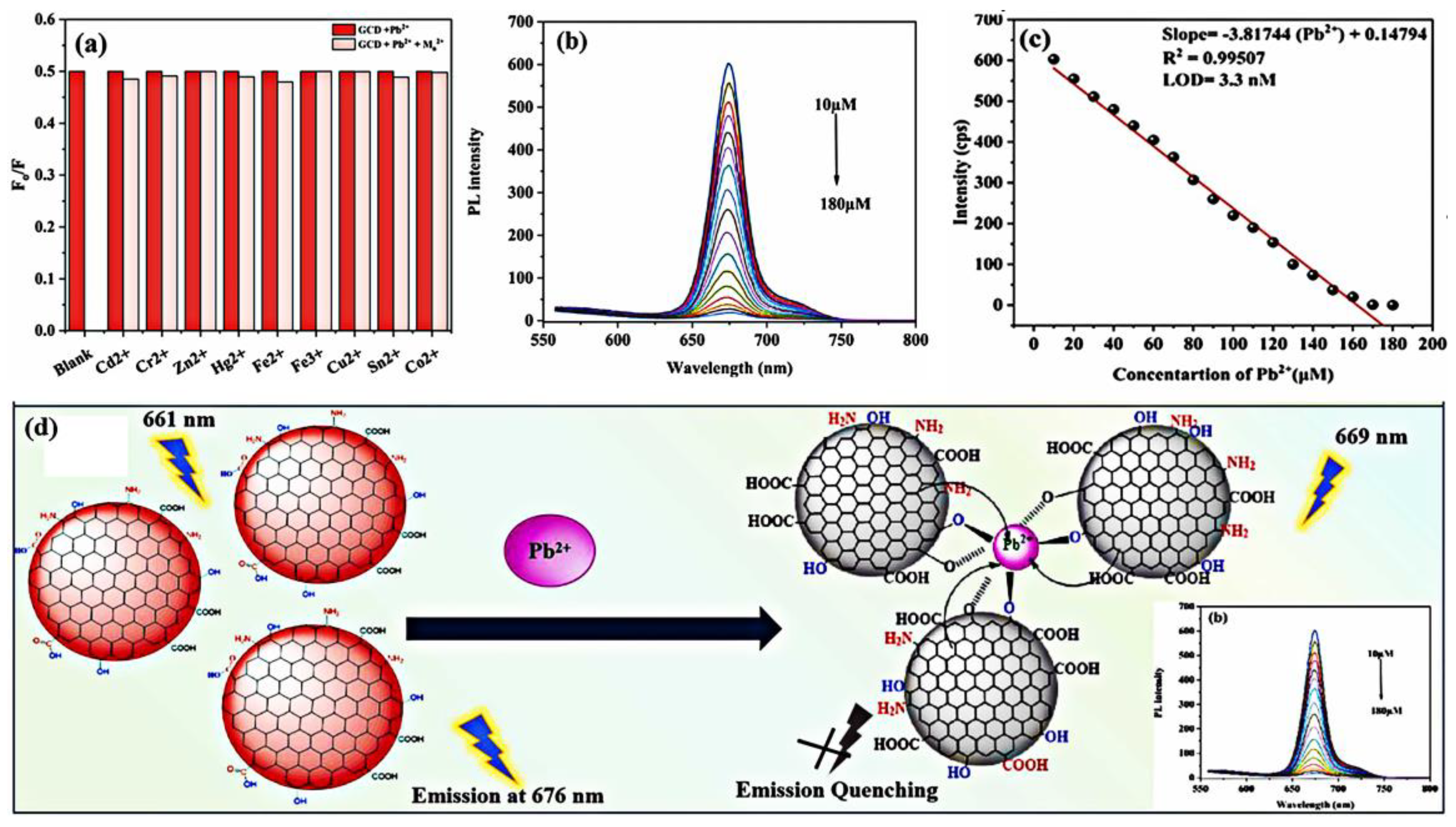
| CQDs | Pb2+ | 10–180 µM | 3.3 | River/lake/tap water | FRET | [115] |
| MPA@CDs | Pb2+ | 0.05–6 µM | 0.051 | Tap water | AIEE | [114] |
| P, Cl-CQDs | Pb2+, gentamicin | 0.0483–14.49 µM | 14.49 | River/tap/mineral water, | Fluorescence quenching | [132] |
| CQDs | Pb2+ | 0.01–1 µM | 0.59 | Mineral/tap/pond water, cellular imaging | Fluorescence quenching | [133] |
| N-CQDs | Pb2+ | 0.05–25 µM | 20 | River/mineral/tap water | SQE | [134] |
| N-CQDs | Pb2+ | 0–200 nM | 9.64 | Cellular imaging, MCF cells | DQE | [135] |
| DMA-GQDs | Pb2+ | 0.01–1 nM | 0.009 | Cerebral spinal fluid of rats | Fluorescence enhancement | [126] |
| GQDs-GO | Pb2+ | 0–400 nM | 0.6 | ~ | PET | [127] |
| DTPAN-fn-GQDs | Pb2+ | 0.5–40 nM | 0.25 | River/tap/well water, paper strips | SQE, DQE | [128] |
| DTP-PPD-fn-GQD | Pb2+ | 3–30 nM | 1.02 | Tap/lake water | SQE | [136] |
| GQDs-AuNPs | Pb2+ | 50–4000 nM | 16.7 | ~ | FRET | [137] |
| S-GQDs | Pb2+ | 0.1–140 µM | 30 | ~ | FRET | [138] |
| QDs-Based Sensors | Analyte(s) | Linearity | LOD (nM) | Applications | Mechanism | Ref. |
|---|---|---|---|---|---|---|
| TGA-CdTe QDs | Ag+ | 5–200 nM | 5 | Lake water | DQE | [146] |
| AgInZnS QDs-NGQDs | Cd2+ | 0.5–100 µM | 28.6 | Lake water | CHEF | [147] |
| L-Asp@CdS QDs | Ag+ | 100–7000 nM | 39 | Drinking water | FRET | [144] |
| CdTe QDs | Zn2+ Cd2+ | 1.6–35 µM 1.3–25 µM | 1200 500 | Tap/spring water | CHEF | [148] |
| N, B-CQDs | Cd2+, L-cysteine | 2.5–22.5 µM | 450 | Human urine | CHEF | [149] |
| urea-ZnO QDs | Cr6+ | 4–1000 µM | 19.53 | ~ | ACQ, DQE | [142] |
| N, B-CQDs | Cr6+ | 1–100 µM | 54, 49, 77 | ~ | DQE | [150] |
| S, N-CQDs | Cr3+ | 0–500 µM | 600 | DI/mineral/tap/river water | Fluorescence quenching | [152] |
| GSH-CdTe QDs | Cr3+ | 0–2 µM | 3 | Vitamin tablets | SQE, DQE | [156] |
| MPA-CdS QDs | Co2+ | 0.04–2 µM | ~ | ~ | SQE | [153] |
| ZnInS QDs | Co2+ | 0.1–100 μM | 99 | ~ | ACQ | [154] |
| CQDs | Co2+ | 0–250 μM | 3010 | Human blood | Fluorescence quenching | [157] |
| N-CQDs | Co2+ | 1–60 μM | 250 | Tap water | SQE | [158] |
| B,N-CQDs | Al3+ Fe2+ | 0–20 μM 0–25 μM | 187 276 | Tap water Ferrous sulfate tablets | AIEE ACQ | [159] |
| N,S-CQDs | Al3+ Fe2+ | 0–20 μM 0–25 μM | 6.99 5.5 | ~ | FE AIEE | [155] |
| ZnS/ZnO QDs | Al3+ | 0.001–8 μM/ 8–100 μM | 1 | Tap/lake water | Fluorescence enhancement | [143] |
| Ag2S QDs | Zn2+ | 1–40 μM | 760 | Peptone/intracellular fluid | Fluorescence enhancement | [160] |
| Ag2S QDs | Cd2+ | 1–40 μM | 546 | Tap/lake water | Fluorescence enhancement | [160] |
| CQDs | Zn2+ | ~ | 6400 | Blood plasma | Fluorescence enhancement | [161] |
| CdTe QDs | Zn2+ Cd2+ | 1.6–35 μM 1.3–25 μM | 1200 500 | Tap/spring water | Fluorescence enhancement | [148] |
| Mn:ZnSe QDs | Cd2+ | 0.02–60 μM | 18 | Tap water | Fluorescence enhancement | [162] |
| QDs-Based Sensors | Selectivity | Linearity | LOD | Applications | Mechanism | Ref. |
|---|---|---|---|---|---|---|
| N, P-CQDs/ Au NPs | carbendazim | 0.005–0.16 µM | 2 nM | Cabbage, apple | FRET | [166] |
| N,CQDs/AuNCs | carbendazim | 1–100 µM 150–1000 µM | 0.83 µM | Strawberry, apple | FRET | [167] |
| S-CQDs/CuNCs | dinotefuran | 10–500 µM | 7.04 µM | Honey | IFE | [168] |
| CdTe-CQDs | glyphosate | 0–1000 nM | 2 pM | Cucumber, capsicum, ginger | PET | [169] |
| CdSe QDs | malathion | 0.908–303 nM | 303 pM | Cabbage leaves | Fluorescence quenching | [170] |
| CdSe/ZnS QDs | organophosphorus | 0.0001–160 μg/mL | 96.7 μg/mL | Tomato | Fluorescence quenching | [171] |
| S-GQDs | omethoate | 4.7–470 µM | 4.7 nM | ~ | Fluorescence enhancement | [172] |
| S-GQDs | carbofuran thiram | 0–225 ppb 0–800 ppb | 0.45 ppb 1.6 ppb | Apple | PET | [173] |
| N-CQDs | thiram paraquat | 10–500 nM 5–100 nM | 7.49 nM 3.03 nM | Semen nelumbinis, coix lacryma-jobi, ginseng | FRET | [174] |
| CdTe QDs | carbaryl | 0–14 μg/mL | 0.12 ng/mL | Apple | Ratiometric fluorescence | [175] |
| CQDs | acephate | 0–100 ng/mL | 0.052 ppb | Pear | IFE | [176] |
| CQDs | chlorpyrifos | 0.02–0.18 μg/mL | 2.7 ng/mL | River water, apple juice | DQE | [177] |
| AP-NSCDs | nitroalkenes | 0.05–10 mg/L | 0.02 mg/L | Rice, orange, cabbage, serum, urine, water | FRET | [178] |
| GQDs/CDs | imidacloprid | 5–4000 nM | 0.823 nM | Banana, apple, cabbage, cucumber | FRET | [179] |
Disclaimer/Publisher’s Note: The statements, opinions and data contained in all publications are solely those of the individual author(s) and contributor(s) and not of MDPI and/or the editor(s). MDPI and/or the editor(s) disclaim responsibility for any injury to people or property resulting from any ideas, methods, instructions or products referred to in the content. |
© 2023 by the authors. Licensee MDPI, Basel, Switzerland. This article is an open access article distributed under the terms and conditions of the Creative Commons Attribution (CC BY) license (https://creativecommons.org/licenses/by/4.0/).
Share and Cite
Wang, Z.; Yao, B.; Xiao, Y.; Tian, X.; Wang, Y. Fluorescent Quantum Dots and Its Composites for Highly Sensitive Detection of Heavy Metal Ions and Pesticide Residues: A Review. Chemosensors 2023, 11, 405. https://doi.org/10.3390/chemosensors11070405
Wang Z, Yao B, Xiao Y, Tian X, Wang Y. Fluorescent Quantum Dots and Its Composites for Highly Sensitive Detection of Heavy Metal Ions and Pesticide Residues: A Review. Chemosensors. 2023; 11(7):405. https://doi.org/10.3390/chemosensors11070405
Chicago/Turabian StyleWang, Zhezhe, Bo Yao, Yawei Xiao, Xu Tian, and Yude Wang. 2023. "Fluorescent Quantum Dots and Its Composites for Highly Sensitive Detection of Heavy Metal Ions and Pesticide Residues: A Review" Chemosensors 11, no. 7: 405. https://doi.org/10.3390/chemosensors11070405