Article

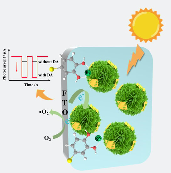
Bi2WO6@g-C3N4 Heterostructure for Cathodic Photoelectrochemical Dopamine Sensor

1
Guangzhou Key Laboratory of Sensing Materials and Devices, Guangdong Engineering Technology Research Center for Photoelectric Sensing Materials and Devices, School of Chemistry and Chemical Engineering, Guangzhou University, Guangzhou 510006, China
2
Guangdong Provincial Key Laboratory of Psychoactive Substances Monitoring and Safety, Anti-Drug Tethnology Center of Guangdong Province, Guangzhou 510230, China
*
Authors to whom correspondence should be addressed.
Chemosensors 2023, 11(7), 404; https://doi.org/10.3390/chemosensors11070404
Submission received: 29 June 2023 / Revised: 14 July 2023 / Accepted: 17 July 2023 / Published: 19 July 2023
(This article belongs to the Special Issue Nanoparticles in Chemical and Biological Sensing)

Abstract

:
A simple and low-cost cathodic photoelectrochemical (PEC) sensor based on Bi2WO6@g-C3N4 was designed for dopamine (DA) detection. The Bi2WO6 nanoflower was first prepared using a simple hydrothermal method followed by the combination with g-C3N4 nanosheet to form the Bi2WO6@g-C3N4 heterostructure. The heterostructure can extend the absorbance to the visible region and accelerate the transfer of charge carriers. Furthermore, DA easily coordinates with exposed Bi3+ on the Bi2WO6 surface and forms the charge-transfer complex to further enhance the cathodic photocurrent. Under optimal conditions, there are two linear relationships between the concentration of DA and photocurrent intensity. The linear ranges are 0.1–10 µM and 10–250 µM, with a sensitive detection limit (LOD) of 28 nM. Notably, the real sample of human blood serum analysis further revealed the accuracy and feasibility of the Bi2WO6@g-C3N4-based PEC platform. Convincingly, the heterostructure of Bi2WO6 and g-C3N4 opened up a new avenue for the construction of DA analysis.

Graphical Abstract

1. Introduction

Dopamine (DA) is an important neurotransmitter that plays a key role in the function of the human central nervous, metabolism, renal, immune and cardiovascular systems [1,2]. DA levels in biological systems have attracted much attention in the clinical field due to abnormal changes in DA concentrations being closely associated with many serious physical and neurological disorders [3,4]. Inadequate levels of DA in the brain may lead to neurological disorders such as schizophrenia and attention deficit hyperactivity disorder [5]. On the other hand, higher levels of DA may lead to pleasure and euphoria [6]. Therefore, the high precision and rapid measurement of DA concentrations are important for the diagnosis and treatment of related disorders.
To date, a number of analytical technologies have been developed for DA detection, such as electrochemiluminescence [7], fluorescence spectrophotometry (FL) [8], high-performance liquid chromatography [9], and electrochemical method (EC) [10,11,12]. It is a pity that most of these methods more or less required sophisticated equipment and were affected by external circumstances and a lack of portability. In recent years, photoelectrochemical (PEC) sensors have blossomed into promising analysis methods with the advantages of EC, such as simple equipment, fast response speed, and being easy-to-operate [13,14]. Owing to the partition of the excitation and detection signal, the PEC sensing exhibits high sensitivity and a lower detection limit than the EC sensor [15,16,17]. And the selection of photoelectric materials is a major key subject in the fabrication of PEC sensors.
Bi2WO6 is an attractive aurivillius oxide that has been widely used in the fields of sensors, photocatalysis, and environmental remediation of wastewater due to its stable physicochemical properties [18,19,20]. However, the low absorption efficiency of pure Bi2WO6 for light and the poor transfer efficiency for electrons are major challenges to further improve the performance of PEC. In the quest for promoting electron(e)-hole(h+) pairs separation and suppressing unnecessary carrier recombination effects, researchers have made great efforts in tuning the physical morphology and regulating the chemical composition. Undoubtedly, the hierarchical structure brings numerous advantages, and researchers have prepared a variety of Bi2WO6 with different morphologies in recent years, such as single-crystal-like [21], helix-like [22], 3D microspheres [23] and 3D flower-like Bi2WO6 [24]. These unique structures can effectively improve the aggregation of nanosheets, thereby exhibiting a larger specific surface area and endowing better photocatalytic performance. Among these 3D structures, the flower-like Bi2WO6 displays a large surface area and outstandingly facilitates the separation of photogenerated charge carriers. Composition tuning mainly focuses on noble mental loading [25], element doping [26], heterostructures [27] and so on. Currently, constructing a heterostructure with two proper energy band matching semiconductors as favorite photoelectrode schemes [28,29,30]. Adhikari et al. prepared Bi2S3/Bi2WO6 hierarchical microstructures that exhibited excellent performance for ofloxacin detection [31]. Zhang prepared Bi2WO6/TiO2 flake nano-heterostructures catalysts, which possessed the high photocatalytic property for the degradation of methylene blue [32]. The above modification method has also demonstrated that the construction of heterojunctions can extend the absorbance to the visible region and more effectively hinder the recombination of carriers, thereby improving the performance of the sensor.
Graphite-like carbon nitride (g-C3N4) has become a research hotspot due to its numerous advantages such as inexpensive, easy synthesis and stable physical and chemical properties and has been widely used in lithium-ion batteries, energy conversion and storage and photocatalysis [33,34,35]. For instance, Zhao et al. established a PEC sensor based on g-C3N4/BiVO4 heterostructure for tetracycline determination [36]. Cao et al. prepared a Cu-BTC MOF/g-C3N4 nanosheet for the detection of non-electroactive glyphosate [37]. The usage of g-C3N4 to design heterojunction indeed greatly improves the performance of the sensor. However, reductive molecules tend to absorb at the n-type-based photoanode/electrolyte interface, which will inevitably affect the intrinsic hole oxidation reaction in real biological sample analysis [38].
Motivated by these concerns, the Bi2WO6 nanoflowers were first prepared using a simple hydrothermal method followed by the combination with g-C3N4 nanosheet to form the Bi2WO6@g-C3N4 heterostructure. The heterostructure showed better PEC performance than pure Bi2WO6, which accelerated the transfer of charge carriers. More importantly, in the presence of DA, DA will replace water molecules and form the charge-transfer complex in concert with exposed Bi3+, which is a key factor for enhanced cathodic photocurrent [39]. Moreover, the Bi2WO6@g-C3N4-based PEC sensor obtained a good recovery in real human serum samples. Thus, the easily fabricated at a low-cost label-free PEC sensor based on Bi2WO6@g-C3N4 heterostructure achieves the selective detection of DA, which opened up a new avenue for the construction of DA analysis.

2. Materials and Methods

2.1. Reagents and Materials

The fluorine-doped tin oxide (FTO) glasses used as the substrates were purchased from Jinge-Wuhan and cut into 15 × 25 mm2 pieces before use. Bismuth nitrate pentahydrate (Bi(NO3)3•5H2O, 99%), sodium tungstate dihydrate (Na2WO4•2H2O) and urea were received from J&K Scientific Ltd. (Beijing, China). Ascorbic acid (AA), DA, uric acid (UA), L-proline (Pro), glutathione (GSH), L-cysteine (Cys), histidine (His) and glucose (Glu) were provided by InnoChem Science & Technology Co., Ltd. (Beijing, China). All reagents were used without further purification.

2.2. Apparatuses

In this work, a scanning electron microscope (SEM, JSM-7001F, Hitachi, Tokyo, Japan) and transmission electron microscope (TEM, JSM-2100F, JEOL, Tokyo, Japan) were used to observe the surface morphology and lattice spacing of the samples. The crystal structure information was studied using the X-ray diffractometer (XRD, Rigaku Ultima IV, Tokyo, Japan) with Cu Kα radiation (λ = 0.15405 nm). X-ray photoelectron spectroscopy (XPS, Thermo Fischer ESCALAB Xi+, Waltham, MA, USA) was employed to characterize the surface chemical states of the samples. The vacuum level of the analysis chamber was 8 × 10⁻¹⁰ Pa, and the excitation source was Al kα rays (hν = 1486.6 eV). UV-vis diffuse reflectance spectra (DRS) of the samples were recorded on a UV-vis spectrophotometer (U-3900, Hitachi, Tokyo, Japan). The Fourier transform infrared (FTIR) spectra were carried out by an FTIR Spectrometer (Thermo Nicolet iS50, Waltham, MA, USA). A 420 nm LED light (Beijing Perfectlight Technology Co., Ltd., Beijing, China) was used as the irradiation source. All electrochemical experiments were carried out on the electrochemical workstation (CHI660E, Shanghai, China).

2.3. Syntheses of Bi2WO6@g-C3N4 Composites

The g-C3N4 was prepared using the reported heat-condensation polymerization method [35]. Quite simply, 10 g of urea was ground and placed in a tube furnace and maintained at a heating rate of 5 °C/min to 550 °C for 4 h. After natural cooling, the resulting pale yellow solid g-C3N4 was ground and prepared for use.
Bi2WO6 nanoflower structures (Bi2WO6 NFs) were synthesized using a conventional hydrothermal method [40]. In a typical process, 0.33 g of Na2WO4•2H2O was dissolved in 20 mL of deionized (DI) water, labeled as solution A. And 0.97 g of Bi(NO3)3•5H2O was prepared in another 20 mL of DI water, labeled as solution B. Solution B was then slowly added dropwise to solution A. The pH of the mixed solution was adjusted to 1 and stirring was continued for another 1 h. Subsequently, the final mixture solution was transferred to a 100 mL Teflon-lined stainless-steel autoclave and heated at 160 °C for 15 h. The reaction was brought to an end, and the reactor was allowed to cool naturally to room temperature. The obtained precipitate was washed through centrifugation with water and ethanol and then collected. The obtained precipitate was dried under a vacuum at 60 °C and ground into homogeneous pellets.
A certain amount of g-C3N4 (5 mg, 10 mg, 15 mg) was dispersed into 20 mL of DI water and sonicated for 1 h. Subsequently, 100 mg of the above-prepared Bi2WO6 NFs powder was weighed into the dispersion and stirred for 1 h. The purpose of stirring is to allow better contact between Bi2WO6 and g-C3N4 to form a more homogeneous complex. Then precipitate was separated through centrifugation and dried. The obtained samples were labeled as BWO-0.05CN, BWO-0.1CN and BWO-0.15CN, respectively.

2.4. Fabrication of Bi2WO6@g-C3N4/FTO PEC Sensor

Next, 100 μL of 2 mg/mL Bi2WO6@g-C3N4 sample was dropped on FTO electrode with an active area of 0.785 cm2, prepared using waterproof tape with a 1.0 cm diameter hole of. The performance of the same Bi2WO6@g-C3N4/FTO electrode was tested under optimal conditions. Bi2WO6@g-C3N4/FTO was placed as the working electrode in a homemade PEC detection cell containing 4 mL of 0.1 M PBS solution, together with Ag/AgCl used as the reference electrode and Pt wire used as the counter electrode to form a three-electrode detection system. The PEC detection was performed on a CHI660e electrochemical workstation, and data were recorded using the current-time (i-t) technique type at a bias potential of 0 V. A 420 nm LED was used as the irradiation source. After the dark current signal was stabilized, the photocurrent signal was collected by switching the light on and off alternately for 10 s three times. Different concentrations of DA were added continuously to the test cell for detection.

3. Results and Discussion

3.1. Morphology Characterization of Bi2WO6@g-C3N4 Composites

The synthetic strategy of the Bi2WO6@g-C3N4 sample is illustrated in Figure 1, which involves two steps. The Bi2WO6 nanoflower was first prepared using a simple hydrothermal method followed by the combination with g-C3N4 nanosheet to form the Bi2WO6@g-C3N4 heterostructure. The morphology, microstructure and lattice structure of BWO-0.1CN heterostructure were first investigated through SEM, TEM and HRTEM. Figure 2A shows that the Bi2WO6 obtained hydrothermally were uniform, and individual particles had a diameter of 2 μm. The higher magnification SEM image in Figure 2B presents that Bi2WO6 particles possess a 3D flower-like structure formed by numerous nanosheets with a thickness of several nanometers, which can greatly increase the surface area and active site of the samples. After the recombination with g-C3N4, it can be seen from Figure 2C that some of the g-C3N4 nanosheets grow on the Bi2WO6 nanoflower, and others disperse between the nanoflowers. Furthermore, the HRTEM image was further employed to confirm the formation of the Bi2WO6@g-C3N4 heterostructure. As shown in Figure 2D, some of the amorphous phases decorate the surface of the high crystallinity phase with an obvious interface, indicating its heterostructure character. The lattice spacing of about 0.315 nm belongs to the spacing of the (113) plane of orthogonal Bi2WO6 [40], while the amorphous phase with no clear lattice fringe is observed mainly due to the disordered state of g-C3N4. In addition, EDS-mapping (Figure 2E) shows that Bi, W, O, C and N elements are uniformly dispersed on the Bi2WO6@g-C3N4, which further clarifies the reliability of the heterostructure.

3.2. Structure and Chemical Compositions of Bi2WO6@g-C3N4 Composites

The crystal structures of the Bi2WO6, g-C3N4, BWO-0.05CN, BWO-0.1CN and BWO-0.15CN were evaluated using XRD analysis, respectively (Figure 3A). The main diffraction peaks of the Bi2WO6 nanoflower at 28.3°, 32.8°, 47.1°, 55.9° and 58.5° in the XRD pattern are indexed to the (131), (200), (202), (133) and (262) planes of the orthogonal Bi2WO6 phase (JCPDS card No. 39-0256) [40]. The two peaks at 13.1° and 27.3° are the typical diffraction peaks of g-C3N4, which belong to the (100) and (002) planes of the C3N4 crystal [40]. However, there is no obvious peak of the g-C3N4 phase in a series of Bi2WO6@g-C3N4 heterostructure samples, which is probably due to the less content and low crystallinity of g-C3N4, and it overlaps with the strong peak of Bi2WO6.
Furthermore, the FT-IR spectra were carried out to investigate the chemical structure of the samples. As shown in Figure 3B, the broad peaks at 3100–3300 cm−1 (the orange area) are assigned to the N-H stretching vibration modes, which are mainly caused by the uncondensed amino groups in the g-C3N4. In addition, the absorption peaks at 1200–1650 cm−1 (the purple area) originate from the representative stretching modes of aromatic C-N and C=N heterocycles. And the featured typical peak located at 811 cm−1 is derived from the characteristic vibration mode of tri-s-triazine units [41]. Moreover, the vibration bands at 579, 729 and 813 cm−1 in the Bi2WO6 sample could be attributed to the Bi-O, W-O and W-O-W bonds, respectively [42]. Obviously, it can be seen that the characteristic peaks of Bi2WO6 and g-C3N4 exist simultaneously in the series of Bi2WO6@g-C3N4 samples, demonstrating the successful construction of the Bi2WO6@g-C3N4 heterojunction.
The surface elemental compositions and valence states of the BWO-0.1CN sample were further confirmed through XPS measurements. As depicted in Figure 4A, the XPS survey spectrum of the BWO-0.1CN confirms the coexistence of Bi, W, O, C and N elements, in which the Bi, W and O elements come from the Bi2WO6 and the C and N elements due to the g-C3N4. Figure 4B–F show the high-resolution XPS spectra of Bi4f, W4f, O1s, C1s and N1s, respectively. The high-resolution of the Bi 4f spectra (Figure 4B) shows a pair of spin-orbit doublet peaks located at 164.67 and 159.38 eV, indicating that Bi exists in BWO-0.1CN in the form of Bi3+ [40]. As for the W4f core-level spectra (Figure 4C), two main peaks at binding energies of 37.76 and 35.63 eV were assigned to W 4f5/2 and W 4f7/2, respectively, suggesting that the W in BWO-0.1CN is in the valence state of W6+ [42]. In addition, the O 1s spectrum at 530.03 and 532.37 eV in Figure 4D is consistent with the different chemical environments of Bi-O and W-O for oxygen elements, and another peak at 530.57 eV corresponds to the OH group at the surface of Bi2WO6. In Figure 4E, the C1s signal can be fitted into three peaks centered at 284.80, 286.35 and 288.58 eV, which correspond to the graphitic carbon (C-C), sp3 coordinated carbon (C-N3) and sp2 hybridized carbon (N-C=N), respectively [43]. Similarly, in the N1s spectrum (Figure 4F), the binding energies at 399.12, 400.09 and 406.68 eV are attributed to the sp2 hybridized nitrogen of C-N=C, the sp3 bridged N atom of the N-C3 group and the terminal amino groups of C-N-H, respectively [43]. Similarly, the different contents (5%, 15%) of g-C3N4 samples BWO-0.05CN and BWO-0.15CN show the adjacent parameters were used to deconvolute Bi4f, W4f, O1s, C1s and N1s core level as shown in Figures S1 and S2. Obviously, the C-N3 and N-C3 groups in the C1s and N1s spectrum prove the existence of triple-s-triazine structure in g-C3N4, which also confirmed the coexistence of g-C3N4 and Bi2WO6 in the BWO-0.05CN, BWO-0.1CN and BWO-0.15CN heterojunction, and this is consistent with the results of the above measurements.

3.3. Construction of the PEC Sensor

The transient photocurrent response of BWO/FTO and BWO-0.1CN/FTO in the presence and absence of DA under light irradiation was carried out in Figure 5A. Not surprisingly, the BWO/FTO shows a faint photocurrent intensity of about 32.5 nA in the absence of DA. Since g-C3N4 embedded in Bi2WO6 nanoflowers, the photocurrent value of BWO-0.1CN (78.7 nA) is stronger than BWO/FTO, and the absorption edge of BWO-0.1CN was between the pure Bi2WO6 and pure g-C3N4 (Figure S3A,B), indicating that the introduction of g-C3N4 nanosheets and the formation of Bi2WO6@g-C3N4 heterojunction are more susceptible to excitation. Likewise, in the presence of 50 µM DA, the instantaneous photocurrent of BWO/FTO (250.9 nA) just accounted for 25.1% of BWO-0.1CN (999.3 nA). It should be pointed out that constructing a Bi2WO6@g-C3N4 heterostructure can more effectively hinder the recombination of carriers, thereby resulting in a remarkable photoelectric signal in return. EIS spectra of BWO/FTO and BWO-0.1CN/FTO are further authenticated by the viewpoint (Figure 5B), where BWO-0.1CN/FTO shows smaller semicircle radii with respect to BWO, indicating that BWO-0.1CN/FTO has a faster transfer rate of electrons.
To improve the fabricated sensor performance, the amounts of g-C3N4 nanosheets in Bi2WO6@g-C3N4 heterojunction were optimized, which are related to the transfer rate of carriers. Therefore, a series of composite materials BWO-0.05CN, BWO-0.1CN and BWO-0.15CN with different contents (5%, 10%, 15%) of g-C3N4 were synthesized. And the photocurrent response of these composites in the presence of 50 µM DA was tested. From Figure S4A, it can be seen that as the doping ratio of g-C3N4 increases, the photocurrent response is gradually enhanced due to the more active sites that are beneficial for the capture of e or h+. Oppositely, the amount of g-C3N4 ratio exceeds 10%, and excessive g-C3N4 nanosheets cover the surface of the flower-like Bi2WO6, which limits the exposure of active sites, resulting in the photocurrent intensity presenting a downward trend. BWO-0.1CN exhibited the highest photocurrent response. Moreover, EIS spectra of BWO-0.05CN, BWO-0.1CN and BWO-0.15CN have further approved the impunity (Figure S4B), where BWO-0.1CN shows the smallest semicircle radii, indicating that BWO-0.1CN has the optimum conductivity. So we chose to construct the sensor with BWO-0.1CN as the photoelectrode material. In addition, the parameter configuration is another factor that may trigger large differences in photocurrent response. Figure S4C depicts the influence of applied potential on the BWO-0.1CN-based sensor platform. The operation voltage of −0.2 V shows the strongest transient photoelectric current, but considering the ease of use in instrument integration, 0.0 V was chosen as the working voltage in the following experiment.

3.4. PEC Sensor for DA Detection

Relying on optimal experimental conditions, the PEC sensor fabricated based on BWO-0.1CN/FTO was used for the quantitative analysis of DA. As shown in Figure 6A, the photocurrent gradually increased with increasing the concentration of DA and eventually reached a plateau. As a matter of fact, there were two linear relationships between the concentration of DA and photocurrent intensity (Figure 6B). The linear ranges were 0.1–10 µM and 10–250 µM, respectively. The linear equations were y1 = 44.47x1 + 123.93 (R2 = 0.9908), y2 = 7.1188x2 + 574.92 (R2 = 0.9922). It is worth noting that the slope was higher at a low concentration of DA because the electron transfer rate had greater change at a low concentration of DA compared to a high concentration [44,45]. And the detection limit was about 28 nM, which was comparable to the previously reported techniques (as shown in Table S1) [16,46,47,48,49,50]. Clearly, the established PEC sensor obtained excellent sensing performance for DA quantitative analysis.

3.5. Selectivity and Stability of the PEC Sensor

In fact, the actual test samples are more complex, and co-interferences may set obstacles to the detection process. In light of the actual requirement, the photocurrent responses of some possible commensal interferents (GSH, Cys, AA, Glu, His, Pro, UA) were compared with DA (Figure 6C), and it was observed that these interferents exhibited negligible PEC responses even at 10 times higher concentrations than DA. Furthermore, the interferer AA, which most readily interferes with DA detection, is only 7.4% of the DA photocurrent response at a tenfold concentration. The stability of the sensor constructed with BWO-0.1CN was verified with repeated irradiation on and off 20 times, with no significant photocurrent changes during the measurements, signifying acceptable stability (Figure 6D). These results indicated that the fabricated BWO-0.1CN-based sensor exhibited satisfactory selectivity and high photochemical stability.

3.6. Real Samples Analysis

In order to investigate the practical application of the BWO-0.1CN-based PEC platform, the determination of DA in the human blood serum samples was carried out. The accuracy of the method was verified using the standard addition method, and DA standard solutions of 0.5 µM, 10 µM and 25 µM were adopted for the DA determination. The satisfactory recoveries (98.5–104%) and precision (RSD < 4%) were obtained from the assay results listed in Table 1, illustrating that the promising application of the BWO-0.1CN-based PEC sensor can be achieved for the detection of DA in real samples.

3.7. PEC Sensing Mechanism

Scheme 1 shows the possible mechanism for the selective detection of DA on the Bi2WO6@g-C3N4/FTO electrode. When the BWO-0.1CN is irradiated with 420 nm light, electrons on the valence band (VB) of g-C3N4 can absorb photons and then transfer to the conduction band (CB) to generate e-h+ pairs. Then, the photo-generated e is rapidly transferred to the CB of Bi2WO6 through the interface between g-C3N4 and Bi2WO6. On the one hand, g-C3N4 nanosheets attached to 3D flower-like Bi2WO6 can not only reduce the diffusion pathway of photogenerated carriers participating in surface reactions but also facilitate the charge transfer in the heterojunction, resulting in enhanced photocurrent response. On the other hand, the abundant Bi3+ exposed on the Bi2WO6 surface may partially coordinate with water molecules due to the unstable bonding between Bi and O atoms [51]. More importantly, in the presence of DA, DA will replace water molecules and form the charge-transfer complex in concert with the exposed Bi3+, leading to a change in the energy positions of the surface state [39]. Therefore, the free CB electrons react rapidly with the electron acceptor (dissolved O2), which is a key factor for enhanced cathodic photocurrent. Thus, a PEC sensor for DA sensitive detection was developed based on the enhanced cathodic photocurrent strategy.

4. Conclusions

In brief, a label-free cathodic PEC sensor employing BWO-0.1CN was simply established to achieve the sensitive and selective detection of DA. The introduction of g-C3N4 nanosheets and the formation of Bi2WO6@g-C3N4 heterojunctions are more susceptible to exciting and improving the transfer efficiency of photogenerated carriers. In addition, DA easily coordinates with exposed Bi3+ on the Bi2WO6 surface and forms the charge-transfer complex to further enhance the cathodic photocurrent. Benefitting from the Bi2WO6@g-C3N4 heterojunction interface, the proposed BWO-0.1CN-based analytical platform was successfully applied to the determination of DA with excellent interference resistance, high sensitivity, favorable reproducibility and good practicality. Such a simple and low-cost PEC platform holds great promise to apply for early DA-related disease diagnosis.

Supplementary Materials

The following supporting information can be downloaded at: https://www.mdpi.com/article/10.3390/chemosensors11070404/s1, Figure S1: The XPS spectra of the BWO-0.05CN (A) Full scan, (B) Bi 4f, (C) W 4f, (D) O 1s, (E) C 1s, (F) N 1s. Figure S2: The XPS spectra of the BWO-0.15CN (A) Full scan, (B) Bi 4f, (C) W 4f, (D) O 1s, (E) C 1s, (F) N 1s. Figure S3: (A) UV-vis diffuse reflectance absorption spectra, (B) the corresponding K-M plot of Bi2WO6, g-C3N4, and BWO-0.1CN.; Figure S4: (A) Photocurrent response of BWO-0.05CN, BWO-0.1CN, and BWO-0.15CN in the presence of 50 µM DA. (B) Effects of the applied potential on photocurrent response of BWO-0.1CN/FTO electrode in 0.1 M PBS (pH = 7.4) containing 50 µM DA. Table S1: Comparison of previous and current DA detection methods. References [16,46,47,48,49,50] are cited in the Supplementary Materials.

Author Contributions

Conceptualization, Z.W.; methodology, Y.S. and F.H.; validation, Y.S. and F.H.; formal analysis, F.H. and D.Q.; investigation, Z.W. and Z.L.; data curation, Z.L.; writing—original draft preparation, D.H. and D.Q.; writing—review and editing, Z.W. and L.N.; supervision, L.N. All authors have read and agreed to the published version of the manuscript.

Funding

This research was funded by the National Key R&D Program of China (2022YFD2100304), National Natural Science Foundation of China (22172040, 21974031), University-Industry Collaborative Education Program of Ministry of Education of China (220605940231526), Guangdong Basic and Applied Basic Research Foundation (2023B1515040004), the Department of Science and Techniques of Guangdong Province (2021A1515010180, 2019B010933001), the Department of Guangdong Provincial Public Security (GZQC20-PZ11-FD084), Science and Technology Projects in Guangzhou (202201000002), Department of Science & Technology of Guangdong Province (ID:2022A156), Guangzhou Municipal Science and Technology Bureau (202102010449) and Tertiary Education Scientific research project of Guangzhou Municipal Education Bureau (202235344), and we thank them for their financial support of this work.

Institutional Review Board Statement

Not applicable.

Informed Consent Statement

Not applicable.

Data Availability Statement

Not applicable.

Conflicts of Interest

The authors declare no conflict of interest.

References

  1. Berke, J.D. What does dopamine mean? Nat. Neurosci. 2018, 21, 787–793. [Google Scholar] [CrossRef] [PubMed]
  2. Zhang, Y.; Xu, M.; Gao, P.; Gao, W.; Bian, Z.; Jia, N. Photoelectrochemical sensing of dopamine using gold-TiO2 nanocomposites and visible-light illumination. Microchim. Acta 2019, 186, 326. [Google Scholar] [CrossRef] [PubMed]
  3. Liu, C.; Gomez, F.A.; Miao, Y.; Cui, P.; Lee, W. A colorimetric assay system for dopamine using microfluidic paper-based analytical devices. Talanta 2019, 194, 171–176. [Google Scholar] [CrossRef] [PubMed]
  4. Segura-Aguilar, J.; Paris, I.; Munoz, P.; Ferrari, E.; Zecca, L.; Zucca, F.A. Protective and toxic roles of dopamine in Parkinson’s disease. J. Neurochem. 2014, 129, 898–915. [Google Scholar] [CrossRef]
  5. Faraone, S.V. The pharmacology of amphetamine and methylphenidate: Relevance to the neurobiology of attention-deficit/hyperactivity disorder and other psychiatric comorbidities. Neurosci. Biobehav. Rev. 2018, 87, 255–270. [Google Scholar] [CrossRef]
  6. Guo, Z.; Seol, M.L.; Kim, M.S.; Ahn, J.H.; Choi, Y.K.; Liu, J.H.; Huang, X.J. Sensitive and selective electrochemical detection of dopamine using an electrode modified with carboxylated carbonaceous spheres. Analyst 2013, 138, 2683–2690. [Google Scholar] [CrossRef]
  7. Zhuang, X.M.; Gao, X.Q.; Tian, C.Y.; Cui, D.L.; Luan, F.; Wang, Z.G.; Xiong, Y.; Chen, L.X. Synthesis of europium(iii)-doped copper nanoclusters for electrochemiluminescence bioanalysis. Chem. Commun. 2020, 56, 5755–5758. [Google Scholar] [CrossRef]
  8. Peng, J.; Han, C.L.; Ling, J.; Liu, C.J.; Ding, Z.T.; Cao, Q.E. Selective fluorescence quenching of papain-Au nanoclusters by self-polymerization of dopamine. Luminescence 2018, 33, 168–173. [Google Scholar] [CrossRef] [PubMed]
  9. Tsunoda, M.; Aoyama, C.; Nomura, H.; Toyoda, T.; Matsuki, N.; Funatsu, T. Simultaneous determination of dopamine and 3,4-dihydroxyphenylacetic acid in mouse striatum using mixed-mode reversed-phase and cation-exchange high-performance liquid chromatography. J. Pharm. Biomed. Anal. 2010, 51, 712–715. [Google Scholar] [CrossRef]
  10. Shin, J.W.; Kim, K.J.; Yoon, J.; Jo, J.; El-Said, W.A.; Choi, J.W. Silver nanoparticle modified electrode covered by graphene oxide for the enhanced electrochemical detection of dopamine. Sensors 2017, 17, 2771. [Google Scholar] [CrossRef] [Green Version]
  11. Zhou, J.; Huang, Y.; Chen, C.; Xiao, A.; Guo, T.; Guan, B.O. Improved detection sensitivity of gamma-aminobutyric acid based on graphene oxide interface on an optical microfiber. Phys. Chem. Chem. Phys. 2018, 20, 14117–14123. [Google Scholar] [CrossRef] [PubMed]
  12. Kokulnathan, T.; Ahmed, F.; Chen, S.M.; Chen, T.W.; Hasan, P.M.Z.; Bilgrami, A.L.; Darwesh, R. Rational confinement of yttrium vanadate within three-dimensional graphene aerogel: Electrochemical analysis of monoamine neurotransmitter (dopamine). ACS Appl. Mater. Interfaces 2021, 13, 10987–10995. [Google Scholar] [CrossRef] [PubMed]
  13. Yan, Y.; Liu, Q.; Du, X.; Qian, J.; Mao, H.; Wang, K. Visible light photoelectrochemical sensor for ultrasensitive determination of dopamine based on synergistic effect of graphene quantum dots and TiO2 nanoparticles. Anal. Chim. Acta 2015, 853, 258–264. [Google Scholar] [CrossRef] [PubMed]
  14. Hun, X.; Wang, S.; Wang, S.; Zhao, J.; Luo, X. A photoelectrochemical sensor for ultrasensitive dopamine detection based on single-layer NanoMoS2 modified gold electrode. Sens. Actuators B Chem. 2017, 249, 83–89. [Google Scholar] [CrossRef]
  15. Ma, W.; Wang, L.; Zhang, N.; Han, D.; Dong, X.; Niu, L. Biomolecule-free, selective detection of o-diphenol and its derivatives with WS2/TiO2-based photoelectrochemical platform. Anal. Chem. 2015, 87, 4844–4850. [Google Scholar] [CrossRef]
  16. Wu, Z.; Han, F.; Wang, T.; Guan, L.; Liang, Z.; Han, D.; Niu, L. A recognition-molecule-free photoelectrochemical sensor based on Ti3C2/TiO2 heterostructure for monitoring of dopamine. Biosensors 2023, 13, 526. [Google Scholar] [CrossRef]
  17. Han, F.J.; Song, Z.Q.; Xu, J.N.; Dai, M.J.; Luo, S.L.; Han, D.X.; Niu, L.; Wang, Z.X. Oxidized titanium carbide MXene-enabled photoelectrochemical sensor for quantifying synergistic interaction of ascorbic acid based antioxidants system. Biosens. Bioelectron. 2021, 177, 112978. [Google Scholar] [CrossRef]
  18. Zhang, J.; Huang, L.; Jin, H.; Sun, Y.; Ma, X.; Zhang, E.; Wang, H.; Kong, Z.; Xi, J.; Ji, Z. Constructing two-dimension MoS2 /Bi2WO6 core-shell heterostructure as carriers transfer channel for enhancing photocatalytic activity. Mater. Res. Bull. 2017, 85, 140–146. [Google Scholar] [CrossRef]
  19. Li, C.; Chen, G.; Sun, J.; Feng, Y.; Liu, J.; Dong, H. Ultrathin nanoflakes constructed erythrocyte-like Bi2WO6 hierarchical architecture via anionic self-regulation strategy for improving photocatalytic activity and gas-sensing property. Appl. Catal. B 2015, 163, 415–423. [Google Scholar] [CrossRef]
  20. Zhang, Z.J.; Wang, W.Z.; Wang, L.; Sun, S.M. Enhancement of visible-light photocatalysis by coupling with narrow-band-gap semiconductor: A case study on Bi2S3/Bi2WO6. ACS Appl. Mater. Interfaces 2012, 4, 593–597. [Google Scholar] [CrossRef]
  21. Li, C.; Chen, G.; Sun, J.; Rao, J.; Han, Z.; Hu, Y.; Zhou, Y. A novel mesoporous single-crystal-like Bi2WO6 with enhanced photocatalytic activity for pollutants degradation and oxygen production. ACS Appl. Mater. Interfaces 2015, 7, 25716–25724. [Google Scholar] [CrossRef] [PubMed]
  22. Zhang, L.; Wang, W.; Zhou, L.; Xu, H. Bi2WO6 nano and microstructures: Shape control and associated visible-light-driven photocatalytic activities. Small 2007, 3, 1618–1625. [Google Scholar] [CrossRef] [PubMed]
  23. Zhang, L.; Wang, W.; Chen, Z.; Zhou, L.; Xu, H.; Zhu, W. Fabrication of flower-like Bi2WO6 superstructures as high performance visible-light driven photocatalysts. J. Mater. Chem. 2007, 17, 2526–2532. [Google Scholar] [CrossRef]
  24. Qi, S.; Zhang, R.; Zhang, Y.; Liu, X.; Xu, H. Preparation and photocatalytic properties of Bi2WO6/g-C3N4. Inorg. Chem. Commun. 2021, 132, 108761. [Google Scholar] [CrossRef]
  25. Liu, J.; Wang, T.; Nie, Q.; Hu, L.; Cui, Y.; Tan, Z.; Yu, H. One-step synthesis of metallic Bi deposited Bi2WO6 nanoclusters for enhanced photocatalytic performance: An experimental and DFT study. Appl. Surf. Sci. 2021, 559, 149970. [Google Scholar] [CrossRef]
  26. Liu, L.; Liu, J.; Sun, K.; Wan, J.; Fu, F.; Fan, J. Novel phosphorus-doped Bi2WO6 monolayer with oxygen vacancies for superior photocatalytic water detoxication and nitrogen fixation performance. Chem. Eng. J. 2021, 411, 128629. [Google Scholar] [CrossRef]
  27. Peng, J.; Zhuge, W.; Liu, Y.; Zhang, C.; Yang, W.; Huang, Y. Photoelectrochemical dopamine sensor Based on Cu-Doped Bi2WO6 micro-Flowers sensitized cobalt tetraaminophthalocyanine functionalized graphene oxide. J. Electrochem. Soc. 2019, 166, B1612–B1619. [Google Scholar] [CrossRef]
  28. Wang, X.; Rong, X.; Zhang, Y.; Luo, F.; Qiu, B.; Wang, J.; Lin, Z. Homogeneous photoelectrochemical aptasensors for tetracycline based on sulfur-doped g-C3N4/n-GaN heterostructures formed through self-assembly. Anal. Chem. 2022, 94, 3735–3742. [Google Scholar] [CrossRef]
  29. Liu, Z.; Zhou, Y.; Yang, L.; Yang, R. Green preparation of in-situ oxidized TiO2/Ti3C2 heterostructure for photocatalytic hydrogen production. Adv. Powder Technol. 2021, 32, 4857–4861. [Google Scholar] [CrossRef]
  30. Zhang, J.; Shang, M.; Gao, Y.; Yan, J.; Song, W. High-performance VS2 QDs-based type II heterostructured photoanode for ultrasensitive aptasensing of lysozyme. Sens. Actuators B Chem. 2020, 304, 127411. [Google Scholar] [CrossRef]
  31. Adhikari, S.; Kim, D.-H. Synthesis of Bi2S3/Bi2WO6 hierarchical microstructures for enhanced visible light driven photocatalytic degradation and photoelectrochemical sensing of ofloxacin. Chem. Eng. J. 2018, 354, 692–705. [Google Scholar] [CrossRef]
  32. Zhang, M. Synthesis of Bi2WO6/TiO2 flake nano-heterostructure photocatalyst and its photocatalytic performance under visible light irradiation. J. Mater. Sci. Mater. Electron. 2020, 31, 20129–20138. [Google Scholar] [CrossRef]
  33. Zhao, B.; Luo, Y.; Qu, X.; Hu, Q.; Zou, J.; He, Y.; Liu, Z.; Zhang, Y.; Bao, Y.; Wang, W.; et al. Graphite-like carbon nitride nanotube for electrochemiluminescence featuring high efficiency, high stability, and ultrasensitive ion detection capability. J. Phys. Chem. Lett. 2021, 12, 11191–11198. [Google Scholar] [CrossRef]
  34. Yang, H.; Zhou, Q.; Fang, Z.; Li, W.; Zheng, Y.; Ma, J.; Wang, Z.; Zhao, L.; Liu, S.; Shen, Y.; et al. Carbon nitride of five-membered rings with low optical bandgap for photoelectrochemical biosensing. Chem 2021, 7, 2708–2721. [Google Scholar] [CrossRef]
  35. Wang, D.; Li, J.; Xu, Z.; Zhu, Y.; Chen, G.; Cui, Z. Synthesis of g-C3N4/NiO p–n heterojunction materials with ball-flower morphology and enhanced photocatalytic performance for the removal of tetracycline and Cr6+. J. Mater. Sci. 2019, 54, 11417–11434. [Google Scholar] [CrossRef]
  36. Zhao, Z.; Wu, Z.; Lin, X.; Han, F.; Liang, Z.; Huang, L.; Dai, M.; Han, D.; Han, L.; Niu, L. A label-free PEC aptasensor platform based on g-C3N4/BiVO4 heterojunction for tetracycline detection in food analysis. Food Chem. 2023, 402, 134258. [Google Scholar] [CrossRef]
  37. Cao, Y.; Wang, L.; Wang, C.; Hu, X.; Liu, Y.; Wang, G. Sensitive detection of glyphosate based on a Cu-BTC MOF/g-C3N4 nanosheet photoelectrochemical sensor. Electrochim. Acta 2019, 317, 341–347. [Google Scholar] [CrossRef]
  38. Xu, Y.; Yu, S.; Zhu, Y.; Fan, G.; Han, D.; Qu, P.; Zhao, W. Cathodic photoelectrochemical bioanalysis. TrAC Trend. Anal. Chem. 2019, 114, 81–88. [Google Scholar] [CrossRef]
  39. Weng, S.; Hu, J.; Lu, M.; Ye, X.; Pei, Z.; Huang, M.; Xie, L.; Lin, S.; Liu, P. In situ photogenerated defects on surface-complex BiOCl (0 1 0) with high visible-light photocatalytic activity: A probe to disclose the charge transfer in BiOCl (0 1 0)/surface-complex system. Appl. Catal. B 2015, 163, 205–213. [Google Scholar] [CrossRef]
  40. Yang, C.; Chai, H.; Xu, P.; Wang, P.; Wang, X.; Shen, T.; Zheng, Q.; Zhang, G. One-step synthesis of a 3D/2D Bi2WO6/g-C3N4 heterojunction for effective photocatalytic degradation of atrazine: Kinetics, degradation mechanisms and ecotoxicity. Sep. Purif. Technol. 2022, 288, 120609. [Google Scholar] [CrossRef]
  41. Wang, Q.; Wang, W.; Zhong, L.; Liu, D.; Cao, X.; Cui, F. Oxygen vacancy-rich 2D/2D BiOCl-g-C3N4 ultrathin heterostructure nanosheets for enhanced visible-light-driven photocatalytic activity in environmental remediation. Appl. Catal. B 2018, 220, 290–302. [Google Scholar] [CrossRef]
  42. Zhu, D.; Zhou, Q. Novel Bi2WO6 modified by N-doped graphitic carbon nitride photocatalyst for efficient photocatalytic degradation of phenol under visible light. Appl. Catal. B 2020, 268, 118426. [Google Scholar] [CrossRef]
  43. Duan, Y.; Li, X.; Lv, K.; Zhao, L.; Liu, Y. Flower-like g-C3N4 assembly from holy nanosheets with nitrogen vacancies for efficient NO abatement. Appl. Surf. Sci. 2019, 492, 166–176. [Google Scholar] [CrossRef]
  44. Zheng, L.; Zhang, H.; Won, M.; Kim, E.; Li, M.; Kim, J.S. Codoping g-C3N4 with boron and graphene quantum dots: Enhancement of charge transfer for ultrasensitive and selective photoelectrochemical detection of dopamine. Biosens. Bioelectron. 2023, 224, 115050. [Google Scholar] [CrossRef]
  45. Kong, W.; Zhu, D.; Luo, R.; Yu, S.; Ju, H. Framework-promoted charge transfer for highly selective photoelectrochemical biosensing of dopamine. Biosens. Bioelectron. 2022, 211, 114369. [Google Scholar] [CrossRef]
  46. Peng, J.; Li, X.; Liu, Y.; Zhuge, W.; Zhang, C.; Huang, Y. Photoelectrochemical sensor based on zinc phthalocyanine semiconducting polymer dots for ultrasensitive detection of dopamine. Sens. Actuators B Chem. 2022, 360, 131619. [Google Scholar] [CrossRef]
  47. Chen, Y.; Li, X.; Cai, G.; Li, M.; Tang, D. In situ formation of (001)TiO2/Ti3C2 heterojunctions for enhanced photoelectrochemical detection of dopamine. Electrochem. Commun. 2021, 125, 106987. [Google Scholar] [CrossRef]
  48. Yao, W.; Guo, H.; Liu, H.; Li, Q.; Xue, R.; Wu, N.; Li, L.; Wang, M.; Yang, W. Simultaneous electrochemical determination of acetaminophen and dopamine based on metal-organic framework/multiwalled carbon nanotubes-Au@Ag nanocomposites. J. Electrochem. Soc. 2019, 166, B1258–B1267. [Google Scholar] [CrossRef]
  49. Liu, S.; Shi, F.; Zhao, X.; Chen, L.; Su, X. 3-Aminophenyl boronic acid-functionalized CuInS2 quantum dots as a near-infrared fluorescence probe for the determination of dopamine. Biosens. Bioelectron. 2013, 47, 379–384. [Google Scholar] [CrossRef]
  50. Hao, Q.; Wang, P.; Ma, X.; Su, M.; Lei, J.; Ju, H. Charge recombination suppression-based photoelectrochemical strategy for detection of dopamine. Electrochem. Commun. 2012, 21, 39–41. [Google Scholar] [CrossRef]
  51. Moser, J.; Punchihewa, S.; Infelta, P.P.; Graetzel, M. Surface complexation of colloidal semiconductors strongly enhances interfacial electron-transfer rates. Langmuir 1991, 7, 3012–3018. [Google Scholar] [CrossRef]
Figure 1. The preparation of Bi2WO6 NFs and Bi2WO6 NFs/g-C3N4 composites.
Chemosensors 11 00404 g001
Figure 2. SEM image of (A,B) Bi2WO6 (BWO), (C) BWO-0.1CN (red circles: Bi2WO6; yellow circles: g-C3N4)(D) HRTEM image and (E) EDS mapping of BWO-0.1CN.
Chemosensors 11 00404 g002
Figure 3. (A) XRD pattern and (B) FT−IR spectra of Bi2WO6, g-C3N4, BWO-0.05CN, BWO-0.1CN, BWO-0.15CN (orange area: 3100–3300 cm−1; purple area: 1200–1650 cm−1; cyan area: 800–820 cm−1; mint tulip area: 500–820 cm−1).
Chemosensors 11 00404 g003
Figure 4. The XPS spectra of the BWO-0.1CN (A) Full scan, (B) Bi 4f, (C) W 4f, (D) O 1s, (E) C 1s, (F) N 1s.
Chemosensors 11 00404 g004
Figure 5. (A) Photocurrent response of BWO/FTO and BWO-0.1CN/FTO in the presence (solid line) and absence (dash line) of 50 µM DA at a bias potential of 0 V. (B) EIS spectra of BWO/FTO and BWO-0.1CN/FTO in 5 mM [Fe(CN)6]3−/4− containing 0.1 M KCl.
Chemosensors 11 00404 g005
Figure 6. (A) PEC response of BWO-0.1CN/FTO in the presence of 0, 0.1 0.5, 1.0, 2.5, 5.0, 10, 25, 50, 100, 150, 250, 500, 1000, 2500 μM DA (from curve a to curve n) in 0.1 M PBS (pH = 7.40) at a bias potential of 0 V. (B) Corresponding calibration curve for DA detection. (C) Selectivity of the proposed PEC sensor for DA. (D) Stability of BWO-0.1CN/FTO in the presence of 50 μM DA after scanning for 20 cycles.
Chemosensors 11 00404 g006
Scheme 1. Proposed PEC sensing mechanism for DA detection on the Bi2WO6@g-C3N4/FTO photoelectrode.
Chemosensors 11 00404 sch001
Table 1. PEC detection of DA in the human blood serum sample.
AnalyteAdded
(µM)
Found
(µM)
Recovery
(%)
RSD (%)
(n = 3)
Human Blood
serum
0.500.52104.03.6
1010.14101.41.4
2524.6298.52.8
Disclaimer/Publisher’s Note: The statements, opinions and data contained in all publications are solely those of the individual author(s) and contributor(s) and not of MDPI and/or the editor(s). MDPI and/or the editor(s) disclaim responsibility for any injury to people or property resulting from any ideas, methods, instructions or products referred to in the content.

