Article

Highly Crystalline Oxidase-like MnOOH Nanowire-Incorporated Paper Dipstick for One-Step Colorimetric Detection of Dopamine

Department of BioNano Technology, Gachon University, Seongnam 13120, Gyeonggi, Republic of Korea
*
Author to whom correspondence should be addressed.
These authors contributed equally to this work.
Chemosensors 2023, 11(7), 382; https://doi.org/10.3390/chemosensors11070382
Submission received: 29 May 2023 / Revised: 3 July 2023 / Accepted: 5 July 2023 / Published: 7 July 2023
(This article belongs to the Special Issue Novel Biosensors for Medical Diagnostics)

Abstract

:
Developing a convenient detection method for dopamine holds a significant incentive due to its high clinical significance. Herein, we synthesize crystalline MnOOH nanowires (MNWs) via a simple solvothermal treatment of KMnO4 and demonstrate that they possess excellent oxidase-like activity owing to the presence of pure Mn3+ sites on the MNWs. Particularly, MNWs catalyze the rapid oxidation of dopamine into aminochromes, which show a vivid brown color. The dopamine oxidase-like activity of MNWs follows the typical Michaelis–Mentenkinetics with excellent storage stability. Based on the affirmative catalytic features, a paper dipstick incorporating MNWs in the detection zone is constructed for the one-step colorimetric detection of target dopamine. By immersing the dipstick into the sample solution for 30 min, the sample spontaneously moves to the detection zone due to capillary force, yielding a brown color proportional to the amount of dopamine, which is quantified from an image acquired using a smartphone. With the MNW-containing solution-based assay and MNW-incorporated paper dipstick, dopamine is successfully determined with high selectivity, sensitivity, and detection precision when using spiked human serum and pharmaceutical dopamine injection samples, respectively. Successful analytical values such as the dynamic linear ranges of 3–60 μM and 0.05–7 mM are achieved with the solution-based assay and paper dipstick, respectively, along with excellent detection accuracy (95–99%) and precision (1.0–3.1%). Hence, we developed a simple and efficient nanozyme-based paper dipstick biosensor for dopamine that can be used in point-of-care testing environments.

1. Introduction

Dopamine is a main catecholamine neurotransmitter produced and stored in dopaminergic neurons in the midbrain [1]. Many cognitive and motor functions are driven by the involvement of dopamine. Thus, abnormally high or low levels of physiological dopamine have been recognized as important markers of many serious symptoms and diseases, such as Parkinson’s disease, Alzheimer’s disease, epilepsy, and schizophrenia [2]. Dopamine has also been considered a neurotoxin because of its oxidation and continuing catabolic reactions to aminochromes, protofibrils, and leukoaminochrome-o-semiquinone radicals, causing detrimental effects on the nervous system [3]. Thus, efficient detection of physiological dopamine is essential. Moreover, as intravenous injection of dopamine hydrochloride has been frequently prescribed for treating many symptoms related to dopamine dysregulation, appropriate analytical methods for the quality control of dopamine dosage are required [4,5].
Several methods have been utilized for the sensitive and reliable determination of dopamine, such as high-performance liquid chromatography [6,7], capillary electrophoresis [8,9], and mass spectroscopy [10]; however, these methods require expensive instrumentation and sophisticated procedures, which are not suitable in resource-limited environments [11]. For simpler manipulation, operated with cheaper equipment, electrochemical, fluorescent, surface plasmon resonance (SPR), and colorimetric sensors have been developed. Electrochemical and fluorescent dopamine sensors generally display high sensitivity; however, their selectivity is frequently affected by co-existing species in physiological fluids, such as ascorbic and uric acids [12,13]. Although SPR sensors are label-free and simple to perform, their sensitivity is often limited owing to the instability of the noble metal nanoparticles involved, which easily aggregate under diverse assay environments [14]. Although colorimetric methods may yield limited sensitivity depending on the recognition capability of color gradients, they provide a naked-eye readout with the simplest procedures, which is highly beneficial for realizing dopamine diagnostics in point-of-care testing (POCT) environments.
Diverse colorimetric methods for the detection of dopamine have been reported. Among them, enzymatic strategies have been the most widely employed because natural enzymes, such as tyrosinase, horseradish peroxidase, laccase, and phenolic oxidase, can catalyze various colorimetric reactions with high selectivity and sensitivity [15]. Nevertheless, the instability of natural enzymes under diverse reaction and storage conditions and their high production and purification costs have hindered their widespread application. In this regard, nanomaterial-based enzyme mimics (nanozymes) have attracted considerable attention due to their superior robustness and stability, high and tunable catalytic activity, and low synthesis costs. Recently, colorimetric dopamine detection strategies based on nanozymes have been intensively studied. For example, peroxidase-like nanozymes, such as BSA-stabilized Au nanoclusters [12] or Pt-decorated boron nitride nanosheets [16], were utilized for the colorimetric detection of dopamine with the involvement of chromogenic substrates, such as 3,3′,5,5′-tetramethylbenzidine (TMB), to produce corresponding color responses. Several nanozymes that catalyze the direct oxidation of dopamine in the presence or absence of H2O2 were also reported. Cu2+ or Fe3+-based nucleoapzymes were proven to catalyze the oxidation of dopamine into aminochrome in the presence of H2O2 [17]. Hybrid nanoflowers incorporating copper phosphate crystals and aromatic phenanthroline derivatives show oxidase-like activity toward several phenolic compounds, including dopamine, in the absence of H2O2 [18]. Hierarchical manganese dioxide-copper phosphate hybrid nanoflowers were reported to show laccase-like activity, enabling the colorimetric detection of dopamine with the involvement of 4-aminoantipyrine as a chromogenic substrate [19]. Although these examples reveal the potential of nanozymes for the development of colorimetric dopamine sensors, further research is required to develop more efficient nanozymes and biosensing devices, which will facilitate their practical applications, particularly in POCT environments, without the involvement of additional chromogenic substrates, oxidizing agents like H2O2, or detection instrumentation.
Herein, we develop a highly efficient oxidase-like nanozyme, manganese oxyhydroxide (MnOOH) nanowires (MNWs), by a simple solvothermal method using potassium permanganate (KMnO4) and dimethylformamide (DMF) as the precursor and reducing agent, respectively. Traditional Mn-based nanozymes showed oxidase-like activity; however, they are primarily in the form of MnO2 or Mn3O4, which contain mixed valences of Mn3+ and Mn4+ [20,21]. Unlike traditional Mn-based nanozymes, the synthesized MNWs are composed of pure Mn3+, which is further reduced and shows higher oxidase-like activity. In this study, we demonstrate that the synthesized MNWs possess high oxidase-like activity, as they quickly oxidize a series of organic substrates, including TMB, to produce a colored end product without any oxidizing agent (e.g., H2O2). Importantly, the MNWs catalyzes the rapid oxidation of dopamine into aminochromes, which exhibit a vivid brown color. Based on the dopamine oxidase-like activity, a paper dipstick containing MNWs on its detection zone is designed and developed, which could be used for the convenient colorimetric determination of dopamine. By using the MNW-incorporated paper dipstick, pharmaceutical dopamine injections are rapidly, selectively, and sensitively determined by simply immersing the device in a sample solution. Compared to those previously reported, MNW-incorporated paper dipsticks may be the most practical and cost-effective dopamine sensors. They also offer long-term stability and adequate detection accuracy and precision; thus, they should find use in facility-limited or POCT environments.

2. Materials and Methods

2.1. Reagents and Materials

KMnO4, DMF, potassium chloride, sodium chloride, magnesium chloride, calcium chloride, glucose, maltose, several amino acids (lysine, cysteine, glycine, alanine, tyrosine, histidine), bovine serum albumin (BSA), acetylcholine, uric acid, ascorbic acid, tetraethyl orthosilicate (TEOS), human serum, TMB, o-phenylenediamine (OPD), 3,3′-diaminobenzidine (DAB), and dopamine hydrochloride were purchased from Sigma-Aldrich (St. Louis, MO, USA). All chemicals were of analytical grade and used without further purification. All solutions were prepared using deionized (DI) water purified using a Milli-Q Purification System (Millipore, Darmstadt, Germany).

2.2. Synthesis and Characterization of MNWs

MNWs were synthesized by the solvothermal method with a slight modification [22]. In brief, 60 mL of DMF was used as the solvent for dissolving 0.8 g KMnO4, after which the mixture was placed in the coil of a 100 mL Teflon-lined pressure vessel. The vessel was placed in a stainless-steel autoclave, which had been adjusted to 120 °C for 10 h to acquire the proper environment for solvothermal processing. After heating at constant pressure, the material was handled by removing the residual precursors. The synthesized solution was centrifuged at 12,000 rpm for 10 min and washed several times with ethanol to collect the precipitate. The MNWs were finally obtained by vacuum drying the precipitates for 1 d at 60 °C.
The morphology of the MNWs was characterized by scanning electron microscopy (SEM; Hitachi S-4700, Tokyo, Japan) and transmission electron microscopy (TEM; FEI, Tecnai, OR, USA). The MNW powder was suspended in water at 0.1 mg/mL and then dropped onto silicon wafers, followed by air drying for SEM analysis. An aqueous solution of MNWs was sonicated for 15 min for TEM analysis, dropped onto carbon-coated copper TEM grids, and air-dried overnight. The techniques of high-resolution TEM (HR-TEM) and energy-dispersive X-ray spectroscopy (EDS) were combined, with the TEM system to observe the crystal structure and constituent elements of the MNWs, respectively. Fourier-transform infrared (FT-IR) spectra of the MNWs were obtained using an FT-IR spectrophotometer (FT/IR-4600, JASCO, Easton, MD, USA). X-ray diffraction (XRD) analysis (D/MAX-2500, Rigaku Corporation, Tokyo, Japan) was employed for an intensive study of the MNWs’ crystallography. The electronic structures of the MNWs were analyzed using X-ray photoelectron spectroscopy (XPS; Sigma Probe, Thermo Scientific, Fitchburg, WI, USA).

2.3. Assessment of the Oxidase-like Activity of MNWs

The oxidase-like activity of MNWs was examined using TMB, OPD, or DAB as a substrate. Colorimetric substrates (100 µL, 5 mM TMB, OPD, or DAB) and MNWs (100 µL, 1 mg/mL) were added to sodium acetate buffer (800 µL, 20 mM, pH 4). After 15 min of incubation at room temperature (RT), the mixture was centrifuged at 10,000 rpm for 2 min, and the supernatant was transferred into a transparent 96-well plate to scan the respective absorbance using a microplate reader (Synergy H1, BioTek, Winooski, VT, USA), serviced by the Center for Bionano Materials Research at Gachon University (Seongnam, Republic of Korea). The dopamine oxidase-like activity of the MNWs, was evaluated using dopamine as the substrate. Briefly, dopamine (100 μL, 10 mM) and MNWs (100 μL, 1 mg/mL) were added to a sodium phosphate buffer (800 μL, 20 mM, pH 8). Afterwards, the mixture was incubated at 40 °C for 15 min. Oxidized dopamine was monitored by measuring the absorbance at 470 nm.
Steady-state kinetic assays were conducted to elucidate the dopamine oxidase-like catalytic activity of the MNWs. In this experiment, dopamine at diverse concentrations (31.25, 62.5, 125, 250, 500, 700, 1000, 1400, and 1800 µM) was mixed with MNWs at 0.1 mg/mL in sodium phosphate buffer (20 mM, pH 8). The reaction was performed in a transparent 96-well plate for recording kinetic behaviors at 40 °C. The kinetic parameters were calculated using the Lineweaver–Burk plot and Michaelis–Menten equation, presented as ν = Vmax × [S]/(Km + [S]), where ν is the initial velocity, Vmax is the maximal velocity, [S] is the substrate concentration, and Km is the Michaelis constant.

2.4. Colorimetric Detection of Dopamine Using a Solution-Based Assay

Dopamine was detected using a solution-based assay in a reaction tube as follows: Dopamine at diverse concentrations (500 μL) was mixed with MNWs (1 mg/mL, 100 μL) in sodium phosphate buffer (50 mM, pH 8, 400 μL), followed by incubation at 40 °C for 15 min. The resulting brown color corresponding to oxidized dopamine (aminochrome) was recorded by measuring the absorbance at 470 nm using a microplate reader. The limit of detection (LOD) values was calculated according to the equation LOD = 3 S/K, where S is the standard deviation of the blank absorbance signals, and K is the slope of the calibration plot. For selectivity evaluation, interfering physiological molecules, such as ions (K, Na, Mg, Ca), carbohydrates (glucose, maltose), amino acids (lysine, cysteine, glycine, alanine, tyrosine, and histidine), proteins (BSA), and other small molecules (acetylcholine, ascorbic acid, and uric acid) were employed with ten times higher concentration (1 mM) than that of the target dopamine (0.1 mM).
The original dopamine concentrations in the human serum samples were determined using a commercial dopamine ELISA kit (Abcam, Waltham, MA, USA). Then, predetermined amounts of dopamine (3, 5, and 10 µM) were added to prepare spiked samples, and the serum was diluted fiftyfold with sodium phosphate buffer. The recovery rate (recovery [%] = measured value/added value × 100) and the coefficient of variation (CV [%] = SD/average × 100) were computed to assess the precision and reproducibility of the solution-based assays.

2.5. Colorimetric Detection of Dopamine Using the MNWs-Incorporated Paper Dipstick

The paper dipstick was designed using AutoCAD 2018 and mass-produced using a laser-cut machine. Whatman chromatography paper (grade 1) was used as the paper substrate, which provides high permeability to aqueous sample solutions, including dopamine. The dipstick consists of a straight flow channel of 3 mm in width connected to a rectangular sample zone and round control and detection zones of 8 mm in diameter (Figure S1). To construct the MNW-incorporated paper dipstick, the detection zone of the dipstick was soaked in an MNW solution (5 mg/mL in sodium phosphate buffer) for 10 min and allowed to dry. Subsequently, 3 µL TEOS (3% in ethanol) was dropped onto the detection zone for tight immobilization. The MNW-incorporated paper dipstick was finally dried at 80 °C for 10 min, which was ready to use for the colorimetric dopamine assay.
Colorimetric dopamine detection using the MNW-incorporated paper dipstick was performed as follows: The sample zone of the dipstick was immersed in the solution containing dopamine for 30 min. During immersion, a brown color corresponding to dopamine oxidized by the immobilized MNWs was produced. The color output could be distinguished by the naked eye; however, for the quantification of color intensity, images were acquired using a smartphone (GALAXY S8 NOTE, Samsung, Republic of Korea). The obtained images were processed using ImageJ software (NIH) and analyzed by converting RGB into grayscale [23].
The developed dipstick was used to determine the concentration of dopamine injection (dopamine hydrochloride, 200 mg/5 mL, Huons, Seongnam, Korea). The dopamine injection was diluted to 250 and 500 mL with water to make solutions of 4.14 and 2.09 mM dopamine, respectively. MNW-incorporated paper dipstick was then immersed in the diluted dopamine solution, and the other procedures were the same as those described above. The recovery rate [recovery (%) = measured value/expected value × 100] and coefficient of variation [CV (%) = SD/average × 100] were determined to assess the precision and reproducibility of the dipstick-based assays.

3. Results and Discussion

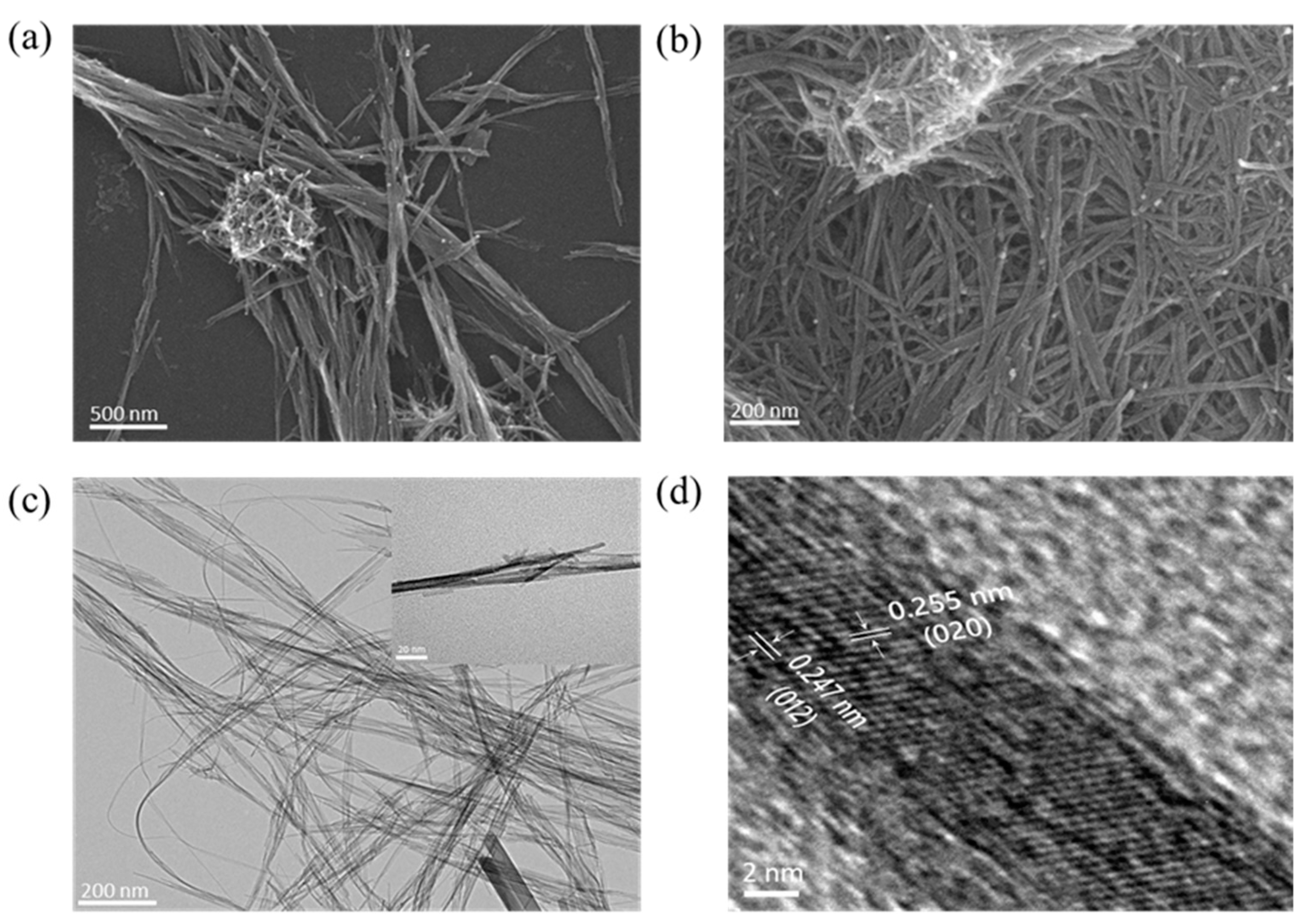
3.1. Synthesis and Characterization of MNWs

The overall process for synthesising MNWs and their application in the colorimetric detection of dopamine are illustrated in Figure 1. MNWs, synthesized via the solvothermal treatment of KMnO4 with DMF, exhibited efficient oxidase-like activity and, in particular, catalyzed the oxidation of the important neurotransmitter dopamine into brown-colored aminochrome without the involvement of any oxidizing agent. Based on dopamine-induced color generation by the MNWs, a paper dipstick was constructed by immobilizing the MNWs in the detection zone. Finally, by merely immersing the MNW-incorporated dipstick into the sample solution containing dopamine, a selective and vivid brown color proportional to the amount of dopamine was produced, distinguished by the naked eye or quantified by processing real images acquired using a smartphone.
Through the reduction of KMnO4 with DMF as a reducing agent, MNWs were obtained. The reaction between KMnO4 and DMF initially generated MnOOH nuclei, and then the continued reduction for 10 h resulted in fully grown MNWs [22]. The synthesized MNWs were characterized using SEM and TEM (Figure 2). The SEM images showed a slim, long-wired morphology with an average length of 344.3 ± 12.8 nm and an average width of 12.7 ± 1.5 nm. The morphology observed in the TEM images is consistent with that observed in the SEM images. The HR-TEM image of the MNWs shows that the crystal structure of the MNWs has uniform lattice fringes on both the vertical and horizontal axes. The lattice spacings of the horizontal and vertical fringes were 0.255 and 0.247 nm, respectively, corresponding to the (020) and (012) planes of the MNWs.
The MNWs were further analyzed by XRD, XPS, FT-IR, and EDS techniques. In the XRD spectra, three sharp diffraction peaks were observed, which correspond to the (–111), (020), and (012) planes and were well aligned with the standard JCPDS database (JCPDS No. 01-074-1632) (Figure 3a). These peaks clearly support that MNWs have a high crystallinity and can be indexed to the monoclinic crystal structure [24,25]. XPS spectra clearly show the presence of the main elements, Mn and O, in the MNWs (Figure 3b–d). The high-resolution Mn 2p was deconvoluted into Mn 2p3/2 (641.7 eV) and Mn 2p1/2 (653.5 eV) peaks, indicating that pure Mn3+ was present in the MNWs [25]. In the high-resolution peak of O 1s, there were two peaks at nearly 529.0 and 531.1 eV, which are assigned to O2− and OH, respectively, which are expected in the ideal MnOOH where the O2− binds to Mn3+ and OH binds to hydrogen [25]. The FT-IR spectra further support the presence of the representative chemical structures (Mn-O and O-H) of the MNWs (Figure S2). The strong shoulder peaks at 590 and 487 cm−1 correspond to the vibrations of Mn-O bonds. The peaks at 1069 and 1150 cm−1 are typically assigned to the O-H bending. The peak at 2151 cm−1,associated with a broad peak at 2760 cm−1, may indicate the vibration of Mn-O bonds [26]. The EDS maps provide evidence of the elemental composition of the MNWs, with two major elements, Mn and O, which are well distributed throughout the material (Figure S3). These characterizations confirm that the MNWs were successfully formed while preserving pure Mn3+ and high crystallinity.

3.2. Investigation of the Dopamine Oxidase-like Activity of MNWs

Because MNWs contain reduced, rather than oxidized, manganese species, (Mn3+) rather than (Mn4+), they are expected to have high oxidase-like activity [27]. In order to demonstrate this theory, the catalytic activity of the MNWs was explored by adding a series of colorimetric substrates, such as TMB, OPD, and DAB, in the absence of H2O2. The experimental results clearly demonstrate that the MNWs catalyzed the oxidation of all the employed substrates by showing the respective colors and absorption spectra corresponding to their oxidized products (Figure 4a). In particular, after a 15-min incubation with MNWs and dopamine (1 mM) in sodium phosphate buffer (pH 8.0), a vivid brown color and the corresponding absorption intensity at approximately 470 nm were detected, which was come from oxidized dopamine, known as aminochrome (Figure 4b) [28]. It was previously reported that the Mn3+ might be essential for oxidizing dopamine into aminochrome via a two-electron transfer process [29]. Thus, MNWs with pure Mn3+ could be efficient dopamine oxidase mimics to produce a brown color through the oxidation of target dopamine. Although dopamine can be self-oxidized at basic pH conditions, it takes quite a long time, up to 20 h [30]. Since our dopamine assay took only 15 min, it is evident that the color generation in our assay comes from the dopamine oxidase-like activity of MNWs. The types of reactive oxygen species (ROS) produced during dopamine oxidation by MNWs were examined to clarify the plausible catalytic mechanism. In order to accomplish this, five different radical scavengers were added during the typical dopamine oxidation reaction with MNWs, to capture specific ROS released during the reaction (Figure S4). The presence of hydroxyl radicals (•OH) was detected using isopropyl alcohol (IA) and methanol (MA), the existence of superoxide anion radicals (O2•−) was detected using benzoquinone (BQ) and superoxide dismutase (SOD), and singlet oxygen (1O2) was detected using sodium azide (SA) [31]. According to the experimental findings, O2•− and 1O2 were more important than •OH in the reactions that convert dopamine to aminochrome by the MNWs.
The effects of several synthetic parameters, including the incubation temperature and time, were examined on the morphology and dopamine oxidase-like activity of the MNWs (Figures S5 and S6). MNWs prepared at 100 °C (incubation for 6 h and 10 h) showed a thicker wire shape and displayed marginally lowered dopamine oxidase-like activity (approximately 90%) than that of the MNWs prepared at 120 °C for 10 h. MNWs prepared at 120 °C for 6 h were relatively uniform nanowires, but their catalytic activity was lower than those of MNWs prepared at 100 °C and at 120 °C for 10 h. MNWs synthesized at 140 °C yielded more diverse morphologies and exhibited a much lower catalytic activity. Based on these observations, we chose 120 °C for 10 h as the optimal synthetic conditions for solvothermal treatment. Preliminary optimizations related to the MNWs concentration, pH, temperature, and reaction time on the dopamine oxidase-like activity of the MNWs were also conducted. The results revealed that 0.1 mg/mL MNWs, pH 8.0, 40 °C, and 15 min reaction time were optimal conditions for the activity of MNWs (Figure S7).
To elucidate the dopamine oxidase-like activity of the MNWs, steady-state kinetic assays were performed by varying the dopamine concentration. From the Michaelis–Menten curve and the corresponding Lineweaver–Burk plot, the Michaelis constant (Km) and maximal reaction velocity (Vmax) were determined to be 0.32 mM and 1.43 µM/min, respectively, which are similar to those of recently reported dopamine oxidase mimic (Figure 4c,d) [32]. Compared with traditional Mn-based nanozymes, the MNWs yielded significantly enhanced kinetic parameters, probably due to their abundant Mn3+ [21]. The kinetic parameters were also comparable to most natural oxidases and oxidase-like nanozymes [33,34,35]. These investigations indicate that the solvothermally synthesized MNWs are potential dopamine oxidase mimics, preserving a sufficiently high affinity toward dopamine and reaction velocity.

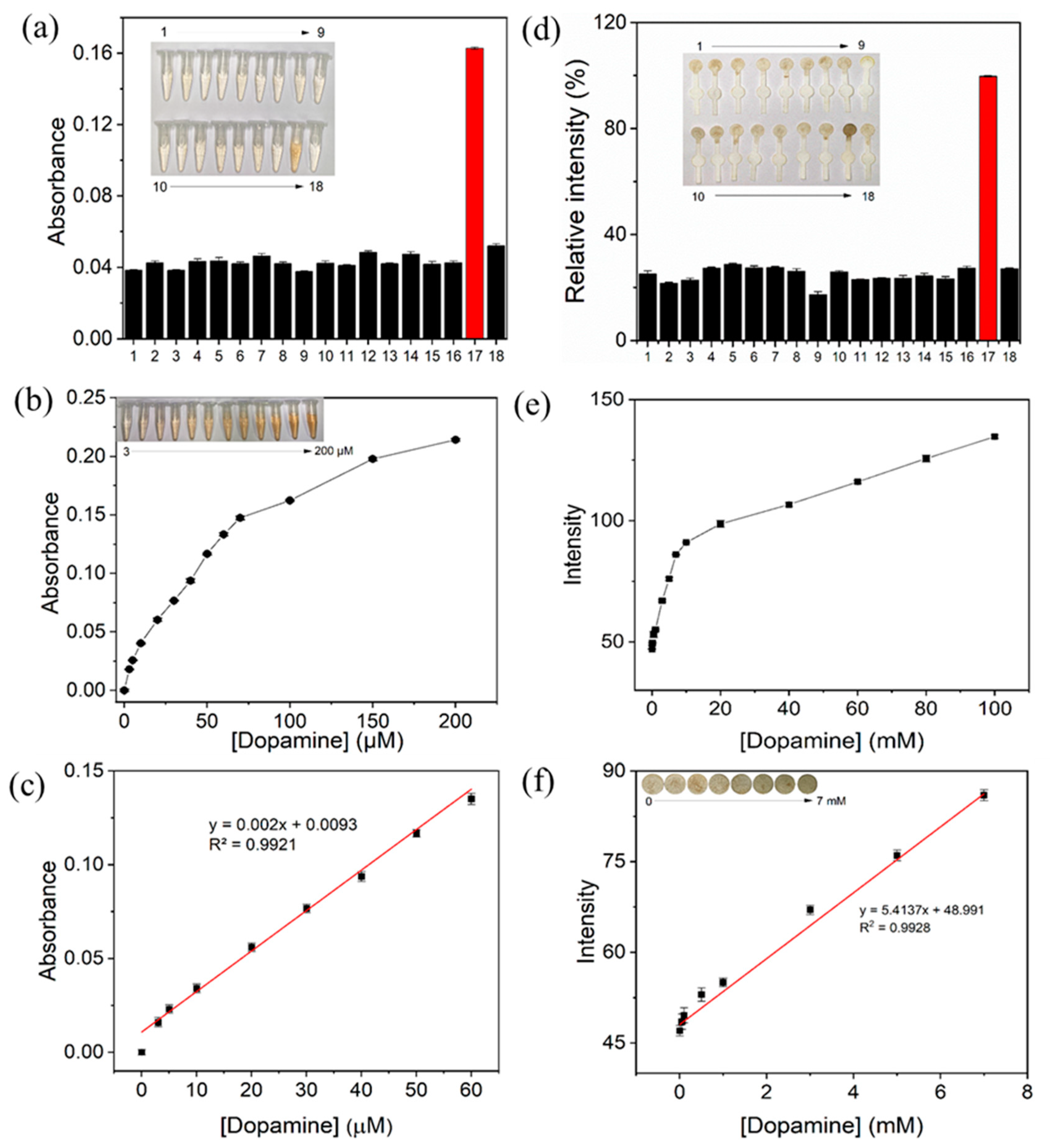
3.3. Colorimetric Detection of Dopamine Using the MNWs-Containing Solution-Based Assay and MNWs-Incorporated Paper Dipstick

The target dopamine levels were determined using an MNWs-containing solution-based assay based on the optimised conditions. Through a simple 15-min incubation at 40 °C, dopamine was selectively determined by the generation of visibly or spectroscopically detected brown color evolution (Figure 5a). In contrast, interfering molecules present in physiological fluids, such as ions (K, Na, Mg, and Ca), carbohydrates (glucose and maltose), small molecules (acetylcholine, uric acid, and ascorbic acid), amino acids (lysine, cysteine, glycine, alanine, tyrosine, and histidine), and proteins (BSA) did not yield any considerable color signal, where they are used at tenfold higher concentrations. These results demonstrated that the MNW-mediated catalytic reaction only produced a specific colorimetric signal from the target dopamine. From the analysis of dose–response curves, LOD was determined to be as low as 0.7 µM with a linear range from 3 to 60 µM (Figure 5b,c). These LOD and linear range values are among the best results of recent reports describing colorimetric dopamine detection and are sufficient to distinguish between normal and patient dopamine levels in human serum (Table S1).
To develop dopamine diagnostics suitable for POCT environments, we designed a simple paper dipstick platform that is portable, storable, disposable, inexpensive to mass-produce with laser cutting, and selective/sensitive by providing a large contrast difference of paper for efficiently visualizing color change [36]. The paper dipstick was composed of three zones: a rectangular sample zone (3 mm × 10 mm), a circular control zone (radius = 4 mm), and a circular detection zone (radius = 4 mm), where the sample solution could be moved by capillary force (Figure S1). To optimize the dopamine detection performance, the fabrication and sensing conditions for the dipstick were optimized, such as the size of the detection zone, type of paper substrate, immobilization agents, and optimal concentration, which helped the tight immobilization of MNWs in the detection zone, the concentration of MNWs employed, and reaction time (Figure S8). Besides, we demonstrated that it took approximately 90 s took for the complete movement of an aqueous solution from the sample zone to the detection zone of the dipstick (Figure S9). Under the optimized conditions, dopamine was selectively detected by showing an intense brown color in its detection zone, whereas a negligible color intensity, similar to that of the negative control, was detected from interfering substances, which were the same as those used in the solution-based assays, even at tenfold higher concentrations (Figure 5d and Figure S10). With increasing dopamine concentrations, the respective color intensities gradually increased. Dose–response curves for dopamine were prepared from real images of dipsticks taken by a conventional smartphone, followed by image processing using ImageJ software. Through the investigation, the LOD was calculated to be 20 µM, with a linear range from 0.05 to 7 mM (Figure 5e,f). Because the LOD and linear range values were higher than the actual dopamine levels in human serum, the developed dipstick may not be suitable for detecting dopamine in human serum. However, the dipstick can be used in the dopamine assay in pharmaceutical formulations, normally used at several µM to mM concentrations [37]. Moreover, it would be beneficial to add a rigid support frame made of materials like glass or plastic, to increase the dipstick’s mechanical strength and facilitate practical applications.

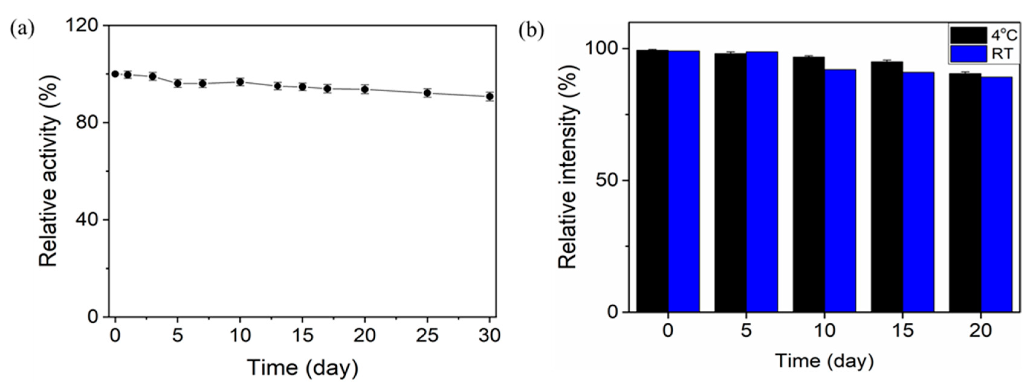
3.4. Practical Applicability of MNWs-Based Colorimetric Detection of Dopamine in Human Serum or Dopamine Injection

Long-term storage stability is important for practical applications. As shown in Figure 6a, the MNWs effectively maintained their initial dopamine oxidase-like activity during one month of storage at RT. The MNWs-incorporated paper dipstick also exhibited impressive storage stability at both 4 °C and RT, by preserving over 90% of initial activity (Figure 6b). These results demonstrate the high stability of MNWs and MNW-incorporated paper dipsticks under conventional storage conditions.
Finally, we demonstrated the practical utility of the MNWs-containing solution-based assay and MNWs-incorporated paper dipstick for the quantitative determination of dopamine in spiked human serum and pharmaceutical dopamine injections, respectively. In the solution-based assay, the original dopamine concentration in human serum was quantified first using a commercial dopamine ELISA kit and was determined to be approximately 0.05 µM. Then, predetermined amounts of dopamine at 3, 5, and 10 µM were spiked in the human serum to make normal, boundary, and high dopamine levels in brain tissue [38]. As a result, the serum dopamine was quantified with excellent precision, yielding CVs in the range of 1.0–3.1% with recovery rates of 95.7–98.5% (Table 1a), verifying the excellent reproducibility and reliability of the method.
An MNW-incorporated paper dipstick was used to quantify the concentration of the dopamine injection. Dopamine injection (dopamine hydrochloride, 200 mg/5 mL) was diluted to 250 and 500 mL with water to make concentrations of 4.14 and 2.09 mM of dopamine, respectively. The two dopamine formulations were precisely and accurately determined using the MNWs-incorporated paper dipstick, yielding CVs in the range of 2.0–3.0% with recovery rates of 97.3–98.1% (Table 1b). These results demonstrate that the developed MNWs-incorporated paper dipstick can serve as a convenient analytical tool for analyzing dopamine in pharmaceutical formulations in POCT environments.

4. Conclusions

In summary, we developed oxidase-like, more precisely, dopamine oxidase-like nanozyme, MNWs, having considerably high catalytic activity, presumably due to the presence of pure Mn3+ throughout the material. MNWs followed typical Michaelis–Menten kinetics with improved storage stability. By immobilizing MNWs on a paper dipstick, the target dopamine was conveniently detected by the naked eye and quantified using real images acquired using a smartphone. MNW-containing solution-based assays and MNW-incorporated paper dipsticks enabled the successful determination of dopamine with excellent selectivity, sensitivity, and reliability in human serum samples and pharmaceutical formulations, respectively. This study serves as a basis for the continued efforts towards developing nanozyme-incorporated paper-based devices, which have significant potential in biosensing areas, particularly in POCT environments.

Supplementary Materials

The following supporting information can be downloaded at: https://www.mdpi.com/article/10.3390/chemosensors11070382/s1. Figure S1: Dipstick platform designed by AutoCAD 2018; Figure S2: FT-IR spectra of MNWs; Figure S3: EDS maps and corresponding elemental ratios of MNWs; Figure S4: Radical scavenging assay for demonstrating the catalytic mechanism of the dopamine oxidase-like activity of MNWs; Figure S5: Dopamine oxidase-like activity of MNWs synthesized at diverse reaction conditions; Figure S6: SEM images of MNWs synthesized at (a)100 °C/6 h, (b) 100 °C/10 h, (c) 140 °C/6 h, (d) 140 °C/10 h, and (e) 120 °C/6 h; Figure S7: Optimization of (a) MNW concentration, (b) pH, (c) Temperature, (d) Reaction time for dopamine oxidase-like activity of MNWs; Figure S8: Optimization for dopamine detection of MNWs-incorporated dipstick. (a) The radius of the detection zone, (b) Types of paper substrate, (c) Immobilizing agent, (d) TEOS concentration, (e) MNW concentration, (f) Reaction time of the assay; Figure S9: Time required for an aqueous solution containing crystal violet dye to move through the dipstick; Figure S10: Selectivity toward dopamine via MNW-incorporated paper dipstick; Table S1: Comparison of LOD and linear range values of MNW-containing solution-based assay to detect dopamine, with those of recent reports describing colorimetric dopamine detection [39,40,41].

Author Contributions

Conceptualization, investigation, writing—original draft preparation, P.B.K.C.; validation, writing—review and editing, T.V.D.; and conceptualization, supervision, writing—review and editing, M.I.K. All authors have read and agreed to the published version of the manuscript.

Funding

This work was supported by a National Research Foundation of Korea (NRF) grant funded by the Korean government (Ministry of Science and ICT [NRF-2023R1A2C2007833]) and by the Gachon University research fund of 2021 (GCU-202102970001).

Institutional Review Board Statement

Not applicable.

Informed Consent Statement

Not applicable.

Data Availability Statement

The data presented in this study are available on request from the corresponding author.

Conflicts of Interest

The authors declare no conflict of interest.

References

  1. Damier, P.; Hirsch, E.C.; Agid, Y.; Graybiel, A. The substantia nigra of the human brain: II. Patterns of loss of dopamine-containing neurons in Parkinson’s disease. Brain 1999, 122, 1437–1448. [Google Scholar] [CrossRef] [PubMed]
  2. Manbohi, A.; Ahmadi, S.H. Sensitive and selective detection of dopamine using electrochemical microfluidic paper-based analytical nanosensor. Sens. Bio-Sens. Res. 2019, 23, 100270. [Google Scholar] [CrossRef]
  3. Paris, I.; Cardenas, S.; Lozano, J.; Perez-Pastene, C.; Graumann, R.; Riveros, A.; Caviedes, P.; Segura-Aguilar, J. Aminochrome as a preclinical experimental model to study degeneration of dopaminergic neurons in Parkinson’s disease. Neurotox. Res. 2007, 12, 125–134. [Google Scholar] [CrossRef] [PubMed]
  4. Carter, J.E.; Johnson, J.H.; Baaske, D.M. Dopamine hydrochloride. In Analytical Profiles of Drug Substances; Elsevier: Amsterdam, The Netherlands, 1982; pp. 257–272. [Google Scholar]
  5. MacCannell, K.L.; McNay, J.L.; Meyer, M.B.; Goldberg, L.I. Dopamine in the Treatment of Hypotension and Shock. N. Engl. J. Med. 1966, 275, 1389–1398. [Google Scholar] [CrossRef]
  6. Janků, S.; Komendová, M.; Urban, J. Development of an online solid-phase extraction with liquid chromatography method based on polymer monoliths for the determination of dopamine. J. Sep. Sci. 2016, 39, 4107–4115. [Google Scholar] [CrossRef]
  7. Hubbard, K.E.; Wells, A.; Owens, T.S.; Tagen, M.; Fraga, C.H.; Stewart, C.F. Determination of dopamine, serotonin, and their metabolites in pediatric cerebrospinal fluid by isocratic high performance liquid chromatography coupled with electrochemical detection. Biomed. Chromatogr. 2010, 24, 626–631. [Google Scholar] [CrossRef]
  8. Vuorensola, K.; Sirén, H. Determination of urinary catecholamines with capillary electrophoresis after solid-phase extraction. J. Chromatogr. A 2000, 895, 317–327. [Google Scholar] [CrossRef]
  9. Bouri, M.; Lerma-García, M.J.; Salghi, R.; Zougagh, M.; Ríos, A. Selective extraction and determination of catecholamines in urine samples by using a dopamine magnetic molecularly imprinted polymer and capillary electrophoresis. Talanta 2012, 99, 897–903. [Google Scholar] [CrossRef]
  10. Syslová, K.; Rambousek, L.; Kuzma, M.; Najmanová, V.; Bubeníková-Valešová, V.; Šlamberová, R.; Kačer, P. Monitoring of dopamine and its metabolites in brain microdialysates: Method combining freeze-drying with liquid chromatography–tandem mass spectrometry. J. Chromatogr. A 2011, 1218, 3382–3391. [Google Scholar] [CrossRef]
  11. Wei, X.; Zhang, Z.; Wang, Z. A simple dopamine detection method based on fluorescence analysis and dopamine polymerization. Microchem. J. 2019, 145, 55–58. [Google Scholar] [CrossRef]
  12. Tao, Y.; Lin, Y.; Ren, J.; Qu, X. A dual fluorometric and colorimetric sensor for dopamine based on BSA-stabilized Aunanoclusters. Biosens. Bioelectron. 2013, 42, 41–46. [Google Scholar] [CrossRef] [PubMed]
  13. Guo, L.; Hu, Y.; Zhang, Z.; Tang, Y. Universal fluorometric aptasensor platform based on water-soluble conjugated polymers/graphene oxide. Anal. Bioanal. Chem. 2018, 410, 287–295. [Google Scholar] [CrossRef] [PubMed]
  14. Baron, R.; Zayats, M.; Willner, I. Dopamine-, l-DOPA-, Adrenaline-, and Noradrenaline-Induced Growth of Au Nanoparticles: Assays for the Detection of Neurotransmitters and of Tyrosinase Activity. Anal. Chem. 2005, 77, 1566–1571. [Google Scholar] [CrossRef] [PubMed]
  15. Liu, X.; Liu, J. Biosensors and sensors for dopamine detection. View 2021, 2, 20200102. [Google Scholar] [CrossRef]
  16. Ivanova, M.N.; Grayfer, E.D.; Plotnikova, E.E.; Kibis, L.S.; Darabdhara, G.; Boruah, P.K.; Das, M.R.; Fedorov, V. Pt-Decorated Boron Nitride Nanosheets as Artificial Nanozyme for Detection of Dopamine. ACS Appl. Mater. Interfaces 2019, 11, 22102–22112. [Google Scholar] [CrossRef]
  17. Biniuri, Y.; Albada, B.; Wolff, M.; Golub, E.; Gelman, D.; Willner, I. Cu2+ or Fe3+ Terpyridine/Aptamer Conjugates: Nucleoapzymes Catalyzing the Oxidation of Dopamine to Aminochrome. ACS Catal. 2018, 8, 1802–1809. [Google Scholar] [CrossRef]
  18. Nag, R.; Rao, C.P. Development and demonstration of functionalized inorganic–organic hybrid copper phosphate nanoflowers for mimicking the oxidative reactions of metalloenzymes by working as a nanozyme. J. Mater. Chem. B 2021, 9, 3523–3532. [Google Scholar] [CrossRef]
  19. Le, T.N.; Le, X.A.; Tran, T.D.; Lee, K.J.; Kim, M.I. Laccase-mimicking Mn–Cu hybrid nanoflowers for paper-based visual detection of phenolic neurotransmitters and rapid degradation of dyes. J. Nanobiotechnol. 2022, 20, 358. [Google Scholar] [CrossRef]
  20. Xue, L.; Jin, N.; Guo, R.; Wang, S.; Qi, W.; Liu, Y.; Li, Y.; Lin, J. Microfluidic Colorimetric Biosensors Based on MnO2 Nanozymes and Convergence–Divergence Spiral Micromixers for Rapid and Sensitive Detection of Salmonella. ACS Sens. 2021, 6, 2883–2892. [Google Scholar] [CrossRef]
  21. Lu, W.; Chen, J.; Kong, L.; Zhu, F.; Feng, Z.; Zhan, J. Oxygen vacancies modulation Mn3O4 nanozyme with enhanced oxidase-mimicking performance for l-cysteine detection. Sens. Actuator B Chem. 2021, 333, 129560. [Google Scholar] [CrossRef]
  22. Li, Z.; Bao, H.; Miao, X.; Chen, X. A facile route to growth of γ-MnOOH nanorods and electrochemical capacitance properties. J. Colloid Interface Sci. 2011, 357, 286–291. [Google Scholar] [CrossRef] [PubMed]
  23. Kumar, T.; Verma, K. A Theory Based on Conversion of RGB image to Gray image. Int. J. Comput. Appl. 2010, 7, 7–10. [Google Scholar] [CrossRef]
  24. Zhang, Y.; Liu, Y.; Guo, F.; Hu, Y.; Liu, X.; Qian, Y. Single-crystal growth of MnOOH and beta-MnO2 microrods at lower temperatures. Solid State Commun. 2005, 134, 523–527. [Google Scholar] [CrossRef]
  25. Wang, Y.; Chen, L.; Chen, M.; Zhong, Z.; Meng, Q.; Xing, W. Ultralight 3D-gamma-MnOOH porous materials fabricated by hydrothermal treatment and freeze-drying. Sci. China-Mater. 2019, 62, 527–535. [Google Scholar] [CrossRef] [Green Version]
  26. Varghese, S.P.; Babu, A.T.; Babu, B.; Antony, R. γ-MnOOH nanorods: Efficient adsorbent for removal of methylene blue from aqueous solutions. J. Water Process Eng. 2017, 19, 1–7. [Google Scholar] [CrossRef]
  27. Zou, Y.; Zhang, M.; Cao, F.; Li, J.; Zhang, S.; Qu, Y. Single crystal MnOOH nanotubes for selective oxidative coupling of anilines to aromatic azo compounds. J. Mater. Chem. A 2021, 9, 19692–19697. [Google Scholar] [CrossRef]
  28. Ouyang, Y.; Fadeev, M.; Zhang, P.; Carmieli, R.; Li, J.; Sohn, Y.S.; Karmi, O.; Nechushtai, R.; Pikarsky, S.; Fan, C. Aptamer-Modified Au Nanoparticles: Functional Nanozyme Bioreactors for Cascaded Catalysis and Catalysts for Chemodynamic Treatment of Cancer Cells. ACS Nano 2022, 16, 18232–18243. [Google Scholar] [CrossRef]
  29. Tang, M.; Zhang, Z.; Sun, T.; Li, B.; Wu, Z. Manganese-Based Nanozymes: Preparation, Catalytic Mechanisms, and Biomedical Applications. Adv. Healthc. Mater. 2022, 11, 2201733. [Google Scholar] [CrossRef]
  30. Salomäki, M.O.; Marttila, L.; Kivelä, H.; Ouvinen, T.; Lukkari, J.O. Effects of pH and Oxidants on the First Steps of Polydopamine Formation: A Thermodynamic Approach. J. Phys. Chem. B 2018, 122, 6314–6327. [Google Scholar] [CrossRef]
  31. Jiang, L.; Han, Y.; Fernández-García, S.; Tinoco, M.; Li, Z.; Nan, P.; Sun, J.; Delgado, J.J.; Pan, H.; Blanco, G.; et al. Multi-functional oxidase-like activity of praseodymia nanorods and nanoparticles. Appl. Surf. Sci. 2023, 610, 155502. [Google Scholar] [CrossRef]
  32. Ouyang, Y.; Biniuri, Y.; Fadeev, M.; Zhang, P.; Carmieli, R.; Vázquez-González, M.; Willner, I. Aptamer-modified Cu2+-functionalized C-Dots: Versatile means to improve nanozyme activities–“Aptananozymes”. J. Am. Chem. Soc. 2021, 143, 11510–11519. [Google Scholar] [CrossRef] [PubMed]
  33. Liang, H.; Lin, F.; Zhang, Z.; Liu, B.; Jiang, S.; Yuan, Q.; Liu, J. Multicopper Laccase Mimicking Nanozymes with Nucleotides as Ligands. ACS Appl. Mater. Interfaces 2017, 9, 1352–1360. [Google Scholar] [CrossRef] [PubMed]
  34. Tran, T.D.; Nguyen, P.T.; Le, T.N.; Kim, M.I. DNA-copper hybrid nanoflowers as efficient laccase mimics for colorimetric detection of phenolic compounds in paper microfluidic devices. Biosens. Bioelectron. 2021, 182, 113187. [Google Scholar] [CrossRef] [PubMed]
  35. Gao, L.; Zhuang, J.; Nie, L.; Zhang, J.; Zhang, Y.; Gu, N.; Wang, T.; Feng, J.; Yang, D.; Perrett, S.; et al. Intrinsic peroxidase-like activity of ferromagnetic nanoparticles. Nat. Nanotechnol. 2007, 2, 577–583. [Google Scholar] [CrossRef]
  36. Huang, L.; Sun, D.W.; Pu, H.; Wei, Q.; Luo, L.; Wang, J. A colorimetric paper sensor based on the domino reaction of acetylcholinesterase and degradable γ-MnOOH nanozyme for sensitive detection of organophosphorus pesticides. Sens. Actuator. B Chem. 2019, 290, 573–580. [Google Scholar] [CrossRef]
  37. Manmana, Y.; Chutvirasakul, B.; Suntornsuk, L.; Nuchtavorn, N. Cost effective paper-based colorimetric devices for a simple assay of dopamine in pharmaceutical formulations using 3,3′,5,5′-tetramethylbenzidine–Silver nitrate as a chromogenic reagent. Pharm. Sci. Asia 2019, 46, 270–277. [Google Scholar] [CrossRef]
  38. Wang, T.; Song, R.; Song, W.; Zhao, Y.; Zhu, K.; Yuan, X. Improvement of mechanical properties for epoxy composites with modified titanate whiskers via dopamine self-oxidation. J. Polym. Res. 2021, 28, 1–11. [Google Scholar] [CrossRef]
  39. Zhu, J.; Peng, X.; Nie, W.; Wang, Y.; Gao, J.; Wen, W.; Selvaraj, J.N.; Zhang, X.; Wang, S. Hollow copper sulfide nanocubes as multifunctional nanozymes for colorimetric detection of dopamine and electrochemical detection of glucose. Biosens. Bioelectron. 2019, 141, 111450. [Google Scholar] [CrossRef]
  40. Zhu, Y.; Yang, Z.; Chi, M.; Li, M.; Wang, C.; Lu, X. Synthesis of hierarchical Co3O4@NiO core-shell nanotubes with a synergistic catalytic activity for peroxidase mimicking and colorimetric detection of dopamine. Talanta 2018, 181, 431–439. [Google Scholar] [CrossRef]
  41. Guo, M.X.; Li, Y.F. Cu (II)-based metal-organic xerogels as a novel nanozyme for colorimetric detection of dopamine. Spectroc. Acta Pt. A-Molec. Biomolec. Spectr. 2019, 207, 236–241. [Google Scholar] [CrossRef]
Figure 1. Schematic illustration of (a) The synthesis of MnOOH nanowires (MNWs) having dopamine oxidase-like activity, (b) Their application on the paper dipstick for the colorimetric detection of dopamine.
Chemosensors 11 00382 g001
Figure 2. SEM images of MNWs dispersed at (a) 1 mg/mL and (b) 5 mg/mL. (c) TEM image of MNWs. (d) HR-TEM image showing the crystal lattice spacing of a single MNW.
Chemosensors 11 00382 g002
Figure 3. (a) XRD spectra of MNWs. (b) Fully scanned XPS of MNWs with high-resolution XPS spectra in the (c) Mn 2p and (d) O 1s regions.
Chemosensors 11 00382 g003
Figure 4. (a) Oxidase-like activity of MNWs in the presence of various substrates (TMB, OPD, and DAB). (b) Dopamine oxidase-like activity of MNW, (c) Michaelis–Menten curve for the dopamine oxidase-like activity of MNWs at various concentrations, (d) Their corresponding Lineweaver–Burk plots (n = 3).
Chemosensors 11 00382 g004
Figure 5. Analytical capability for the determination of dopamine. (a,b) Selectivity toward dopamine (c,d) The dose-response curves (e,f) The corresponding linear calibration plots, using MNW-containing solution-based assay and MNWs-incorporated paper dipstick, respectively. Numbers 1 to 18 in the inset images represent K+, Na+, Mg2+, Ca2+, glucose, maltose, acetylcholine, uric acid, ascorbic acid, lysine, cysteine, glycine, alanine, tyrosine, histidine, BSA, dopamine, and negative control, respectively.
Chemosensors 11 00382 g005
Figure 6. Storage stability of (a) MNWs at RT, (b) MNWs-incorporated paper dipstick at 4 °C and RT.
Chemosensors 11 00382 g006
Table 1. Detection precision of (a) MNW-containing solution-based assay and (b) MNW-incorporated paper dipstick for the quantitative determination of dopamine, in spiked human serum and pharmaceutical injection, respectively.
(a) MNWs-Containing Solution-Based Assay
Original Dopamine (μM)Added (μM)Found (μM) a ± SD bRecovery (%)CV c (%)
32.99 ± 0.0398.031.00
0.0554.97 ± 0.1598.423.02
109.62 ± 0.1095.721.04
(b) MNWs-Incorporated Paper Dipstick
Dopamine Formulation (mM)Found (mM) d ± SDRecovery (%)CV (%)
4.144.03 ± 0.0897.341.99
2.092.05 ± 0.0698.092.93
a Mean of three replicates; b standard deviation (SD); c coefficient of variation (CV). d Mean of three replications.
Disclaimer/Publisher’s Note: The statements, opinions and data contained in all publications are solely those of the individual author(s) and contributor(s) and not of MDPI and/or the editor(s). MDPI and/or the editor(s) disclaim responsibility for any injury to people or property resulting from any ideas, methods, instructions or products referred to in the content.

