Review

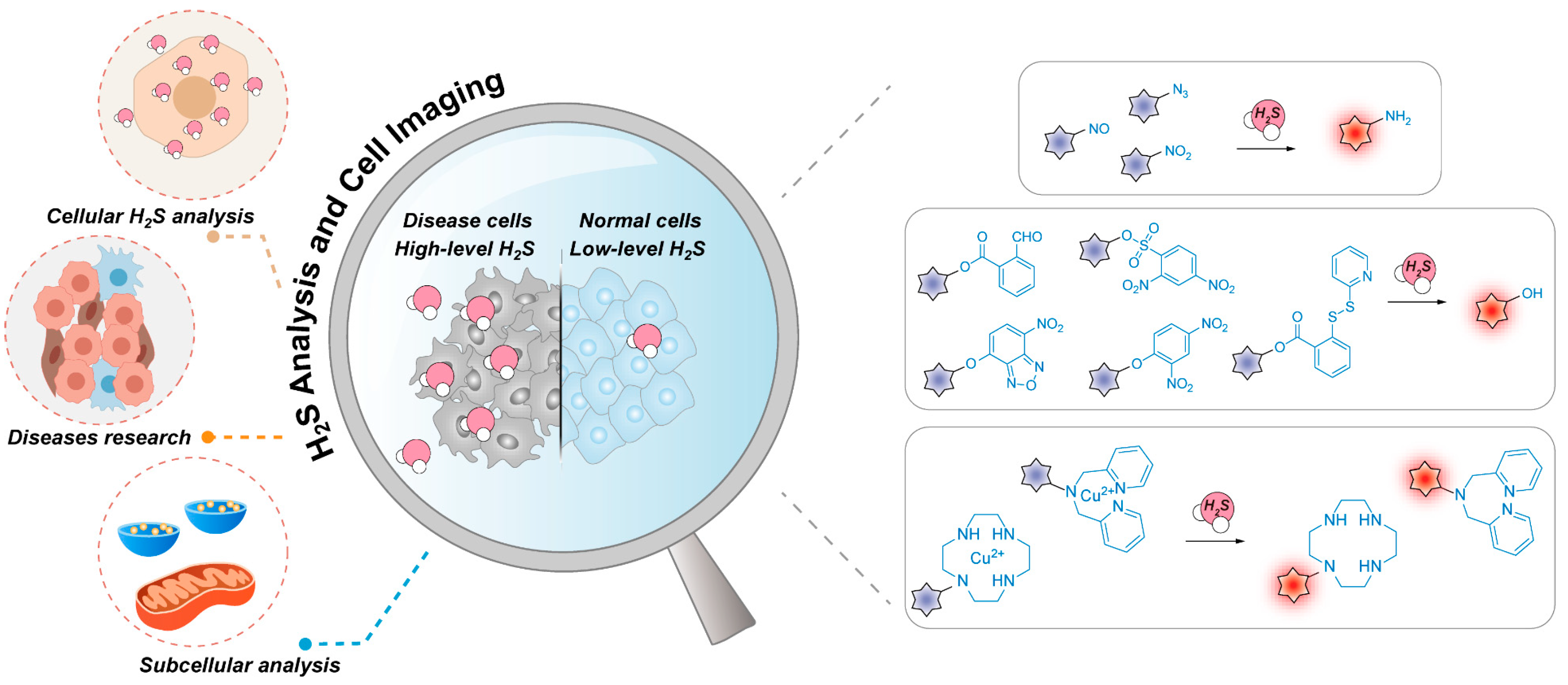
Recent Progress in Fluorescent Probes for the Detection and Research of Hydrogen Sulfide in Cells

Center of Excellence for Environmental Safety and Biological Effects, Beijing Key Laboratory for Green Catalysis and Separation, Department of Chemistry, Beijing University of Technology, Beijing 100124, China
*
Author to whom correspondence should be addressed.
These authors contributed equally to this work.
Chemosensors 2023, 11(6), 333; https://doi.org/10.3390/chemosensors11060333
Submission received: 22 April 2023 / Revised: 25 May 2023 / Accepted: 1 June 2023 / Published: 4 June 2023

Abstract

:
Hydrogen sulfide (H2S) is a gaseous signaling molecule that plays an important role in regulating various physiological activities in biological systems. As the fundamental structural and functional unit of organisms, cells are closely related to the homeostasis of their internal environment. The levels of H2S in different organelles maintain a certain balance, and any disruption of this balance will lead to various functional abnormalities that affect the health of organisms. Fluorescent imaging technology provides unique merits, such as simplicity, non-invasiveness, and real-time monitoring, and has become a powerful approach for the detection of molecules in biological systems. Based on the special physicochemical properties of H2S, numerous H2S-specific fluorogenic probes have been designed with different recognition mechanisms that enable rapid and accurate detection of H2S in cells. Therefore, this review briefly illustrates the design strategies, response principles, and biological applications of H2S-specific fluorescent probes and aims to provide relevant researchers with insight for future research.

Graphical Abstract

1. Introduction

Hydrogen sulfide (H2S) is a gaseous signaling molecule that regulates various physiological activities in biological systems [1]. It is naturally produced by enzymes such as cystathionine γ-lyase (CSE), cystathionine β-synthetase (CBS), and 3-mercaptopyruvate sulfurtransferase (3-MST) that catalyze sulfur-containing substances such as cysteine (Cys) and homocysteine (Hcy) in living systems [2,3]. H2S content varies in different tissues and organs of organisms [4,5,6], which reflects the corresponding health status. As a mediator of the inflammatory response [7,8,9], H2S affects physiological activities and pathological processes in vivo. The disruption of H2S balance in internal circulation is an important predisposing factor for many diseases, such as Alzheimer’s disease [10], Down’s syndrome [11], Parkinson’s disease [12], asthma [13], and so on [14,15]. Therefore, the detection of endogenous H2S is critical for the study of biological and pathological processes in vivo.
A cell is the basic structure and functional unit of an organism. The biological activity of organisms is closely related to cellular homeostasis [16]. Notably, the vital movement of cells requires the joint operation of various organelles [17]. Studies have found that H2S not only plays an important role in various organelles [18,19,20], but also plays a key role in various cell signaling pathways [21,22]. Therefore, the development of simple, effective, and precise methods to detect H2S in biological systems is critical for better understanding its subcellular distribution and functions in various physiological and pathological processes.
The traditional methods for the detection of H2S mainly include gas chromatography [23,24], colorimetry [25], electrochemical methods [26], etc., but these methods inevitably show some shortcomings. The pre-treatment process of the above methods is too complicated, which hinders the application of real-time H2S monitoring in vivo. In addition, the low selectivity and sensitivity to H2S also limit their further application in vivo [27,28]. Therefore, in order to overcome the above problems, it is necessary to develop fast, efficient, and real-time H2S monitoring technology to realize sensitive monitoring of H2S in cells. In recent years, through the unremitting efforts of researchers, the development of fluorescence imaging technology has brought hope for the detection of molecules in biological systems. Its non-invasiveness and quick detection of biomarkers have been pursued by researchers [29]. Selective activatable probes can be designed to specifically detect target biomarkers, providing effective tools for the visualization of H2S in cells and in vivo [30,31,32].
In recent years, the rapid development of fluorescence imaging technology has led to the emergence of a series of H2S fluorescence probes with excellent properties, high efficiency, and accuracy, enabling real-time monitoring of H2S [33]. Furthermore, many fluorescent probes have been developed for H2S detection in different organelles, achieving systematic research on physiological and pathological processes at the subcellular level [34,35]. Although an abundance of literature briefly summarizes the design principles of H2S probes, their applications at the cellular level have not been elaborated in detail [36,37]. Therefore, this short review aims to summarize novel H2S probes with various design strategies and their applications in biological research at the cellular level, so as to provide readers with a deeper understanding of H2S probe design strategies and to develop novel H2S probes for further clinical applications (Scheme 1).

2. H2S Probe

Organisms are complex systems where various molecules coexist and collaborate. Therefore, the detection of H2S in vivo is susceptible to interference from other bioactive substances, such as persulfides (RSSH) [38] and reactive oxygen species (ROS) [39]. In view of the crucial role of H2S in biological systems, a new type of H2S-responsive fluorogenic probe with good biocompatibility, specificity, and optical properties is urgently needed to facilitate the detection of H2S and further study of its physiological properties. H2S has strong reduction ability and nucleophilicity, which are the main basis for designing H2S response fluorogenic probes. Based on the activatable fluorogenic platform, a variety of fluorescent probes have been designed for detecting H2S with different reaction strategies.

2.1. Probes Based on H2S Reduction Reactions

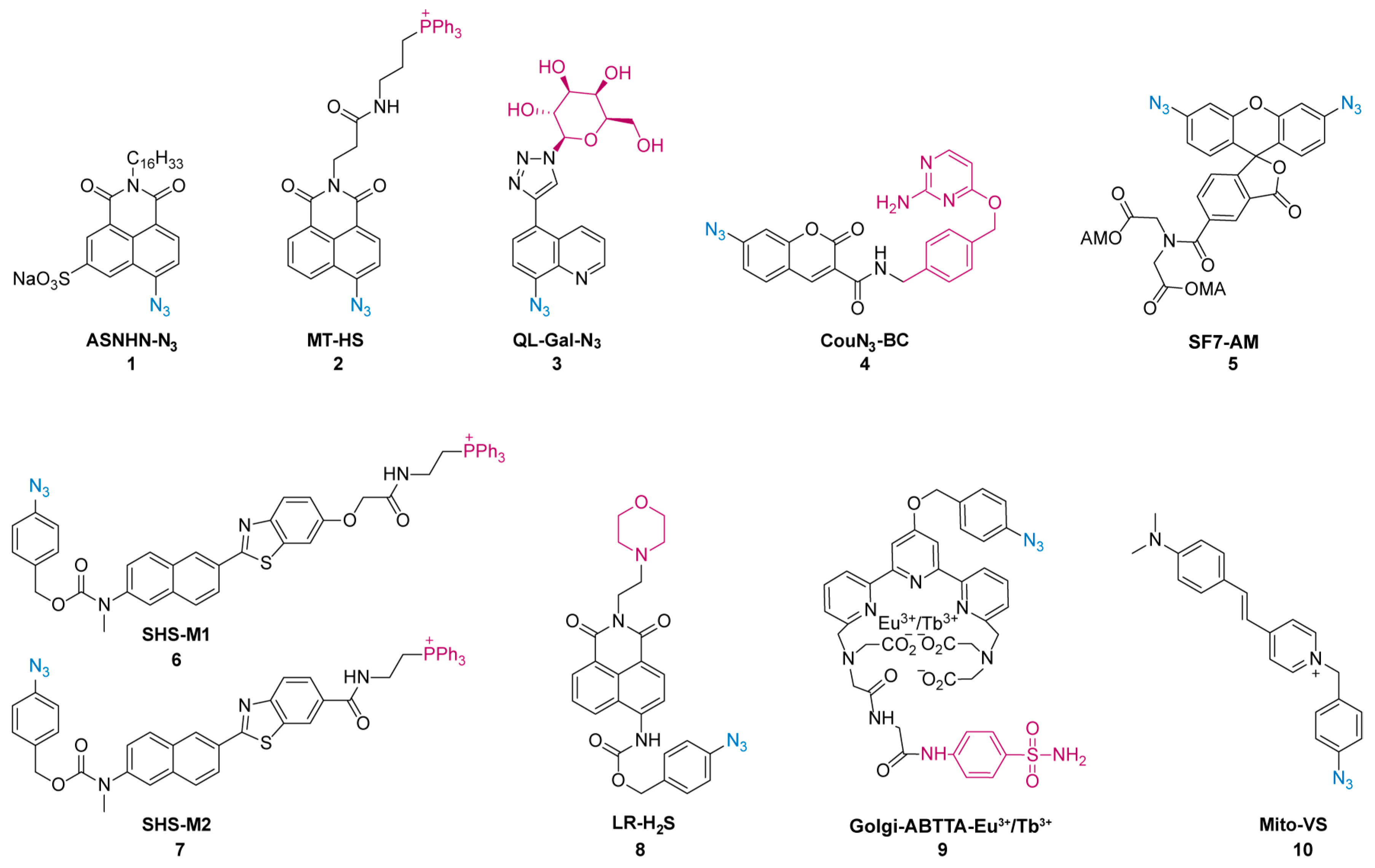
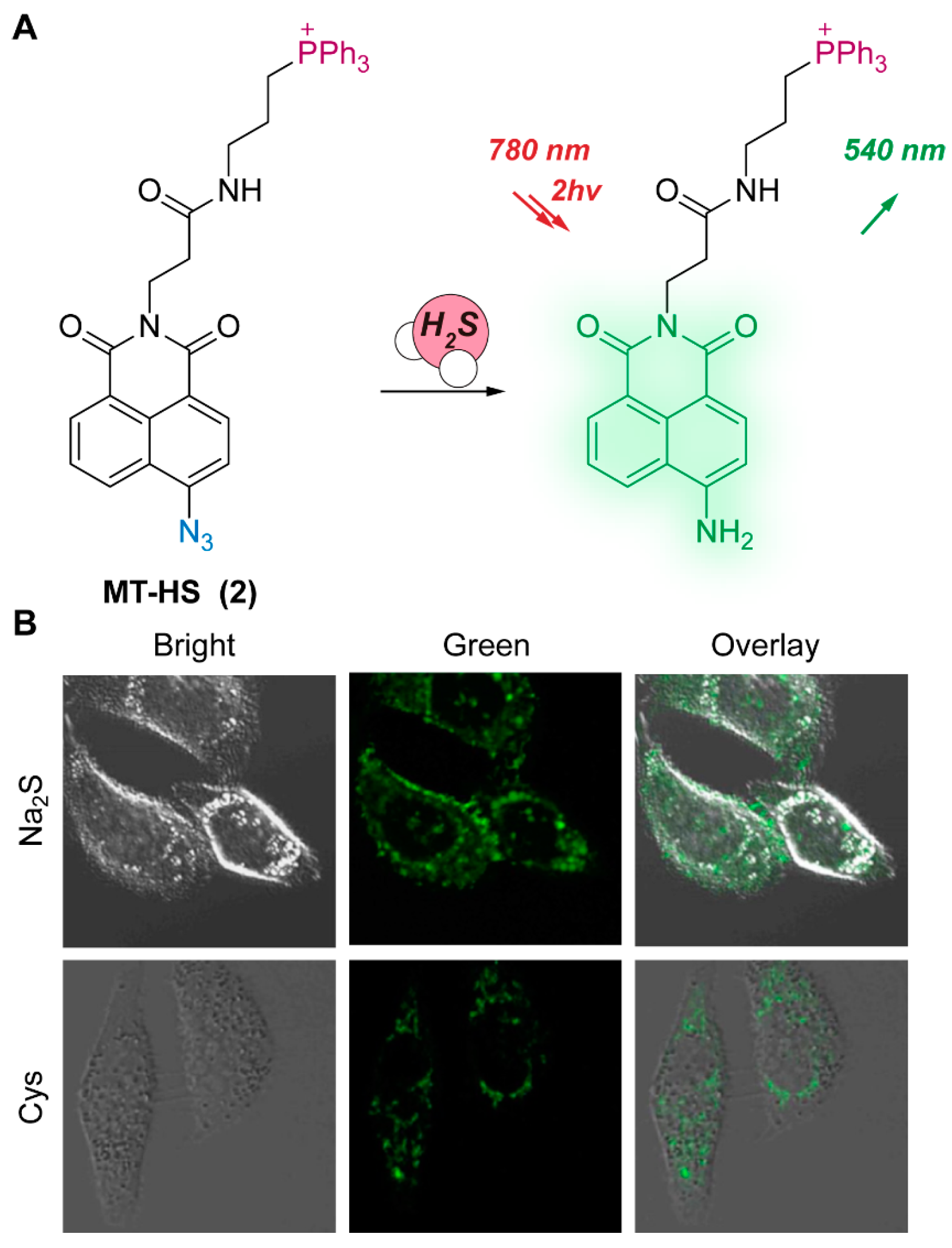
2.1.1. Azide-Based H2S Probes

As a bioorthogonal functional group, azide can be used to modify the chemical structure of various fluorescent dyes because of its convenient assembly. In addition, its efficient fluorescence quenching effect and good biocompatibility have been widely used. Due to the reducibility of H2S, the electron-withdrawing azide group will be converted to an electron-donating amino group by H2S, restoring the D-π-A structure with high-emissive fluorescence and thereby achieving sensitive detection of H2S. Notably, the selectivity of azide-based fluorogenic probes for H2S is higher than that of reactive sulfur, oxygen, and nitrogen species. The chemical structures of azide-based H2S probes introduced in this review are listed below (Figure 1).
In 2017, Wang et al. designed the two-photon Probe 1 [40] to visualize the intracellular and intercellular H2S transfer processes. Probe 1 contains a H2S-responsive azide group, a long-chain hydrophobic alkyl for anchoring the cell membrane, and a sulfonate to increase the hydrophilicity. Fluorescence imaging of endogenous H2S in RAW 264.7 cells showed that fluorescence was mainly distributed in the plasma membrane rather than the cytoplasm, confirming its specific localization on the cytomembrane and the sensitive detection of intracellular H2S release. In the same year, Lin et al. designed the azide-caged naphthalimide fluorescent Probe 2 [41] for the detection of mitochondrial H2S (Figure 2A). When Probe 2 encounters high-level H2S, the azide group transforms into an electron-donating amino group, which activates the probe with an intramolecular charge transfer (ICT) effect, emitting intense fluorescence at 540 nm (40 times). Due to its excellent responsiveness and the introduced triphenylphosphonium cations, Probe 2 shows mitochondrial distribution and is able to rapidly detect endogenous and exogenous H2S, resulting in notable fluorescence imaging effects in cells (Figure 2B). In addition, Probe 2 can effectively detect H2S in liver tissue using two-photon microscopy. Similarly, in order to improve the targeting of hepatocytes, Wang et al. introduced a galactose group into the quinoline fluorophore to obtain Probe 3 [42], which was effectively localized to hepatocytes through the specific recognition of the overexpressed asialoglycoprotein receptor (ASGPR). The low detection limit of fluorescent probes is crucial for the detection of trace substances and has significant impacts on cells and in vivo imaging. Probe 3 is an excellent probe for H2S detection, with a reaction rate of only 1 min and a low detection limit of 126 nM in aqueous solution. Moreover, confocal imaging on different cell lines confirmed that Probe 3 can accurately and specifically detect H2S in HepG-2 cells, making it a promising tool for detecting H2S in cells and further investigating H2S-related biological functions.
Protein labeling technology enables subcellular imaging of H2S in different organelles. Wang et al. reported an anchored fluorescent Probe 4 [43] for selective imaging of H2S in mitochondria and nuclei, respectively. Probe 4 includes the H2S-activated fluorophore 7-azido coumarin (CouN3) and additional O2-benzylcytosine (BC) as a CLIP-label substrate. To verify the biological applicability of the technology, two plasmids, pCLIP-H2B and pCLIP-COX8A, that can instantaneously express CLIP fusion proteins were used to locate probes in the nucleus and mitochondria, respectively. Confocal fluorescence results indicated that the probe can effectively target the mitochondria and nucleus, respectively, and accurately detect changes in subcellular H2S levels. By modifying the chemical structure of the probe, the optical properties of the probe can be effectively improved, which is conducive to the applications of probes in complex cellular environments. For this purpose, Lin et al. developed a series of H2S probes based on rhodamine 110 dyes. In particular, Probe 5 [44] exhibits excellent optical performance and can visually detect H2S generated by vascular endothelial growth factor (VEGF)-stimulated human umbilical vein endothelial cells (HUVECs). This finding confirms that H2S production is dependent on VEGF receptor 2 (VEGFR2) and CSE.
In some cases, the direct addition of an azide group to a fluorophore is challenging. Therefore, the strategy of adding a self-immolative group between the fluorophore and the azide group has been proposed to facilitate the design and preparation of probes with excellent properties. Based on this approach, Kim et al. reported two ratiometric two-photon Probes 6 and 7 [45]. After reacting with H2S, the azide group was reduced to an amino group, and then the two-photon fluorophore was released, accompanied by a gradual change in fluorescence from blue to yellow. Notably, Probe 7 showed two-photon fluorescence and mitochondrial targeting properties, and can monitor the level of mitochondrial H2S in living cells through changes in ratio fluorescence (FYellow/FBlue). The probe was then further applied to investigate the role of CBS in the intracellular synthesis of H2S (Figure 3), as well as pathological studies of Parkinson’s disease. Based on the same strategy, Liu et al. designed a ratiometric two-photon Probe 8 [46] to detect lysosomal H2S. Due to its good biocompatibility and excellent two-photon properties, the probe enabled the visualization of both endogenous and exogenous H2S in the lysosomes of living cells.
In 2022, Yuan et al. reported a multifunctional Probe 9 [47], which consisted of three parts: the introduced 4-azidobenzyl as the H2S responsive site, the Eu3+/Tb3+ complex as the luminophore for ratiometric fluorescence response, and the sulfanilamide moiety responsible for targeting the Golgi apparatus. Owing to its unique selectivity, Probe 9, combined with time-gated luminescence (TGL) technology, was utilized for the detection of H2S under the interference of other biologically active substances. Therefore, Probe 9 can quantitatively detect H2S in cells.
Dual-response fluorescent probes enable the simultaneous detection of multiple molecules in cells, which facilitates the investigation of complex cascaded pathways. Li et al. reported a dual-response fluorescent Probe 10 [48] for simultaneous detection of H2S and viscosity in mitochondria. The increased viscosity limits the intramolecular rotation of the probe, which then emits an intense red fluorescence. In addition, when the probe is encountered with high-level H2S, H2S mediates the reduction of azide to amino, releasing an uncaged fluorophore and emitting green fluorescence. Probe 10 was able to synchronously monitor mitochondrial viscosity and H2S levels by dual-channel fluorescence imaging in living cells and confirmed crosstalk between viscosity and H2S in mitochondria.
Therefore, the utilization of the strong electron-withdrawing azide as the recognition site of fluorescent probes offers significant advantages for achieving highly sensitive and specific detection of H2S, with ultra-low detection limits in the nanomolar range. However, it should be noted that the majority of probe reactions necessitate the addition of organic solvents as co-solvents, which poses challenges for imaging studies in cells and needs to be addressed.

2.1.2. Nitro-/Nitroso-Based H2S Probe

The nitro group is a strong electron-withdrawing group and can be used as a quenching group. Based on the strong reducibility of H2S, the quenching group nitro will be reduced to an amine, thus realizing strong turn-on fluorescence and sensitive detection of H2S. Hu et al. selected the nitro group as the recognition site and designed a ratiometric NIR fluorescent Probe 11 [49] for the detection of H2S. After the reduction of the nitro group to an electron-donating amino group, the reductive product of the ICT process releases strong fluorescence at 650 nm, accompanied by a decrease in fluorescence at 565 nm. The combination of ratio fluorescence and NIR emission not only reduces background infection, but also reduces false results caused by photobleaching, enabling accurate detection of endogenous and exogenous H2S in cells. The chemical structures of nitro-/nitroso-based H2S probes introduced in this review are listed below (Figure 4).
Similarly, Hua et al. designed a H2S-activated Probe 12 [50] employing N-annulated perylene and then introduced triphenylphosphonium salt or morpholine to design two organelle-targeting Probes 13 and 14 [50] (Figure 5A). All three probes can quantitatively detect low concentrations of H2S in cells. Probes 13 and 14 exhibit excellent ratiometric fluorescence and biocompatibility, making them suitable tools for visual imaging of H2S in mitochondria and lysosomes (Figure 5B). Aromatic nitroso compounds are reactive intermediates in biological reactions and are widely used in chemical and biological research. A flavylium derivative fluorescent Probe 15 [51] with nitroso as a recognition group was designed to detect H2S. When the probe encounters H2S, the sensitive fluorescence can be activated rapidly. With its good biocompatibility and optical properties, the probe can achieve fluorescence imaging of endogenous or exogenous H2S in cells, making it an important tool for studying H2S-related physiological processes.

2.2. Probes Based on H2S Nucleophilic Reactions

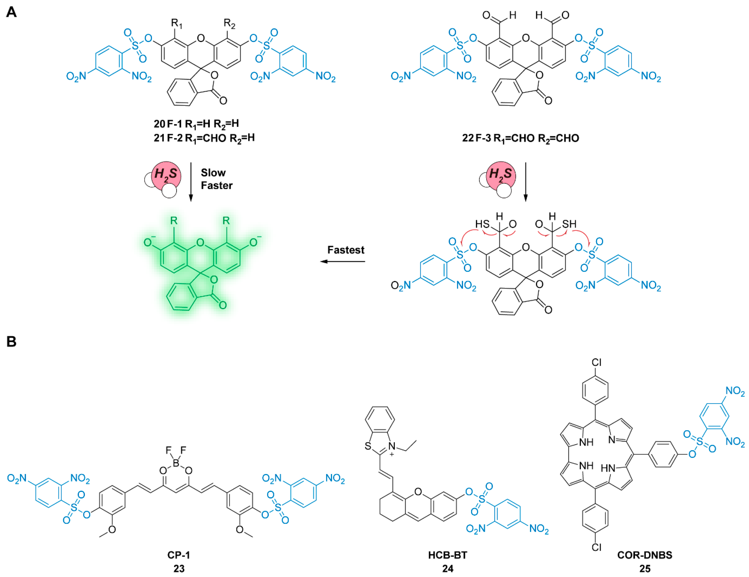
2.2.1. 2,4-Dinitrophenyl (DNP)-Based H2S Probe

In addition to reducibility, the nucleophilicity of H2S is also an important property in the design of fluorescent probes. DNP is a potent electron-withdrawing group that can efficiently quench fluorescence by covalently linking to the modifiable phenolic group in the fluorophore. When the nucleophilic H2S is added, the resulting mercaptan undergoes intramolecular nucleophilic substitution, and the probe releases free fluorophores with potent fluorescence. Therefore, DNP-based fluorescent probes are easy to prepare and have excellent quenching effects, so DNP is widely used in the design of various fluorescent probes. Yuan et al. designed two dual-excitation ratiometric fluorescent Probes 16 and 17 [52] based on coumarin and rhodamine. Those probes can eliminate the interference of microenvironments and improve the accuracy of fluorescence measurement. Fluorescence imaging of H2S in living cells under different excitations showed excellent dual-excitation ratiometric imaging in mitochondria, which is expected to be applied to fluorescence imaging of intracellular H2S in biological systems. The chemical structures of DNP-based H2S probes introduced in this review are listed below (Figure 6 and Figure 7).
NIR fluorescent probes can overcome the interference of scattering and auto-fluorescence and are more conducive to sensitive imaging in cells. Therefore, Jiang et al. designed a NIR fluorescent Probe 18 [53] with a selective mitochondrial localization function for the detection of H2S in cells. After reacting with H2S, the probe emits significant fluorescence at 720 nm, which can quickly respond to H2S in mitochondria. Then, Mito-Tracker rhodamine 123 was used for the co-localization experiment, and the results showed that Probe 18 had good mito-targeting. Cell imaging demonstrated that Probe 18 was suitable for the detection of endogenous and exogenous H2S with significant fluorescence effects. In addition, the level of H2S within the endoplasmic reticulum plays a crucial role in various biological and physiological processes. Li et al. developed three pyrimidine-based fluorescent probes for targeted detection of H2S in the endoplasmic reticulum. The most suitable fluorophore L1 was selected by optical analysis, and Probe 19 [54] with good lipophilicity and a fast response to H2S was obtained (Figure 7A). Lipophilicity enables the probe to penetrate cells and improve the detection efficacy, which facilitates the detection of H2S in the endoplasmic reticulum (Figure 7B). Probe 19 can be utilized to detect H2S in the cellular environment and also facilitate research on the physiological activities of H2S within the endoplasmic reticulum (Figure 7C).
Figure 7. (A) Fluorescent Probe 19 for sensing H2S. (B) Endoplasmic reticulum colocalization fluorescence imaging of Probe 19. (C) Endoplasmic reticulum-targeted Probe 19 for visualizing Cys-induced endogenous H2S in cells. Reproduced with permission from Ref. [54]. Copyright 2020 Elsevier.
Chemosensors 11 00333 g007
2,4-Dinitrobenzenesulfonyl (DNPS) is a H2S-responsive quencher that can be connected to fluorescent dyes through DNP and sulfonic acid groups. The chemical structures of DNPS-based H2S probes introduced in this review are listed blow (Figure 8 and Figure 9). Chen et al. designed three fluorescent Probes 20–22 [55] based on the fluorescein fluorophore and promoted the H2S nucleophilic reaction by utilizing the positive influence of ortho aldehyde groups, so that DNPS could leave quickly, and the fluorescence of the probe could be restored. In addition, Probe 22 with two aldehyde groups can form thiazolidine enantiomers with biothiols, which can inhibit the fluorescence of fluorescein and improve the selectivity for H2S (Figure 8A). Similarly, using DNPS groups, chemical probes with different performance can be designed to meet the monitoring needs of biological systems. Therefore, Zhu et al. designed Probe 23 based on a curcumin core with a BF2 group, which can distinguish between two DNPS groups as a regioselective fluorescence probe. The probe has two DNPS groups, and the one DNPS group is solely used to quench fluorescence, while the other DNPS reacts with H2S to form a stable fluorescence intermediate. The selectivity of Probe 23 [56] for H2S is more than 10,000 times higher than that of biothiols, and its excellent H2S visualization ability is confirmed by cell fluorescence imaging. Xu et al. obtained NIR Probe 24 [57] by connecting hemicyanidin with benzothiazole groups, which can achieve the detection of H2S content in cells (Figure 9). In another work, a H2S-responsive NIR fluorescent Probe 25 [58] uses 5,15-di(4-chlorophenyl)-10-(4-hydroxylphenyl)-corrole as the fluorophore, which has excellent NIR fluorescence and low cytotoxicity and realizes the specific detection of H2S in cells.

2.2.2. 7-Nitro-1,2,3-benzoxadiazole (NBD)-Based H2S Probe

NBD is similar to H2S-specific DNP, and its excellent characteristics can also be responsible for fluorescence quenching and H2S-specific recognition. After incubating with H2S, the nucleophilicity of H2S causes the cleavage of NBD from the probe, releasing H2S concentration-dependent fluorescence signals. NBD-based probes have the advantages of fast reaction speed and good biocompatibility and are widely used in biological systems. The chemical structures of the NBD-based H2S probes introduced in this review are listed below (Figure 10). Li et al. designed a coumarin-based Probe 26 [59] with an NBD group as the cage group and introduced triphenylphosphonium salt as a localization group for specific detection of mitochondrial H2S. After reacting with H2S, the fluorescence of the probe at 565 nm was decreased, while the fluorescence of coumarin at 415 nm was increased. Due to the self-calibration function of ratiometric fluorescence, the dual-channel F415 nm/F565 nm signal enables more accurate and quantitative analysis of H2S levels. Cell imaging confirmed that the probe could visualize H2S in the mitochondria of cells. Similarly, Probe 27 [60] was designed using naphthalimide derivative as the fluorophore. Free Probe 27 did not show fluorescence due to the strong fluorescence quenching effect of NBD but showed fluorescence enhancement (68 times) at 528 nm after the addition of Na2S. The probe has stable fluorescence characteristics and can be used for confocal microscopy imaging of intracellular H2S, which proves that the probe can effectively detect exogenous H2S in cells.
A NIR fluorescent Probe 28 [61] was designed using a long π-conjugated rhodamine dye and an NBD quenching group. When reacting with H2S, the fluorescence intensity is increased 10 times at 660 nm, and the quantum yield of the redshift product reaches 0.29. The probe kinetic test showed that the reaction rate k2 was 29.8 M−1S−1, and the detection limit was 0.27 µM, demonstrating that the probe had a rapid response to H2S. The positive charge of the probe allows for its mitochondrial location, enabling fast and efficient NIR fluorescence imaging of H2S at the subcellular level. Li et al. reported a fluorescent Probe 29 for simultaneous detection of H+ and H2S, and then introduced a morpholine to improve its lysosome location [62]. The fluorescence of the probe can only be turned on in the presence of H+ and H2S simultaneously. Probe 29 could detect H2S within the pH range of the lysosome, thus realizing the accurate location of acidic lysosomes (Figure 11). Feng et al. improved the previous NBD-type probe (HBT-NBD) and obtained highly efficient and specific Probes 30 and 31 [63]. Probe 30 is not suitable for physiological applications due to its instability under alkaline conditions. In contrast, Probe 31 exhibits excellent stability at physiological pH. The incorporation of aldehyde and ortho-methyl groups introduces reversible reaction sites and steric hindrance effects, enhancing its selectivity for H2S compared to other biothiols. Moreover, due to its low pKa value, H2S is more likely to perform a nuclear reaction with a probe than other biothiols under physiological pH conditions. Consequently, Probe 31 is more suitable for monitoring H2S levels in living cells.
The fluorescent probes based on DNP and NBD exhibit sensitive fluorescence changes upon H2S nucleophilic attack, enabling effective detection and imaging of H2S. However, the selectivity of these probes is hampered by interference from endogenous nucleophilic thiols in living cells. To address this challenge, the probes can be customized by implementing strategies such as spatial hindrance and auxiliary groups (such as ortho-benzaldehyde).

2.2.3. Disulfide Bond-Based H2S Probe

H2S is nucleophilic and can also attack disulfide bonds. Therefore, disulfide bonds are introduced into the chemical structure of fluorescent dyes as H2S recognition sites. When H2S nucleophilic attacks and disulfide bonds break, mercaptan intermediates are formed, and then intramolecular cyclization occurs between the intermediates and esters, releasing fluorescence groups and restoring fluorescence signals. The chemical structures of bisulfide bond-based H2S probes introduced in this review are listed below (Figure 12). Ye et al. designed two kinds of two-photon fluorescent probes, Probes 32 and 33 [64], based on naphthalimide fluorescent dyes and disulfide bond-responsive groups. They were linked to 2,2-disulfide benzoic acid by ester bonds and amides, respectively, and introduced morpholine groups into the target lysosomes. Probe 32 itself has only weak fluorescence. After the nucleophilic attack of H2S, the disulfide bonds break, and the fluorescent product is released, emitting intense fluorescence. However, Probe 33 is attached to disulfides by an amide bond, and after the disulfide bond breaks, the thiol intermediate cannot continue the cyclization process, which will quench the fluorescence of the probe. Both single photon and two-photon cell imaging experiments showed that Probe 32 could achieve H2S fluorescence imaging, which confirmed its application value in biological cells. Probe 34 [65] is a ratiometric fluorescent probe based on ICT and the excited intramolecular proton transfer (ESIPT) effect based on solvation effects. It has good photostability at physiological pH and can be used to achieve accurate and rapid quantitative detection of H2S by ratiometric fluorescence I525 nm/I495 nm. The probe can achieve significant fluorescence imaging ratios with different concentrations of H2S through different channels in cells. The importance of CBS and CSE for H2S production has been confirmed in endogenous H2S imaging experiments.
The nucleophilicity of H2S allows it to activate disulfide-based fluorescent probes, but endogenous biothiols cannot be ignored as interfering species. The substitution of sulfur with selenium (-S-Se-) can effectively enhance the selectivity of such probes for H2S, enabling the convenient design of a wide range of probes suitable for H2S detection. In 2017, Xian et al. designed a series of Washington Red (WR) NIR dyes with large Stokes shifts (>110 nm) and modified the hydroxyl group of the selected WR6 by disulfide bonding to obtain Probe 35 [66]. Compared with Probe 35, Probe 36 [66] uses Se to replace the sulfur atom in the disulfide bond, which improves anti-interference ability against biological mercaptans, enhances specificity against H2S, and successfully realizes fluorescence imaging of H2S in cells. Probe 36 overcomes the weak anti-interference ability of previous WSP and Sep series probes and allows for better adaptation to bioimaging (Figure 13). In the same year, Yin et al. prepared a water-soluble ratio fluorescence Probe 37 by introducing 2-(pyridin-2-yldisulfanyl) benzoic into 4-hydroxycoumarin [67]. In 2020, Probe 38 [68] was designed based on the structure of dicyanoisophorone. Both probes have outstanding fluorescence characteristics in vitro and good biocompatibility, enabling their application in the intracellular detection of H2S.

2.2.4. Other Nucleophilic-Based of H2S Probes

Due to the nucleophilic nature of H2S, various nucleophilic reactions can be carried out, such as nucleophilic substitution, nucleophilic addition, etc. Based on this strategy, probes with different structures can also be designed to meet the needs of various types of cell imaging. The chemical structures of other nucleophilic-based H2S probes introduced in this review are listed below (Figure 14). Han et al. developed a cyanine dye-based turn-off fluorescent Probe 39 [69]. The strong fluorescence of the probe itself is turned off under the nucleophilic attack of H2S. Thus, using fluorescence or UV–visible spectroscopy, the probe can even detect H2S in serum. Cell imaging experiments showed that the fluorescence of Probe 39 could be effectively turned off against H2S, and NIR fluorescence emission characteristics reduced the signal interference in biological samples. Similarly, Probe 40 [70] also uses cyanine dyes as fluorophore and selects 2-carboxybenzaldehyde as the H2S reaction site. Based on the tautomerism of ketones and enols in the nucleophilic addition product of Probe 40, a ratiometric fluorescence probe is designed to reduce environmental and instrumental interference. In the imaging of exogenous H2S, the pixel intensity was only 0.0768. With the extension of reaction time with H2S, the pixel intensity increased to 0.7091. In vitro imaging confirmed that Probe 40 could locate mitochondria and detect cellular H2S in the NIR region.
In 2020, Zhang et al. designed the NIR Probe 41 [71] based on O-carboxybenzaldehyde to realize the fluorescence detection of endogenous/exogenous H2S in the endoplasmic reticulum. The excellent organelle localization of Probe 41 confirmed the important physiological role of H2S in endoplasmic reticulum stress. Probe 42 [72] selected N-methyl-2-methoxyaniline moiety as the NO response site, while 4-nitrobenzenethiol-substituted boron dipyrromethene could respond to H2S. NO and H2S cycles can be dynamically visualized by the 645 nm channel (NO) and the 936 nm NIR II channel (H2S). Imaging experiments on colonic smooth muscle cells and HepG-2 cells showed that Probe 42 can indeed dynamically monitor NO and H2S within the cells, facilitating the study of the signal transduction between NO and H2S (Figure 15).
In addition to the mentioned H2S fluorescent probes, it is important to consider additional recognition sites during probe design, while taking into account the specific chemical properties of the fluorophore itself. This approach allows for the development of more versatile H2S fluorescent probes with improved sensitivity and specificity.

2.3. Probes Based on Metal and H2S Reactions

Due to its special affinity for Cu2+, H2S can effectively combine with Cu2+ to form CuS precipitates with Ksp about 10−45 (25 °C, water) [36]. When H2S and Cu2+ react rapidly, Cu2+, which quenches the fluorescence, immediately leaves, and the probe resumes the potent fluorescence. Fluorescent dyes designed using this mechanism allow for rapid detection of H2S. The chemical structures of metal-based H2S probes introduced in this review are listed below (Figure 16). In 2011, Sasakura et al. designed four fluorescent probes, Probes 43–46 [73], by incorporating fluorescein and a heterocyclic copper. Azazamacrocycles can form a stable metal complex with Cu2+, which has an obvious quenching effect on the fluorophore. Probe 44 has the best optical properties and strong anti-interference ability, enabling selective fluorescence imaging of endogenous/exogenous H2S within cells. Yang et al. also designed three triarylboron-based Probes 47–49 [74] with different numbers of diphenylamine and cyclen, which can chelate Cu2+ according to the affinity of cyclen to Cu2+, thus inhibiting the ICT process and effectively quenching the probe fluorescence. The finite aggregates induced by probe and nonchromphoric analyte can effectively inhibit the aggregation-caused quenching (ACQ) effect and enhance the photostability of the probe. The selected Probe 48 has good membrane permeability and excellent two-photon properties, which can accurately image endogenous H2S in mitochondria. Using the fluorescence lifetime microscopy (FLIM) technique, it can be clearly shown that H2S is uniformly distributed within mitochondria. Similarly, Huang et al. designed two Cu(II)-cyclen-based BODIPY fluorescent Probes 50 and 51 [75] with fluorescence emission wavelengths of 765 and 680 nm, respectively. The detection limit of H2S by Probe 50 is as low as 80 nM in vitro. In cell imaging, the probe showed excellent fluorescence signals in response to cellular H2S. Yuan et al. designed Probe 52 [76] based on the time-gated luminescence (TGL) technique of luminescent lanthanide complexes. Upon the reaction with H2S, the chelated Cu2+ leaves, and the fluorescence of Eu3+ at 610 nm is significantly enhanced (62 times), while the fluorescence of Tb3+ at 540 nm does not change significantly. A proportional TGL cell fluorescence imaging experiment showed that the addition of Cu2+ can effectively quench fluorescence. In addition, the probe can be used for rapid and quantitative detection of intracellular H2S, showing that TGL technology is suitable for fluorescence imaging of biological systems rich in biological background autofluorescence interference.
The metal-based probes exhibit rapid responses, but their biocompatibility is a significant concern. To mitigate these issues, the design of efficient sites capable of strong chelation with Cu2+ should be prioritized, thereby preventing the binding of Cu2+ with various cellular proteins and minimizing the potential toxicity of Cu2+ to living cells. In addition, the impediment posed by the metabolic mechanisms of CuS precipitation in cells should be given careful consideration.

3. Summary and Outlook

As an important biological signaling molecule in biological systems, H2S plays a unique role in various physiological and pathological activities, and the level of intracellular H2S is closely related to the homeostasis of the biological environment. Therefore, it is extremely important to develop techniques that can detect H2S in organisms, especially in cells that play various physiological functions. Fluorescence imaging probes have good biocompatibility and chemical stability, which is suitable for the application of biological systems. This review carefully analyzes fluorescent probes based on the physicochemical properties of H2S and their applications in intracellular H2S detection and H2S-related biological processes, providing ideas and strategies for designing functional fluorescent probes for H2S. However, there are still some challenges that hinder the development of H2S-activated fluorescent probes, such as the following: (1) the resolution of most current cell imaging approaches is more than 200 nm, which makes it difficult to achieve accurate detection of intracellular molecular processes. Therefore, there is an urgent need to develop high-resolution cell imaging probes and techniques to obtain high-resolution information on intracellular H2S to facilitate further studies of physiological mechanisms. (2) Fluorescent probes are usually easy to photobleach, making it difficult to meet the demand for long-term dynamic observation of H2S. Therefore, developing fluorescent dyes with excellent light stability and strong anti-bleaching ability is necessary to achieve continuous analysis of intracellular H2S fluctuation and H2S-related pathways. (3) Fluorescent probes for H2S detection should also be combined with other techniques to study physiological processes and gain a deeper understanding of the physiological significance of H2S signal transduction. So far, researchers are continuing their exploration of the detection of cellular H2S, hoping to develop more fluorescent probes with excellent characteristics suitable for cellular H2S research in the future.

Author Contributions

Writing—original draft preparation, W.L. and Y.Z.; Writing—review and editing, S.X.; Writing—review & editing, Supervision, Funding acquisition, D.S. All authors agreed to the published version of the manuscript.

Funding

This research was funded by the National Natural Science Foundation of China (No. 22274005, U2067214, U2167222), the Beijing Municipal Education Commission-Beijing Natural Science Foundation Joint Funding Project (KZ202010005006), and the Scientific Research Project of Beijing Educational Committee (KM202110005014).

Institutional Review Board Statement

Not applicable.

Informed Consent Statement

Not applicable.

Conflicts of Interest

The authors declare no competing financial interest.

References

  1. Gadalla, M.M.; Snyder, S.H. Hydrogen sulfide as a gasotransmitter. J. Neurochem. 2010, 113, 14–26. [Google Scholar] [CrossRef] [Green Version]
  2. Huang, C.W.; Moore, P.K. H2S Synthesizing Enzymes: Biochemistry and Molecular Aspects. In Chemistry, Biochemistry and Pharmacology of Hydrogen Sulfide; Moore, P.K., Whiteman, M., Eds.; Springer International Publishing: Cham, Switzerland, 2015; pp. 3–25. [Google Scholar]
  3. Singh, S.; Padovani, D.; Leslie, R.A.; Chiku, T.; Banerjee, R. Relative Contributions of Cystathionine β-Synthase and γ-Cystathionase to H2S Biogenesis via Alternative Trans-sulfuration Reactions. J. Biol. Chem. 2009, 284, 22457–22466. [Google Scholar] [CrossRef] [Green Version]
  4. Shibuya, N.; Tanaka, M.; Yoshida, M.; Ogasawara, Y.; Togawa, T.; Ishii, K.; Kimura, H. 3-Mercaptopyruvate Sulfurtransferase Produces Hydrogen Sulfide and Bound Sulfane Sulfur in the Brain. Antioxid. Redox Signal. 2008, 11, 703–714. [Google Scholar] [CrossRef]
  5. Madden, J.A.; Ahlf, S.B.; Dantuma, M.W.; Olson, K.R.; Roerig, D.L. Precursors and inhibitors of hydrogen sulfide synthesis affect acute hypoxic pulmonary vasoconstriction in the intact lung. J. Appl. Physiol. 2011, 112, 411–418. [Google Scholar] [CrossRef] [PubMed] [Green Version]
  6. Sun, H.-J.; Wu, Z.-Y.; Nie, X.-W.; Wang, X.-Y.; Bian, J.-S. Implications of hydrogen sulfide in liver pathophysiology: Mechanistic insights and therapeutic potential. J. Adv. Res. 2021, 27, 127–135. [Google Scholar] [CrossRef]
  7. Li, L.; Bhatia, M.; Zhu, Y.Z.; Zhu, Y.C.; Ramnath, R.D.; Wang, Z.J.; Anuar, F.B.M.; Whiteman, M.; Salto-Tellez, M.; Moore, P.K. Hydrogen sulfide is a novel mediator of lipopolysaccharide-induced inflammation in the mouse. FASEB J. 2005, 19, 1196–1198. [Google Scholar] [CrossRef] [PubMed]
  8. Vincenzo, B.; Emma, M.; Roderick, J.F.; Giuseppe, C.; Mauro, P. Annexin A1 Mediates Hydrogen Sulfide Properties in the Control of Inflammation. J. Pharmacol. Exp. Ther. 2014, 351, 96. [Google Scholar] [CrossRef] [Green Version]
  9. Liu, Q.; Zhong, Y.; Su, Y.; Zhao, L.; Peng, J. Real-Time Imaging of Hepatic Inflammation Using Hydrogen Sulfide-Activatable Second Near-Infrared Luminescent Nanoprobes. Nano Lett. 2021, 21, 4606–4614. [Google Scholar] [CrossRef]
  10. Giuliani, D.; Ottani, A.; Zaffe, D.; Galantucci, M.; Strinati, F.; Lodi, R.; Guarini, S. Hydrogen sulfide slows down progression of experimental Alzheimer’s disease by targeting multiple pathophysiological mechanisms. Neurobiol. Learn. Mem. 2013, 104, 82–91. [Google Scholar] [CrossRef] [PubMed]
  11. Kamoun, P.; Belardinelli, M.-C.; Chabli, A.; Lallouchi, K.; Chadefaux-Vekemans, B. Endogenous hydrogen sulfide overproduction in Down syndrome. Am. J. Med. Genet. Part A 2003, 116A, 310–311. [Google Scholar] [CrossRef] [PubMed]
  12. Wang, M.; Zhu, J.; Pan, Y.; Dong, J.; Zhang, L.; Zhang, X.; Zhang, L. Hydrogen sulfide functions as a neuromodulator to regulate striatal neurotransmission in a mouse model of Parkinson’s disease. J. Neurosci. Res. 2015, 93, 487–494. [Google Scholar] [CrossRef]
  13. Wang, P.; Zhang, G.; Wondimu, T.; Ross, B.; Wang, R. Hydrogen sulfide and asthma. Exp. Physiol. 2011, 96, 847–852. [Google Scholar] [CrossRef]
  14. Magierowski, M.; Jasnos, K.; Kwiecien, S.; Drozdowicz, D.; Surmiak, M.; Strzalka, M.; Ptak-Belowska, A.; Wallace, J.L.; Brzozowski, T. Endogenous Prostaglandins and Afferent Sensory Nerves in Gastroprotective Effect of Hydrogen Sulfide against Stress-Induced Gastric Lesions. PLoS ONE 2015, 10, e0118972. [Google Scholar] [CrossRef] [Green Version]
  15. Jha, S.; Calvert, J.W.; Duranski, M.R.; Ramachandran, A.; Lefer, D.J. Hydrogen sulfide attenuates hepatic ischemia-reperfusion injury: Role of antioxidant and antiapoptotic signaling. Am. J. Physiol.-Heart Circ. Physiol. 2008, 295, H801–H806. [Google Scholar] [CrossRef] [Green Version]
  16. Satori, C.P.; Henderson, M.M.; Krautkramer, E.A.; Kostal, V.; Distefano, M.M.; Arriaga, E.A. Bioanalysis of Eukaryotic Organelles. Chem. Rev. 2013, 113, 2733–2811. [Google Scholar] [CrossRef] [Green Version]
  17. Xu, W.; Zeng, Z.; Jiang, J.-H.; Chang, Y.-T.; Yuan, L. Discerning the Chemistry in Individual Organelles with Small-Molecule Fluorescent Probes. Angew. Chem. Int. Ed. 2016, 55, 13658–13699. [Google Scholar] [CrossRef] [PubMed]
  18. Fu, M.; Zhang, W.; Wu, L.; Yang, G.; Li, H.; Wang, R. Hydrogen sulfide (H2S) metabolism in mitochondria and its regulatory role in energy production. Proc. Natl. Acad. Sci. USA 2012, 109, 2943–2948. [Google Scholar] [CrossRef] [PubMed] [Green Version]
  19. Cheung, N.S.; Peng, Z.F.; Chen, M.J.; Moore, P.K.; Whiteman, M. Hydrogen sulfide induced neuronal death occurs via glutamate receptor and is associated with calpain activation and lysosomal rupture in mouse primary cortical neurons. Neuropharmacology 2007, 53, 505–514. [Google Scholar] [CrossRef]
  20. Wei, L.; Kan, L.-Y.; Zeng, H.-Y.; Tang, Y.-Y.; Huang, H.-L.; Xie, M.; Zou, W.; Wang, C.-Y.; Zhang, P.; Tang, X.-Q. BDNF/TrkB Pathway Mediates the Antidepressant-Like Role of H2S in CUMS-Exposed Rats by Inhibition of Hippocampal ER Stress. Neuro Mol. Med. 2018, 20, 252–261. [Google Scholar] [CrossRef]
  21. Abe, K.; Kimura, H. The possible role of hydrogen sulfide as an endogenous neuromodulator. J. Neurosci. 1996, 16, 1066. [Google Scholar] [CrossRef] [PubMed] [Green Version]
  22. Filipovic, M.R.; Zivanovic, J.; Alvarez, B.; Banerjee, R. Chemical Biology of H2S Signaling through Persulfidation. Chem. Rev. 2018, 118, 1253–1337. [Google Scholar] [CrossRef]
  23. Vitvitsky, V.; Banerjee, R. Chapter Seven—H2S Analysis in Biological Samples Using Gas Chromatography with Sulfur Chemiluminescence Detection. In Methods in Enzymology; Cadenas, E., Packer, L., Eds.; Academic Press: Cambridge, MA, USA, 2015; Volume 554, pp. 111–123. [Google Scholar]
  24. Radford-Knoery, J.; Cutter, G.A. Determination of carbonyl sulfide and hydrogen sulfide species in natural waters using specialized collection procedures and gas chromatography with flame photometric detection. Anal. Chem. 1993, 65, 976–982. [Google Scholar] [CrossRef]
  25. Bae, J.; Choi, M.G.; Choi, J.; Chang, S.-K. Colorimetric signaling of hydrogen sulfide by reduction of a phenylseleno-nitrobenzoxadiazole derivative. Dye. Pigment. 2013, 99, 748–752. [Google Scholar] [CrossRef]
  26. Kahyarian, A.; Nesic, S. H2S corrosion of mild steel: A quantitative analysis of the mechanism of the cathodic reaction. Electrochim. Acta 2019, 297, 676–684. [Google Scholar] [CrossRef]
  27. Li, H.; Fang, Y.; Yan, J.; Ren, X.; Zheng, C.; Wu, B.; Wang, S.; Li, Z.; Hua, H.; Wang, P.; et al. Small-molecule fluorescent probes for H2S detection: Advances and perspectives. TrAC Trends Anal. Chem. 2021, 134, 116117. [Google Scholar] [CrossRef]
  28. Ibrahim, H.; Serag, A.; Farag, M.A. Emerging analytical tools for the detection of the third gasotransmitter H2S, a comprehensive review. J. Adv. Res. 2021, 27, 137–153. [Google Scholar] [CrossRef] [PubMed]
  29. Bezner, B.J.; Ryan, L.S.; Lippert, A.R. Reaction-Based Luminescent Probes for Reactive Sulfur, Oxygen, and Nitrogen Species: Analytical Techniques and Recent Progress. Anal. Chem. 2020, 92, 309–326. [Google Scholar] [CrossRef] [PubMed]
  30. Yang, M.; Fan, J.; Du, J.; Peng, X. Small-molecule fluorescent probes for imaging gaseous signaling molecules: Current progress and future implications. Chem. Sci. 2020, 11, 5127–5141. [Google Scholar] [CrossRef] [Green Version]
  31. Jiao, X.; Li, Y.; Niu, J.; Xie, X.; Wang, X.; Tang, B. Small-Molecule Fluorescent Probes for Imaging and Detection of Reactive Oxygen, Nitrogen, and Sulfur Species in Biological Systems. Anal. Chem. 2018, 90, 533–555. [Google Scholar] [CrossRef]
  32. Guo, L.-Y.; Zhao, J.-S.; Peng, H.-S. Fluorescent Probes for Sensing and Imaging Biological Hydrogen Sulfide. Anal. Sens. 2022, 2, e202200025. [Google Scholar] [CrossRef]
  33. Li, J.; Su, Z.; Yu, C.; Yuan, Y.; Wu, Q.; Liu, J.; Peng, B.; Hu, W.; Lu, X.; Yu, H.; et al. Recent progress in the development of sensing systems for in vivo detection of biological hydrogen sulfide. Dye. Pigment. 2021, 192, 109451. [Google Scholar] [CrossRef]
  34. Gilbert, A.K.; Pluth, M.D. Subcellular Delivery of Hydrogen Sulfide Using Small Molecule Donors Impacts Organelle Stress. J. Am. Chem. Soc. 2022, 144, 17651–17660. [Google Scholar] [CrossRef] [PubMed]
  35. Montoya, L.A.; Pluth, M.D. Organelle-Targeted H2S Probes Enable Visualization of the Subcellular Distribution of H2S Donors. Anal. Chem. 2016, 88, 5769–5774. [Google Scholar] [CrossRef] [Green Version]
  36. Zhou, L.; Chen, Y.; Shao, B.; Cheng, J.; Li, X. Recent advances of small-molecule fluorescent probes for detecting biological hydrogen sulfide. Front. Chem. Sci. Eng. 2022, 16, 34–63. [Google Scholar] [CrossRef]
  37. Luo, Y.; Zuo, Y.; Shi, G.; Xiang, H.; Gu, H. Progress on the reaction-based methods for detection of endogenous hydrogen sulfide. Anal. Bioanal. Chem. 2022, 414, 2809–2839. [Google Scholar] [CrossRef]
  38. Cuevasanta, E.; Lange, M.; Bonanata, J.; Coitiño, E.L.; Ferrer-Sueta, G.; Filipovic, M.R.; Alvarez, B. Reaction of Hydrogen Sulfide with Disulfide and Sulfenic Acid to Form the Strongly Nucleophilic Persulfide*♦. J. Biol. Chem. 2015, 290, 26866–26880. [Google Scholar] [CrossRef] [Green Version]
  39. Chauhan, P.; Jos, S.; Chakrapani, H. Reactive Oxygen Species-Triggered Tunable Hydrogen Sulfide Release. Org. Lett. 2018, 20, 3766–3770. [Google Scholar] [CrossRef]
  40. Fu, Y.-J.; Yao, H.-W.; Zhu, X.-Y.; Guo, X.-F.; Wang, H. A cell surface specific two-photon fluorescent probe for monitoring intercellular transmission of hydrogen sulfide. Anal. Chim. Acta 2017, 994, 1–9. [Google Scholar] [CrossRef]
  41. Deng, B.; Ren, M.; Wang, J.-Y.; Zhou, K.; Lin, W. A mitochondrial-targeted two-photon fluorescent probe for imaging hydrogen sulfide in the living cells and mouse liver tissues. Sens. Actuators B Chem. 2017, 248, 50–56. [Google Scholar] [CrossRef]
  42. Shen, D.; Liu, J.; Sheng, L.; Lv, Y.; Wu, G.; Wang, P.; Du, K. Design, synthesis and evaluation of a novel fluorescent probe to accurately detect H2S in hepatocytes and natural waters. Spectrochim. Acta Part A Mol. Biomol. Spectrosc. 2020, 228, 117690. [Google Scholar] [CrossRef] [PubMed]
  43. Chen, J.; Zhao, M.; Jiang, X.; Sizovs, A.; Wang, M.C.; Provost, C.R.; Huang, J.; Wang, J. Genetically anchored fluorescent probes for subcellular specific imaging of hydrogen sulfide. Analyst 2016, 141, 1209–1213. [Google Scholar] [CrossRef] [PubMed] [Green Version]
  44. Lin, V.S.; Lippert, A.R.; Chang, C.J. Cell-trappable fluorescent probes for endogenous hydrogen sulfide signaling and imaging H2O2-dependent H2S production. Proc. Natl. Acad. Sci. USA 2013, 110, 7131–7135. [Google Scholar] [CrossRef] [PubMed] [Green Version]
  45. Bae, S.K.; Heo, C.H.; Choi, D.J.; Sen, D.; Joe, E.-H.; Cho, B.R.; Kim, H.M. A Ratiometric Two-Photon Fluorescent Probe Reveals Reduction in Mitochondrial H2S Production in Parkinson’s Disease Gene Knockout Astrocytes. J. Am. Chem. Soc. 2013, 135, 9915–9923. [Google Scholar] [CrossRef] [PubMed]
  46. Feng, W.; Mao, Z.; Liu, L.; Liu, Z. A ratiometric two-photon fluorescent probe for imaging hydrogen sulfide in lysosomes. Talanta 2017, 167, 134–142. [Google Scholar] [CrossRef] [PubMed]
  47. Kong, D.; Song, B.; Yuan, J. A Golgi Apparatus-Targetable Probe Based on Lanthanide Complexes for Ratiometric Time-Gated Luminescence Detection of Hydrogen Sulfide. Eur. J. Inorg. Chem. 2023, 26, e202200642. [Google Scholar] [CrossRef]
  48. Li, S.-J.; Li, Y.-F.; Liu, H.-W.; Zhou, D.-Y.; Jiang, W.-L.; Ou-Yang, J.; Li, C.-Y. A Dual-Response Fluorescent Probe for the Detection of Viscosity and H2S and Its Application in Studying Their Cross-Talk Influence in Mitochondria. Anal. Chem. 2018, 90, 9418–9425. [Google Scholar] [CrossRef]
  49. Wu, Q.; Yin, C.; Wen, Y.; Zhang, Y.; Huo, F. An ICT lighten ratiometric and NIR fluorogenic probe to visualize endogenous/exogenous hydrogen sulphide and imaging in mice. Sens. Actuators B Chem. 2019, 288, 507–511. [Google Scholar] [CrossRef]
  50. Zhang, X.; Tan, H.; Yan, Y.; Hang, Y.; Yu, F.; Qu, X.; Hua, J. Targetable N-annulated perylene-based colorimetric and ratiometric near-infrared fluorescent probes for the selective detection of hydrogen sulfide in mitochondria, lysosomes, and serum. J. Mater. Chem. B 2017, 5, 2172–2180. [Google Scholar] [CrossRef]
  51. Chen, H.; Gong, X.; Liu, X.; Li, Z.; Zhang, J.; Yang, X.-F. A nitroso-based fluorogenic probe for rapid detection of hydrogen sulfide in living cells. Sens. Actuators B Chem. 2019, 281, 542–548. [Google Scholar] [CrossRef]
  52. Yuan, L.; Zuo, Q.-P. FRET-Based Mitochondria-Targetable Dual-Excitation Ratiometric Fluorescent Probe for Monitoring Hydrogen Sulfide in Living Cells. Chem.–Asian J. 2014, 9, 1544–1549. [Google Scholar] [CrossRef]
  53. Zhao, X.-j.; Li, Y.-t.; Jiang, Y.-r.; Yang, B.-q.; Liu, C.; Liu, Z.-h. A novel “turn-on” mitochondria-targeting near-infrared fluorescent probe for H2S detection and in living cells imaging. Talanta 2019, 197, 326–333. [Google Scholar] [CrossRef] [PubMed]
  54. Wang, H.; Hu, L.; Xu, B.; Chen, H.; Cai, F.; Yang, N.; Wu, Q.; Uvdal, K.; Hu, Z.; Li, L. Endoplasmic reticulum-targeted fluorogenic probe based on pyrimidine derivative for visualizing exogenous/endogenous H2S in living cells. Dye. Pigment. 2020, 179, 108390. [Google Scholar] [CrossRef]
  55. Lv, J.; Wang, F.; Qiang, J.; Ren, X.; Chen, Y.; Zhang, Z.; Wang, Y.; Zhang, W.; Chen, X. Enhanced response speed and selectivity of fluorescein-based H2S probe via the cleavage of nitrobenzene sulfonyl ester assisted by ortho aldehyde groups. Biosens. Bioelectron. 2017, 87, 96–100. [Google Scholar] [CrossRef]
  56. Yang, B.; Su, M.-M.; Xue, Y.-S.; He, Z.-X.; Xu, C.; Zhu, H.-L. A selective fluorescence probe for H2S from biothiols with a significant regioselective turn-on response and its application for H2S detection in living cells and in living Caenorhabditis elegans. Sens. Actuators B Chem. 2018, 276, 456–465. [Google Scholar] [CrossRef]
  57. Li, B.; Mei, H.; Wang, M.; Gu, X.; Hao, J.; Xie, X.; Xu, K. A near-infrared fluorescent probe for imaging of endogenous hydrogen sulfide in living cells and mice. Dye. Pigment. 2021, 189, 109231. [Google Scholar] [CrossRef]
  58. Lu, G.; Gao, Y.; Wang, X.; Zhang, D.; Meng, S.; Yu, S.; Zhuang, Y.; Duan, L. A near-infrared fluorescent probe based on corrole derivative with large Stokes shift for detection of hydrogen sulfide in water and living cells. Dye. Pigment. 2022, 204, 110445. [Google Scholar] [CrossRef]
  59. Jia, X.; Li, W.; Guo, Z.; Guo, Z.; Li, Y.; Zhang, P.; Wei, C.; Li, X. An NBD-Based Mitochondrial Targeting Ratiometric Fluorescent Probe for Hydrogen Sulfide Detection. ChemistrySelect 2019, 4, 8671–8675. [Google Scholar] [CrossRef]
  60. Pak, Y.L.; Li, J.; Ko, K.C.; Kim, G.; Lee, J.Y.; Yoon, J. Mitochondria-Targeted Reaction-Based Fluorescent Probe for Hydrogen Sulfide. Anal. Chem. 2016, 88, 5476–5481. [Google Scholar] [CrossRef] [PubMed]
  61. Ismail, I.; Chen, Z.; Ji, X.; Sun, L.; Yi, L.; Xi, Z. A Fast-Response Red Shifted Fluorescent Probe for Detection of H2S in Living Cells. Molecules 2020, 25, 437. [Google Scholar] [CrossRef] [PubMed] [Green Version]
  62. Ou-Yang, J.; Jiang, W.-L.; Tan, K.-Y.; Liu, H.-W.; Li, S.-J.; Liu, J.; Li, Y.-F.; Li, C.-Y. Two-photon fluorescence probe for precisely detecting endogenous H2S in lysosome by employing a dual lock system. Sens. Actuators B Chem. 2018, 260, 264–273. [Google Scholar] [CrossRef]
  63. Ding, S.; Feng, W.; Feng, G. Rapid and highly selective detection of H2S by nitrobenzofurazan (NBD) ether-based fluorescent probes with an aldehyde group. Sens. Actuators B Chem. 2017, 238, 619–625. [Google Scholar] [CrossRef]
  64. Li, G.; Ma, S.; Tang, J.; Ye, Y. Lysosome-targeted two-photon fluorescent probes for rapid detection of H2S in live cells. New J. Chem. 2019, 43, 1267–1274. [Google Scholar] [CrossRef]
  65. Cao, T.; Teng, Z.; Gong, D.; Qian, J.; Liu, W.; Iqbal, K.; Qin, W.; Guo, H. A ratiometric fluorescent probe for detection of endogenous and exogenous hydrogen sulfide in living cells. Talanta 2019, 198, 185–192. [Google Scholar] [CrossRef]
  66. Chen, W.; Xu, S.; Day, J.J.; Wang, D.; Xian, M. A General Strategy for Development of Near-Infrared Fluorescent Probes for Bioimaging. Angew. Chem. Int. Ed. 2017, 56, 16611–16615. [Google Scholar] [CrossRef]
  67. Kang, J.; Huo, F.; Yin, C. A novel ratiometric fluorescent H2S probe based on tandem nucleophilic substitution/cyclization reaction and its bioimaging. Dye. Pigment. 2017, 146, 287–292. [Google Scholar] [CrossRef]
  68. Wu, Q.; Huo, F.; Wang, J.; Yin, C. Fluorescent probe for detecting hydrogen sulfide based on disulfide nucleophilic substitution-addition. Spectrochim. Acta Part A Mol. Biomol. Spectrosc. 2020, 238, 118437. [Google Scholar] [CrossRef]
  69. Wu, X.; Shi, J.; Yang, L.; Han, J.; Han, S. A near-infrared fluorescence dye for sensitive detection of hydrogen sulfide in serum. Bioorganic Med. Chem. Lett. 2014, 24, 314–316. [Google Scholar] [CrossRef]
  70. Wang, X.; Sun, J.; Zhang, W.; Ma, X.; Lv, J.; Tang, B. A near-infrared ratiometric fluorescent probe for rapid and highly sensitive imaging of endogenous hydrogen sulfide in living cells. Chem. Sci. 2013, 4, 2551–2556. [Google Scholar] [CrossRef]
  71. Shu, W.; Zang, S.; Wang, C.; Gao, M.; Jing, J.; Zhang, X. An Endoplasmic Reticulum-Targeted Ratiometric Fluorescent Probe for the Sensing of Hydrogen Sulfide in Living Cells and Zebrafish. Anal. Chem. 2020, 92, 9982–9988. [Google Scholar] [CrossRef] [PubMed]
  72. Zhu, T.; Ren, N.; Liu, X.; Dong, Y.; Wang, R.; Gao, J.; Sun, J.; Zhu, Y.; Wang, L.; Fan, C.; et al. Probing the Intracellular Dynamics of Nitric Oxide and Hydrogen Sulfide Using an Activatable NIR II Fluorescence Reporter. Angew. Chem. Int. Ed. 2021, 60, 8450–8454. [Google Scholar] [CrossRef]
  73. Sasakura, K.; Hanaoka, K.; Shibuya, N.; Mikami, Y.; Kimura, Y.; Komatsu, T.; Ueno, T.; Terai, T.; Kimura, H.; Nagano, T. Development of a Highly Selective Fluorescence Probe for Hydrogen Sulfide. J. Am. Chem. Soc. 2011, 133, 18003–18005. [Google Scholar] [CrossRef] [PubMed]
  74. Liu, J.; Guo, X.; Hu, R.; Liu, X.; Wang, S.; Li, S.; Li, Y.; Yang, G. Molecular Engineering of Aqueous Soluble Triarylboron-Compound-Based Two-Photon Fluorescent Probe for Mitochondria H2S with Analyte-Induced Finite Aggregation and Excellent Membrane Permeability. Anal. Chem. 2016, 88, 1052–1057. [Google Scholar] [CrossRef] [PubMed]
  75. Wu, H.; Krishnakumar, S.; Yu, J.; Liang, D.; Qi, H.; Lee, Z.-W.; Deng, L.-W.; Huang, D. Highly Selective and Sensitive Near-Infrared-Fluorescent Probes for the Detection of Cellular Hydrogen Sulfide and the Imaging of H2S in Mice. Chem.–Asian J. 2014, 9, 3604–3611. [Google Scholar] [CrossRef] [PubMed]
  76. Tang, Z.; Song, B.; Ma, H.; Shi, Y.; Yuan, J. A ratiometric time-gated luminescence probe for hydrogen sulfide based on copper(II)-coupled lanthanide complexes. Anal. Chim. Acta 2019, 1049, 152–160. [Google Scholar] [CrossRef]
Scheme 1. The mechanism and application of H2S-activated fluorogenic probes.
Chemosensors 11 00333 sch001
Figure 1. Chemical structures of azide-based Probes 1–10.
Chemosensors 11 00333 g001
Figure 2. (A) Two-photon fluorescence Probe 2 for H2S detection. (B) Probe 2 for H2S detection in cells. Reproduced with permission from Ref. [41]. Copyright 2017 Elsevier.
Chemosensors 11 00333 g002
Figure 3. (A) Schematic diagram and (B) fluorescence spectra of Probe 7 in response to H2S. (C) Fluorescence imaging of cells after different treatments using Probe 7. Reproduced with permission from Ref. [45]. Copyright 2013 American Chemical Society.
Chemosensors 11 00333 g003
Figure 4. Chemical structures of nitro-/nitroso-based Probes 11–15.
Chemosensors 11 00333 g004
Figure 5. (A) Fluorescent Probes 12–14 for H2S detection. (B) Confocal fluorescence imaging of subcellular H2S in living cells with Probes 13 and 14. Reproduced with permission from Ref. [50]. Copyright 2017 The Royal Society of Chemistry.
Chemosensors 11 00333 g005
Figure 6. Chemical structures of DNP-based Probes 16–18.
Chemosensors 11 00333 g006
Figure 8. (A) Schematic illustration and comparison of Probes 20–22 for H2S detection. (B) Chemical structure of DNPS-based Probes 23–25.
Chemosensors 11 00333 g008
Figure 9. (A) Fluorescent Probe 24 for H2S detection. (B) Fluorescence imaging of endogenous and exogenous H2S in cells. Reproduced with permission from Ref. [57]. Copyright 2021 Elsevier.
Chemosensors 11 00333 g009
Figure 10. Chemical structures of NBD-based Probes 26–31.
Chemosensors 11 00333 g010
Figure 11. (A) Fluorescent Probe 29 for H2S detection under acidic condition. (B) fluorescence imaging of H2S in cells after different treatments. Reproduced with permission from Ref. [62]. Copyright 2018 Elsevier.
Chemosensors 11 00333 g011
Figure 12. Chemical structures of disulfide bond-based Probes 32–38.
Chemosensors 11 00333 g012
Figure 13. (A) The structures of NIR fluorescent Probes 35, 36. (B) Response mechanisms of WSP and Sep probes to H2S. (C) Fluorescence imaging of endogenous H2S in cells using Probe 36. Reproduced with permission from Ref. [66]. Copyright 2017 Wiley-VCH Verlag GmbH & Co. KGaA, Weinheim.
Chemosensors 11 00333 g013
Figure 14. Chemical structures of other nucleophilic-based Probes 39–42.
Chemosensors 11 00333 g014
Figure 15. (A) Fluorescent Probe 42 for the detection NO and H2S. (B) Fluorescence imaging of Probe 42 for the detection of NO and H2S in cells. Reproduced with permission from Ref. [72]. Copyright 2021 Wiley-VCH GmbH.
Chemosensors 11 00333 g015
Figure 16. Chemical structures of metal-based Probes 43–52.
Chemosensors 11 00333 g016
Disclaimer/Publisher’s Note: The statements, opinions and data contained in all publications are solely those of the individual author(s) and contributor(s) and not of MDPI and/or the editor(s). MDPI and/or the editor(s) disclaim responsibility for any injury to people or property resulting from any ideas, methods, instructions or products referred to in the content.

